the cell - teachers of india
TRANSCRIPT

16 – 19 May 2013
"The Cell"
Azim Premji Foundation, District institute,
Rudrapur, Udham Singh Nagar

1
Overview
A four days science workshop on the concept of “Cell” was organized in Azim Premji Foundation,
Udham Singh Nagar, district institute in which 31 upper primary and high school science teachers and
one DIET faculty participated. This workshop was designed and facilitated by biology resource persons
across the foundation from different locations like Subhash Goswami and Sandhya Sahu from
Chhattisgarh, Prakash S. Shivran from Tonk, Mohammed Javed Siddiqui from Bhopal, Meenakshi Pant
from Dehradun, Vikas Chandra Roy and Deepak Rawat from Uttarkashi, Shiv Pandey from Udham
Singh Nagar and Nishi Khandelwal from Digantar Jaipur.
Cell is an appropriate concept to start a course of biology in order to understand the life processes and
its complexities in a simple way. It is imperative to have an in depth understanding of cell as they are
the basic units that form living organisms and execute their functioning. On reviewing text-books of
various states wherein the concept of Cell is dealt in, it was found that there are various gaps in the
way the content is being presented in the text books.
Many interwoven concepts and alternative conceptions are associated with the theme “cell” which calls
for developing an exhaustive resource and churning on the same that can be helpful in honing the
understanding of teachers and better learning of children in turn. This workshop on “cell” attempts to
explore the entire amazing world inside a cell locating the context in its historical perspective and
keeping in view the true spirit of science learning.
Objectives and Goals of the workshop
To minimize the gaps produced between the concepts of cell in science textbooks.
To assist teachers in realizing the importance of experimentation in testing out and validating
own hypotheses and accommodating new learning out of those processes.
To show how an experimental approach has allowed great progress in understanding the
function of cell by incorporating historical perspective and microscopic activities.
To revenue critical feedback on content and process flow of module for further improvement.
Wider perspective of the workshop was to come out with enriched knowledge, more scope of
experimental processes and pedagogical practices for target group. This workshop had also exhibited
the validation of scientific knowledge formation, and scientific processes and method with an emphasis
on forming space for curiosity and the questions. In this workshop we were inclined to create the
thoughts that hands-on activity is core of the subject and we would go through a lot of joyful
experiments initiating curiosity with microscopy, variety of material for observing the variety of cells
and wide range of staining possibilities.

2
Day - 1 (16 May 2013)
Session - 1
Introduction of Participants
The day 1 started with an activity with participants, where they formed a circle and provided folded
paper strips contained in a bowl. Each paper strip had unfinished lyrics of a popular Hindi movie song,
everyone had to choose one of the strip randomly and every participant had to locate his/her partner
having the other segment of same song by softly singing his/ her line while roaming around the hall.
Once every participant including facilitators had a partner then they were asked to introduce each
other to the larger group by welcoming his/her partner with a bunch of flowers and furnishing details
on following pointers:
Name of the Participant
From where the participant has came
What are his/her views regarding science teaching in schools under current educational scenario.
The participants came in pairs and introduced each other to the larger group by furnishing details
according to the above pointers.
Reflections of Participants on Science Education
1. Science develops new understanding and curiosities in students.
2. Science teaching should be concerned with immediate environment, teaching of the
subject generates curiosity among the students and the role of teacher is to pacify their
curiosities accordingly
3. Science teaching should be experiment based. I am of the opinion that student shall fully
comprehend the concept like chemical reactions/equations in a more concrete way.

3
4. Science should be learned through activities which is normally absent in the
schools.
5. In these types of workshop we learn good things but we shall be able to
deliver these ideas and concepts to the student through improvised
pedagogy.
6. Science develops new thoughts, teaching of this subject in class should be
activities based.
7. Though science teaching must be practical/activity based in class but this is
only possible when there is availability of instruments and other requirements.
What can we do if these things are not found in schools?
2. Science teaching open new frontiers for learning and gives an opportunity to
understand abstract terms in a more concrete form.
3. We always discuss that we should learn science by doing but seldom found
someone giving space to children for that.
4. Science can’t’ be understand by rote or repetition of the facts.
5. Science is everywhere, where we can’t see the science? We have only made
havoc about science and its teaching; I only know that this should be taught
as per the nature of the subject.
6. Science teaching should be focused to develop scientific temperament in the
students.
7. Science teaching should be taught with addition of some questions like
‘what’, ‘when’, ‘why’ etc.
8. Science should not be taught like literature, but should be for understanding.
End of the discussion was marked by a two liner from a participant:
“Kaun kahta hai aasmaan mein suraakh ho nahi sakta,
tabiyat se ek pathar to uchaalo yaaron.”

4
Reflections of participants regarding sub concepts taught by them in various classes
In class 6th, we teach about Unicellular and Multicellular organisms
Difference between plant and animal cell
Study of Mitochondria and its basic functions
In class 9th we teach about various organelles, difference between multicellular and unicellular
organisms, Eukaryotic and prokaryotic cells followed by tissue grade organization.
In class 8th there is description about five type of microorganisms
Some teachers also raised issues related to design of science textbooks these days.
Session - 2
Context Setting
Participants were warmly welcomed on taking initiative to
participate in the workshop followed by a brief
description of foundation activities and expansion of its
institutes in various states. Facilitator provided an insight
regarding the basic procedure adopted by the foundation
in coming up with these kind of thematic workshops
accompanied with properly developed resource material.
Facilitator built up the context further around following
broad heads:
The amount of effort and rigor put up in the process through participation of teachers and
involvement of students.
Content mapping of different state text books in order to come up with proper content coverage
and its logical flow.
Discussion and analysis of the difficulties faced by
teachers while teaching concepts related to cell.
Guided discussion with student groups of different
standards to know their alternate conceptions.
Session - 3
Why do we study cell?
Facilitator initiated a discussion by drawing attention of participants towards the progression of theme
“Cell” in textbooks, where initial concepts about cell starts from class 6th and goes up to class 12th and
beyond with increased intricacies at each higher level where more and more adjoining concepts start
gluing to an initial concept. Facilitator asked the participants what kind of sub concepts they deal in
classrooms across various classes while dealing the concept of Cell as a whole.
Facilitator raised a question for the participants by asking them “Why do we need to teach these
concepts in our classrooms?” The question ignited a discussion resulting into some broad reflections.

5
Reflections of participants on “Why do we need to teach concepts related to Cell”
There is some kind of Psychological basis for this
To know if there is a difference between the real cell and the one shown in text books
There is a need to understand that cell is the basic and structural unit of life
Every organism is raised by assemblage of basic units – This can be an objective in
teaching above concepts
It helps in understanding How living things have formed
It is also essential to know about Microscopes
To know the genetic basis of certain diseases, behaviors and traits.
To know the levels of cell organization in the living world.
It is essential to know what kind of work and functions are going inside the cell to
understand the whole organism.
Session - 4
Introduction to Microscope and its technique
Session’s objective:-
To know about discovery of microscope and
its types
To know the function of each part of
microscope and their adjustment/settings
To understand the working principle of
compound microscope and image formation
To recognize how microscope magnify the
image of the object
To understand the preparation of slide
Facilitator started the session by emphasizing the role of microscopes in cell study and how important
is to know the parts and handling techniques of microscopes before starting up with any kind of
observation. After this facilitator demonstrated how a compound microscope can be sequentially
dismantled followed by its reassembling and maintenance instructions.
Following points were covered while discussion on microscopes:
Dismantling points in a microscopes and its role.
Magnification power of objectives and eye pieces

6
Calculation of total magnification and limit of
resolution received through switching objectives and
ocular lenses
Role of reflector (Concave and plain mirror
sides)
Role of diaphragm
Image formation inside the microscope
Use of stage and stage clips
Moving parts of microscope
Maintenance of objectives
Types of lenses used in the ocular and
objectives
Meaning of compound microscope (use of ocular and objective)
This was followed by a Power Point presentation on the evolution of microscopes starting from basic
design of first microscope to the new modern multifunctional microscopes. The PPT covered following
slides:
All Participants were told to draw a picture of cell on the provided A4 sheets as they visualize it before
moving on to the session of observation after the PPT presentation. Photographs of these sketches
were taken, so that it can be used during large group discussion after the observation.
Session – 5
Observation through Microscopes
After the PPT presentation participants were briefed about the types of locally available material they
would be provided with so that they can make section/whole of the mterial out of it and observe them
under microscope by staining with appropriate stains. The large group was split up into 8 subgroups,
with respective co facilitator with each group.
Following materials were provided at each table:
The water net (Hydrodictyon)

7
Slide of Diatoms made by
participants
Stains like Saffranine and Methylene blue
Garlic, onion, tomato, capsicum, pond water
The session began with a discussion on how to
prepare temporary slides of the given materials. An
idea of locally available and standardized stains was
also given. Participants were surprised to know that
materials like Coffee, tea, Alta or Mahavar (a pinkish
dye used for coloring the feet by married women)
etc. could be used for staining. But then some of the
participants wanted to know, when sections can be
stained by local dyes, why do we need to use
standardized stains which are expensive and not
easily available. Facilitator discussed about the use
and importance of specific dyes for specific purpose
The sub
groups
were oriented in step-wise preparation of slides by their co
facilitators. The groups were briefed by their facilitators on
usage of stains, glycerin and coverslip. They were also
oriented in focusing of microscope while observing the
slide. For most of them it was an entirely new experience.
Some of them had never even touched a Microscope.
Observing the slides of various cells, prepared by them was
so inspiring to them that they went on making and
perfecting their slides one after the other. Also observing
some living micro-organisms like Paramecium moving
about in a drop of Pond water under microscope was like
an achievement for them. The thrill and excitement of the
participants during the entire session could be easily felt by us.
After seeing all the given material one of the participant was
keen to see his blood cells and he took one drop of his own
blood and observed under the microscope!!!!
Session - 6
Large group discussion on Observation
Having seen different kind of cells under the microscope all the
subgroups returned in the large group. Facilitator asked the
individual sub groups to draw the pictures of various cells
which they have seen during observation session. After all the
sub groups have drawn the pictures, facilitator projected the
Stomata in Rhoeo leaf peel
Participant’s cheek cells
8
All the living things are made
up of cells
images of previously drawn pictures of cell by the
participants. Participants were quite amazed to see
that there was a stark difference between them.
This was followed by a large group discussion
resulting in following reflections:
Why one type of cell is printed in text book
while there are a lot variety of cells available in
nature.
We have seen cells in aggregation while in
books only one individual cell is shown
We were not able to see all the components
of cell as shown in text books
We all were totally based on books but
seeing cells under microscope is a real experience.
Handling the microscope used to generate fear in us, but now we are quite confident that we will
use the skill extensively in our classrooms.
The cell structure was different in all given materials.
The first day was called off by the facilitator by circulating a
reading on “Observation of Cells” containing basic information
about microscopes and procedure related to observe different
kind of locally available material.
With discussion, the MANTRA for the first day emerged out
as…….
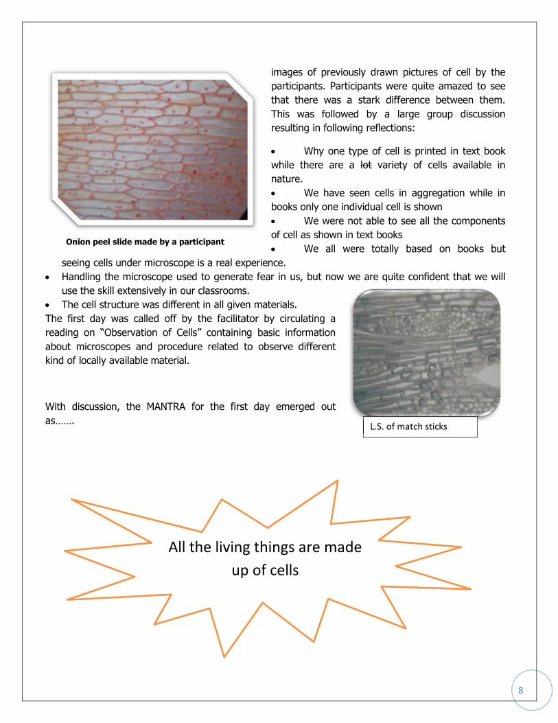
Onion peel slide made by a participant
L.S. of match sticks

9
Slide showing 3D structure of cell
Day - 2 (17 May 2013)
The day commenced at 9:30 morning with summation of the
previous day session. The facilitator briefed the day one by three
points.
1. Cells are not flattened; it is a box like structure which
contains different components or parts of cell. The
facilitator recalled the incident where a participant had
made a slide of tomato peel where the cells were looking
somewhat bulged on the edges giving a feeling of 3D
specifications of cell. Facilitator explained that we can try
and observe the 3D structure of cells by dimming the
light through adjustment and focusing on the edges of
cells.
2. Secondly the facilitator discussed the different cells observed by the participants in day one.
Power point presentation “Kaisi Dikhi Koshika” was shown to them. By showing the power point
presentation with pictures of some plant cells and animal cells facilitator established that all
organisms are made up of cells. The participants were quite amazed to see the photographs of
slides made by them, this give them a sense of satisfaction which was visible on their faces.
3. After showing variety of cells through PPT facilitator added that under the microscope most of
us were able to see a group of cells rather than a segregated cell as generally shown in
textbooks. Facilitator sketched a picture of stomata opening and asked the participant; how
many cells they are able to see here. The participants replied one, two and three and also said
the cell boundaries and cytoplasm and nucleus are the points under which the cell is to be
called as single cell. The facilitator summarized that in some cell nucleus and cytoplasm are not
clear or absent then in such cases cell boundary is the basis to identify a single cell.
Session 1
Cell Size and shape:
Session Objectives:
1. Understand that some organisms are
unicellular while others are multicellular.
2. Appreciate that cells are of varied size and
shapes.
3. Appreciate that the cell is the structural unit of
life.

10
Before the activities participants Reflections
It is time taking methods, can’t be done
easily in the school.
It’s possible in bigger cell like onion but
not with the cell of stems, roots and
leaves.
This method is not applicable for Class 6,
7 & 8.This can be used in the higher
classes.
The objective of the session was to measure the size of a single cell. Facilitator initiated the session by
saying that all of us have observed different types of cell in the previous day session, if I ask you how
you can say that single cell of aloe vera was bigger, equal or greater than the size of a single tomato
cell. Some participants said that we can easily make out through comparison that one cell is bigger and
other is smaller in size. Facilitator further probed that if we have very less difference in size of two cells
so that they appear same in size then what we can do to ascertain the size. Then some of the
participants said that somehow we have to measure them.
After this facilitator talked about the size ranges of certain cells or bacterial species given in the book
with a measurement scale along with their photograph.
What might be the method of measuring the sizes of cell and giving them an average range? What is
the basis of this measurement? Do we have any technique to measure one cell? Can the measurement
of the cell be done in the classroom?
Facilitator further defines two methods of measurement of cell that can be done in the classrooms.
Graph paper method introduced to the
participants, which is cost effective and
provides approximate size of the cell. The
second method which is costly but has more
accuracy than the graph method called
Micrometry was also familiarized by the
facilitator to the group.
Thereafter facilitator put forward a question
‘What is the basic requirement for
measurement? Participants replied scale and
Standard unit. In continuation graph slide
was shown to the participants and
preparation of graph slide and measurement of each big and small box were told to them. The method
to observe and measure a single cell by graph was discussed with the participants.
After this procedure and calculation part of micrometry was discussed with the participants through a
power point. The facilitator discussed calibration (the stage micrometre, oculomicrometer, its
adjustment with microscope, calculating the least count) and steps to measure a cell.
Facilitator added that graph paper method as discussed is quite simple and can be easily executed in
classroom with children but micrometry is for the sake of our knowledge. The facilitator told the
participants that micrometry is a way to find out the size of a particular cell more finitely, that’s why
some calculation is involved and it’s for the sake of our knowledge.
The facilitator formed the 6 groups of the participants for the activity. They have been given proper
instructions to measure the size of the cell of different specimen like onion, tomato with graph method
11
firstly and two sets of microscope were arranged for oculomicrometery. The participants were asked to
do individually. They involved very enthusiastically and those who did explaining the others.
Facilitator summated the session by explaining how knowing cell size have opened great avenues in
research and science. After this a comparative flash animation were shown, where variety of cells with
their finite size starting from a crystal of salt to deadly viruses to carbon atoms was beautifully
compared. It was also discussed that how to calculate actual size of cell by observing magnified image
of the object through microscope. In this way facilitator directed the participants to look inside the cell.
Session 2
Inside the cell
Session Objectives:
1. Observe different cell organelles
2. Cell organelles are different from cell inclusions.
3. Understand that all the cells not necessarily own all the
cellular organelles as depicted in the ‘typical cell in the
text-books
4. Understand that same cell organelle also have different
structures in different organism/ species. For example-
Shape of chloroplast in spirogyra and Ulothrix
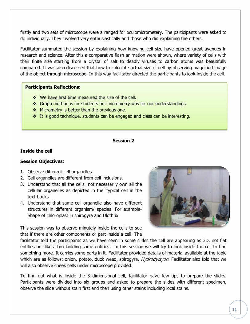
This session was to observe minutely inside the cells to see
that if there are other components or part inside a cell. The
facilitator told the participants as we have seen in some slides the cell are appearing as 3D, not flat
entities but like a box holding some entities. In this session we will try to look inside the cell to find
something more. It carries some parts in it. Facilitator provided details of material available at the table
which are as follows: onion, potato, duck weed, spirogyra, Hydrodyctyon. Facilitator also told that we
will also observe cheek cells under microscope provided.
To find out what is inside the 3 dimensional cell, facilitator gave few tips to prepare the slides.
Participants were divided into six groups and asked to prepare the slides with different specimen,
observe the slide without stain first and then using other stains including local stains.
Participants Reflections:
We have first time measured the size of the cell.
Graph method is for students but micrometry was for our understandings.
Micrometry is better than the previous one.
It is good technique, students can be engaged and class can be interesting.

12
There was lot of excitement in the subgroups.
Participant were able to see spirally tangled wires of
series made by chloroplast in spirogyra, movement of
round chloroplasts in duckweed, some kind of needles
in the sap of Tradescantia , net like networking in
Hydrodyctyon , cell membrane & cytoplasm in cheek
cells and starch granules in potato.
After the observation participants returned in larger
group and draw the observed structures. Every
participant explained what he/she has observed in detail.
With discussion, the MANTRA of the second day emerged out as……..
Participants Reflections
Participants were found observing the nucleus,
chloroplast, cytoplasm, cell membrane, cell wall
in the provided specimen.
Participants asked how we can teach cell in the class.
They noticed that they had seen chloroplast only in the text
Book so far not in reality like today.
The position of cell parts varies from cell to cell was notice
by the participants.
After observing the stained and without stained slides they noticed that stained slide is clear
and the parts were easy to identify.
Is cell made up of living and non - living substances?
Cell is made up of many parts
13
Day - 3 (18 May 2013)
Session - 1
Day-3 began with discussing the work done on previous day during the session Inside the Cell.
Facilitator discussed the observations done by the participants. Since some photographs of the slides
prepared by the participants were captured during this session, these photographs were also projected.
Facilitator put forth the question; were the observed organelles structure alike in different plants?
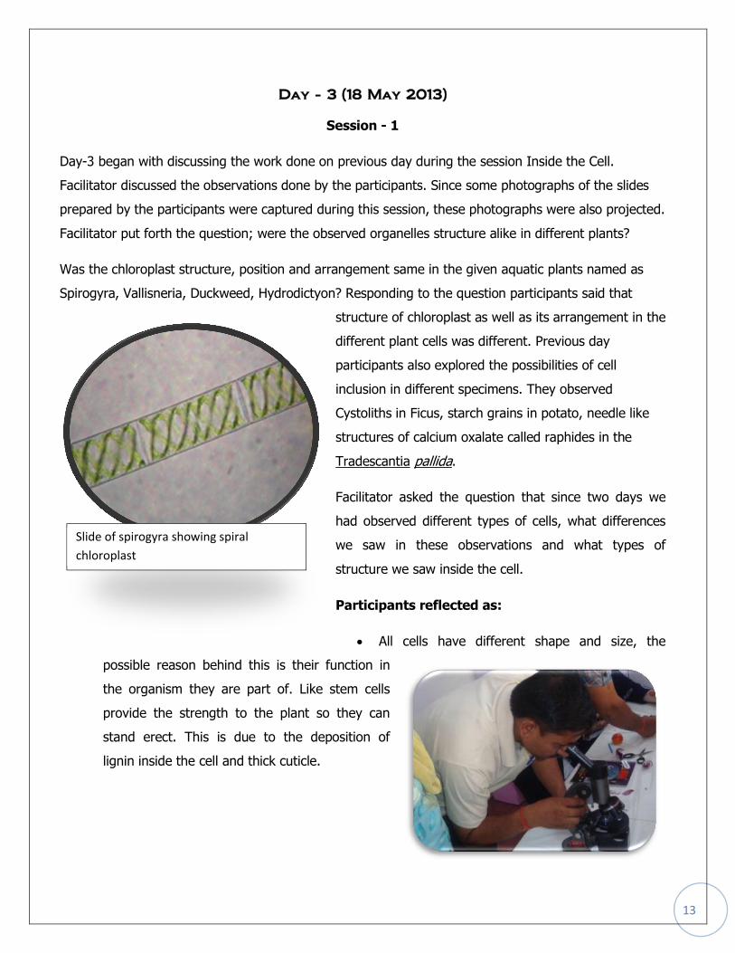
Was the chloroplast structure, position and arrangement same in the given aquatic plants named as
Spirogyra, Vallisneria, Duckweed, Hydrodictyon? Responding to the question participants said that
structure of chloroplast as well as its arrangement in the
different plant cells was different. Previous day
participants also explored the possibilities of cell
inclusion in different specimens. They observed
Cystoliths in Ficus, starch grains in potato, needle like
structures of calcium oxalate called raphides in the
Tradescantia pallida.
Facilitator asked the question that since two days we
had observed different types of cells, what differences
we saw in these observations and what types of
structure we saw inside the cell.
Participants reflected as:
All cells have different shape and size, the
possible reason behind this is their function in
the organism they are part of. Like stem cells
provide the strength to the plant so they can
stand erect. This is due to the deposition of
lignin inside the cell and thick cuticle.
Slide of spirogyra showing spiral
chloroplast

14
Position of the cell within the organism also
affects its shape and size.
Thereafter facilitator put forth the question that does all
cells have chloroplast. To this question participants’
response was obviously no and they said that it
depends on the function of the particular cell. Each
plant part has its own characters and functions,
therefore, chloroplast founds where food is prepared.
Facilitator requested to the participants to ponder upon
the reasons behind the variations of chloroplast in
different plant cells like in onion bulb chloroplast is not present whereas in duckweed they are plenty.
Their position also varies within the cell like Vallisneria contains chloroplast around margins whereas in
spirogyra it is arranged in spiral form and in Zygnema it is star shaped.
Participants’ reflections:
The shape and size of cell organelles is related with their functions; just similar to the variation
of cell shape and size.
Due to the limitations of light microscope we are not able to tell about the shape of organelles.
One participant had opinion that chloroplast is in liquid state.
Since all cell organelles are present in the liquid cytoplasm. So they can move within the
cytoplasm.
Cells also produces some byproducts or stored materials like starch grains, needles and stones.
They may be helpful for plants.
One participant said that chloroplast doesn’t
found in Amar bail (Cuscuta) since it gets food
from another plant.
Fungi also does not possess chloroplast
Root and some underground stems too doesn’t
have chloroplast.
Facilitator concluded the session while providing
different examples that all cell organelles are not
Cystolith in Ficus leaf

15
found in all cells as depicted in a typical cell. Structure and number of the cell organelles may also
differ in different cells. The difference between the cell organelle and cell inclusion was also talked in
brief.
Session – 2
Peep in to the cell
Session Objectives:
1. Understand that the cell is the functional unit of life. (Function at two level-for its own survival and
for the organism).
2. Appreciate that cell is a dynamic entity. (in terms of
cell organelle movement)
3. Understand the importance of Typical’ cell as shown
in the Biology text-book
The session was initiated with explaining that the cell
contains many organelles but what they do inside the
cell? This was concluded by discussion that each and
every organelle has specific function to perform and
they contribute in the complete functioning of cell. Organelles work in coordination within the cell and
for the organism all the body cells work in coordination. For siting the examples two PPT was shown.
In first PPT coordination of cell organelles like nucleus, endoplasmic reticulum, ribosome and Golgi
body was explained while in other PPT explains coordinated work of chloroplast , mitochondria and
peroxisomes. With this discussion the facilitator moved on asking that how the functions of organelles
were found out by the scientists. After few reflections one reading on discovery of the functions of cell
nucleus titles “Kaun Taya karata hai topi ka roop” by
Sushil Joshi was shared, participants were requested to
read the paper and share their thoughts on the same.
Afterwards a question was raised among participants
“Does all Cell organelles have their specific positions
within cell? Or they can change their position. After
participants reflection few short video as amoeba

16
engulfing another organism and digest them, binary
fission in paramecium, paramecium doing
osmoregulation and cyclosis in paramecium to show
the dynamicity of cell and cell organelles, was
showed to the participants and facilitator explained
that the organelles in the cell are not static they are
flexible to move.
Facilitator also discussed about what alternate
concepts could generate in students mind due to
giving analogy to onion peal cells with wall made by brick.
Session – 3
Formulation of cell theory
Session Objectives:
1. Demonstrate/ understand the chronological events related to the development of cell theory
2. Appreciate the de-mystifying the theory of spontaneous generation
3. Understand the scientific basis for toppling a theory
Facilitator began with recapitulating the objectives of previous sessions and the process adopted for
them. Thereafter, he asked participants to divide themselves in to six groups. Each group was provided
with some pictures and same number of captions. Facilitator asked participants to match the caption
with its respective picture and then try to make a story while arranging these pictures in order to the
event took place. For completion of this activity 30 minutes were provided to the participants.
Participants took around 40 minutes to match the captions
with related pictures and then making a story. All the
groups could have managed to arrange the given pictures in
their right sequence (chronological order). This group
activity was followed by discussion on nature of science.
Wherein the facilitator discussed about the steps followed in
investigating any problem related to nature as making

17
hypothesis, performing experiment, doing observations,
taking readings/notes, classifying the
observations/readings and reaching to the conclusion.
He made it clear that these steps are not rigid to follow
as mentioned here. He also said that the nature of
science is tentative, he clarified that no
conclusion/theory is last in science. Science is based on
evidences, if one wants to disprove any theory s/he has
to produce evidences against that. after this discussion,
we switched over to next session on cell division.
Session – 4
Cell Division
Session Objectives:
1. Understand how new cells are formed
2. Observation and Identification of mitotic stages
3. Demonstrate mitotic stages through onion root tip experiment
4. Understand the significance of mitosis and meiosis in relation to growth, repair and reproduction.
The discussion on this session began with
linking up the video shown in the session;
peep in to cell. Facilitator said that you all saw
what happens in the paramecium (organelles
movement and cell division) in the video.
Followed to this facilitator asked the question
why cells divide or what is the importance of
cell division? Responding to the questions
participants said that:
Mitotic stages shown in onion root tip slide
made by participants

18
Facilitators’ observations: They showed their enthusiasm while preparing the slides, as they
followed the instructions precisely the slides were very clear. In most of the slides some stages were
visible which were captured in the camera to show the participants.
For organism growth (mitosis takes place)
For continuity of the species cell division is essential
Thereafter facilitator asked that is it possible for us to see the cell division/stages of cell division, is it
possible to show the cell division in the classroom with the available resources? He moved on sharing
that we can see the cell division in onion root tips (newly growing). Facilitator asked participants to
divide in eight groups and gave instructions for preparing the slides which were as followed:
After this discussion participants divided in the groups and began work to prepare the slide
.
General Instructions for preparing the
slide
First we should clean slide and cover
slip
Put the material on slide.
Put the cover slip on slide with care to
avoid the bubbles
Use the side clips to fix the slide on the
stage of microscope
If anyone prepare the good/clearly
visible slide to make it permanent fix it
with the help of nail paint
Now see the slide with microscope. In
case of blurred vision in microscope
clean the objective lens. This blurred
vision would have occurred because of
two reasons
o The lens might have touched
with uncovered slide
o While adjusting the microscope
our own finger might have
touched with the lenses
Clean the objective lens with the help
of xylene and fresh tissue paper/cloth
Instructions for preparing the slide to see
the stages of cell division
Cut 1 mm root tip and keep it aside
Now cut 2 mm root from the same root
Put this 2 mm section in 1N HCl for one
to one and a half minute
Take out the section from HCl and put it
in acetocarmine solution for 15-20
minutes
Heat the section with the solution for
one minute, while heating move it
continuously for preventing the root
cells to damage
Put the section on slide and take some
drops of 45% glacial acetic acid
Put the cover slip on slide and tap on it
slowly with rubber headed of pencil.
When the root get spread see the slide
under the microscope
See the stages on the posters pasted on
the table and match which stage is
visible in the prepared slide

19
Day - 4 (19 May 2013)
The day was started by the recapitulation of work done in
previous days. It was shared that in previous days we had
tried to develop an understanding of cell organelles and other
cell inclusions found in cell sap and we had also tried to
observe all stages of mitosis. In addition to that we had also
observed the things other than cell organelles which are
called as cell inclusions.
Session - 1
Cell division Continued……
First we applauded the participants for preparing good slides. Since the instructions were followed
thoroughly so prepared slides were very good and one or more stages of mitosis were clearly visible in
most of the slides. Thereafter, we have asked them individually to share about the stages seen by
her/him in her/his slide. Most of the participants shared that
1. Stage one prophase and stage two metaphase have been seen in their slides.
2. One group shared that they saw stage three that is anaphase.
3. One group was even able to see the fourth, telophase stage.
The photograph of mitotic stages captured in previous day was also projected to show the participants.
After that we had discussed with the participants each stage of mitosis as well as stage wise changes
occurs during the process. This explanation discussion was followed by the videos on cell division. One
video was animated and another was live video on cell division (mitosis only). We reinforced the
concept that cell divide occurs in a continuous fashion, for our ease we have break this continuous
process in four different and distinct stages.
After showing that video we had simply put forth a question, “why one cell divides into two”?
Responding to this, participants said that for growth cell division is essential. Subsequently we have
asked them the second question, in plants what are the regions where continuous growth takes place.
One of the participant’s responded that it occurs in stem and root. One participant added that
organisms do also need repair and maintenance for which they need new cells, hence continues cell
division.

20
There was an interesting query that does cell divides after death also? Participants had variety of
opinion, somewhere believing that cell continue divide for some time even after organism die. The
reason given behind this that the height and length of hair got increased after death of human being.
This created a platform for facilitator to inculcate the nature of science, and how we could have used
scientific logic to resolve the problems.
Session – 2
Cell Death and its Significance
Session Objectives:
1. To understand how apoptosis is essential for continuing life in multicellular organisms.
2. To understand the basic underlying process and necessary conditions for apoptosis.
3. To differentiate between Necrosis and Apoptosis.
The session begun with the question that as we have
discussed that cell divides and organism grows, does
cell die too? Most of the participants said that yes cell
dies. To bring the session at its next level two questions
were floated
1. Can we quote some examples of cell death?
2. What is the link between the cell death and
organism death?
Responding to this the participants’ said that:
Leaving the layer of skin (Kenchul) by snake and leaving the tail by lizard is an example of cell
death
Dead cell can be seen in nails and hoofs
RBCs are regularly growing and dying
Dead cells come out from the body as dirt during bath
Next question was posed by facilitator, why cells die?’ there was very straight answer that everything
has definite life and after that period cell almost dies. During discussion there was a question what
would happen when one goes into comma, in this case cell division takes place or not?.

21
To conclude the discussion an article from Sandarbh “ koshika ki mritu jaruree hai…..jeevan ke leye “
was distributed to all participants which spokes about the cell death and its significance.
Participant’s reflection was as below-
The reading explains the need of cell death which is a programmed phenomenon and is useful
for the organism.
The reading creates new domain of understanding of cell death
The knowledge about inhibition of carcinogenic cell which leads to cancer due to uncontrolled
growth was gained.
Cell death is also important for those cells which are get infected by pathogens like virus etc.
This reading further added a guided discussion on the disappearance tadpole tail during
metamorphosis process. All the participants agreed that adult frog never came out from egg but a
tailed Tadpole developed first. They were also thinking that the tadpole actually dropped its tail same
as lizards do. To clarify we explained some of the stages of the limbs growth and tail decomposition in
tadpole and displayed a video on apoptosis in tadpole. Video explained how the tail of tadpole
disappears when it develops in a frog. During discussion (after looking the video of metamorphosis of
frog), some of the participants also shared that vanishing of tail in tadpole leads to the development of
limbs. Absorption of tail during metamorphosis was also discussed in detail with some of the example
that what is the exact meaning of absorption? This came in discussion that during this process, as it is
controlled death of the cells by the organism itself; the important things of dying cells were consumed
by the organism.
Session - 3
Origin of Cell
This session was basically designed to give some
glimpse of contrasting theories came during time
span to explain the origin and evolution of life in the
form of cell. How the debates on these theories has
crossed the barriers of one belief and proved the
present theory.

22
The session initiated with reflecting on last three days
proceedings. Facilitator said that we have seen number of
cells, tried to look what is inside the cell, built an
understanding on functions of the different cell organelles
found inside the cell and just we talked about cell death.
But can we think about “kahan se aai hogi pahli Cell?”
(How the first cell originated?) After getting the
participants’ responses, facilitator requested to all the participants to read a write-up regarding origin
of cell.
This session was quite important as this is a rigid belief in Indian sub-continent that the life is
originated through some eternal power. In fact the teachers were very possessive about this belief. We
have discussed about the experiment done by the Urey and Miller, which has established the link of
origin of life on earth. In this session we tried to understand that though we have few theories
regarding origin of life but still this is a matter of extreme curiosity and scientist community is regularly
trying to know something more in this.
Now, we have shifted to some more discussion about why we study cell, which we have paused at first
day. Then we came back on the question of a participant “Is ostrich egg is single cell or it is a group of
cells” It was concluded that any unfertilized egg remains single celled until cleavage starts in it which
leads to convert the egg into multi cellular structure.
Session - 4
Feedback and Future planning
After this session we were going to end up the session. With closing discussion, we have taken some
oral feedback of the workshop and some reflections in written also. They have shared that the thing
which they have missed in college life, find here and
thus make them curious too. Some of the teachers
were also shared that a school level workshop should
be designed for science kit provided by the govt. One
thing was very much interesting that is, in written, we
have also designed a question for participation in our

23
next module, everyone has given his/her nomination for the upcoming workshops.
FACILITATORS RETROSPECT:
1. Questions for the participants should be reframed and to be presented in such a way that force
the participants to voice out themselves.
2. For preparation of the workshop three days should be kept.
3. Due to lack of time one session was dropped. The group need to once again reframe the
session/ topic ‘Cell organelles’ for transaction which is something longer.
4. The sessions on cell shape and size and observation of cell can be merged.
5. PDM need to be re-designed.
6. Graph activity should be made easier and interesting.
7. Micrometry session should be done more practically than theory and we should avoid technical
term like least count.
8. There shouldn’t be repetition of materials/specimen in the sessions.
9. Summation points need to be strengthened.
10. We should expand the time for discussion in each session.
11. Story board activity went good. But we have to rethink about its session placing. If it will come
after cell division, as all of the postulates of cell theory have been discussed here, therefore it
will be more logical to keep this session after cell division.