the brain by mechanical means were of three types: concussion
TRANSCRIPT

TRAUMA TO THE NERVOUS SYSTEMHunterian Lecture delivered at the Royal College of Surgeons of England
on29th July 1965
byA. K. Ommaya, F.R.C.S., F.A.C.S.
Associate Neurosurgeon, National Institute of Neurological Diseasesand Blindness, Bethesda, U.S.A.
MORE THAN TWO HUNDRED years ago, John Hunter taught that injuries tothe brain by mechanical means were of three types: concussion, compres-sion, and brain wounds with loss of substance. He emphasized that" these three kinds of injury will produce symptoms similar to each other ".With typical Hunterian acumen, he emphasized the importance of con-cussion. " When there is depression of bone or extravasation, thesymptoms of concussion are lost, though it may be at the bottom of all"(Hunter, 1841).
This statement brings out very clearly the important fact that the basicproblem of head injuries is concussion. Hunter also realized what oftenrequires reiteration, that concussion is not invariably a benign and transientform of injury to the nervous system, but rather that it is the local responseof such tissues to trauma. It is disquieting to realize that our under-standing of this response, its mechanics and its management have pro-gressed very slowly in all these years.
Considerable advances have been made in recognizing and treating thesurgically remediable complications of head injury such as depressedfractures, intracranial haematomas of various types, and infections.However, we have not achieved equally significant progress in under-standing cerebral concussion. The management of patients with closedhead injuries remains empirical and the resultant mortality, though lessthan 20 years ago, is high. More importantly, the quality of survivalafter severe head injuries could be improved. Two important limitingfactors in advancing these aims are as follows: first, the lack of a suitableexperimental model upon which correlations between the physical andbiological phenomena of the trauma can be made in a rigorous manner.Secondly, the lack of adequate series of unselected head injury patientsin whom observations have been made in a uniform manner so as to makecomparison of cases and evaluation of management statistically meaning-ful. The reason for this has been the relative absence ofplanned prospec-tive studies of head injuries as opposed to retrospective analyses of caserecords (McIver et al., 1958; Lindgren, 1960; Lewin, 1953; Gurdjian andWebster, 1958).
In this lecture I shall try to trace the results of 24 years' work on methodsaimed at resolving these limitations to our understanding of the effects oftrauma to the nervous system.
317
A. K. OMMAYA
The experimental studyThis was based on the rational approach to the mechanics of brain
trauma outlined by Holbourn (1943), " that damage to the brain is a con-sequence, direct or indirect, of the movements, forces and deformationsat each point in the brain. The movements, forces and deformations arenot independent; so it is sufficient to express everything in terms ofdeformations. These are woiked out with strict adherence to Newton'slaws of motion, but with approximations to the constitution of shapeof the skull and brain. Hence further advances can only come frommaking better approximations."
It should be realized such deformations cannot be directly measuredand thus we have to use the measurement of such variables as velocity andforce of impact, acceleration, and intracranial pressure, and deduce themechanical behaviour of the brain assuming certain properties for itsstructure. To cite " acceleration-deceleration " or " compression "
per se as the mechanism of brain concussion is scarcely more meaningfulthan to cite the rise in temperature in the object striking the head. Theseare but measurable indices monotonically related to the severity of trauma.The three pseudo-alternative " mechanisms " are scarcely better, i.e.instantaneous intracranial pressure changes; excitation of a train ofpressure waves in the skull and cavitation. Fundamentally, these aremerely three idealizations of the same phenomena. It is difficult todistinguish rationally among the assumptions and approximations under-lying these three " mechanisms " (Shea, 1964).
MethodsEighty monkeys were used in this study. These were naive mature
animals, unselected for sex and ranging in body weight between 3 and 6kilograms. Each animal was examined and tested by a veterinarian anddeclared healthy and free from diseases, including tuberculosis, prior toexperimental use. All experiments were conducted under generalanaesthesia produced by intravenous Pentobarbital Sodium, 15 mgm./kilobody weight, not exceeding a total dose of 150 mgm. After surgicalpreparation, the animal was maintained at a lesser degree of anaesthesia,wherein voluntary movements when undisturbed were minimal, super-ficial reflexes and aversive responses to noxious stimuli were present. Thislevel was sustained when necessary by repeated intravenous injections of"Brevital " (methohexital sodium), usually not exceeding 5 mgm.
Details of the surgical preparation, instrumentation and completeapparatus specifications cannot suitably be given in this lecture. Thesewill be published separately. The following is a brief summary of thephysiological and physical measurements made. A Gilson polygraphrecorded arterial blood pressure from the femoral artery and cerebrospinalfluid pressure from the lumbo-thoracic C.S.F. space via unbonded straingauge transducers. Electrocardiographic recordings were taken from
318
TRAUMA TO THE NERVOUS SYSTEM
standard leads on this polygraph as well as on a Grass 8-channel E.E.G.machine on which one channel of respiration (via a pneumotachograph)and six electro-encephalographic channels could also be recorded. ThisE.E.G. was obtained from four to six extradural electrodes screwed intothe skull.
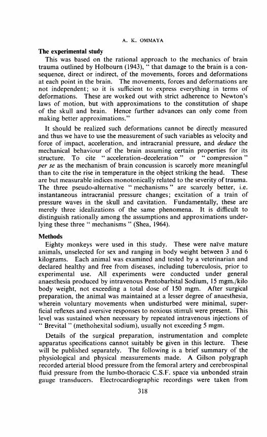
Linear acceleration of the head was obtained from a miniature (1 gram)piezo-electric accelerometer mounted by screws directly to the skull.Instantaneous changes in intracranial pressure were measured by semi-conductor strain gauges mounted in 5 mm. trephine holes in the skulland held in place in a watertight fashion by steel screws and dental cement.Experimental head injury of varying degree was produced by an air-powered gun firing a captive piston having a mass = 0.086 slug and bearingat the striking surface a rubber tip = 1 square inch. The velocity of thepiston was directly measured from a voltage generated by a magnet withinthe piston passing through a coil. A bonded strain gauge was incorpor-ated in the striking tip of the piston and this enabled the direct measure-ment of the force of impact with the head. The output from the velocity,acceleration, force and pressure transducers was fed through suitableamplifiers into a tape recorder having a frequency response up to 10 kilo-cycles. The tape record of this data could then be played back at anychosen speed through an ultraviolet recording oscillograph in order toobtain a faithful visual record of the impact phenomena. Figure 1illustrates the general arrangement of the apparatus. The contouredprimate chair was specially designed to allow complete mobility in position-ing the animal for impact. In the foreground and above the chair can beseen two high-speed motion cameras which allowed motion pictures atspeeds around 4,000 frames per second. Motion analysis of such filmsagainst the reference grid seen in the background allowed calculation ofthe displacement of the head in two axes (x and y) and of the consequenttangential velocity attained by the head after each blow.
Other experiments included radiographic studies using carotidangiography by a technique which has been published (Ommaya et al.,1964; Rockoff and Ommaya, 1964) and a modification of the Shelden-Pudenz-Restarski-Craig technique of a lucite calvarium for direct observa-tion of brain movement during impact (Shelden et al., 1944). Experi-mental cerebral concussion was defined as the loss of voluntary movement,and aversive responses to ear pinch when these had been present con-sistently immediately before impact. The duration of concussion wasdefined as being equivalent to the duration of the loss of such responses.This criterion was found to be much more reliable than the corneal reflex.In addition to the measured indices of piston and head velocity (feet persecond), linear head acceleration (g = 32 feet per second2), impact force(lb.) and intracranial pressure changes (p.s.i. = lb. per square inch-1 p.s.i. = 51.7 mm. Hg) calculated values for kinetic energy (I mV2) in
319
A. K. OMMAYA
foot-lb. and for impulse (f't2fdt) in lb.-sec. were all correlated with the
production of concussion as defined above. Each animal was struck avarying number of blows in different positions and under varying con-ditions as outlined later. However, only the first blows for each animalstruck either in the midoccipital regions, or midfrontal regions, and whichdid not producefracture, were considered for purposes of statistical analysis
Fig. 1. Apparatus for experimental head injury in the primate. Note thecompressed air gun on the right, the polygraph in the right foreground, themonkey seated in the adjustable chair, and the two high-speed cameras.
of the relation of concussion to each of the impact energy measurementsdescribed. Immediately after non-lethal experimental head injury, eachmonkey was allowed to regain consciousness and observed in isolation andin company with its peers for neurological and behavioural abnormalitiesfor varying intervals of time. Any evidence of infection or discomfortwas suitably treated.At the end of each experiment the animals were sacrificed at varying
intervals provided the head injury produced was not immediately fatal.All animals were sacrificed by perfusion of the major arteries with isotonicsaline and 10 per cent formalin in saline under general anaesthesia pro-
320
TRAUMA TO THE NERVOUS SYSTEM
duced by Pentobarbital Sodium. Post-mortem observations were madeof scalp injuries and fractures, haematomas or evidence of haemorrhage,brain swelling, contusion and laceration. To this macroscopic study wereadded observations of blood-brain barrier breakdown as indicated byultraviolet fluorescence of sodium fluorescine given intravenously andhistological studies of routinely stained sections taken at various sites ofthe brain and upper cervical cord.
RESULTSIt did not appear that the actual site of impact on the head was a crucial
factor in the production of concussion for occipital and frontal blows.The important factor was the efficiency of impact, i.e. whether or not thepiston impacted squarely or delivered a glancing blow. The site of impactwas crucial, however, for the production of skull fracture. Thus blowsto the vertex produced fractures most easily, while blows to the occipitalregion (inion) were least likely to do so. The frontal and temporal regionswere intermediate between the other regions in this regard. For purposesof this study, occipital blows were studied primarily.A maximum of five successive blows were given to some of the animals.
An interesting feature was the difficulty in producing a state of severeclosed head injury with prolonged unconsciousness lasting more than afew hours in these experiments. Thus the blow (or blows) resulted in oneof three things. First, no effect at all (sub-concussive blow); secondly,a concussive blow from which the animal recovered completely, usuallyin less than one hour. Finally, a severe concussion with or without skullfracture could occur. These animals usually died within 2-3 hours afterthe blow. In only 4 out of 80 monkeys (5 per cent) was there a severeconcussion with survival up to 24 hours. It proved impossible to produceskull fracture without concussion under our experimental conditions.On the other hand, fatal concussion without skull fracture could be ob-tained, when death usually occurred after a short interval ranging from afew minutes to a few hours.The usual sequence of events after a concussive blow in monkeys con-
sisted of immediate generalized flaccid paralysis, areflexia and loss ofresponse to noxious stimuli. In a few animals, the first effect of the blowafter a brief stilling was a tonic phase of opisthotonic posturing, occasion-ally followed or preceded by a few clonic jerks of facial muscles or ex-tremities. This type of response was followed by the flaccid response asdescribed above. In the recovery phase it was quite usual to see responsesto noxious stimuli return before the corneal reflex was definitely elicitable.In most cases both responsiveness and reflex actions returned together.However, the corneal reflex would often temporarily be unobtainableafter initial return without concurrent changes in responsiveness to noxiousstimuli, or vital signs. On the other hand, if responsiveness were abolishedafter initial return, reflex responses were also abolished and the animal
321
A. K. OMMAYA
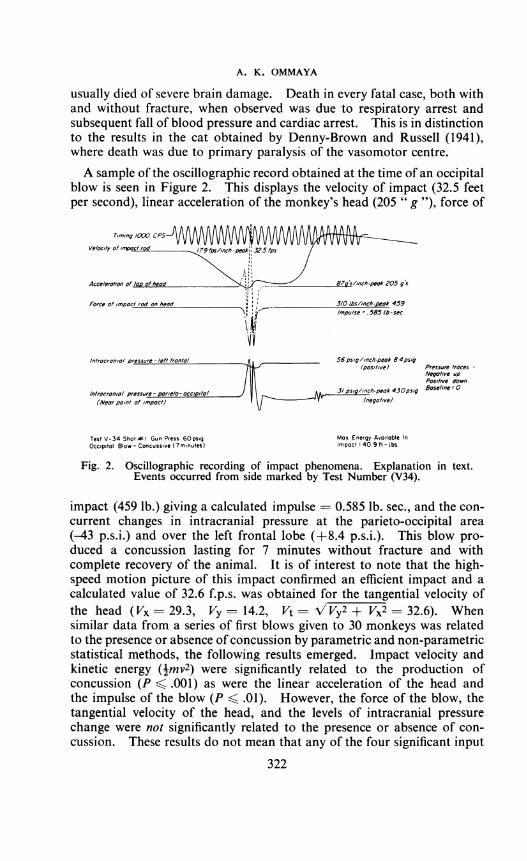
usually died of severe brain damage. Death in every fatal case, both withand without fracture, when observed was due to respiratory arrest andsubsequent fall of blood pressure and cardiac arrest. This is in distinctionto the results in the cat obtained by Denny-Brown and Russell (1941),where death was due to primary paralysis of the vasomotor centre.A sample of the oscillographic record obtained at the time of an occipital
blow is seen in Figure 2. This displays the velocity of impact (32.5 feetper second), linear acceleration of the monkey's head (205 " g "), force of
Timing /000 CPS
Velocity of impac ro 9fps ,nch.peak| Slps
Acceleration of top of head \ i87snch-peak 205 g's
Force of Impact rod on heod 3/0 lbsllnch-peak 459Impulse .585 lb -sec
Introcronial pressure -leftfrontol 56psiglInch-peak 8 4ps/g(positive) Pressure troces
Negative upPositive down
In/racranial pressure rieta acci i/al 3~~~/psiglinch-peak 430posig Baseline 0Introcronlol pressure - parleto-occip/l/ J|h pck4apl os/c(Neorpoanl at impoct) (negotive)
Test V-34 Shot I Gun Press 60psig Max Energy Available InOccipital Blow -Concussive (7minutes) Impact - 409 fit- Lbs
Fig. 2. Oscillographic recording of impact phenomena. Explanation in text.Events occurred from side marked by Test Number (V34).
impact (459 lb.) giving a calculated impulse - 0.585 lb. sec., and the con-current changes in intracranial pressure at the parieto-occipital area(-43 p.s.i.) and over the left frontal lobe (+8.4 p.s.i.). This blow pro-duced a concussion lasting for 7 minutes without fracture and withcomplete recovery of the animal. It is of interest to note that the high-speed motion picture of this impact confirmed an efficient impact and acalculated value of 32.6 f.p.s. was obtained for the tangential velocity ofthe head (Vx = 29.3, Vy = 14.2, Vt -/Vy2 + Vx2 = 32.6). Whensimilar data from a series of first blows given to 30 monkeys was relatedto the presence or absence of concussion by parametric and non-parametricstatistical methods, the following results emerged. Impact velocity andkinetic energy (Imv2) were significantly related to the production ofconcussion (P < .001) as were the linear acceleration of the head andthe impulse of the blow (P < .01). However, the force of the blow, thetangential velocity of the head, and the levels of intracranial pressurechange were not significantly related to the presence or absence of con-cussion. These results do not mean that any of the four significant input
322

TRAUMA TO THE NERVOUS SYSTEM
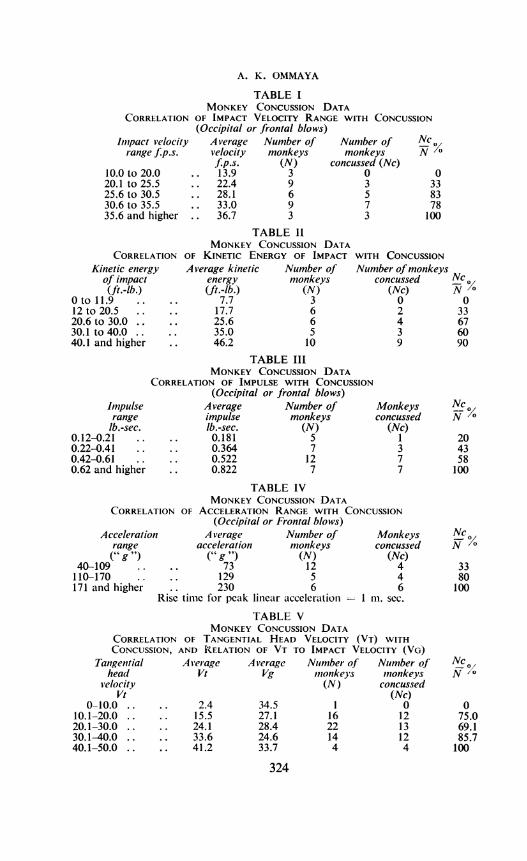
measurements can be indicated as the mechanism of concussion, nor arethey equally useful in their only legitimate use, i.e. as a precise index of theseverity of a blow in producing concussion and other effects of experi-mental head injury. From a consideration of Newton's Laws of Motion,the best index would appear to be the impulse. Thus of the four significantindices (impact velocity, kinetic energy, impulse and acceleration) onlythe impulse can be usefully specified independent of the impacting massesand related to the energy input to the head. The impulse thereforeprovides one with an index which can be used in any experimental situa-tion and levels of this index can be studied precisely. This point is wellbrought out in Tables I to V. From this data it is also evident that itis erroneous to speak of concussion as an " all-or-none" type of phe-nomenon related to a specific level ofenergy input. The tables show clearlythe range of energy levels at which concussion occurs. Here, too, itwould appear that impulse is the best index as indicated in the relativelysmooth progression of increased percentage of monkeys concussed withincreasingly higher levels of impulse. (Impulse levels below 0.12 are notconcussive.) The non-significance of the tangential velocity of the headwould also vitiate against the true significance of the impact velocity underour experimental conditions.
It is also clear from these results that to relate experimental cerebralconcussion to " thresholds " of velocity such as the often-quoted valuesofimpact velocities of 28 f.p.s. (Denny-Brown and Russell, 1941), 29.4 f.p.s.(Gurdjian and Webster, 1943) and 30.0 f.p.s. (White et al., 1943) isessentially inadequate. There is only a rough correspondence for thisindex as can be seen in Table I. Thus in the range of impact velocities =20.1 to 25.5 f.p.s. (average = 22.4) three out of nine animals were con-cussed (33 per cent), in the range 25.6 to 30.5 f.p.s. (average = 28.1)five out of six animals were concussed (83 per cent), but in the range 30.6to 35.5 f.p.s. (average = 33.0) only seven out of nine (78 per cent) monkeyswere concussed. Concussion in 100 per cent animals was not obtaineduntil the impact velocity exceeded 35.6 f.p.s. However, even this figurecannot reliably be taken as a threshold for 100 per cent concussion in thatit is meaningful only when the efficiency of the impact and the masses ofpiston and head are specified. As stated before, the same reasoning leadsus to use the impulse of the blow as the best index for the energy of impact.The dissipation of this energy in the head was measured by three
indices: the velocity of the head (Vx, Vy and Vt), the acceleration of thehead and the changes in intracranial pressure. The tangential headvelocities for the first blow to each animal are summarized in Table V.There was no statistically significant relation between the changes inintracranial pressure and concussion, taking into account both amplitudeand duration of the peak pressure at a point almost opposite the site ofimpact. The importance of relating the velocity of impact (Vg) to theactual velocity of the head is brought out in Table V, where it can be seen
323
A. K. OMMAYA
TABLE IMONKEY CONCUSSION DATA
CORRELA [ION OF IMPACT VELOCITY RANGE WITH CONCUSSION(Occipital or frontal blows)
loipacirang4
10.0 to 2(20.1 to 2'25.6 to 3(30.6 to 3'35.6 and
CORRELKinetic ener,
of impact(ft.-lb.)
0 to 11.912 to 20.5 . .
20.6 to 30.0 ..30.1 to 40.0 . .
40.1 and higher
Impulserange1b.-sec.
0.12-0.210.22-0.410.42-0.610.62 and higher
CORREL,
Acceleratirange(" g ")
40-109110-170171 and higher
Ivelocitye fp.s.
0.05.50.55.5higher
A veragevelocity.f.p.s.13.922.428.133.036.7
Number ofmonkeys
(N)39693
Number ofmonkeys
concussed (Nc)03573
NcN0
0338378100
TABLE 11MONKEY CONCUSSION DATA
ATION OF KINETIC ENERGY OF IMPACT WITIgy Average kinetic Number of Nun
energy monkeys(ft.-lb.) (N)
7.7 317.7 625.6 635.0 546.2 10
TABLE IIIMONKEY CONCUSSION DATA
CORRELATION OF IMPULSE WITH CONCUSSION(Occipital or frontal blows)
Average Number ofimpulse monkeyslb.-sec. (N)0.181 50.364 70.522 120.822 7
A
2l
i CONCUSSIONnber ofmonkeysconcussed
(Nc)02439
Monkeysconcussed
(Nc)1377
TABLE IVMONKEY CONCUSSION DATA
TION OF ACCELERATION RANGE WITH CONCUSSION(Occipital or Frontal blows)
n A verage Number of Monkacceleration monkeys concus,("g") (N) (Ne'
73 12 4129 5 4230 6 6
Rise time for peak linear acceleration - 1m. sec.
eyssed
TABLE VMONKEY CONCUSSION DATA
CORRELATION OF TANGENTIAL HEAD VELOCITY (VT) WITHCONCUSSION, AND RtELATION OF VT TO IMPACT VELOCITY (VG)
Tangential Average Average Nuimber of Number ofhead Vt Vg monkeys monkeys
velocity (N) concussedVt (Nc)
0-10.0 .. .. 2.4 34.5 1 010.1-20.0 .. .. 15.5 27.1 16 1220.1-30.0 .. .. 24.1 28.4 22 1330.1-40.0 .. .. 33.6 24.6 14 1240.1-50.0 .. .. 41.2 33.7 4 4
324
Nc%N/033676090
NcN_0/
204358100
NecN0
3380100
NcN'0o
075.069.185.7100

TRAUMA TO THE NERVOUS SYSTEM
that the highest average impact velocity of the piston (34.5 f.p.s.) happenedto produce the lowest average head tangential velocity (2.4 f.p.s.) in oneanimal with no concussion. On the other hand an average impactvelocity of 33.7 f.p.s. was related to an average head velocity of 40.9 f.p.s.in four animals, all of whom were concussed. The velocity of the head inthe X and Y axes considered individually were not related to concussionin any better fashion, and statistically the actual velocity attained by thehead was not significantly related to concussion. On the other handthe acceleration of the head was statistically significant when relatedto the onset of concussion under our experimental conditions.
In summary, it would appear that knowledge of the impulse of theimpact and the acceleration of the head are two reliable and statisticallysignificant indices which can be used to relate the input and dissipationof the energy of the blow to the production of experimental concussion.
Physiological effectsA concussive blow has definite effects on the respiration, blood pressure,
C.S.F. pressure, electrocardiogram and electro-encephalogram. Theseeffects are briefly summarized as follows:1. RespirationAn apnoeic pause lasting for a few seconds is usually followed by a
change in respiratory pattern. This can be either irregularity of rate andamplitude or both. In fatal concussions or severe concussion associatedwith fracture, the apnoeic pause is usually continued and the animal diesin respiratory paralysis or the return of an irregular pattern precedes asecondary apnoea and death. Artificial respiration administered as thesole therapeutic effort in such cases for as long as three hours does notreverse the situation.2. Electro-encephlalograin
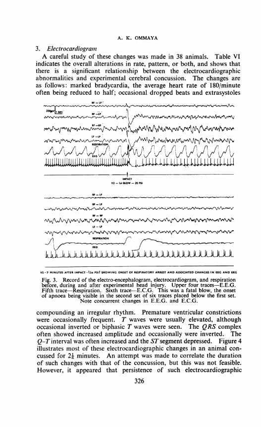
This was recorded in 14 animals in this series and in 18 animals in aprevious series (Ommaya et al., 1964). Concussion was produced in21 of these 32 animals. The electro-encephalographic changes werefairly consistent. Thus in concussed animals an immediate onset of high-amplitude slow activity could be seen, primarily in the parietal areasbilaterally. The high amplitude of these waves would soon decrease,often before the end of concussion, although the rate would remainpersistently slow. Flattening of the record as a primary event was notnoted in these experiments although this was invariably present when com-plications such as fracture or haemorrhage occurred. Occasionally a flatslow record would replace the high-amplitude activity as a later phenom-enon and usually after the end of concussion. In a few animals distinctspikes suggesting epileptiform activity were seen. However, these werenot related to visible convulsive activity.
Figure 3 displays the E.E.G. changes in a severely concussed monkey;concurrent changes in respiration are also seen.
325
A. K. OMMAYA
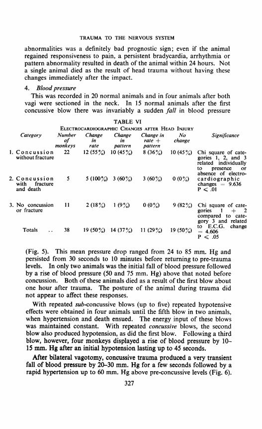
3. ElectrocardiogramtA careful study of these changes was made in 38 animals. Table VI
indicates the overall alterations in rate, pattern, or both, and shows thatthere is a significant relationship between the electrocardiographicabnormalities and experimental cerebral concussion. The changes areas follows: marked bradycardia, the average heart rate of 180/minuteoften being reduced to half; occasional dropped beats and extrasystoles
RF - LF
LF-LP
-4IAPACt
V2 - Ist SLOW -20 PSI
IF - LF
LF - LP
m ~~~~~~RESPIRATON
EKG
1AAkh hOS JJJJtJJJJ.ULJLV2 -7 MINUTES AFTER IMPACT -k2a PSI) SHOWING ONSET OF RESPIRATORY ARREST AND ASSOCIATED CHANGES IN EEG AND EKG
Fig. 3. Record of the electro-encephalogram, electrocardiogram, and respirationbefore, during and after experimental head injury. Upper four traces-E.E.G.Fifth trace-Respiration. Sixth trace-E.C.G. This was a fatal blow, the onsetof apnoea being visible in the second set of six traces placed below the first set.
Note concurrent changes in E.E.G. and E.C.G.
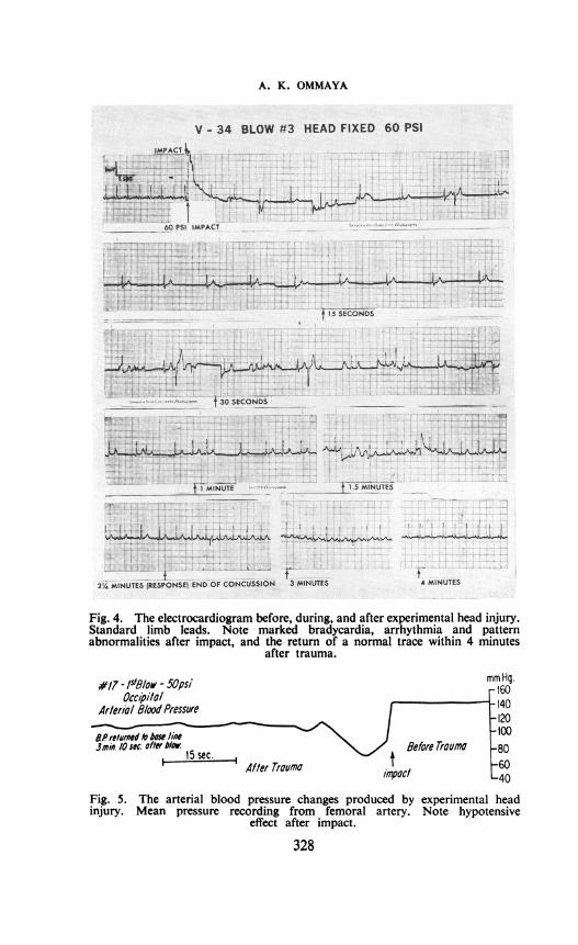
compounding an irregular rhythm. Premature ventricular constrictionswere occasionally frequent. T waves were usually elevated, althoughoccasional inverted or biphasic T waves were seen. The QRS complexoften showed increased amplitude and occasionally were inverted. TheQ-T interval was often increased and the ST segment depressed. Figure 4illustrates most of these electrocardiographic changes in an animal con-cussed for 21 minutes. An attempt was made to correlate the durationof such changes with that of the concussion, but this was not feasible.However, it appeared that persistence of such electrocardiographic
326
TRAUMA TO THE NERVOUS SYSTEM
abnormalities was a definitely bad prognostic sign; even if the animalregained responsiveness to pain, a persistent bradycardia, arrhythmia orpattern abnormality resulted in death of the animal within 24 hours. Nota single animal died as the result of head trauma without having thesechanges immediately after the impact.4. Blood pressure
This was recorded in 20 normal animals and in four animals after bothvagi were sectioned in the neck. In 15 normal animals after the firstconcussive blow there was invariably a sudden fall in blood pressure
TABLE VIELECTROCARDIOGRAPHIC CHANGES AFTER HEAD INJURY
Category Number Change Change Change in No Significanceof in in rate + change
monkeys rate pattern pattern1. Concussion 22 12 (55%) 10 (450%) 8 (36o%) 10 (45O%) Chi square of cate-
without fracture gories 1, 2, and 3related individuallyto presence orabsence of electro-
2. Concussion 5 5 (100%) 3 (60%) 3 (60%) 0 (0%) cardiographicwith fracture changes = 9.636and death P < .01
3. No concussion 11 2 (18%) 1 (9%) 0 (0%) 9 (82%) Chi square of cate-or fracture gories 1 + 2
compared to cate-gory 3 and related
Totals .. 38 19 (50%) 14 (37%) 11(29%) 19 (50%) to-EC-G. changeP < .05
(Fig. 5). This mean press-ure drop ranged from 24 to 85 mm. Hg andpersisted from 30 seconds to 10 minutes before returning to pre-traumalevels. In only two animals was the initial fall of blood pressure followedby a rise of blood pressure (50 and 75 mm. Hg) above that noted beforeconcussion. Both of these animals died as a result of the first blow aboutone hour after trauma. The posture of the animal during trauma didnot appear to affect these responses.With repeated sub-concussive blows (up to five) repeated hypotensive
effects were obtained in four animals until the fifth blow in two animals,when hypertension and death ensued. The energy input of these blowswas maintained constant. With repeated concussive blows, the secondblow also produced hypotension, as did the first blow. Following a thirdblow, however, four monkeys displayed a rise of blood pressure by 10-15 mm. Hg after an initial hypotension lasting up to 45 seconds.
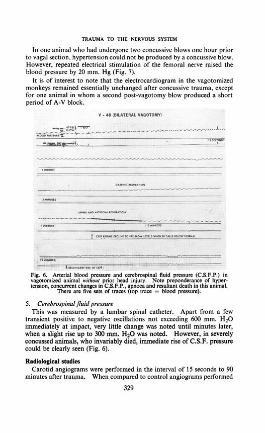
After bilateral vagotomy, concussive trauma produced a very transientfall of blood pressure by 20-30 mm. Hg for a few seconds followed by arapid hypertension up to 60 mm. Hg above pre-concussive levels (Fig. 6).
327
A. K. OMMAYA5 < __ t t l g . W s t *.'.'. -@ [email protected], S =' . l..z. = im _, :e =.. sm ,-,.n.'.n"! S>>W wE | | | | ; on 5 .?. s; .--X G-s Er,^ t ; i - - s| | Z_ @ | E @s .' li h a 2 .:s. _. X . mm 5 iZ . Z . BR R-u i S | |Zt^EX es iitX i r ? .Jv ? .r; ! . T ? W3 2 - . : 1 ]S ffi i S I ? t;EZ x _g | g Z t t -3 b - . i;ffi>i D 1 ^ et. !1-_ I 1|1 | 11 11 ! s N 190 ., .,- Ziti Fi! 1 ! w 7T j Ws, ;':m<:<*3>-X7 '_| | ! S X x E B S Z iX E E X Yv iX j-s L1F :.1 s1;x e er X 2 tl,rejati.B,;i *-| l | l 1|11 i 0 5 11 1 i 5t 3i - 01 i i av_ . . . . . .._ . _. - . ... ' .- - t lv ' . ' _ ,__i............. .......... 0_, ..
.a......4~P4
* i....U.. ..... k-a-- = . 1 jj'
*~~~~~~~~~~~ &UN.' ........................;-0 @..Ww
Fig. 4. The electrocardiogram before, during, and after experimental head injury.Standard limb leads. Note marked bradycardia, arrhythmia and patternabnormalities after impact, and the return of a normal trace within 4 minutes
after trauma.
#17 - Isl8low - 50psi mm Hg.Occi,oilo/ r'6o
Attrlerio glood Pressure I - 140
BPreure to howAN 13min. /Osoc after°1O.5 sec. g/deforerrauma -80
After Trauma imBoreT6ou
Fig. 5. The arterial blood pressure changes produced by experimental headinjury. Mean pressure recording from femoral artery. Note hypotensive
effect after impact.
328
1.!-. r!.flR.1-,.t..,-":r..,..r-::I:.:1J7
ri LWACT
TRAUMA TO THE NERVOUS SYSTEM
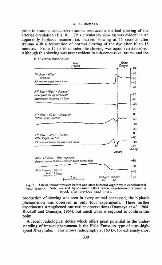
In one animal who had undergone two concussive blows one hour priorto vagal section, hypertension could not be produced by a concussive blow.However, repeated electrical stimulation of the femoral nerve raised theblood pressure by 20 mm. Hg (Fig. 7).
It is of interest to note that the electrocardiogram in the vagotomizedmonkeys remained essentially unchanged after concussive trauma, exceptfor one animal in whom a second post-vagotomy blow produced a shortperiod of A-V block.
(LAIURAL YA4W I@ UY)........... ...
........ ..
Fig. 6. Arterial blood pressure and cerebrospinal fluid pressure (C.S.F.P.) invagotomized animal without prior head injury. Note preponderance of hyper-tension, concurrent changes in C.S.F.P., apnoea and resultant death in this animal.
There are five sets of traces (top trace = blood pressure).
5. Cerebrospinal fluid pressureThis was measured by a lumbar spinal catheter. Apart from a few
transient positive to negative oscillations not exceeding 600 mm. H20immediately at impact, very little change was noted until minutes later,when a slight rise up to 300 mm. H20 was noted. However, in severelyconcussed animals, who invariably died, immediate rise of C.S.F. pressurecould be clearly seen (Fig. 6).
Radiological studiesCarotid angiograms were performed in the interval of 15 seconds to 90
minutes after traunma, When compared to control angiograms performed
329
A. K. OMMAYA
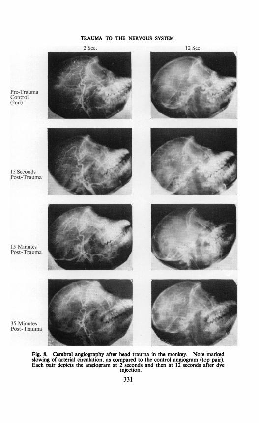
prior to trauma, concussive trauma produced a marked slowing of thearterial circulation (Fig. 8). This circulatory slowing was evident in anapparently biphasic manner, i.e. marked slowing at 15 seconds aftertrauma with a restoration of normal clearing of the dye after 10 to 15minutes. From 15 to 90 minutes the slowing was again re-established.Although this slowing was never evident in sub-concussive trauma and the
V/- t3 Ar/eri/l glood Pressure
/5/ 8/ow - 60psiOccip/onl'
BP returned 1o bose fIne In 4 mln
2n' 8/ow - 70psi - Occipi/ol8/ow givel during persis/onlhypo/ension followi'ng /slb/ow
3rd 8/ow - 80psi - Occipi/olBefore Vogol Secl/on
41h 8/ow - 80psi- Fron/olAfler Vogol Sec/ione P refurned to bose line of/er 2min. 30 sec
impoctAf/er 4th 8/ow - Posl Vogo/omyBefore, during 8 of/er Femorol Nerve s/imu/o/ion
8 Vol frequency = 60/secdOdelay 5 1msec t I
15sec s/mu/us stimu/usI1off on
80
-60
40
-20
Fig. 7. Arterial blood pressure before and after bilateral vagotomy in experimentalhead trauma. Note marked hypotensive effect when vagotomized animal is
struck after previous head injury.
production of slowing was seen in every animal concussed, the biphasicphenomenon was observed in only four experiments. These furtherexperiments strengthened our earlier observations (Ommaya et al., 1964;Rockoff and Ommaya, 1964), but much work is required to confirm thispoint.A recent radiological device which offers great potential in the under-
standing of impact phenomena is the Field Emission type of ultra-high-speed X-ray tube. This allows radiography at 150 kv. for extremely short
330
TRAUMA TO THE NERVOUS SYSTEM
2 Sec. 12 Sec.
Pre-TraumaControl(2nd)
15 SecondsPost-Trauma
15 MinutesPost-Trauma
35 MinutesPost-Trauma
Fig. 8. Cerebral angiography after head trauma in the monkey. Note markedslowing of arterial circulation, as compared to the control angiogram (top pair).Each pair depicts the angiogram at 2 seconds and then at 12 seconds after dye
injection.
331
A. K. OMMAYA
exposure time (to 70 nanoseconds) and repeated exposures can be madedown to intervals of 30 microseconds. This permits arrest of any move-ments in tissues during impact or other energy-loading situations. Pre-liminary results with angiography and intracerebral radio-opaque markersshow that the data thus obtained is reliable. We hope quite shortly topublish full details of the actual displacements in intracranial contents
PRE-TRAUMA
I
DURING TRAUMA
f,
..10rktu^150 KV 70 NANO SECONDS CEREERAt ANGOGRAPHE
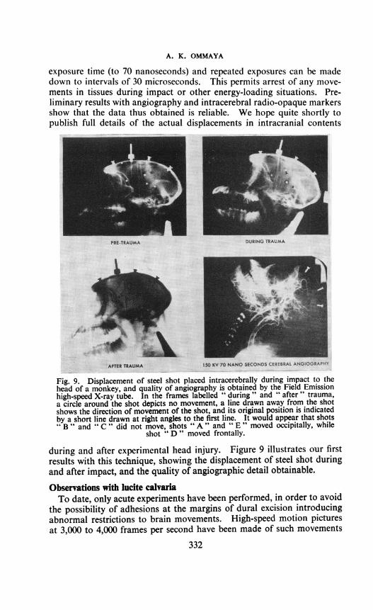
Fig. 9. Displacement of steel shot placed intracerebrally during impact to thehead of a monkey, and quality of angiography is obtained by the Field Emissionhigh-speed X-ray tube. In the frames labelled " during" and " after" trauma,a circle around the shot depicts no movement, a line drawn away from the shotshows the direction of movement of the shot, and its original position is indicatedby a short line drawn at right angles to the first line. It would appear that shotsB"" and " C " did not move, shots " A " and " E " moved occipitally, while
shot " D " moved frontally.
during and after experimental head injury. Figure 9 illustrates our firstresults with this technique, showing the displacement of steel shot duringand after impact, and the quality of angiographic detail obtainable.
Observations with lucite calvariaTo date, only acute experiments have been performed, in order to avoid
the possibility of adhesions at the margins of dural excision introducingabnormal restrictions to brain movements. High-speed motion picturesat 3,000 to 4,000 frames per second have been made of such movements
332
TRAUMA TO THE NERVOUS SYSTEM
during and after impacts to the head, and have essentially confirmed theobservations of Pudenz and Shelden (1946). Thuis at impact the skullmoves away and the brain lags behind. The brain then swirls to followthe head, appearing to rotate on an axis, passing through the centre ofgravity of the head. Maximal movements seem to be found in the fronto-parietal areas. These movements are almost identical to those predictedfor the brain by Holbourn (1943). We also confirmed the marked damp-ing of these movements by fluid by exaggerating the " sub-arachnoidspace" under the lucite dome.
Behavioural studiesAn attempt was made to develop an analogue to testing for amnesia
in man, by training the naive monkey in a large series of visual discrimina-tions until immediately before head injury. The degree of loss of suchlearned discriminations and the rate of re-learning after experimentalcerebral concussion was proposed as an index of the disruption of memorymechanisms by the trauma. The experiment could be controlled for theeffect of anaesthesia, surgery and other interventions. However, theresults of a preliminary testing of this analogue were not conclusive in thatthe post-traumatic interval before testing was too long. Further workalong these lines is being pursued.
General behavioural observations of all animals recovering from traumawere made for periods up to one year. No significant physical or neuro-logical abnormalities were seen. Post-traumatic epilepsy was neverseen to occur. Isolated and colony behaviour in terms of feeding, sex, fearand aggressive-passive behaviour with peers remained unaltered. Particu-lar attention for the appearance of any evidence of the Kluver-Bucysyndrome (" psychic blindness ", oral tendencies, hypermetamorphosis forvisual stimuli, loss of fear and aggression, increased sexual activity,hyperphagia) revealed no evidence for any of its manifestations.
Pathological observationsIn a previous study (Ommaya et al., 1964) an attempt had been made to
correlate the production of experimental cerebral concussion with theleakage of intravenously administered sodium fluorescein into the brainstem and upper cervical cord. However, after further study it would appearthat this is not a constant occurrence. Fluorescence of the cut surfaceof the brain stem under ultraviolet light certainly did not occur with non-concussive trauma, but it was also absent in many animals who hadsustained definite concussion. In those concussed animals in whomfluorescence was seen, it was usually limited to a few millimetres from thesurface of the brain stem rather than uniformly present over the wholesurface (cf. Figures 11 to 15 in Ommaya et al., 1964). The occurrence offluorescence below the impact point, its presence in a fan-shaped manneralong the posterior parasagittal areas, alongside the falx, occasionallystraddling the central sulci, and the rarity of its appearance over " contre-
333

A. K. OMMAYA
coup" areas were confirmed. Macroscopic observations, other thanthose mentioned above, were also made for evidence of fracture, clots,haemorrhage, contusions and lacerations. Animals with skull fracturewere not considered in the statistical evaluation of impact-concussionrelationships. There was nothing of novel merit in the observations onfracture. On the other hand the occurrence of haemorrhage and itsdistribution was noteworthy. First, there was not a single case of signifi-cant acute or chronic extra- or subdural haemorrhage over the convexityof the cerebral or cerebellar hemispheres in those animals not sustaining afractured skull. Acute subdural haemorrhage without fracture waslimited to the base, particularly around the pons and Sylvian fissures.This was in most cases associated with subarachnoid haemorrhage also inthe same areas. Definite contusions and lacerations, other than thosebelow fracture sites, were extremely unusual, and when present wereusually below the impact point, around the pons, on the medial surfacesalong the falx, and in the Sylvian fissures and the adjacent temporal andorbital-frontal cortex. True " contra-coup " lesions, i.e. lesions " oppo-site " to the point of impact, were significantly rare. Including the caseswith skull fracture, only four out of 80 animals displayed such lesions.Moreover, the distribution of these " contra-coup " changes was similarto the contusional changes described above, i.e. related more to the presenceof such structures as the dural and bony projections and partitions ratherthan to any geometric relationship to the point of impact.
Microscopic studies have to date been limited to haematoxylin and eosinpreparations of paraffin sections taken from all anatomical situations in10 animals sacrificed at intervals of 30 minutes to nine months after headinjury. These sections have not revealed any changes of note. Moreextensive studies including Nissl, silver and myelin stains will be reportedseparately. A study using Marchi techniques is being undertaken underthe direction of Dr. Sabina Strich, and this will be reported separately.The experimental model for controlled head injury in the monkey de-
scribed above has been developed with two intentions. First, to establishmethods for prevention or protection against the effects of impact, i.e.the prophylaxis for head injuries. Secondly, to minimize these effects andprevent complications or to treat such effects and complications so that themorbidity and mortality of the experimental head injury is reduced to theminimum. The next stage would then be to establish scaling methods toapply these findings in man, both as prophylaxis for individuals in whomthe hazard of trauma is always present, and in the management of patientswith trauma to the nervous system. The model itself is valid inasmuch asthe onset of concussion as defined is significantly related to the physicalvariables measured. It will also be possible to define, under identicalexperimental conditions, a 50 per cent and 99 per cent " lethal dosage "of energy input under a variety of conditions, which would produce fatalconcussion without fracture of the skull. This would provide a tool for
334

TRAUMA TO THE NERVOUS SYSTEM
testing both prophylactic and mortality reducing therapeutic techniqueswith a reasonable chance for success. It would appear, however, that themorbidity of most head injuries in man cannot be adequately reproducedby experimental techniques in the monkey. Thus, after repeated traumathe majority of our animals survived with no obvious sequelae of note.Only a few (less than 10 per cent) succumbed as a direct result of thetrauma, and of these the majority had suffered compound depressedfractures.
Preliminary testing for protection against head trauma consisted ofestablishing the importance of movement at the cranio-spinal junction inthe genesis of concussion. Thus recent experimental work by Hollisterand his colleagues (1958) has indicated that stretch of the neck can produce" concussion " in the cat. Martinez and his colleagues (1963) have shownthat experimental whiplash in rabbits (150 " g " linear acceleration in 20to 120 m.sec.) can produce brain injury with surface contusions and basalhaemorrhages without impact to the head. In a recent paper Liss (1965)has reported the death of a young swimmer who, after a racing turninvolving voluntary hyperflexion, torsion and hyperextension of the headon the neck, rapidly died with agonal dyspnoea, convulsions and apnoea.Post mortem showed a congested brain, medullary oedema and petechialperivascular haemorrhages at C2-3 within the grey columns, without bonyinjury. In a previous study we reported the protective effect of a cervicalcollar against experimental concussion by head impacts in the monkey(Ommaya et al., 1964). This observation was accordingly tested againand would appear to hold true. These facts have raised the very importantpractical point that crash helmets may, in themselves, constitute a hazardunder certain conditions. Protective helmets have been designedprimarily as energy absorbing or deflecting devices (Cairns and Holbourn,1943; Lewin and Kennedy, 1956; Snively and Chichester, 1961; Gurdjianet al., 1964). But by adding further weight and by shifting the centre ofgravity up and forward, a heavy helmet, such as is worn by pilots andmotorcyclists, increases the moment of inertia about the cervical pivots.This increases the tensile and sheer stresses in the brain and cervical cordunder conditions allowing acceleration and free movement of the head onthe neck (indirectly, by a whiplash or flexion effect or directly by impactto the head). Existing helnet design has not taken the above into con-sideration, and our effort in the field of prophylaxis of cerebral concussionis to establish definite recommendations concerning the importance ofthese factors and ways of making provision for them.As mentioned before, our model of experimental cerebral concussion
will allow the testing of therapeutic measures to reduce the mortality ina controlled manner. We are currently planning to undertake this forhypothermia, hyperoxygenation and steroid therapy. The problems ofreducing morbidity, however, cannot be suitably studied in this experi-mental model. For this reason clinical data from patients suffering with
335
A. K. OMMAYA
head injuries is the only available source of information on which furtheradvances can be made. Unfortunately, as pointed out by a previouslecturer in this College, the standards of accurate and uniform recordkeeping for cases of head injury in these times of uneasy peace very seldomreach that attained in many centres during the war (Jennett, 1961). Inorder to obtain statistically meaningful clinical data it was essential,therefore, to develop a system of recording, classifying and analysing suchdata in as uniform and precise a fashion as possible. This has been themain effort of our clinical study.
The clinical studyExisting diagnostic classifications of closed head injury are not satis-
factory. Most of them are based on the definition given by Munro (1938),which add little to the facts known by John Hunter. Thus brief loss ofconsciousness without other signs after head injury is called concussion;a similar condition but with bloody spinal fluid is called cerebral contusionor laceration-but this term is also used to describe the patient withneurological deficits and more prolonged unconsciousness; if the C.S.F.pressure is raised the diagnosis of " cerebral oedema " is advanced. If adepressed fracture or intracranial haematoma is found, this diagnosisdominates the clinical picture. Yet it is obvious that the loss of conscious-ness which underlies all these diagnoses may be identical to that calledconcussion. Moreover, the " severity" of a head injury can have twointerpretations. Thus there are the prognostic danger signals ofimmediate surgical importance, such as pyrexia, compound skull fracture,respiratory abnormality, neurological deficits, convulsions, worsening ofthe level of consciousness after initial improvement, rising blood pressurewith falling pulse rate, and bloody C.S.F. But these surgical signpostsmay have no relationship to the ultimnate severity of disability producedby head injtury (Denny-Brown, 1945b). Thus the clinical diagnoses incommon usage are at best unreliable approximations to a small part ofthe overall picture. The work of Symonds (1928, 1962), Ritchie Russell1932, 1961), and Denny-Brown (1945b) laid the foundations for a morelogical approach to the classification of closed head injuries. The inten-tions of our clinical study were, therefore, to develop on these foundationsa system of classifying such head injuries which would have greaterdiagnostic precision, to establish a more precise relationship betweendiagnosis and prognosis as opposed to the unreliable prognostic valueof existing diagnostic categories, to standardize tests for early recognitionof compli:-ations, to determine the best management for these complica-tions and for the basic problem of trauma to the nervous system, i.e.cerebral concussion.
The basic tool for clinical research is the medical record or history ofthe patient whose disease is being studied. The efficiency of this tool isin direct proportion to the reliability with which descriptions in one
336
'I RAUMA TO THE NERVOUS SYSTEM
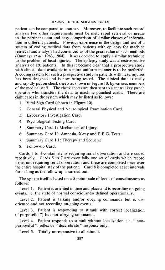
patient can be compared to another. Moreover, to facilitate such recordanalysis two other requirements must be met: rapid retrieval or accessto the pertinent data and easy comparison of similar classes of informa-tion in different patients. Previous experience in the design and use of asystem of coding medical data from patients with epilepsy for machineretrieval and analysis had convinced us of the great value of such methods(Ommaya et al., 1963, 1964). It was decided to apply a similar techniqueto the problem of head injuries. The epilepsy study was a restrospectiveanalysis of 150 patients. In this it became clear that a prospective studywith clinical data available in a more uniform manner is to be preferred.A coding system for such a prospective study in patients with head injurieshas been designed and is now being tested. The clinical data is easilyand rapidly put on check sheets as shown in Figure 10, by various membersof the medical staff. The check sheets are then sent to a central key punchoperator who transfers the data to machine punched cards. There areeight cards in the system which may be listed as follows:
1. Vital Sign Card (shown in Figure 10).2. General Physical and Neurological Examination Card.3. Laboratory Investigation Card.4. Psychological Testing Card.5. Summary Card I: Mechanism of Injury.6. Summary Card II: Amnesia, X-ray and E.E.G. Tests.7. Summary Card III: Therapy and Sequelae.8. Follow-up Card.
Cards I to 4 contain items requiring serial observation and are codedrepetitively. Cards 5 to 7 are essentially one set of cards which recorditems not requiring serial observation and these are completed once overthe entire hospital stay of the patient. Card 8 is completed at set intervalsfor as long as the follow-up is carried out.
The system itself is based on a 5-point scale of levels of consciousness asfollows:
Level 1. Patient is oriented in time and place and is recordinsg on-goingevents, i.e. the state of normal consciousness defined operationally.
Level 2. Patient is talking and/or obeying commands but is dis-oriented and not recording on-going events.
Level 3. Patient is responding to stimuli with correct localization(" purposeful ") but not obeying commands.
Level 4. Patient responds to stimuli without localization, i.e. " non-purposeful ", reflex or " decerebrate " response only.
Level 5. Totally unresponsive to all stimuli.
337

A. K. OMMAYA
The initial effort of this coding system is to relate the duration of suchlevels of consciousness, and the rate of transfer from Level 5 to 1, to theduration of post-traumatic amnesia and other neurological and be-havioural sequelae of head injury in terms of the overall disability after
CARD - VITAL SIGN CARD
Identification Infarmatian'I.- 3 Stucly, Card, Hosp. No.4.-11 IHasp.
Case Na.12 16 Month, Day, Year
Ma. Day Y r.17.-20 Zl1lZl1 Time
Level of Consciousness0 No Info.L Oritn ted2 Talking with Confusion1 Responding with Localizing4 Responding without Localizing5 Totally Unresponsive
23.24 Pupillary Status
No Info.R> L. Reactive
02 R>L-Unreective03 L > R Rctive
04 L>R.Unreactive05 Both Large-Reactive
Both Large-Unreective07 Both Small reactive08 Both Small-Unreective09 Both Normal. Reactive10 Both Normel . Unreective
25 Comeal Reflex
fi No Info.1 Present Bilat.-2 Absent Bilat3DAbsent R Only_4Absent L Only
Blood Pressure
26E28[ l lSystolic29 31 f II Diastolic
Puls and Respiration32 34 1 Pulse35- 36 11 IRespi ration
Fig. 10. The " Vital Sign" card of the head injury coding system. Numberswithin the larger rectangles refer to the punched card column numbers, while the
numbers in the small rectangles are the categories within each column.
trauma. The end point will be the speed and quality of return to economicindependence for each patient. Intercurrent surgical and medical com-
plications are being carefully studied and related to this scheme. Pre-liminary data suggest that the system is practical, and within a few yearswe hope to provide answers to the problems of diagnostic and prognosticprecision. This study also allows the evaluation of current managementof such patients in a more rigorous manner and the statistical analysismade facile by such techniques will soon provide firm guidelines to a morerational therapy.
338
21
22 Motor Activity and Attitude0 No Info.
Normal2 Quiet and Motionless3 Intermittent Overectivity4 Constant Ovoractivity
Extensor Attitude-Const.Extensor Attitude-Intermit.Flexor Spasms
8 Movment of Head Only
38 -41 __F. Tomperature-Rectal - in*C.
TRAUMA TO THE NERVOUS SYSTEM
In the course of this study we have been able to study in detail a numberof cases of very severe head injury with prolonged disturbance of con-sciousness. Of particular interest to us has been certain re-integrativephenomena displayed by such patients, as they slowly struggle towardsfull consciousness. By this is meant an often distorted and irregularlyaccelerated reproduction of ontogenetic development. The patient with asevere head injury appears to retrace in his general behaviour, as wellas in his neurological pattern, his own growth and maturity. This wouldappear to be more clearly seen in young adults than at the two extremesof age. A few investigators (Goldstein, 1942; Wadeson, 1966) haveemphasized the importance of understanding such general behaviour interms of a physiological frame of reference related to the attempt of thepersonality to assert itself after disruption by trauma. Disturbances ofmemory are the most obvious reason for behavioural difficulties and theseare often related to damage in the temporal and rhinencephalic structures(Drachman and Ommaya, 1964; Serafetinides and Falconer, 1963; Gleesand Griffith, 1952; Scoville and Milner, 1957). The retrieval of lostmemories appears to parallel the re-emergence of the personality, and forthis reason the duration of post-traumatic amnesia is a good index ofseverity of brain injury (Russell and Smith, 1961). But this is not theentire story. The following case history is of interest in showing howmany factors are to be considered and how much intellectual and be-havioural recovery is possible even when brain damage is severe. It alsodepicts the re-integrative emergence of the patient's adult personalitythrough stages of apparent " infancy" and " childhood ".
National Institutes of Health Case No. 05-22-33. A 22-year-old, right-handed,married female who sustained a severe closed head injury without skull fracture, afterher car collided with a truck. After 24 hours in a state of restless unconsciousness,not responding purposefully to stimuli (Level 4), increasing intracranial pressurenecessitated bitemporal craniotomies (in two stages) with removal of bilateral subduralhaematomas and excision of the inferior temporal gyrus on the right, and 4j cm. ofsuperior, middle and inferior temporal gyri on the left side as measured from the tipof the temporal pole to the vein of Labbe. This was done for pulverized brain inthese regions. By the fifth day after head injury, she was at Level 3 (responding tostimuli purposefully) and in three weeks had attained Level 2 (talking, but disoriented).From this time on she exhibited slow improvement and at one month her motherdescribed her as " as if she was six years old". Childhood habits were reasserted,e.g. rubbing side of mouth continuously, and in addition oral tendencies appeared.She masturbated frequently, occasionally smeared herself with faeces, and displayeda voracious appetite. Improvement in her behaviour was noted in a step-like increasein her speech, dressing, toilet and interest in self-appearance. Her writing was initiallylettered and childlike, and as her behaviour became more adult so did the handwriting.Her parents and nurses were referred to as " teachers ". She recognized her parentsbefore her husband of six weeks, and insisted on being called by her childhood nickname.Her husband was first recognized as a boy friend and fiance before being acceptedas her husband. She would play with crayons and colour books and talked about" Winnie the Pooh ". She remained in Level 2 for over two months and, when finallyoriented in space and time, was found to have a post-traumatic amnesia of threemonths with a retrograde amnesia of only a few seconds. Her personality at thisstage was that of a rather emotionally labile teenager. Pre-traumatic I.Q. had beennoted at 140 (full scale Wechsler). Psychological testing after orientation was estab-lished, and three months after injury, showed results as follows: Full Scaie I.Q. = 88,
339
A. K. OMMAYA
Verbal I.Q. = 100, Performance I.Q. = 77, Memory Quotient = 68, M.M.P.I. wasnormal.A pneumo-encephalogram done at this stage showed a uniformly dilated ventricular
system, cisternal and subarachnoid spaces, with the third ventricle 1.3 cm. wide in theA.P. view. She continued to improve, and 14 months after injury was able to runher household singlehanded, but had significant bouts of depression and anxiety.Testing for memory revealed no impairment of short-term learning (recent memory)and on repeated I.Q. testing the results were as follows: Full Scale I.Q. = 117, VerbalI.Q. = 125, Performance I.Q. = 102, Memory Quotient = 112, but the M.M.P.I.showed high score on the " depression " scale.Whether this residual personality impairment is related to the temporal lobe damage
and intellectual deficit is difficult to say. However, it is of interest to note that althoughher P.T.A. was three months, this patient had increased her levels of consciousnessvery rapidly from Level 5 to Level 2 within three weeks. We are pursuing furtherthis relation of rates of improvement in levels of consciousness to the P.T.A. andfinal disability after head injury, but it would certainly appear to be of some prognosticvalue.
Considerations of time and space do not allow a full recital of ourresearch into diagnostic and therapeutic measures in the management ofhead injuries, but two techniques have proven particularly useful.
1. The use of R.I.S.A. introduced into the C.S.F. and its distributionas recorded by external scintillation scanning has proven of extremevalue in the accurate diagnosis of C.S.F. rhinorrhea (Ommaya, 1964;Di Chiro et al., 1964). Leakages of C.S.F. after trauma are always apotent source of meningitis and there is increasing realization that, evenin the absence of frank C.S.F. leakage, meningitis may develop in patientswith fractures in the anterior cranial fossa (Jefferson and Lewtas, 1963).To define such actual and potential pathways of infection, intrathecaladministration of only 100 pc of R.I.S.A. and scanning immediately overthe head can usually display the site of such leakages (Fig. 11). In com-bination with the clinical and X-ray information, a more accurate diag-nosis allows a more precise surgical repair to be performed. The use ofR.I.S.A. cisternography and ventriculography to define other abnorm-alities of C.S.F. dynamics after head injury remains to be investigated.
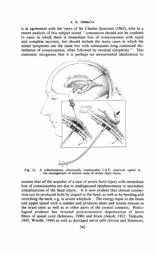
II. The problem of raised intracranial pressure and its managementin severe closed head injuries is often a vexed issue. The use of intra-venous urea and other pressure-reducing agents is attended by certainhazards when the diagnosis is not clearly established. Thus C.S.F.pressure may be raised because of cerebral oedema, sinus thrombosis, orsinus obstruction by a fracture producing hydrocephalus without oedema.Cerebral oedema may be caused by contusions or laceration, followingremoval of clots, develop due to metabolic and electrolyte disturbances,or be precipitated by other cerebrovascular lesions. While investigationsare pursued it is cccasionally of value to monitor intracranial pressure orestablish drainage of the subdural or subarachnoid space. Open techniquessuffice for a few hours, but for longer periods we have suggested an in-dwelling subcutaneous C.S.F. reservoir (Ommaya, 1963). This is seenin Figure 12. Continuous pressure recordings during treatment withdrugs such as mannitol and urea, withdrawal of C.S.F. and subdural
340

TRAUMA TO THE NERVOUS SYSTEM
fluid and introduction of drugs into these spaces is as easy as performing ahypodermic injection. Using such a device we have been able to showthat the recommended dose of urea (0.5 to 1.5 Gm./kilogram body weight)for reducing intracranial pressure is perhaps excessive. With such dosesthe pressure is reduced to extreme negative values, whereas 0.25 Gm. perkilogram body weight is quite adequate in reducing high pressures to alevel just below normal.
9~~~~~~~~~~~~~~~~~~~~~~~.
Fi. 11. R.I.S.A. cisternography in a case of C.S.F. rhinorrhea due to anteriorcranial fossa fracture. Note the pathway for the leakage displayed by the positive
scanning of the whole head.
In conclusion, what practical suggestions can be given for a more usefuldefinition of concussion? The classical definition of this " violentshaking " of the central nervous system is that it is a transient and reversibleparalysis of nervous action characterized by progressive recovery there-after (Trotter, 1924; Denny-Brown, 1945a). But this definition does notfit all the facts. The residual post-traumatic amnesia of a true cerebralconcussion is never a " transient and reversible " phenomenon. Experi-mental, clinical and pathological evidence would suggest that concussionis not an " all or none " response of the central nervous system, as it maywell be in peripheral nerves. Rather it is a graded response, from thevery mild, leaving minimal traces, to the very severe and irreversible. This
341
A. K. OMMAYA
is in agreement with the views of Sir Charles Symonds (1962), who in arecent analysis of this subject stated " concussion should not be confinedto cases in which there is immediate loss of consciousness with rapidand complete recovery, but should include the many cases in which theinitial symptoms are the same but with subsequent long continued dis-turbance of consciousness, often followed by residual symptoms ". Thisstatement recognizes that it is perhaps an unwarranted idealization to
Fig. 12. A subcutaneous, chronically implantable C.S.F. reservoir useful inthe management of certain cases of severe head injury.
assume that all the sequelae of a case of severe head injury with immediateloss of consciousness are due to undiagnosed epiphenomena or secondarycomplications of the head injury. It is now evident that clinical concus-sion can be produced both by impact to the head, as well as by bending andstretching the neck, e.g. in severe whiplash. The energy input to the brainand upper spinal cord is sudden and produces sheer and tensile stresses inthe brain stem as well as in other parts of the cranial contents. Patho-logical evidence has revealed post-concussive degeneration of nervefibres of spinal cord (Schmaus, 1890) and brain (Jakob, 1912; Tedeschi,1945; Windle, 1948) as well as damaged nerve cells (Groat and Simmons,
342
TRAUMA TO THE NERVOUS SYSTEM
1950; Groat et al., 1945; Tedeschi, 1945; Windle and Groat, 1945; Windleet al., 1944). The recent work of Strich (1961) is pertinent in that mildertrauma could well produce lesser degrees of axonal damage.
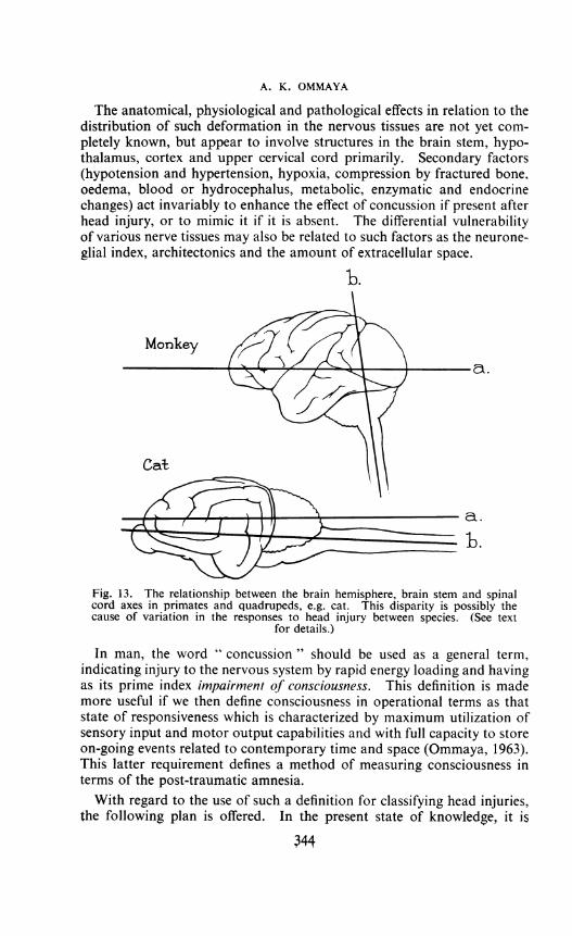
It is apparent from a consideration of the evidence presented that themechanism of concussion is not yet established. But our resultssupport the theory of Holbourn (1943), and further work will be neededeither to support or replace this theory with a better approximation. Theobvious conflict of experimental data from various workers is probablyrelated to three factors. First, the lack ofuniform terminology to describethe data. Secondly, the use of quadrupeds and lower animal species inwhich the relation of the brain hemispheres to the brain stem and cervicalcord are quite different when compared to the primates. This is illus-trated schematically in Figure 13, and raises the extremely importantissue of doubt in transferring concussion data from lower species to man.The similarity between the primate brain-cord geometry and that of manwould perhaps make such extrapolations more permissible. Suchdifferences may also explain the markedly opposite type of blood-pressurechange obtained in our monkeys as compared to that in the cat and dog.The third and possibly fundamental reason for conflicting data may
also serve to explain a curious and not completely explicable fact. Thusit is known that animals with smaller brains can tolerate much higher " g "loading (acceleration) as compared to larger brained animals. Thus micecan tolerate 100 to 1,000 times the "g" level as compared to man(Kornhauser and Lawton, 1961). It would appear that this is not simplyrelated to the mass-size ratio and that the larger brain is sensitive to muchlower levels of energy as compared to smaller brains. We would suggestthat this may be due to certain mechanical factors in the brain itself. Thusit has been shown that as brain weight increases from mouse throughmonkey to man, there is an equivalent increase of chloride, and sucrosespaces (probably related to the extracellular space) (Bourke et al., 1965).Similarly, the number of neurons per cubic millimetre of brain decrease asthe brain weight increases (Cobb, 1965). Finally, the number of glialcells per neuron (glia-to-neuron index) increases from small to largebrains (Friede, 1954; Hawkins and Olszewski, 1957). All this wouldsuggest that the smaller brains of lower animals tolerate injury betterbecause they are more compact. The less compact, looser structure of thelarger brains deforms more readily when energy is applied. These factsenable us to provide the following definition of the mechanism of con-cussion and attempt to relate it to this phenomenon and the other sequelaeof head injury in man.
Sheer or tensile deformation within the brain and cervical cord, pro-duced by sudden impact or indirect acceleration of the head, results indysfunction of neural elements. This dysfunction may be reversible to adegree or it may be completely irreversible in proportion to the amount ofenergy absorbed.
343
A. K. OMMAYA
The anatomical, physiological and pathological effects in relation to thedistribution of such deformation in the nervous tissues are not yet com-pletely known, but appear to involve structures in the brain stem, hypo-thalamus, cortex and upper cervical cord primarily. Secondary factors(hypotension and hypertension, hypoxia, compression by fractured bone,oedema, blood or hydrocephalus, metabolic, enzymatic and endocrinechanges) act invariably to enhance the effect of concussion if present afterhead injury, or to mimic it if it is absent. The differential vulnerabilityof various nerve tissues may also be related to such factors as the neurone-glial index, architectonics and the amount of extracellular space.
b.
MonkeyX
Cat
a.
bj.
Fig. 13. The relationship between the brain hemisphere, brain stem and spinalcord axes in primates and quadrupeds, e.g. cat. This disparity is possibly thecause of variation in the responses to head injury between species. (See text
for details.)
In man, the word " concussion " should be used as a general term,indicating injury to the nervous system by rapid energy loading and havingas its prime index impairment of consciousness. This definition is mademore useful if we then define consciousness in operational terms as thatstate of responsiveness which is characterized by maximum utilization ofsensory input and motor output capabilities and with full capacity to storeon-going events related to contemporary time and space (Ommaya, 1963).This latter requirement defines a method of measuring consciousness interms of the post-traumatic amnesia.With regard to the use of such a definition for classifying head injuries,
the following plan is offered. In the present state of knowledge, it is344
TRAUMA TO 'IHE NERVOUS SYSTEM
essential that wherever possible clinical entities related to head injuryshould be described rather than labelled. Terms such as comatose,contusion, brain stem lesion, etc., should be avoided. Thus all headinjuries should be initially classified, when first seen, in two main cate-gories, concussive or non-concussive. Each of these should then bequalified as falling in one of four classes, i.e. with or without fractureof the skull, and with or without complications. The complications are tobe considered in two groups: (1) the local response, including vascular,parenchymal, bony, C.S.F. leakage, infection, thermoregulatory, meta-bolic and endocrine complications; (2) the general response. This in-cludes complications related to the airway, chest, shock, multiple injuries,fat embolism and systemic metabolic and endocrine disturbances.
This initial diagnosis will be completed during the course of the patient'sstay in hospital. A second diagnosis must then be made after a minimumof one year follow-up (or death) in which the total disability and length ofpost-traumatic amnesia are related to the initial diagnosis. By doing thisconsistently for a very large series of patients, using our coding system,we hope to arrive at more quantitative values for diagnosis that can bemade very early after the onset of the head injury syndromes and thusguide our management and prognosis more accurately. In this effort, wewill investigate particularly the role of vascular complications whoseimportance has been advanced by our observations on the changes inblood pressure, electrocardiogram and in the cerebral angiogram.
I will end by remarking that perhaps I have dwelt very little on thepurely surgical aspects of head injuries. My reason for this is simplythat the techniques of neurosurgery play a well-established but relativelyminor role in less than 10 per cent of all cases. The other 90 per cent ofpatients from their onset, however, are automatically potential candidatesfor surgery until proven otherwise. It behoves us, as surgeons, to beaware, not only of the moment when surgery will save or salvage a life,but also as physicians to ensure everything possible to guarantee the bestsurvival from man's oldest disease-trauma. John Hunter's advice tosurgeons operating on the cranium is well worth recalling, displaying as itdoes that combination of surgical wisdom and utter honesty that was somuch his style. " We should scalp carefully . .. and yet I own I cannotalways call to mind this caution at all times when operating" (Hunter,1841).
ACKNOWLEDGEMENTSThe work of many of my colleagues has enabled this lecture to be pre-
pared. The past support and continuing collaboration of the following isgratefully recognized:
Mr. Richard Mahone and Mr. Arthur Hirsch of the Personnel Pro-tection Branch, David Taylor Model Basin, U.S.N.; Dr. John Coe, Dr. T,
345
A. K. OMMAYA
Krueger, Dr. Eugene Flamm and Mr. Stephen Garrell of the Branch ofSurgical Neurology, National Institute of Neurological Diseases andBlindness, National Institutes of Health, have worked on various aspectsat various stages. Dr. S. David Rockoff of the X-ray Department hasprovided radiological techniques and advice. Mrs. D. Sadowsky andDr. James Mosimann of the Biometrics Branch, N.I.N.D.B., N.I.H.,have given freely of their abilities in designing the coding system andtesting the data. Dr. Igor Klatzo, Dr. Donald Tower and Dr. MaitlandBaldwin have provided valuable help, criticism and advice at all stages.Accelerated development of the experimental work has been ensured bycollaborative arrangements now established between the Branch ofSurgical Neurology, N.I.N.D.B., N.I.H., Laboratory of Biophysics,Naval Medical Research Institute, United States Navy (Dr. DavidGoldman) and the David Taylor Model Basin, Personnel ProtectionBranch (R.M. and A.H.), with the additional financial support providedby the Bureau of Weapons, United States Navy, to whom gratitude isexpressed.
REFERENCESBOURKE, R. S., GREENBERG, E. S., and TOWER, D. B. (1965) Amer. J. Physiol. 208, 682.CAIRNS, H1., and HOLBOURN, H. (1943) Brit. Med. J., 1, 591.COBB, S. (1965) Arch. Neurol. (Chic.), 12, 555.DENNY-BROWN, D. (1945a) Physiol. Rev. 25, 296.
(1945b) J. Amer. med. Ass. 127, 429.and RUSSELL, W. R. (1941) Brain, 64, 93.
Di CHIRO, G., REAMES, P. M., and MATTHEWS, J. R. (1964) Neurology, 14, 185.DRACHMAN, D. A., and OMMAYA, A. K. (1964) Arch. Neurol. (Chic.) 10, 411.FRIEDE, R. (1954) Acta anat. (Basel) 20, 240.GLEES, P., and GRIFFITH, H. B. (1952) Mschr. Psychiatr. Neurol. 123, 193.GOLDSTEIN, K. (1942) After effects ofbrain injuries in war; their evaluation and treatment.
London, Heinemann.GROAT, R. A., and SIMMONS, J. Q., III (1950) J. Neuropath. exp. Neurol. 9, 150.
WINDLE, W. F., and MAGOUN, H. W. (1945) J. Neurosurg. 2, 26.GURDJIAN, E. S., and WEBSTER, J. E. (1943) Surg. Gynec. Obstet. 76, 623.
(1958) Head injuries mechanism, diagnosis andmanagement. Boston: Little, Brown.
HODSON. V. R., HARDY, W. G., PATRICK, L. M., and LISSNER,H. R. (1964) J. Trauma, 4, 309.
HAWKINS, A., and OLSZEWSKI, J. (1957) Science, 126, 75.HOLBOURN, A. H. S. (1943) Lancet, 2, 438.HOLLISTER, N. R., JOLLEY, W. P., HORNE, R. G., and FRIEDE, R. (1958) WADC
Technical Report 58-193. Astia Document No. AD 203385.HUNTER, J. (1841) Lectures on the principles of surgery, with notes by James F. Palmer.
Philadelphia: Haswell, Barrington and Haswell.JAKOB, A. (1912) Histol. histopath. Arb. Grosshirnrinde, 5, 182.JEFFERSON, A., and LEWTAS, N. (1963) Acta radiol. (Stockh.) 1, 118.JENNETT, W. B. (1961) Ann. Roy. Coll. Surg. Engi. 29, 370.KORNHAUSER, M., and LAWTON, R. W. (1961) Proc. 4th AFBMD/STL Symp., Ohio,
J, 386.LEWIN, W. (1953) Brit. mtied. J. 1, 1239.
and KENNEDY, W. (1956) Brit. med. J. 1, 1253.LINDGREN, S. 0. (1960) Acta chir. scand. Suppl. 254.LIss, L. (1965) Neurology, 15, 675.MARTINEZ, J. L. (1963) Pap. Amer. Soc. Mech. Engrs., 63-WA-281.MUNRO, D. (1938) Craniocerebral injuries. New York, Oxford Univ. Press.McIvER, I. N., LASSMAN, THOMSON, C. W., and MCLEOD, G. (1958) Lancet, 2, 544,
346
TRAUMA TO THE NERVOUS SYSTEM
OMMAYA, A. K. (1963) Lancet, 2, 983.(1963) Med. Ann. D.C. 32, 18.(1964) Neurology, 14, 106.and BALDWIN, M. (1963) Proc. Soc. Brit. Neurol. Surg. 68,ROCKOFF, D. S., and BALDWIN, M. (1964) J. Neurosurg. 21, 249.and SADOWSKY, D. (1964) Epilepsia, 5, 192.
PUDENZ, R. H., and SHELDEN, C. H. (1946) J. Neurosurg. 3, 487.ROCKOFF, S. D., and OMMAYA, A. K. (1964) Amer. J. Roentgenol. 91, 1026.RUSSELL, W. R. (1932) Brain, 55, 549.
-and SMITH, A. (1961) Arch. Neurol. (Chic.) 5, 4.SCHMAUS, H. (1890) Virchows Arch. 122, 326 and 470.SCOVILLE, W. B., and MILNER, B. (1957) J. Neurol. Neurosurg. Psychiatr. 20, 11.SERAFETINIDES, E. A., and FALCONER, M. A. (1963) Brain, 86, 333.SHEA, J. J. (1964) Cosmic, Inc. Report No. 75: The Mechanics of Brain Concussion.
Prepared for Personnel Protection Branch, Structural Mechanics Laboratory,David Taylor Model Basin. Washington, D.C.
SHELDEN, C. H., PUDENZ, R. H., RESTARSKI, J. S., and CRAIG, W. M. (1944) J.Neurosurg. 1, 67.
SNIVELY, G. G., and CHICHESTER, C. 0. (1961) Stapp Autonmotive Crash and FieldDemonstration Conference, Ohio, 5, 182.
STRICH, S. J. (1961) Lancet, 2, 443.SYMONDS, C. P. (1928) Brit. med. J. 2, 829.
(1962) Lancet, 1, 1.TEDESCHI, C. G. (1945) Arch. Neurol. Psychiat. 53, 333.TROTTER, W. (1924) Lancet, 1, 935.WADESON, R. (1966) " Ego and Central Nervous System Functions. A Frame of
Reference," in Perspectives in Biology.WHITE, J. C., BROOKS, T. R., GOLDTHWAIT, J. C., and ADAMS, R. D. (1943) Ann. Surg.
118, 619.WINDLE, W. F. (1948) Anat. Rec. 100, 725.
and GROAT, R. A. (1945) Anat. Rec. 93, 201.and Fox, C. A. (1944) Surg. Gynec. Obstet. 79, 561.
THE WORSHIPFUL COMPANY OF BARBERSTHE LORD MAYOR OF LONDON, Sir Lionel Denny, M.C., laid the Founda-tion Stone of the new Barber Surgeons' Hall on 6th November 1966.The new hall is in Monkwell Square, close to London Wall.The Master of the Company, the Rev. George Turner, M.A., thanked
the Lord Mayor, who is the senior past-Master of the Barbers' Company,for so graciously performing this important ceremony. The Chaplainof the Company, the Rev. B. W. Ottaway, delivered a dedicatory prayerwhich concluded the ceremony, after which the Master, Wardens andCourt of Assistants went to the Mansion House for a glass of sherry tocelebrate a very important occasion.The Barbers' Company, which ranks No. 17 in the livery companies of
the City of London, has been in existence since 1308, when the first Masterwas installed.
Barber Surgeons' Hall in Monkwell Street was built by Inigo Jones in1634. It was surrounded by a herb-garden in which the Barber-Surgeonscultivated their samples. It was this garden which no doubt saved thatportion of building which escaped the Great Fire of London in 1666.During the second World War the whole of Barber Surgeons' Hall wascompletely destroyed. C. W.
347