the body anatomy and physiology lesson 4 skeletal...
TRANSCRIPT
Monday 26th Feb 2018
The Body – Anatomy and Physiology
Lesson 4 – Skeletal System
Functions and Structure

Muscular System

Muscular System

Skeletal System Extension – What
are the roles of
tendons, cartilage
and ligaments?
Skeletal System Extension – What
are the roles of
tendons, cartilage
and ligaments?
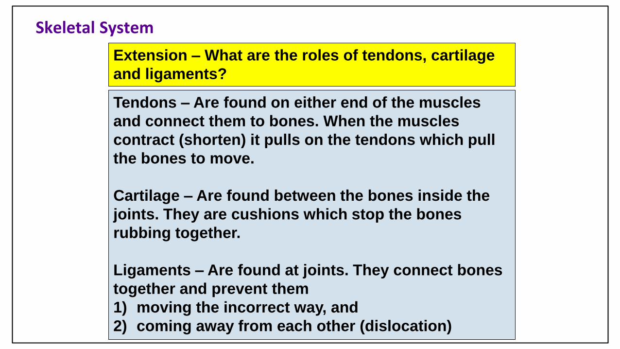
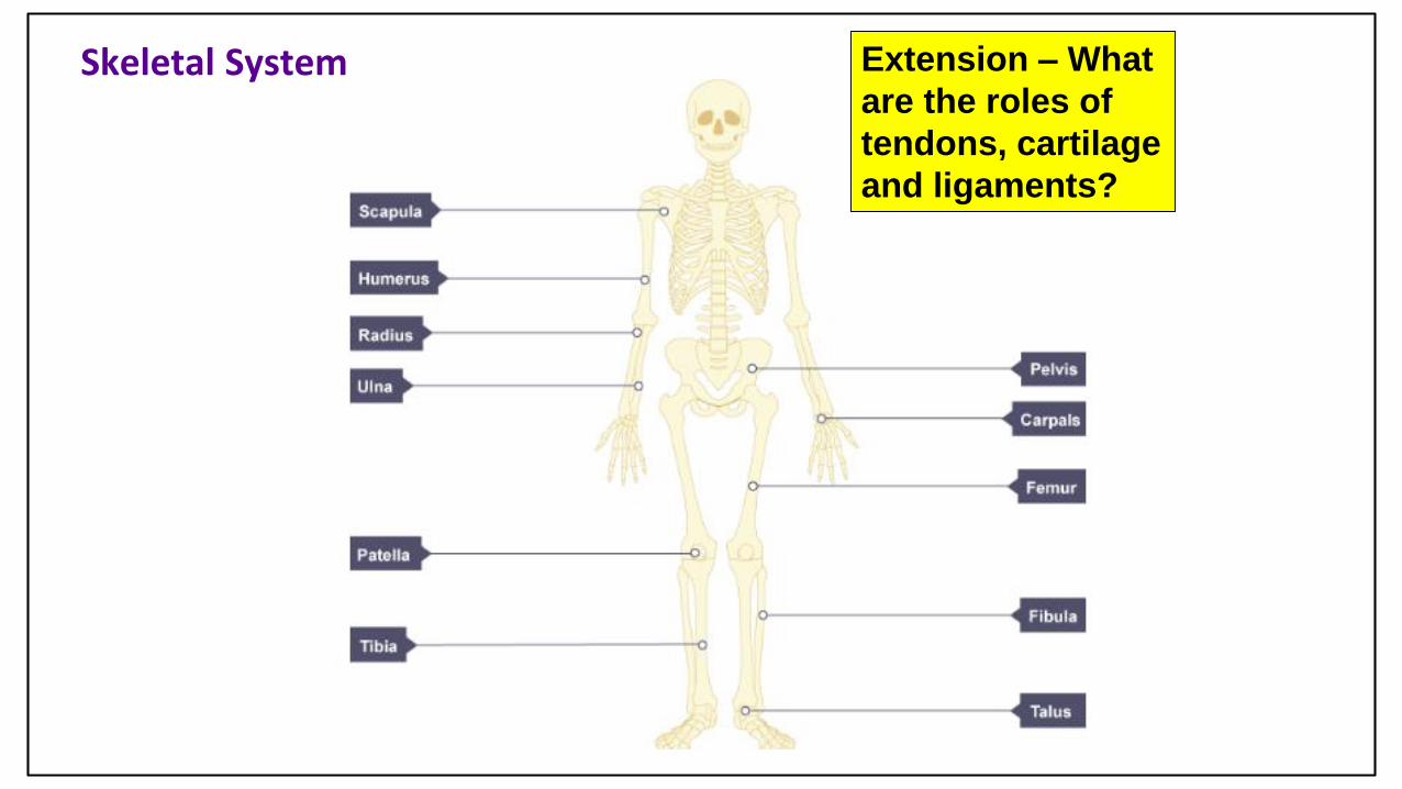
Skeletal System
Extension – What are the roles of tendons, cartilage
and ligaments?
Tendons – Are found on either end of the muscles
and connect them to bones. When the muscles
contract (shorten) it pulls on the tendons which pull
the bones to move.
Cartilage – Are found between the bones inside the
joints. They are cushions which stop the bones
rubbing together.
Ligaments – Are found at joints. They connect bones
together and prevent them
1) moving the incorrect way, and
2) coming away from each other (dislocation)

Circulatory System
R LExtension –
What is
cardio-
hypertrophy?
Circulatory System
Extension –
What is
cardio-
hypertrophy?

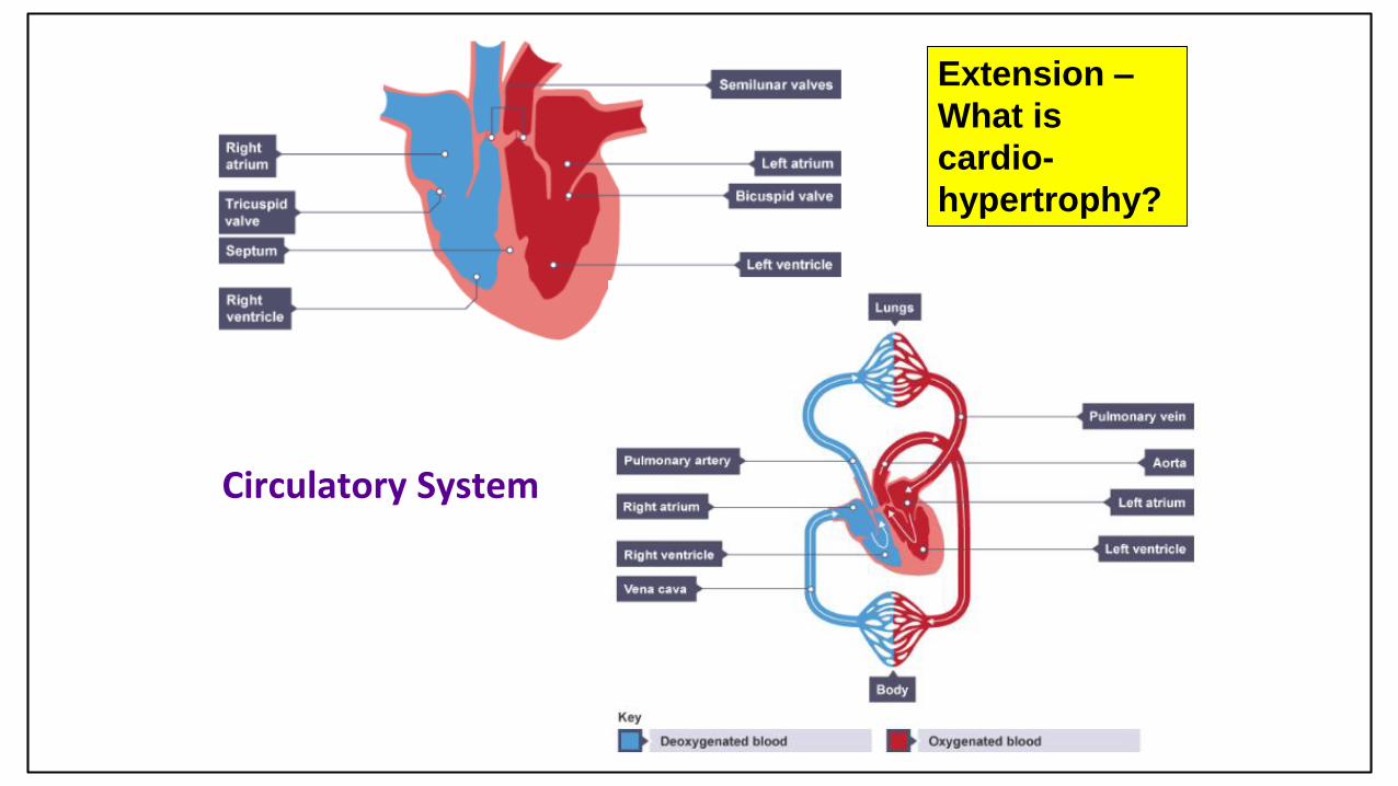
Circulatory System
Extension
What is cardio-hypertrophy?
Cardiac hypertrophy is the enlargement/ thickening of the
heart walls as a result of training. The heart is a muscle,
so once trained through aerobic exercise for a prolonged
period of time, it becomes bigger and stronger just like
any other muscle.
As a result the heart
becomes more efficient
as it can pump more
blood per beat (increased
stroke volume) and
therefore it doesn’t
have to beat as much
(decreased heart rate).

Respiratory System Extension – how
and where does
the oxygen enter
the blood stream?

Respiratory System Extension – how
and where does
the oxygen enter
the blood stream?

Respiratory System
Extension – how and where does the oxygen enter the
blood stream?
The air passes from the nose/ mouth down to the
lungs and into the bronchioles.
At the end of the bronchioles are tiny air sacs called
alveoli.
Surrounding each are capillaries (tiny blood vessels).
The walls between the
alveoli and the capillaries
are only one cell thick
which means the oxygen
and carbon dioxide can
pass through easily
(diffusion)
The structure and functions of the musculo-skeletal system
Learning objectives
To be able to describe the functions of the skeleton.
To be able to recognise and label a skeleton.
To understand the different joint classifications.
To be able to describe and analyse the different types of joint movements and how they are used during sporting movements.
To be able to label the voluntary muscles in our body
To explain the term ‘antagonist pair’ and provide examples

Bones
The adult skeleton has 206 bones and provides the framework for all movement.
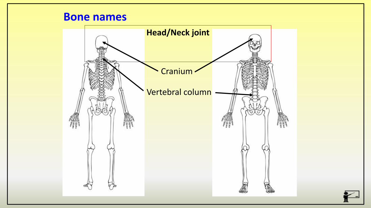
Bone names
Cranium
Vertebral column
Head/Neck joint

Bone names
Scapula
Humerus
Shoulder joint
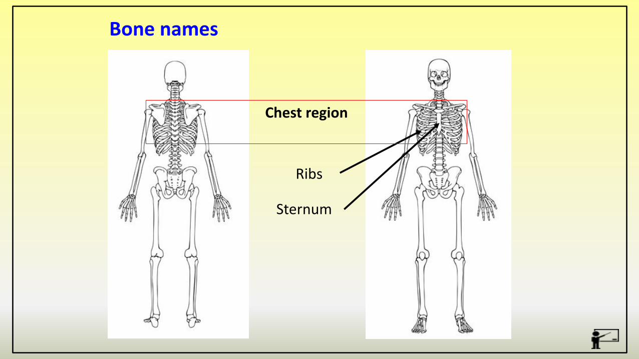
Bone names
Ribs
Sternum
Chest region
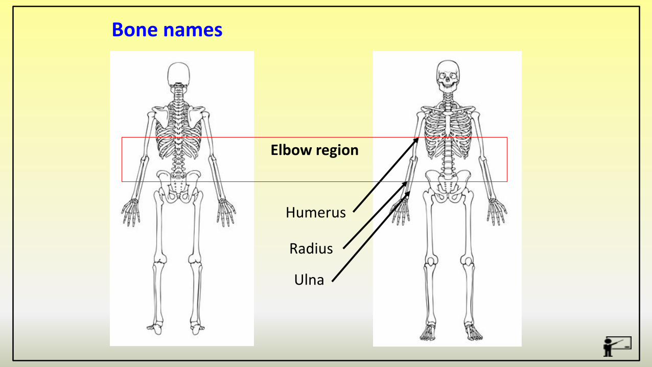
Bone names
Humerus
Radius
Elbow region
Ulna

Bone names
Femur
Pelvis
Hip joint
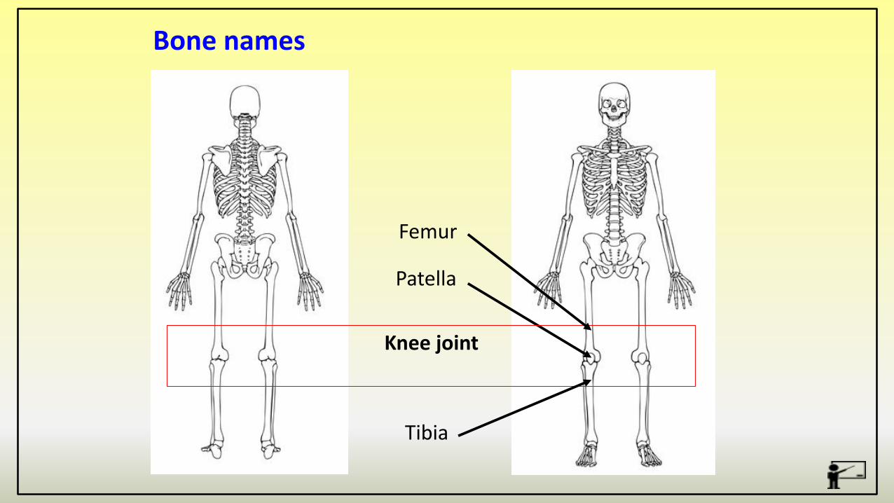
Bone names
Patella
Tibia
Knee joint
Femur
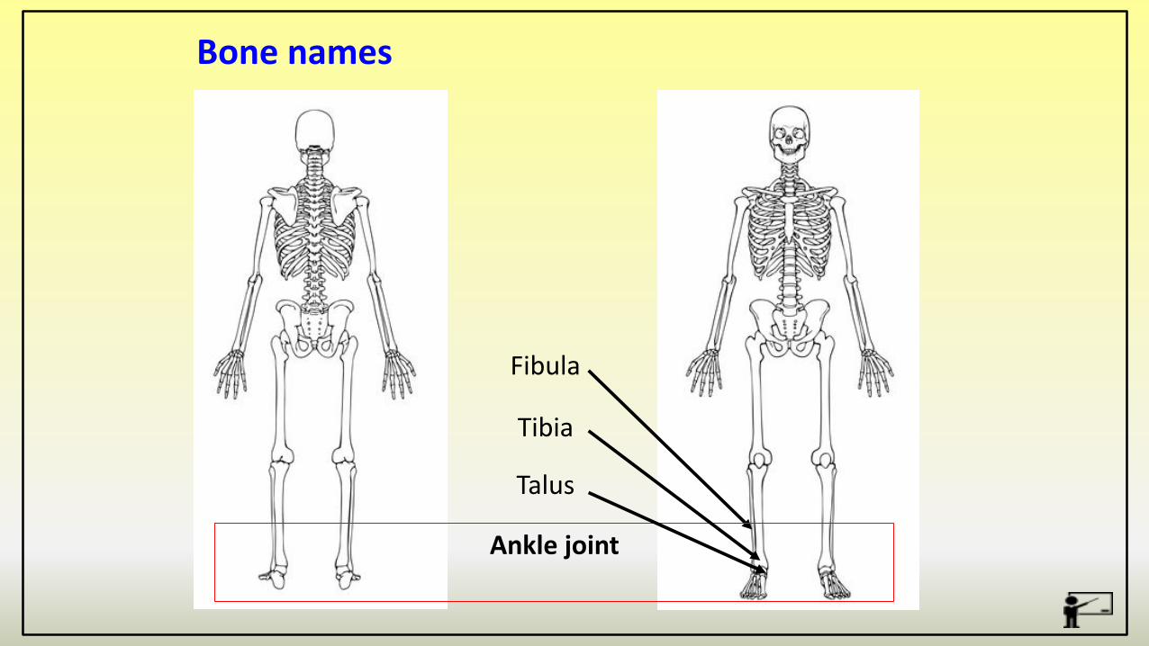
Bone names
Fibula
Tibia
Ankle joint
Talus

The skeleton performs many functions in the body -
3. Movement – Muscles are attached to the bones and once contracting allows movement.
2. Protection –The skeleton protects delicate parts of the body (organs)
1. Support –The skeleton supports the muscles.
5. Mineral storage – vital minerals such as Calcium and Phosphorus are stored in bones.
Functions of the skeleton
4. Structural shape and points for muscle attachment – The skeleton gives us our shape/size and creates levers.
6. Blood cell production –blood cells are made in the bone marrow.
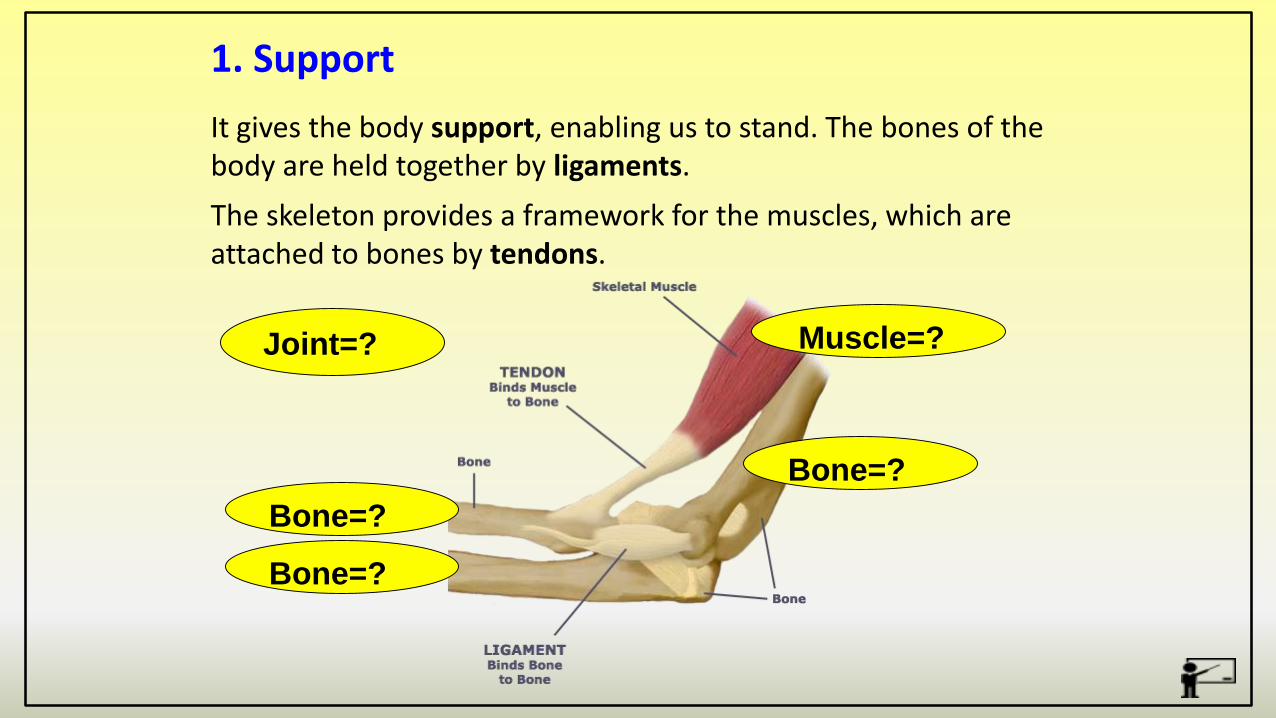
It gives the body support, enabling us to stand. The bones of the body are held together by ligaments.
The skeleton provides a framework for the muscles, which are attached to bones by tendons.
1. Support
Bone=?
Joint=?
Bone=?
Bone=?
Muscle=?

Some of our body parts, such as the brain, are very delicate and need protection.
Bones can protect body parts from impactsand injuries.
2. Protection
Which bone/s protect the following major organs –
Brain –
Heart –
Lungs –
Spinal Cord -
Cranium
Sternum/Ribs
Ribs, Scapula
Vertebrae Column

Bones come together at joints which is where movement occurs.
Muscles are firmly attached to bones via tendons forming levers to allow for sporting movements. The shape and type of the bones determine the amount of movement that occurs For example-Short bones in fingers enable finer controlled movements Long bones in arms and legs enable gross movement
3. Movement
The skeleton acts as a framework and gives the body its shape.the skeleton provides a point of attachment for muscles – when muscles contract they pull the bones
4. Structural Shape & Muscle Attachment

The minerals in your bones serve two main functions.
Minerals transform spongy bone matrix into a rigid structure
and in turn increase density and strength.
Your bones also function as a mineral storage depot,
releasing dissolved calcium, phosphorus and magnesium
into your bloodstream if needed.
5. Mineral Storage
6. Blood cell productionThe ends of long bones and some other bones including the ribs, humerus, femur and even vertebrae bones, contain red bone marrow.
This is where the red blood cells are produced which carry oxygen.

A joint is….
“A joint is a place where two or more bones meet”
Structure of the skeleton
The skeletal system has a number of joints which are responsible for the huge range of movement.
There are several different types of joint in the body which allow different types of movement.
We are concerned with freely moveable joints OR Synovial Joints

Bones are divided into a number of different categories all with different roles in the body.
Structure of the skeleton
Type Description Example Function
Long Long thin shape FemurHumerusTibiaMetatarsals etc
Responsible for a lot of movement – act as levers.Red Blood Cell production.
Short Light, small and very strong
Carpals in the wristTarsals in the foot
Support lots of weight and allow balance
Flat Thin flat bones – lot of surface area
ScapulaCranium PelvisSternum
Protection of organs/delicate body parts, large surfacearea allow large connection for musclesIrregular Specially shaped for
a particular purpose
VertebraePatella
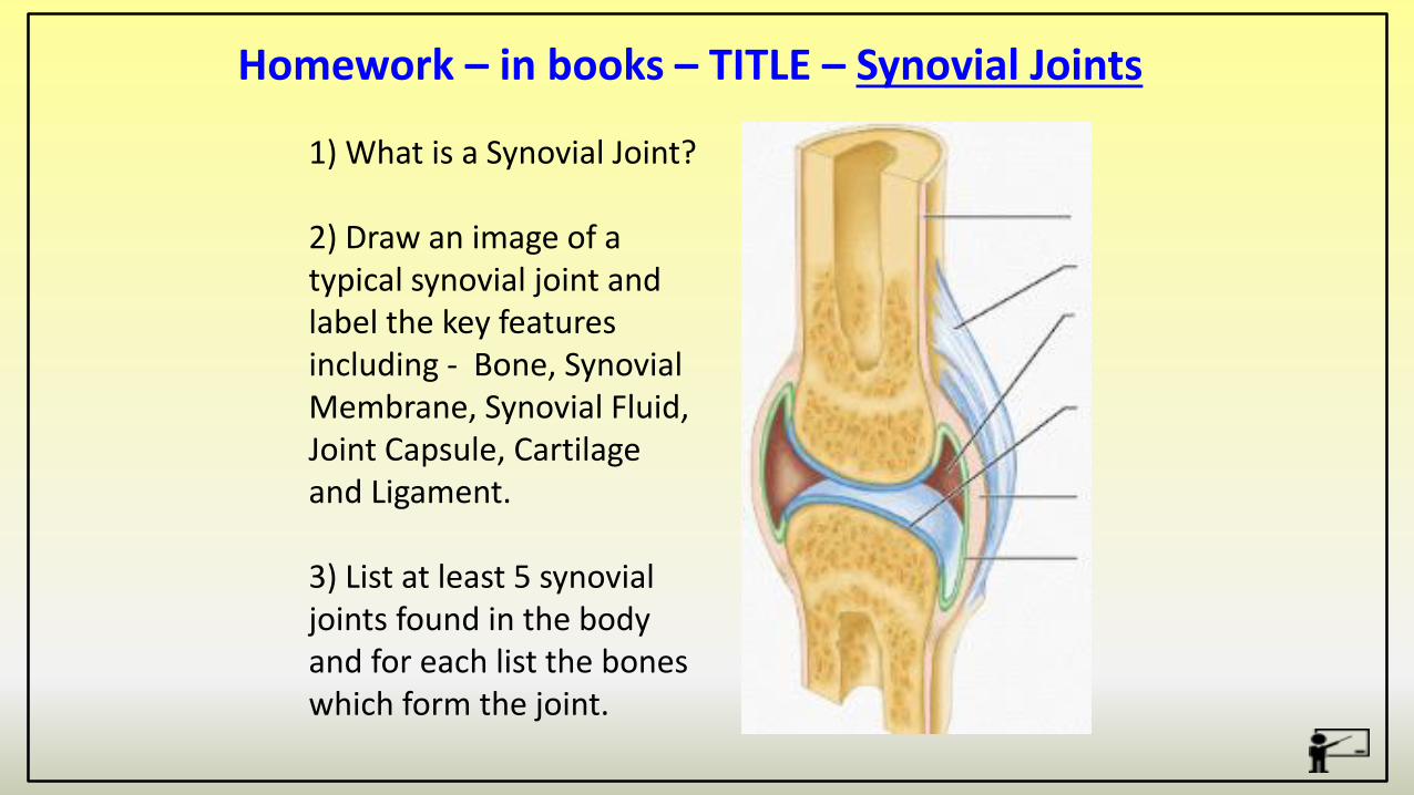
Homework – in books – TITLE – Synovial Joints
1) What is a Synovial Joint?
2) Draw an image of a typical synovial joint and label the key features including - Bone, Synovial Membrane, Synovial Fluid, Joint Capsule, Cartilage and Ligament.
3) List at least 5 synovial joints found in the body and for each list the bones which form the joint.