the biomechanics of the
TRANSCRIPT

The Biomechanics of the
Human Lower Extremity
DR.AYESH
BSPT, PP.DPT. M.Phil*

Hip joint
One of the largest and most stable joint:The hip joint
Rigid ball-and-socket configuration(Intrinsic stability)
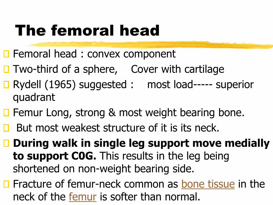
The femoral head
Femoral head : convex component
Two-third of a sphere, Cover with cartilage
Rydell (1965) suggested : most load----- superior quadrant
Femur Long, strong & most weight bearing bone.
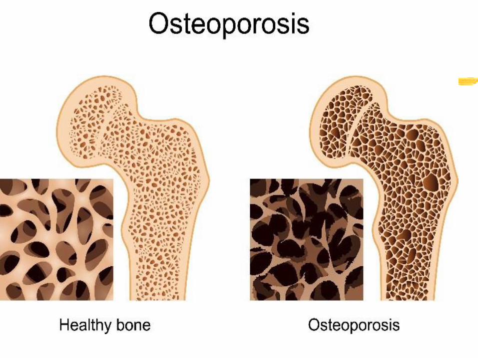
But most weakest structure of it is its neck.
During walk in single leg support move medially to support C0G. This results in the leg being shortened on non-weight bearing side.
Fracture of femur-neck common as bone tissue in the neck of the femur is softer than normal.

Acetabulum
Concave component of ball and socket joint
Facing obliquely forward, outward and downward
Covered with articular cartilage
Provide static stability
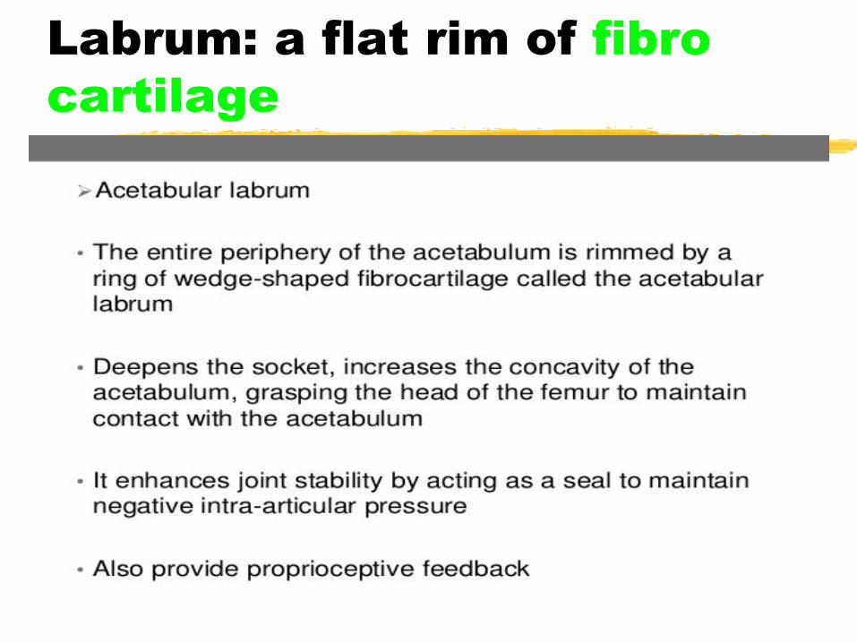
Labrum: a flat rim of fibro
cartilage

Acetabulum
Also contain Transverse acetabular ligament
provide stability
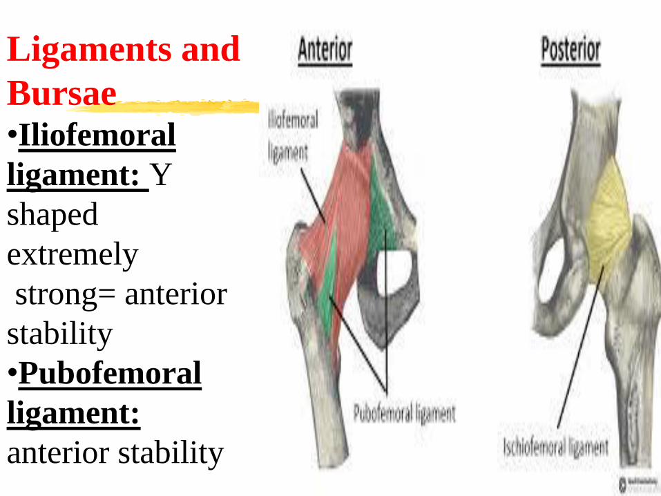
Ligaments and
Bursae •Iliofemoral
ligament: Y
shaped
extremely
strong= anterior
stability
•Pubofemoral
ligament:
anterior stability

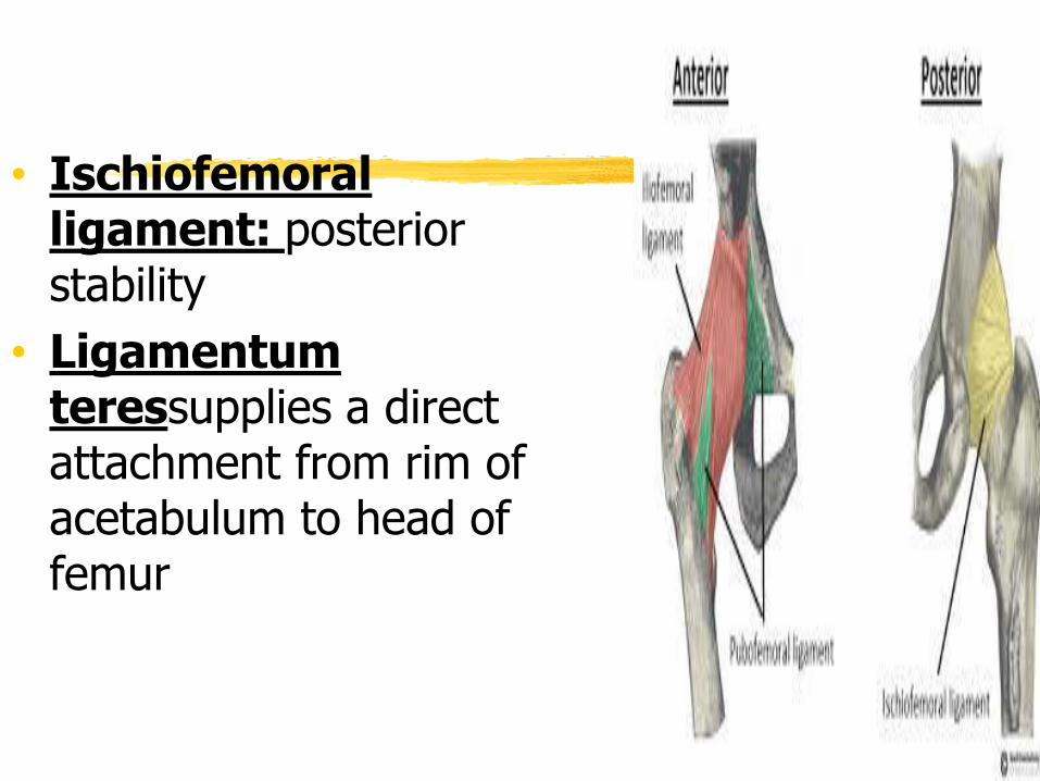
• Ischiofemoral ligament: posterior stability
• Ligamentum teressupplies a direct attachment from rim of acetabulum to head of femur
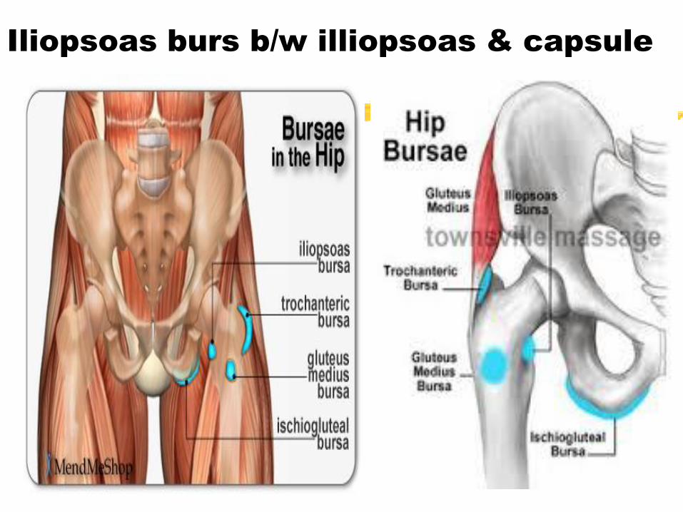
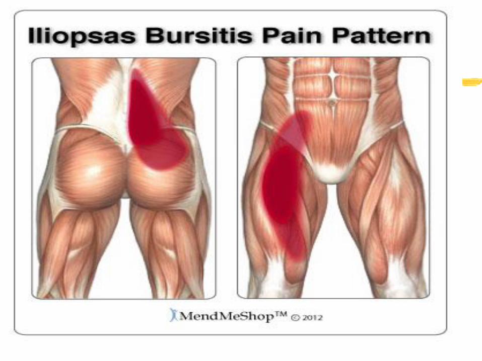
Iliopsoas burs b/w illiopsoas & capsule


Deep trochanteric bursa b/w G.maximus
& G.trochanter

Trochantric bursitis
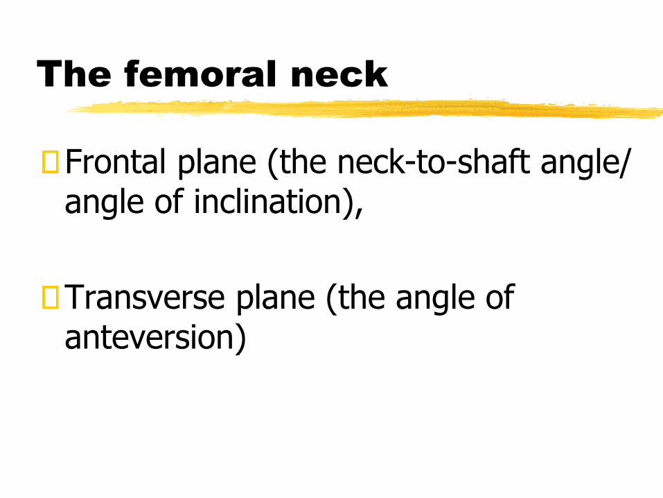
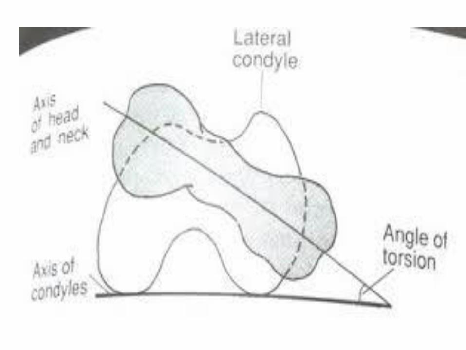
The femoral neck
Frontal plane (the neck-to-shaft angle/ angle of inclination),
Transverse plane (the angle of anteversion)

Neck-to-shaft angle :
125º, vary from 90º to 135º
Effect : lever arms

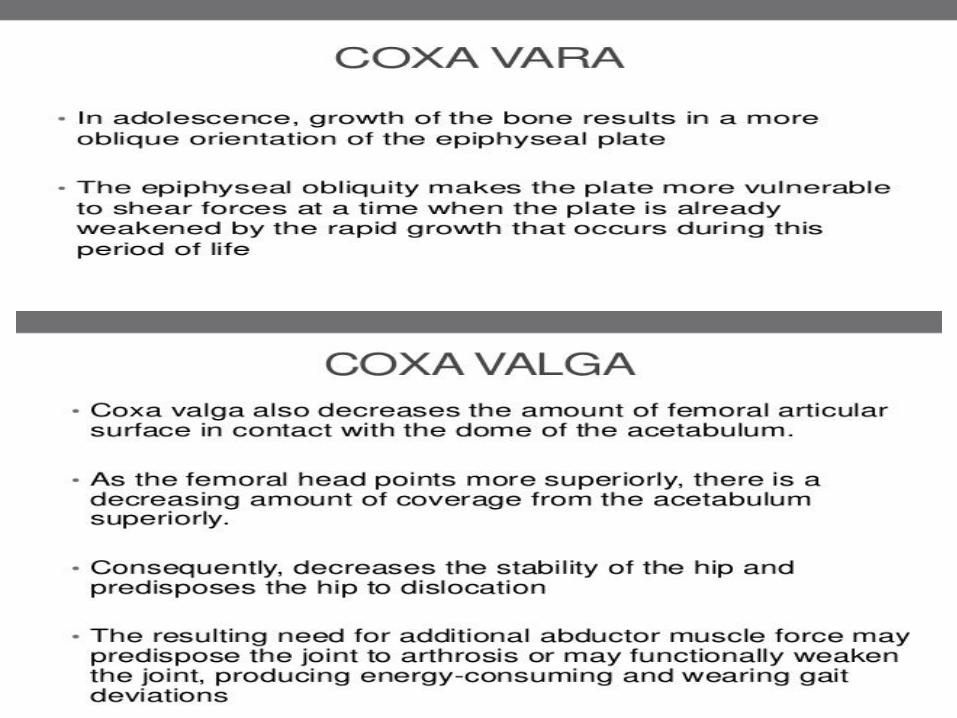
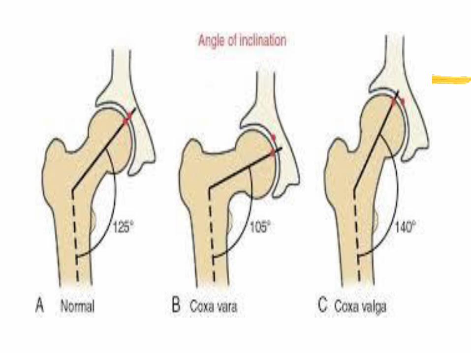
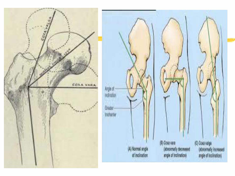


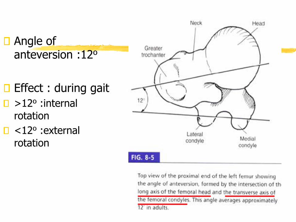
Angle of anteversion :12º
Effect : during gait
>12º :internal
rotation
<12º :external
rotation


a

Structure of the Hip
The pelvic girdle includes the two ilia and
the sacrum,. It can be rotated forward,
backward, and laterally to optimize
positioning of the hip.
Femoral
head
Femur
Acetabulum
Ilium
Sacrum
Pubis
Ischium


KinematicsRang of motion in all three planes: sagittal, frontal, transverse
0~140 0~300~15 0~250~90 0~70

Movements at the Hip
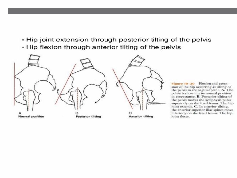
movements of the femur are facilitated by pelvic tilt
Pelvic tilt direction Femoral movement
posterior flexion
anterior extension
lateral (to opposite abduction
side)
Movements at the Hip
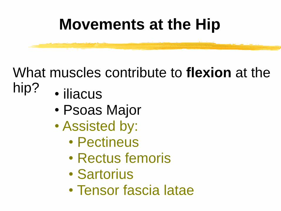
What muscles contribute to flexion at the hip? • iliacus
• Psoas Major• Assisted by:
• Pectineus• Rectus femoris• Sartorius• Tensor fascia latae

Movements at the Hip
extension at the hip joint?
•Gluteus maximus
•Hamstrings
• Biceps Femoris
• Semimembranosus
• Semitendinosus

Movements at the Hip
abduction at the hip joint
• gluteus medius
• assisted by:
• gulteus minimus

Movements at the Hip
adduction at the hip joint?
• adductor magnus
• adductor longus
• adductor brevis
• assisted by:
• gracilis

Movements at the Hip
lateral rotation at the hip joint?
•Piriformis
•Gemellus superior
•Gemellus inferior
•Obturator internus
•Obturator externus
•Quadratus femoris
Movements at the Hip
medial rotation at the hip joint?
• gluteus minimus
•Assisted by:
• TFL
•Semimembranosus
•Semitendinosus
•Gluteus medius

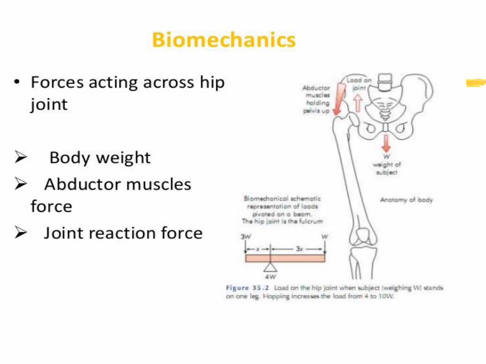
LOADS ON THE HIP
❑Highly specialized and well designed
❑Compressive forces due to following::::
•Amount of load (more than ½ of body weight
above hip +tension in surrounding muscles)..
•Effect of speed→increase speed increase load.,
Foot wear also effect load.
Load decrease due to smooth gait pattern & soft
heel strike…
•Training surface………..
•Painful conditions……….

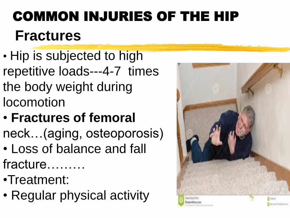
COMMON INJURIES OF THE HIP
Fractures
• Hip is subjected to high
repetitive loads---4-7 times
the body weight during
locomotion
• Fractures of femoral
neck…(aging, osteoporosis)
• Loss of balance and fall
fracture………
•Treatment:
• Regular physical activity

Injuries contd…
CONTUSIONS
•Anterior aspect muscles--- prime
location for direct injury in Contact
sports----→Internal hemorrhaging---
→Appearance of bruises mild to
severe
❑Uncommon but serious
complication as syndrome in which
internal hemorrhage-→compression
on nerves, vessels, muscle----
→tissue death

Injuries contd…
STRAINS
•Hamstring strain…….late
stance or late swing phase as
ecentric contraction.
(simultaneous hip flexion
&knee extension)
•Groin Strain….forceful thigh
movement in abduction
causes strain in adductors(ice
hockey players

KNEE BIOMECHANICS

Structure of the Knee
Modified hinge joint. Formed by Tibofemoral & patello femoral joitTibiofemoral joint?
• Dual condyloid
articulations between
medial and lateral
condyles of tibia and
the femur; composing
the main hinge joint of
the knee
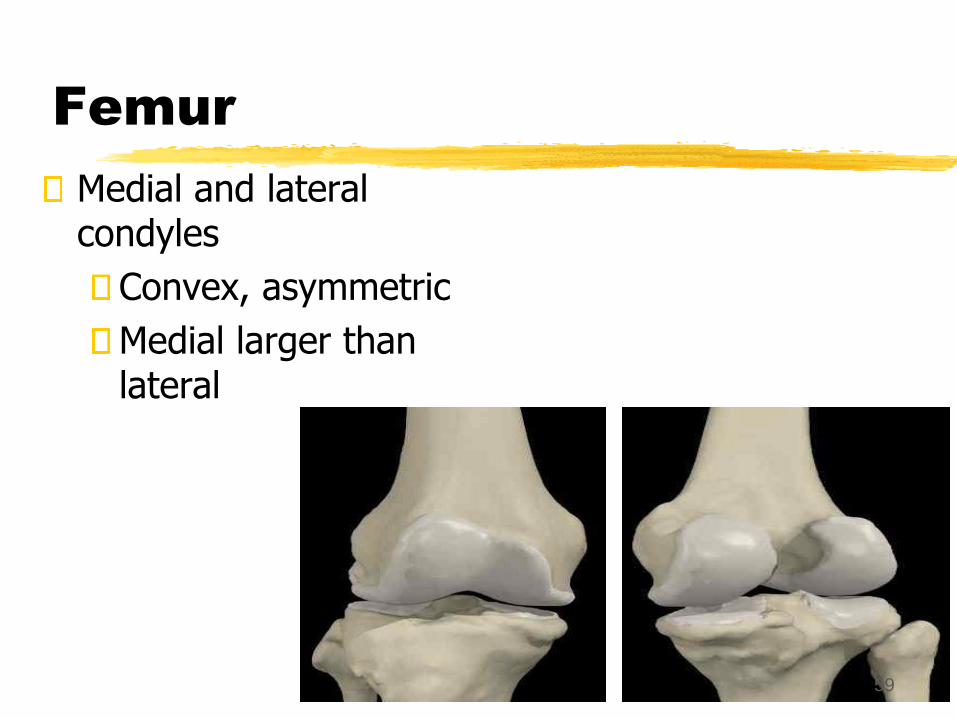
Femur
Medial and lateral condyles
Convex, asymmetric
Medial larger than lateral
59

Tibia
Medial tibial condyle: concave
Lateral tibial condyle: flat or concave
Medial 50% larger than lateral
1
60
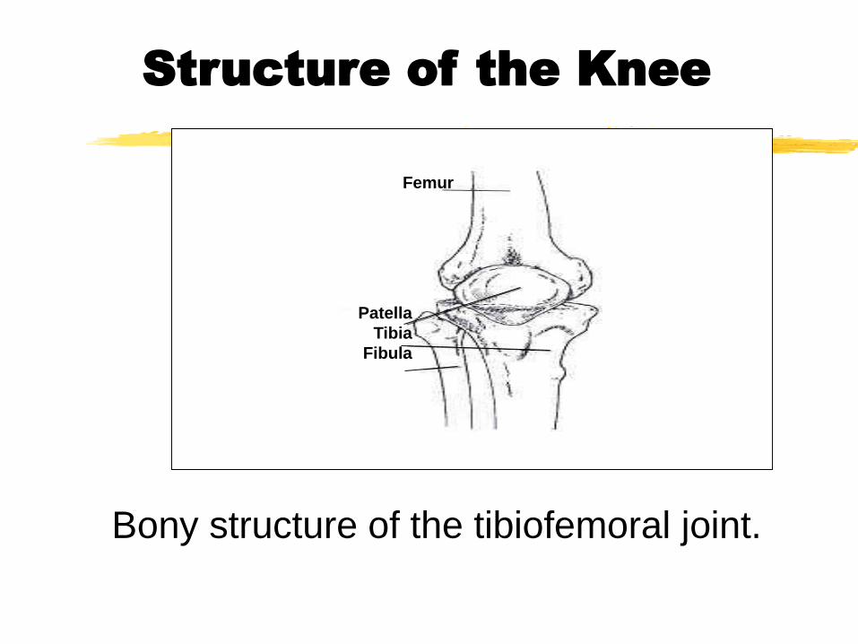
Structure of the Knee
Bony structure of the tibiofemoral joint.
Patella
Tibia
Fibula
Femur

Structure of the Knee
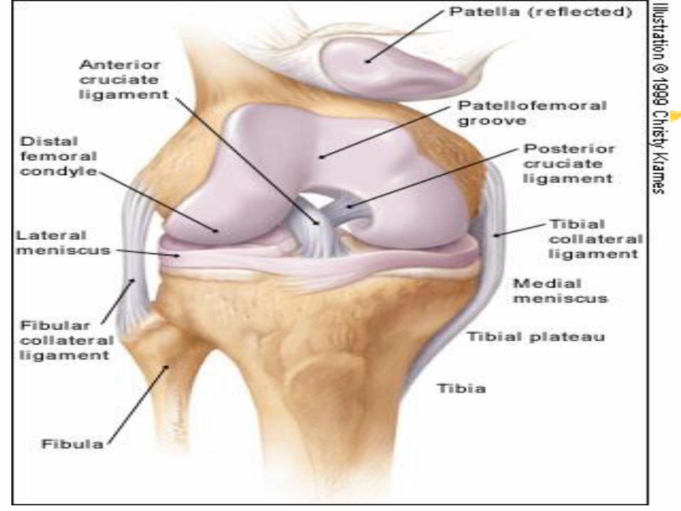
• The menisci of the knee. Medial meniscus is also attached
directly to the medial collateral ligament
Lateral meniscus
Posterior cruciate ligament
Transverse ligament
Anterior cruciate ligament
Medial meniscus
Superior view

• The menisci of the knee. •Deepens the articulating depression of tibial plateaus• Load transmission and shock absorption•Without menisci the weight of the femur would be concentrated to one point on the tibia and stress may reach up to 3 times• Increased likelihood of degenerative conditions
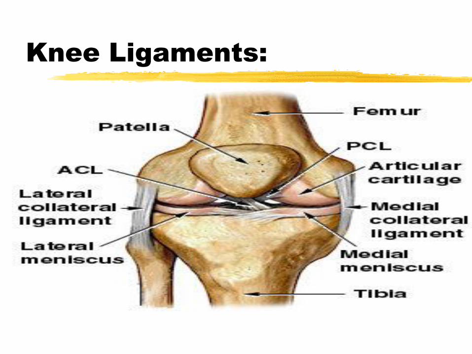
Knee Ligaments:

Medial & lateral stabilizers
(mostly ligaments)
Ligaments
most important static & dynamic stabilizers by ligaments & muscles
tensile strength - related to composition of muscle
Primary valgas restraint -57-78% restraining moment of knee(MCL)
Tense in lateral rotation, lax in flexion
Lateral side
LCL
Primary Varus restraint
lax in extension
Taut in medial rot
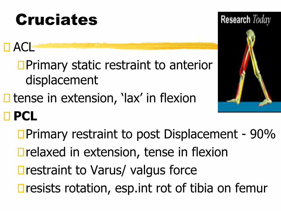
Cruciates
ACL
Primary static restraint to anterior displacement
tense in extension, ‘lax’ in flexion
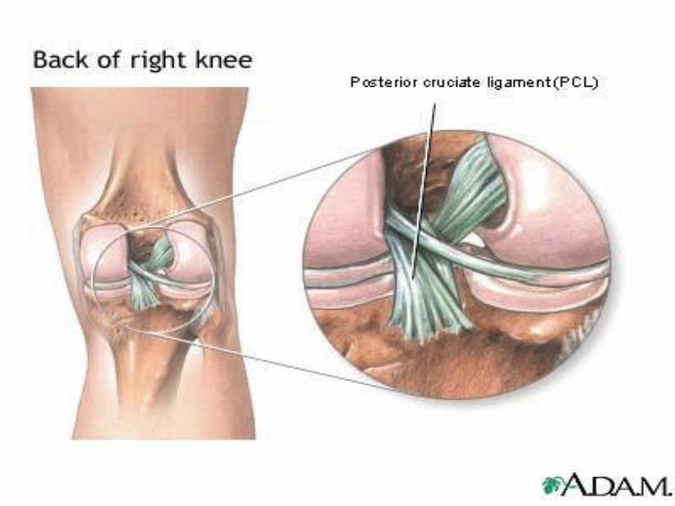
PCL
Primary restraint to post Displacement - 90%
relaxed in extension, tense in flexion
restraint to Varus/ valgus force
resists rotation, esp.int rot of tibia on femur
Structure of the Knee
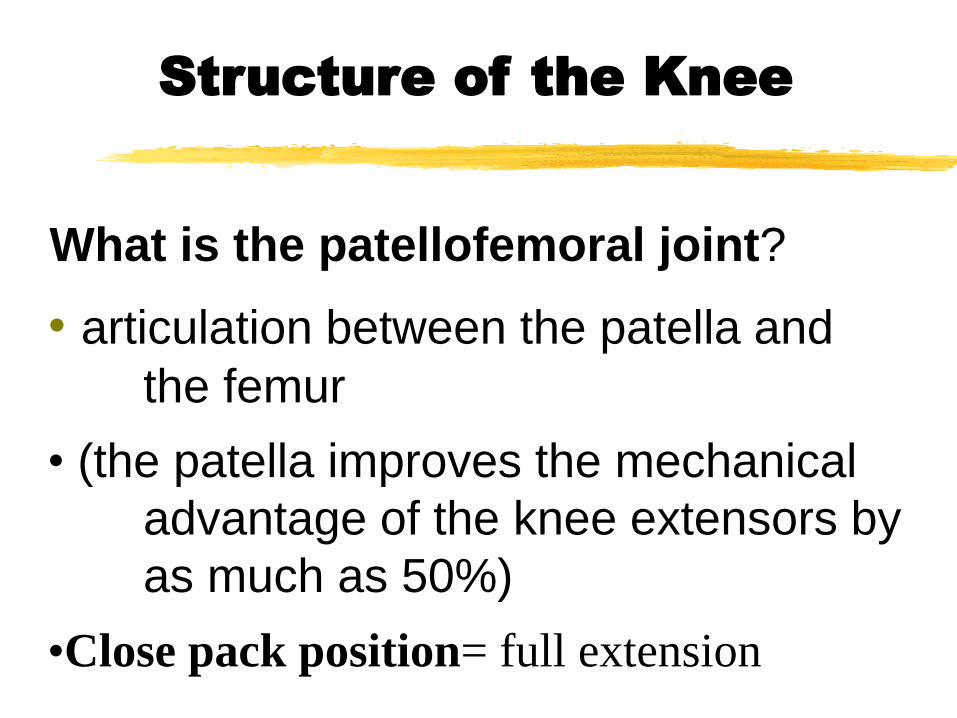
What is the patellofemoral joint?
• articulation between the patella and
the femur
• (the patella improves the mechanical
advantage of the knee extensors by
as much as 50%)
•Close pack position= full extension

Other Important Structures
Articular cartilage1/4 inch thick
tough and slick
Patella and patellar
tendon
Tibial tuberoscity
Patellofemoral groove
Patella acts like a
fulcrum to increase the
force of the quadriceps
muscles
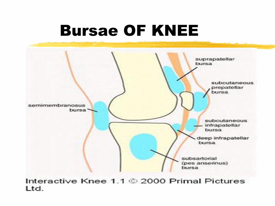
Bursae OF KNEE

Patellofemoral joint motion
Gliding movements== 7 cm in vertical direction
Superior glide
Inferior glide
Lateral and medial shifting (v.little)

Muscles
Quadriceps -
extension
Hamstrings -
flexion
IT band from the
gluteus maximus
and tensor fascia
latae

Screw home –locking mechanism:
A key element to knee stability for standing upright, is the rotation between the tibia and femur.
It occurs at the end of knee extension, between full extension (0 degrees)&20 degrees of knee flexion.
The tibia rotates internally during the swing phase and externally during the stance phase.
External rotation occurs during terminal degrees of knee extension and results in tightening of both cruciate ligaments, which locks the knee.
The tibia is then in the position of maximal stability with respect to the femur.
Vice versa by poplitius muscle
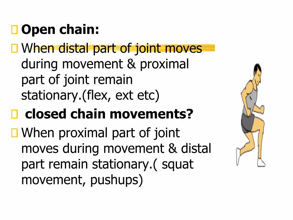
Open chain:
When distal part of joint moves during movement & proximal part of joint remain stationary.(flex, ext etc)
closed chain movements?
When proximal part of joint moves during movement & distal part remain stationary.( squat movement, pushups)

Loads on the knee joint
Tibiofemoral joint:
Compression loading more in stance phase
Shear loading= tendency of the femur to displace anteriorly on tibial plateaus(glide)
Knee flexion angle exceeding than 90 degree result in larger shear forces.
Full squats not recommended for novice athletes


forces at Patellofemoral
joint
1/3rd of body weight compressive forces during normal walking
3 times the body weight during stair climbing--→High compressive forces
during knee flexion
Squatting highly stressful to the knee complex


Common Injuries of the Knee
& Lower Leg
Knee Anatomy

Patella Fractures
Result from direct blow such as knee hitting dashboard in MVA, fall on flexed knee, forceful contraction of quad. Muscle.
Transverse fractures most common
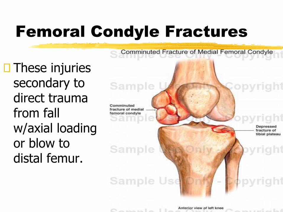
Femoral Condyle Fractures
These injuries secondary to direct trauma from fall w/axial loading or blow to distal femur.
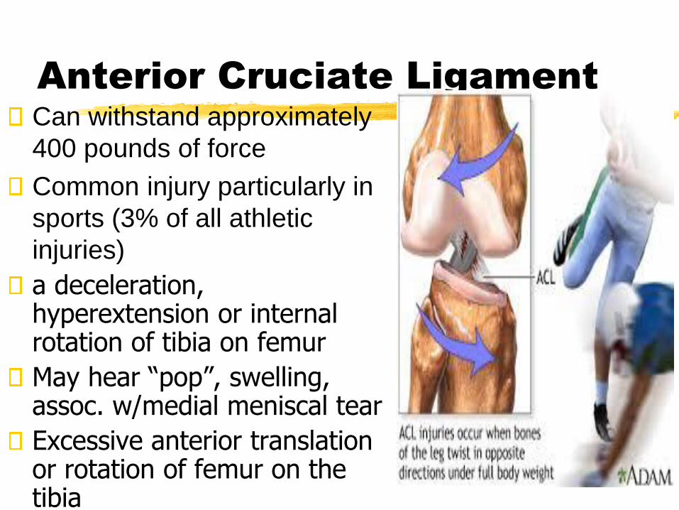
Anterior Cruciate LigamentCan withstand approximately
400 pounds of force
Common injury particularly in
sports (3% of all athletic
injuries)
a deceleration, hyperextension or internal rotation of tibia on femur
May hear “pop”, swelling, assoc. w/medial meniscal tear
Excessive anterior translation or rotation of femur on the tibia
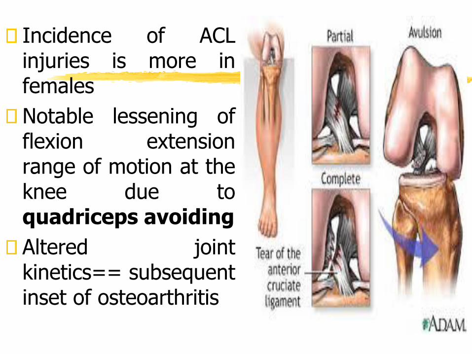
Incidence of ACLinjuries is more infemales
Notable lessening offlexion extensionrange of motion at theknee due toquadriceps avoiding
Altered jointkinetics== subsequentinset of osteoarthritis
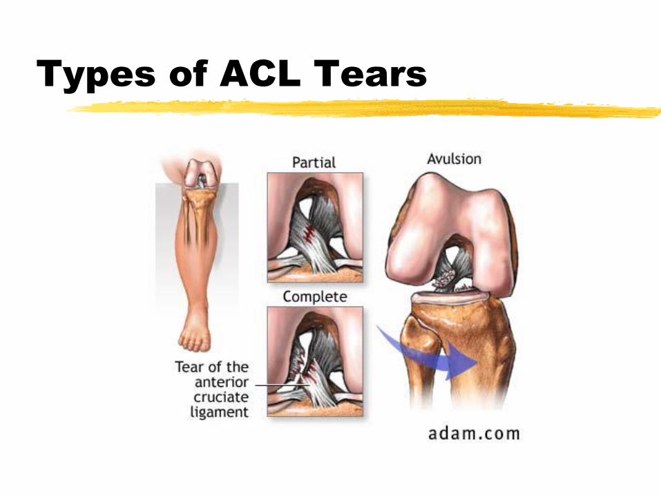
Types of ACL Tears
Surgical repair through middle third of patellar tendon
Notable weakness in quadriceps, impaired joint range and proprioception
Muscle inhibition: inability to activate all motor units of a muscle during maximal voluntary contraction
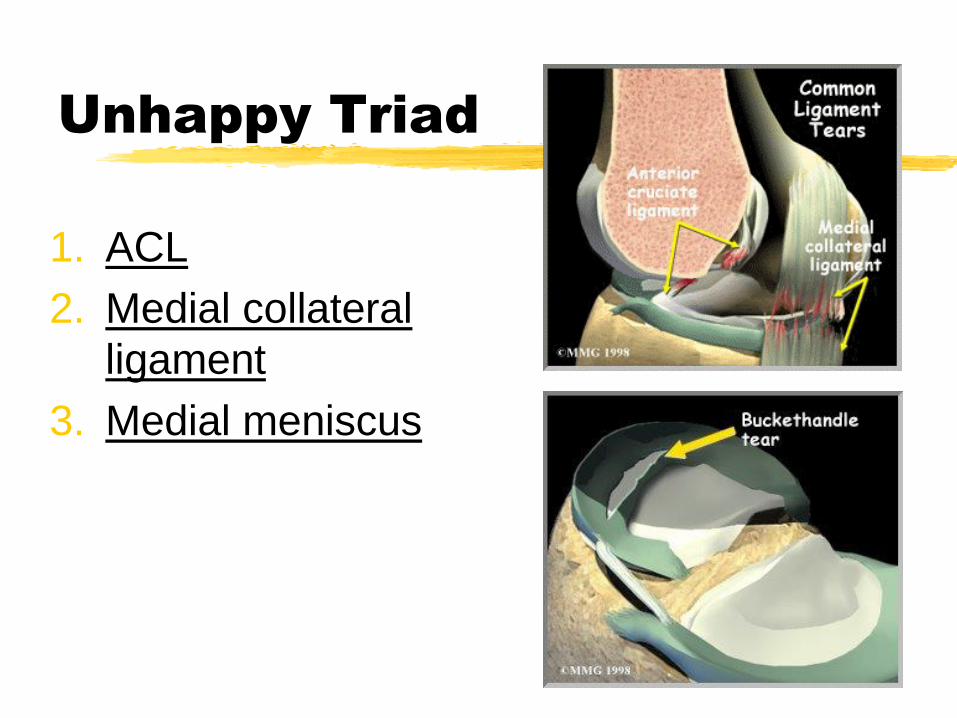
Unhappy Triad
1. ACL
2. Medial collateral
ligament
3. Medial meniscus

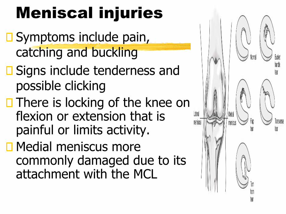
Meniscus InjuriesOne of the most commonly injured parts of the knee
Mechanism is usually squatting or twisting maneuvers. OR
Meniscal tears occur during twisting motions with the knee flexed
Also, they can occur in combination with other injuries such as a torn ACL (anterior cruciate ligament).
Older people can injure the meniscus without any trauma as the cartilage weakens and wears thin over time, setting the stage for a degenerative tear.
Combination injuries
Meniscal injuries
Symptoms include pain, catching and buckling
Signs include tenderness and possible clicking
There is locking of the knee on flexion or extension that is painful or limits activity.
Medial meniscus more commonly damaged due to its attachment with the MCL

Posterior Cruciate
Ligament
Less common than ACL injury
Mechanism is hyperflexion of knee with foot plantarflexed
PCL sprains usually occur because the ligament was pulled or stretched too far, anterior force to the knee, or a simple misstep.

PCL Injuries Impact with dash board during motor vehicle accident
Direct force on proximal anterior tibia
PCL injuries disrupt knee joint stability because the tibia can sag posteriorly.
The ends of the femur and tibia rub directly against each other, causing wear and tear to the thin, smooth articular cartilage.
This abrasion may lead to arthritis in the knee.
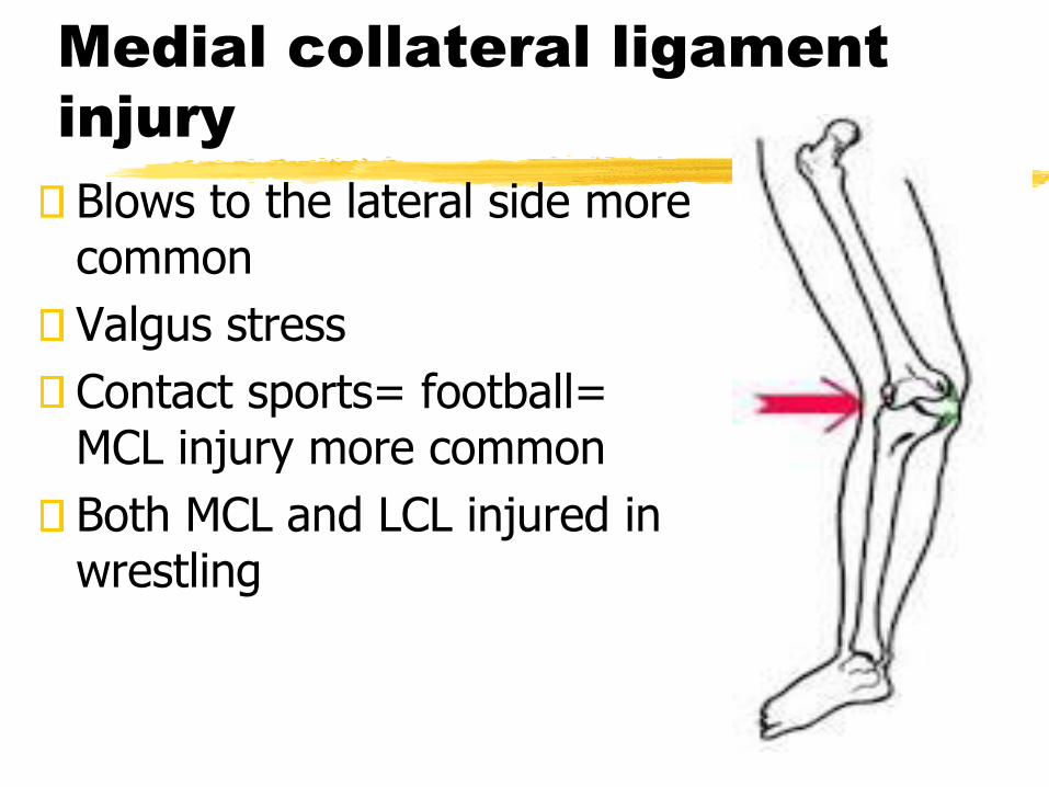
Medial collateral ligament
injury
Blows to the lateral side more common
Valgus stress
Contact sports= football= MCL injury more common
Both MCL and LCL injured in wrestling


Prophylactiion:
knee bracing
To prevent knee ligament injuries in contact sports….(Matter of contention
Protection from torsional loads
Reduced sprinting speed and earlier onset of fatigue

Patellar Tendonitist
Due to high deceleration or eccentric forces of the quadriceps at the knee during landing
As you land the hamstrings cause your knee to flex to absorb the shock of impact
In order to control or decelerate the flexion produced by the hamstrings, the quadriceps muscles contract eccentricly
Eccentric contractions occur as the muscle is being lengthened or stretch
Eccentric contractions produces high amounts of force, and therefore stress to the patellar tendon

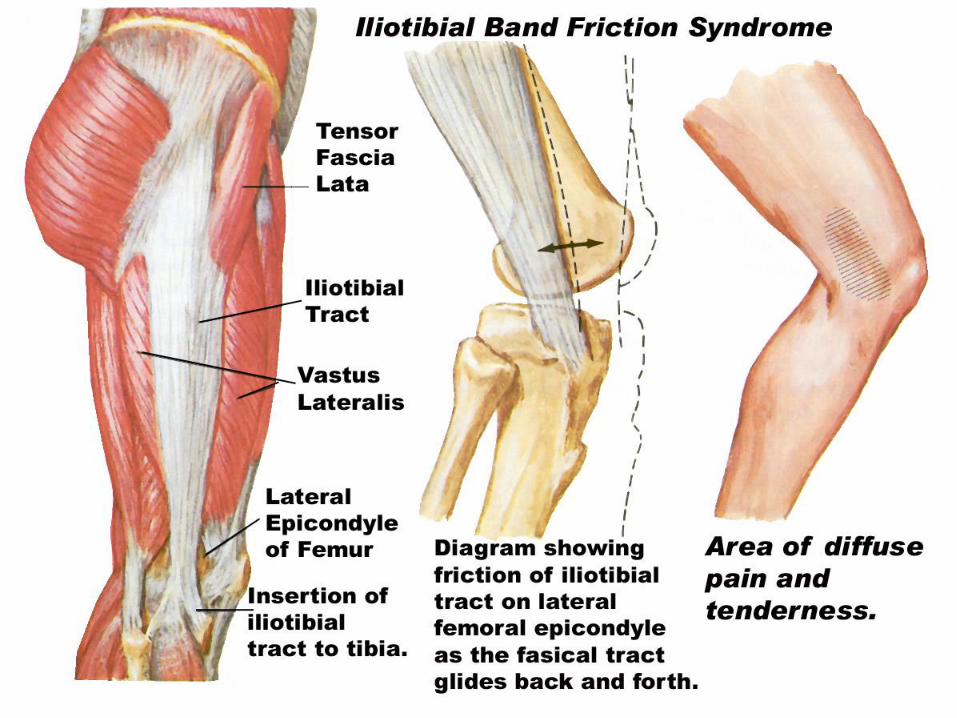
Iliotibial band friction
syndromeFriction of posterior edge of Iliotibial band against the lateral condyle of the femur during foot strike
Very common in distance runners, hence referred as runner’s knee
Training errors and anatomical malalignments
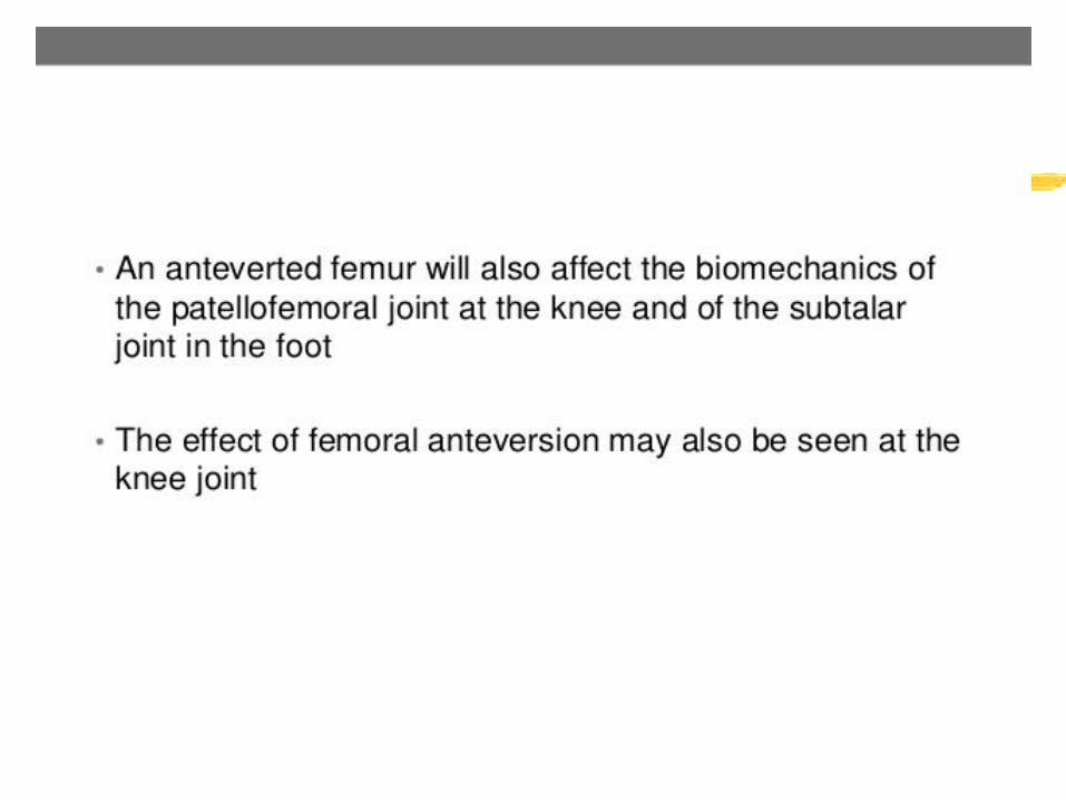
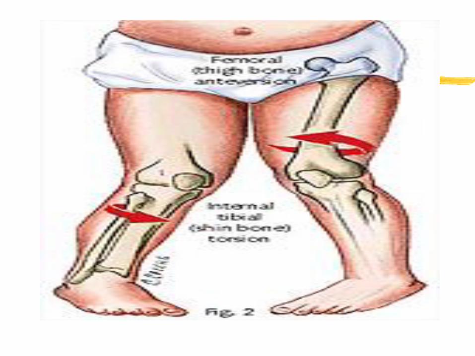
Excessive tibial lateral torsion, femoral anteversion, genu valgum, genu varum, increased Q angle etc,
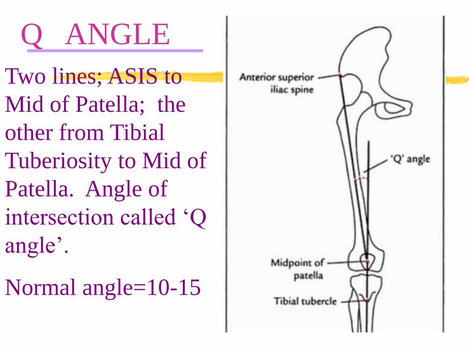
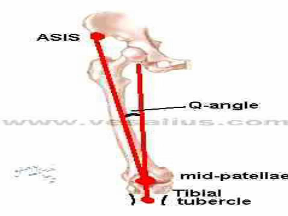
Q ANGLE
Two lines; ASIS to
Mid of Patella; the
other from Tibial
Tuberiosity to Mid of
Patella. Angle of
intersection called ‘Q
angle’.
Normal angle=10-15


❖The greater the Q
angle, the greater the
tendency to move
the patella laterally
against the lateral
femoral condyle.
❖A large Q angle
plus strong quad
contraction can
dislocate pat.
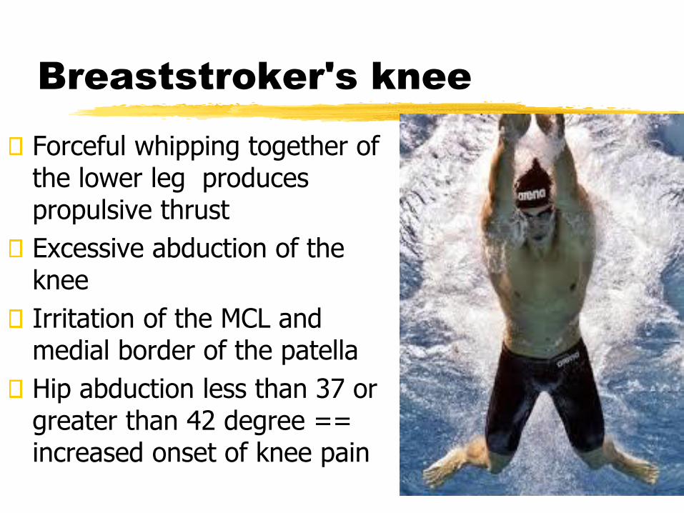
Breaststroker's knee
Forceful whipping together of the lower leg produces propulsive thrust
Excessive abduction of the knee
Irritation of the MCL and medial border of the patella
Hip abduction less than 37 or greater than 42 degree == increased onset of knee pain

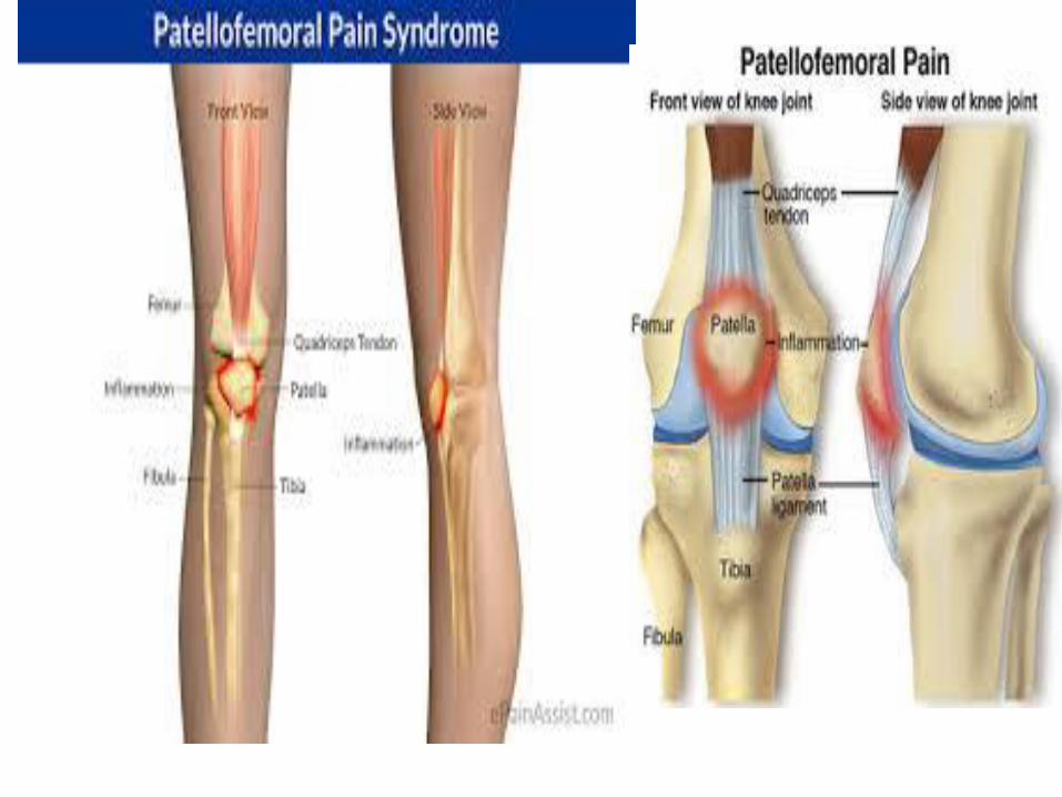
Patellofemoral pain
syndrome
Painful Patellofemoral joint motioninvolving anterior knee pain after activitiesrequiring repeated flexion at the knee
Anatomical malalignments
Weak Vastus Medialis Oblique and VastusLateralis strong
Large Q angle responsible
Patellar maltracking

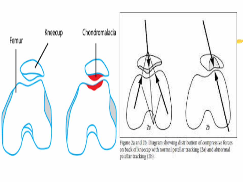


Chondromalacia PatellaeOveruse syndrome of patellarcartilage characterized bysoftening & fissuring of thearticular cartilage of the patella
Caused by aging and mechanicaldefects that include::: patello-femoral malalignments whichleads to tracking abnormality ofpatella putting excessive lateralpressure on articular cartilage
Seen in young active women,pain worse w/stair climbing andrising from a chair
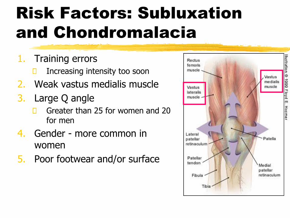
Risk Factors: Subluxation
and Chondromalacia
1. Training errors
Increasing intensity too soon
2. Weak vastus medialis muscle
3. Large Q angle
Greater than 25 for women and 20 for men
4. Gender - more common in women
5. Poor footwear and/or surface
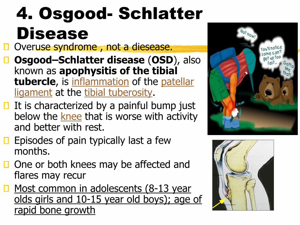
4. Osgood- Schlatter
DiseaseOveruse syndrome , not a diesease.
Osgood–Schlatter disease (OSD), also known as apophysitis of the tibial tubercle, is inflammation of the patellar ligament at the tibial tuberosity.
It is characterized by a painful bump just below the knee that is worse with activity and better with rest.
Episodes of pain typically last a few months.
One or both knees may be affected and flares may recur
Most common in adolescents (8-13 year olds girls and 10-15 year old boys); age of rapid bone growth

Shin Splints
Generalized pain alongthe anterolateral orposteromedial aspectof the lower leg iscommonly known asshin splints
Overuse injury oftenassociated withrunning, dancing onthe hard surface andrunning uphill
Structure of the Ankle
Tibiotalar joint
• Hinge joint where the convex surface of
the superior talus articulates with the
concave surface of the distal tibia
• considered to be the ankle joint
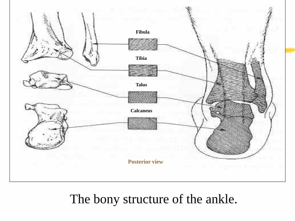
Structure of the Ankle
The bony structure of the ankle.
Fibula
Tibia
Talus
Calcaneus
Posterior view

Movements at the Ankle
Dorsiflexion at the ankle
• Tibialis anterior
• extensor digitorum longus
• peroneus tertius
• assisted by:
• extensor hallucis longus

Movements at the Ankle
plantar flexion at the ankle
• Gastrocnemius
• soleus
• assisted by:
Tibialis posterior, Plantaris, peroneus
longus, peroneus brevis ,flexor
hallucis longus,, flexor digitorum longus
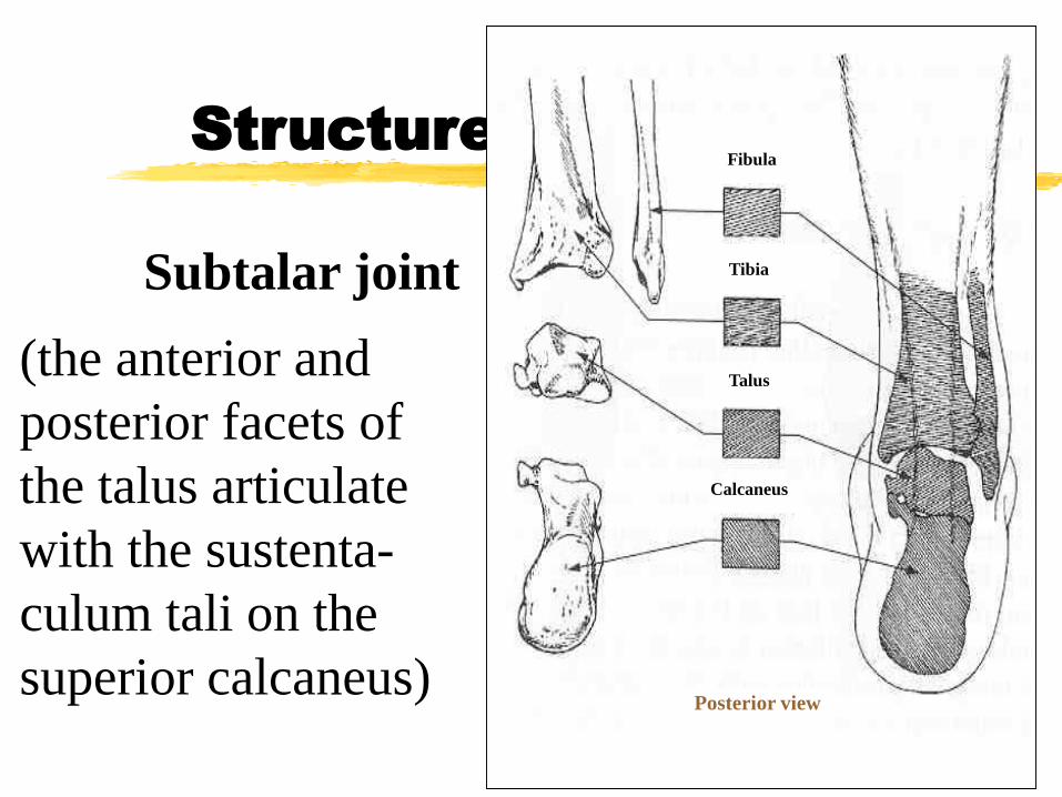
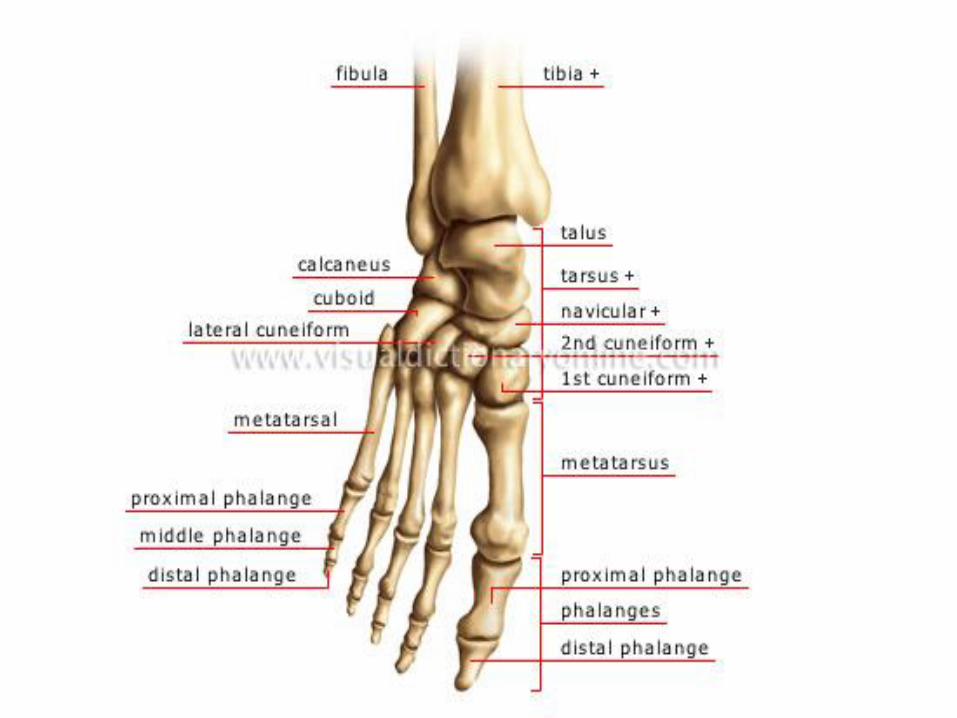
Structure of the Foot
Subtalar joint
(the anterior and
posterior facets of
the talus articulate
with the sustenta-
culum tali on the
superior calcaneus)
Fibula
Tibia
Talus
Calcaneus
Posterior view
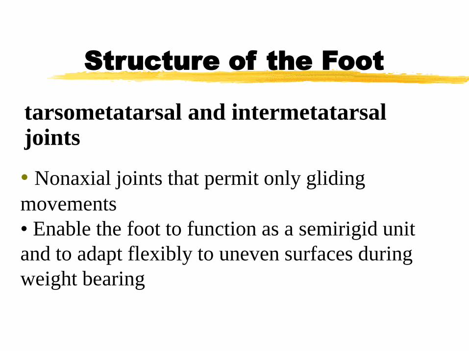
Structure of the Foot
tarsometatarsal and intermetatarsal joints
• Nonaxial joints that permit only gliding
movements
• Enable the foot to function as a semirigid unit
and to adapt flexibly to uneven surfaces during
weight bearing
Structure of the Foot
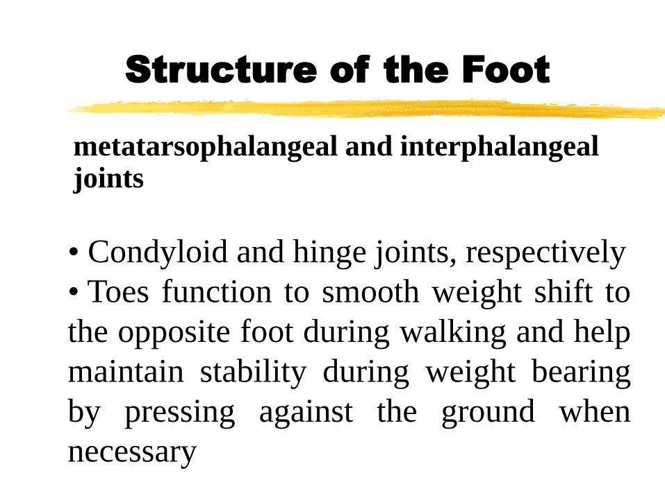
metatarsophalangeal and interphalangeal joints
• Condyloid and hinge joints, respectively
• Toes function to smooth weight shift to
the opposite foot during walking and help
maintain stability during weight bearing
by pressing against the ground when
necessary

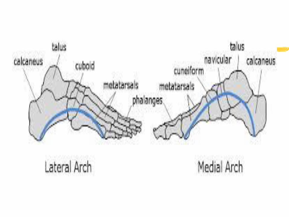
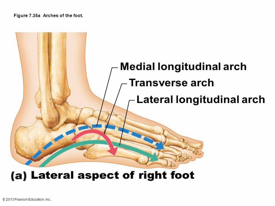
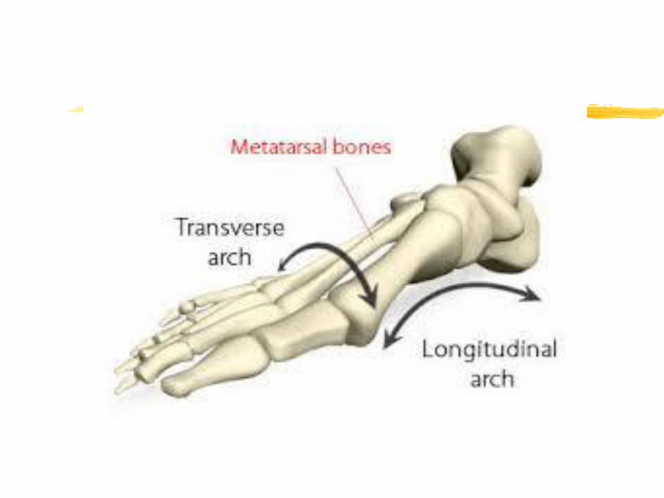
Structure of the Foot
plantar arches
• The medial and lateral longitudinal
arches stretch form the calcaneus to the
metatarsals and tarsals
• The transverse arch is formed by the
base of the metatarsal bones

Plantar Fascia
• Thick bands of fascia that
cover the plantar aspects of
the foot
• During weight bearing=
mechanical energy is stored
in the stretched ligaments,
tendons, and plantar fascia of
the foot.
• This energy is released to
assist with push-off of the
foot from the surface.

Structure of the Foot
The plantar fascia.
Lateral view
Plantar view
Plantar fascia
Movements of the Foot
Toe flexion and extension
• Flexion - flexor digitorum longus,
flexor digitorum brevis, lumbricals,
Interossei
• Extension - extensor hallucis longus,
extensor digitorum longus, extensor
digitorum brevis
Movements of the Foot
Inversion and eversion
• Inversion - Tibialis posterior, Tibialis
anterior
• Eversion - peroneus longus,
peroneus brevis, assisted by peroneus
tertius

Common injuries of the
ankle and foot
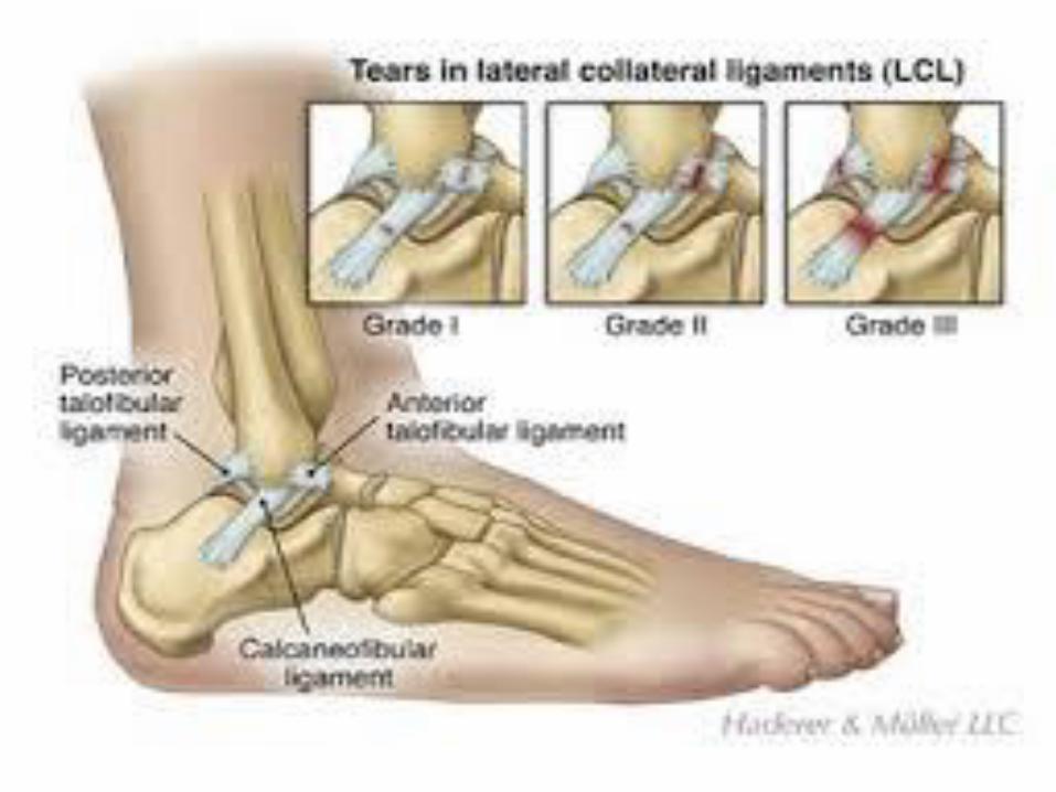
•Ankle injuries
•Inversion sprains= stretching or rupture
of lateral ligaments (ATFL, PTFL, CFL)
•Medial = deltoid ligament very strong
(ATTL, PTTL, TCL, TNL)
•Ankle bracing or taping (Mild injury
treatment)
Ankle sprainGreater inversion increases the potential for over-stretching of the lateral ligaments.
Most sprains involve the lateral ligaments from excessive inversion.
Deltoid ligament is sprained less often (25% of ankle sprains)
Of the lateral ligments, the ATFL is sprained the most often followed by the CFL
Sprains occur most often with the foot in plantar flexion and inversion
The medial malleolus is shorter than the lateral mallelous so there is naturally more inversion than eversion.


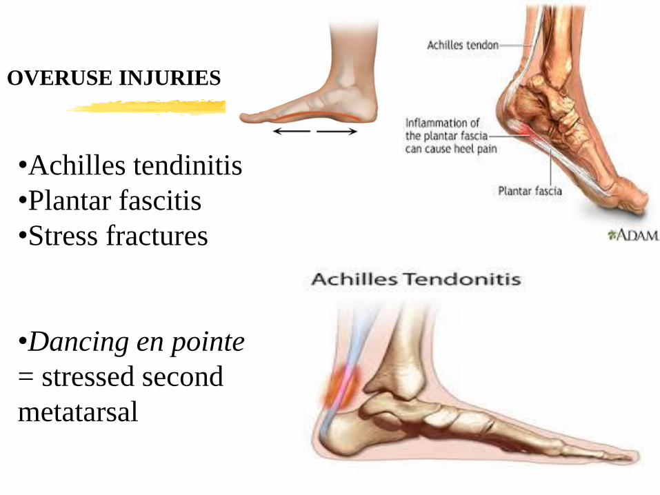
OVERUSE INJURIES
•Achilles tendinitis
•Plantar fascitis
•Stress fractures
•Dancing en pointe
= stressed second
metatarsal

Alignment anomalies of the
foot
•Forefoot
Valgus
•Forefoot Varus
•Hallux Valgus
•Hallux Varus
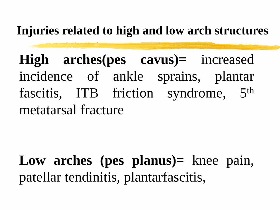
Injuries related to high and low arch structures
High arches(pes cavus)= increased
incidence of ankle sprains, plantar
fascitis, ITB friction syndrome, 5th
metatarsal fracture
Low arches (pes planus)= knee pain,
patellar tendinitis, plantarfascitis,

140