the biology of karyolysus dilloni n. sp. from rana aurora
TRANSCRIPT

University of the Pacific University of the Pacific
Scholarly Commons Scholarly Commons
University of the Pacific Theses and Dissertations Graduate School
1961
The biology of karyolysus dilloni n. sp. from rana aurora draytoni The biology of karyolysus dilloni n. sp. from rana aurora draytoni
Wayne Aldo Sorsoli University of the Pacific
Follow this and additional works at: https://scholarlycommons.pacific.edu/uop_etds
Part of the Zoology Commons
Recommended Citation Recommended Citation Sorsoli, Wayne Aldo. (1961). The biology of karyolysus dilloni n. sp. from rana aurora draytoni. University of the Pacific, Thesis. https://scholarlycommons.pacific.edu/uop_etds/1473
This Thesis is brought to you for free and open access by the Graduate School at Scholarly Commons. It has been accepted for inclusion in University of the Pacific Theses and Dissertations by an authorized administrator of Scholarly Commons. For more information, please contact [email protected].
THE BIOLOGY OF KARYOLYSUS DILIA>lU N. SP.
FROM RANA AURORA DRAYTON!
A Thesis
Presented to
the Faculty of the
Department of Biological Sciences
University of the Pacific
In Partial Fulfillment
of the Re~11rements for the Degree
Master of Arts
by
Wayne Aldo Sorsoli "'
June 1961

TABLE OF CONTENTS
PAGE
INTRODUCTION • • • • • • • • • • • • • • • • • • 1
HISTORY • • • • • • • • • • • • • • • • • • 2
MATERIALS AND HETliODS • • • • • • • • • • • • • • • 4 RESULTS • • • • • • • • • • • • • • • • • • • • • • • 8
Diagnosis • • • • • • • • • • • • • • 8
Parasites of the Peripheral Blood • • • • • • • 8
Visceral Parasites • • • • • • • • • • 0 • • • • 10
• 11 The Vertebrate Lii'e Cyole • • • • • • • • • • • •
The Invertgbrate Life Cycle • • • • • • • • • • 11
Geographical Distribution • • • • • • • • • • • • 12
Art11'1o1al Infootion • • • • • • • • • • • • 13
Results of Cultures • • • • • • • • • • • • • • • 13
DISCUSSION • • • 0 • • • • • • • • • • • • • • • • • • • 17
SUMHARY • • • • • • • • • • • • • • • • • • • • • • • 23
LITERA~URE CITED • • • • • • • • • • • • • • • • • • • • 27

LIST OF FIGURES
PLATE PAGE
I . Illuetpations of typical forms of Katyolysus
d1llon1 found in the peripheral blood • • • • 25
II . Illustrations of typioal forms of Karyolysus
dillon1 inhabiting the endothelial cells of
the 11 ver • • • • • • • • • • • • • • • • • • 26

AOKNOW LEOOMRNT
I am deeply indebted and grateful to Dr . Donald L.
TAhmann, Associate Professor of Zoology, University of the
Pacific, for his assistance and inspiration which contributed
substantially to the writing of t his paper .
I must also thank Mr . Gary J. Drusoa for his help in
collecting the animals used in the study and Miss Patricia A.
Amiolc for her assistance in typing the rough dra!'ts of this
paper.
INTHODUCTION
While investigating various Amphibia of Central
California for blood inhabiting parasites, the writer found
a parasite in tho blood of the Rod-legged Frog (B!n! ~~ draytoni ) t-1hich exhibited the characteristics of Karyolysus,
yet was different from any described; this same parasite had
been previously observed by Dr . D. L. Leruaann, Associate
Professor of Zoology, University of the Pacifio. In order to
increase the knowledge of this form, further 1nvestigat1.ons
were undertaken.
The first step was to determine the life cycle in the
vertebrate host and, if possible, to find the invertebrate
host and to elaborate the developmental stages in the vector.
Due to the difficulty in maintaining the parasito in
the host animal for the purpose of study over prolonged
periods of time, and the desire to concentrate them in large
numbers for metabolic studies, attempts were made to culture
them in vitro . Addi.tionally, it was deemed necessary to
determine the geographic distribution as well as the
specificity of the host for the parasite .

liT STORY
Labbe{ { 1894) established the genus Karyolysus and
included those forma which are found within the erythrocytes
and ara capable of destruction of the host cell nucleus.
Many of the descriptions of species of tl~s genus have been
baaed on the examination of blood smears of peripheral blood
without attention to the life cycle and the site of
schizogony . Reichenow (1921) found that the lytic activity
of the parasite was of doubtful value generically and
demonstrated that ~~olysus could be different iated from
other haemogregarine-like species on the basis that
schizogony takes place in the endothelial cells of the viscera
rathar than in the erythrocytes . The merozoites formed in
this manner penetrate erythrocytes and develop into
gametocytes .
1-iost of the species of this genus are reptilian
parasites and few authentic species of Karyolysus have been
described from amphibians , Stebbins (1905) described~·
clam.atae from .!llm!. olami tans and B.• pipiens,, but Lehmann
{1959a) relegated this species to the genus Haemogregarina as
schizogony occurred in the erythrocytes rather thnn in the
visceral endothelial cells. Sanders (1928 ) and Brandt (1936)
reported Karyolysps sp. from frogs but made their diagnosis
on the basis of peripheral blood smears alone. Da Ctmhe. and

3
Muniz (1927) determined the life cycle of Haemogregarina
leptodactyli Lesage 1900 and found that the parasites dis
played n Karyolysus type of schizogony in the endothelial
cells of the viscera. Scorza, et al. (1956) noted that both
lis• darlinsi Leger 1918 and llg. aquai Phisal1x 1930 possessed
two types of schizogony, one occurring in the endothelial
cells while the other occurred in tho cells of the viscera.
Lohmann (1959b) described K. sonomae from B!B! boyli .
Littl e work has been done on the determination of the
vector or the invertebrate life cycle; however, Reiohenow
(1921) f ound the v&ctor of ~· laoert@rum Danilewsky 1886,
parasitizing the lizard Lacerta muralis, to b~ a mito .
Hyland (1950 ) found the chigger-mite of the genus Hqnnomania
parasitizing amphibians in North Carolina . No references
were found in the literature regarding cultivation or
attempted artificial infections of these forms .

~~TERIALS AND METHODS
The host frogs (~ aurora draytoni) were collected
from Rolland ' s Pond, Dillon Beach, California . Infection was
diagnosed by clipping a digit and making a smear of the
peripheral blood; all such preparu tions trere a ta:tned tri th
Giemsa ' s stain following fixation in methanol. The extra
cellular forms were easily seen, when there was a high rate
of infection, by using lOx oculars and the lOx objective;
however, in those instances of light infection careful
examination of the entire slide was necessary. It was
necessary to use lOx oculars and a 93x oil immersion
objective to determine the presence of intra-cellular gameto
cytes as these appeared as clear areas 1n the cytoplasm or the cell while using lower manificat1on and could be easily
overlooked. With infection determined, smears and i mpressions
were made of the liver, spleen, lunga , heart, kidneys , bone
marrow and brain. All preparations \.Jere stained with
Wright ' s stain, Giemsa ' a stain follol-Ting fixation in methanol,
or Giemsa ' s stain following fixation in Schaudinn' s fluid.
'I'he descriptions are made from Giomsa' s stained films and
impr•assions.
In attempts at cultivation, sterile procedure was used
to transfer blood and tissue of the animal to the medium.
This wan accomplished by the following procedures: The frog
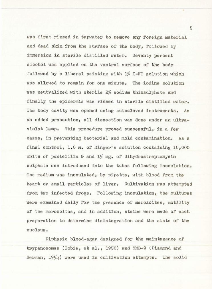
5 \-Tas first rinsed :tn tnpwe.ter to remove any foreign r11aterial
and dead akin from the surface of the body, followed by
immersion in sterile d1.st1lled watel"• Seventy percent
alcohol was applied on the ventral surface of the body
followed by a liberal painting with 1% I-KI solution which
was allm~ed to remain for one minute . The iodine solution
was neutralized l-Ii th sterile 2% sod:tum thiosulphate ancl
finally the epidermis was r·1naed in sterile distilled water .
The body cavity was opened using autoclaved ins tit\lments . As
an added precaution, all dissection was dona under an ultra
violet lamp . This procedure proved successful, in a few
oases, in preventing bacterial and mold contamination. As a
f:i.nal control, 1. 0 m. of lH.nger ' s solution containing 10,000
units of penicillin G and 15 rng . of dihydrostreptomycin
sulphate was introduced into the tubes following inoculation.
The medium was inoculated, by pipette, with blood from the
heart or small particles of liver. Cultivation was att~mpted
fl:tom tl-ro infected frogs. Following inoculation, the cultures
were examined daily for the presence of merozoites, motility
ot the merozoites, and in addition, stains were made or each
preparation to determine disintegration and the state of the
nucleus.
D1phasio blood-agar designed for the maintenance of
trypanosomas (Tobie, et al., 1950) and SNB-9 (Diamond and
Herman, 1954) were used in cultivation attempts. The aolld

phase of Tobie's was prepared by dissolving 1.5 g . llaoto
beef (Difoo), 2.5 g . Bacto-peptone (Difco), 4.0 g. sodium
chloride, and 7.5 g . Bacto-agar (Difoo) in 500 ml. distilled
water; the preparation was dispensed in 10 ml. quantities 1n
sorew top culture tubes. The tubes were then autoclaved at
15 lbs. pressure for 20 minutes and cooled until they could
be comfortably held in the hand; 1 . 0 ml . oitrated human
blood was added, mixed by rotation, and the preparation was
allowed to cool in a slanted position . The liquid phase
was prepared using the following proportions: 8 . 0 g. NaCl,
0 , 2 g. KCl, 0 . 2 g. CaC12 , 0 . 3 g KH2Po4, 2.5 g . dextrose and
1000 ml . of distilled water . After being autoolaved at 15
lbs . pressure for 15 minutes, it was added to the solid
phase, using sterile technique, in 10 ml. quantities .
6
SNB-9 ~dium was prepared as follows: dissolve 1 .8 g .
Baoto-agar (Difco ) , 1. 8 g . Bacto-neopeptone (Difco) and
0. 49 g . NaCl in 90 ml . distilled water; dispense in 10 ml .
quantities in screw top culture tubes and autoclave at 15 lbs . pressure for 15 minutes. When cool to the touch,
1. 0 ml . of citrated human blood is added, mixed by rotation,
and the preparation allowed to solidify in a slanted posi.tion.
The liquid phase was prepared in the same manner except that
the agar was omitted. The liquid was added to the slant,
aseptically, in 1. 0 ml . quantities. Motility was determined
by the use of a hanging drop preparation .

In attempts at artificial infection, the aseptio
mothod outlined above was employod, and a sntall portion oi'
liver was removed to sterile saline (0 .7%) solution . The
tissue was macerated by gently pinching with blWlt end
forceps. After mixing by rotation, 1.0 ml. of tl~ resulting
turbid solution was injected by syringe into the peritoneal
cavity of a previously determined uninfected E!n! aurora
draytoni.
7

RESULTS
Diagnosis
Karyolysus dilloni n . sp.
A blood inhabiting sporozoan with schizogony occUI•ring,
predominately, in the endothelial oells of the liver
oap1llar1os, and, to a lesser extent, in the endothelial
oells of the spleen . Typically , each schizont divides to
form a pair of equal sized merozoites; some large achizonts
may produce four merozoites . Intra-erythrooyt:tc gametocytes
are found in the peripheral and visceral blood.
Hoe t: .lliill!, aurora drayton!
Locality: Rolland Pond, Dillon Beach,
California
Location: Circulatory system
Parasites of the Peripheral Blood
Observations of Giemsa 's stained thin smears of the
periphoral blood of ~ aurora draytoni revealed the prosenoG
of extra-cellular and intra-cellular 11 haemogregarines 11 in four
of nine frogs examined during t~ spring of 1961 . The
merozoites ( Pl . I, Fig . l) were slander and averaged 16 .88
{13.87-19. 3) f in length and 2.17 (1.43-4. 29 ) ~ 1n width.
The nucleus was centrally located, composed of dark staining

9
granules and appeared to lack a discreet membrane; no
endosome was observed in the nucleus in any of the stages of
the life cycle . In the many merozoites observed, no pattern
of granules within the nucleus was aeon. The nucleus cwvered
an area within the organism of 2 . 17 (1.43-4. 29) ~ in width by
1.6 (1 .43-2 . 1) fL in length. The nucleus was the only
structure which l'raa not, to some extent, resistant to stain.
The number of morozoites varied between the hosts; in
the peripheral blood or highly tnf~ated individuals up to
tt-relve rnerozoites were seen in one high dry field (43x) while
in others the entire slide had to be searched to find a
single parasite.
Observations of citrated hanging drops revealed that
the merozoi tea were motile. 'I'hey moved with a peculiar
"flowing" motion, s1.milar to that of flatworms , and the cell
membrane appeared flexible as twisting and sidewise motion
was also observed. There appeared to be two vacuoles in the
oxtra-cellhlar forms, located anterior and posterior and in
close proximity to the nucleus; these structures, however,
were not seen in stained preparations and were om1tted from
the figures.
The intra-cellular forms ~tere found to be mature and
immature recently penetrated gametocytea. The reoently
penetrated gemetooytes (Pl. I, Fig . 3) were of similar size
and shape as the extra-cellular form, while mature gametooytes

10
(Pl. I, Pig. 4) were club-shaped with the nucleus in the
broad extremity. They were 14. 7 ( 13.6-15 . 9 ) f' in length by
3.4 (2. '7-4.4 ) ~ in l-Iidth. Both the mature and immature
gamotocytes were found in a clear space in the cytoplasm o~
the host erythrocyte. While there was no discreet nuclear
membr•ane, the granules of the nucleus appeared in rather dis
tinct patterns . What was assumed to be the macrogametocyte
( Pl. I, Fig. 5) possessed a mvre rounded and discreet nucleus
and the granules were darker stained and apparent in distinct
groups within the nucleus . The supposed m1orogametocyte
(Pl. I, Fig . 6) possessed a nucleus in which the granules
were scattered and leas distinct. Size was not a criterion
in the distinction of maorogamotocytes from n1icrogametooytes .
Visceral Parasites
The asexual or sohizogon1o forms were found in
endothelial oells primarily in the liver and only rarely in
the spleen. The early trophozoites resembled the merozoites
in both size and shape. The sohizonts (Pl . II, Fig . 1) were
sausage shaped and measured 16 . 35 (14. 56-17. 16) ~ in length
by 3 .. 15 (2 . 71-3.43) f' in width. Generally the scW.zonts 1-1ere
found singly in the endothelial cells, but double, triple,
and quadruple infections were occasionally noted. Large
sausage-shaped sohizonts ( Pl. II, f•,ig . 3) measuring 3.86
(3 . 51-4. 0) f- in width by 15 . 73 (14. 23-17 . 10) f' in length were

found also parasitizing the endothelial cells of the liver .
Bxtra-cellular merozoitos, muturc and immature gumotocytas
11
in erythrocytes- as well as the asexual fonns were encountered
in the liver.
!_he Vertebr,ate Life Cycle
The earliest stage of' the schizogonic cycle to be seen
was the trophozoite . Thie stage Pasides in endothelial cells
of the liver and enlarges to yield a schizont. Prior to
cytoplasmic division in the normal sized schizont, the
nucleus divides into two products which lie in tandem at
right angles to the long axis of the body. The schizont
then divides into a pair of merozoites by a longitudinal
constriction of the cytoplasm, (Pl. II , Jl1ig. 2) . The large
sausage-shaped sohizonts divide once, by the same method, to
form t\oro equal sized secondar'Y bodies (Pl. II, Ii'1g. !~) which
in turn divide again into a pair of merozoites that are
similsr in size and shape to those produced by the smaller
schizonts (Pl. II, Fig. 5). The merozoi tes thus forn1ed then
leave the endothelial cells to become free extra-cellular
bodies in the peripheral blood .
The Invertebrate Life Cycle
The invertebrate host in the life oyclo of K. &illoni
was not determined; however, leeches of the genus

12
Aotinobdella were :round parasitizing salamanders ( 'l1arioha
torosa) in the same pond . Following the observation or an
u~in:t'eoted :t'rog being fed upon by a leech, a smear was made
of the leach which was stained and examined for the presence
of infective forms of li• dilloni. None woro found in th~
preparationl however, crithidia, leishmania, and trypanosome
forms of Trypanosoma barbari Lehmann, 1952 were found .
Examination ot the frog at various later dates fatled to
reveal the presence of infection; however, it is possible
that the leech did not take a blood meal, as few blood cells
were found in the stained preparation and thus had not had
the opportunity to infect the frog . Attempts to artificially
in.fes t frogs with leeches falled as the leeches refus(ld to
attach and feed .
Geographical Distribution
In the process of determlning the nature of t he
paras1. te of· Rana aurora drayton!, othei' 8Jllphibians were
investigated for inf'ection. '.tWenty Hyla regilln and n1ne
Tarioha torosa collected from the Rame pond wero checked but
no haemoaporozonns were found. None of the ~yla possesaed
a1 ther external or internal parasit~es, but all of the Taricha
were parasitized by an eotoparasiti c leech, Actinobdella sp. ,
and also by a haemoflgellate, Trypanosoma barbari. In
addi t ion, four Bu~ro boreas from the same area, but not t he

13
same pond, t•ere examined with negative results. Two Dioamp
todon ensatt~s from the sal'lle water shed were examined and
found to be parasitized by Cytoamoeba ~· but were not the
hosts of Karyolyaus, ExaminaM.on or the peripheral blood or
eight .B!m!. aurora drayton:!. rrom a small oreal< so1110 400 yards
south of Rolland Pond showed that these were not parasitized
by Ka;ryolysus.
Artificial Infection
Two un:l.nfected frogs were injected intraporitoneally
with 1.0 ml. of the macerated liver on successive days. They
were examined for infection by means of Giemsa's stained thin
smears at three, six, fourteen, and twenty-one days after
:l.njection. No infection could be detected,
Results or Cultures
J.'rog Number 1:
SNB-9 Medium - Blood inoculated. No antibiotics.
lst day - Motile Merozoites observed.
2nd day - The culture was badly contaminated
with bacteria. 30,000 units of
penicillin G and 25 mgs. dihydro
streptomycin sulphate added.
Merozoites observed, motility
undetermlned. Stained preparation

railed to reveal a nucleus.
3rd day - Contamination undiminished so the
culture was destroyed .
SNB-9 Medium - Liver inoculated. No antibiotics.
14
1st day - Motile merozoites observed . Culture
slightly contaminated so 15,000 units
penicillin G and 15 mgs. streptomycin
added.
2nd day - Twisting movement or a single mero
zoite was observed. Tho contamination
was reduced.
3rd day - Merozoites were observed but no
motion. Stained preparation railed
to show a nucleus but did reveal
disintegrating forms .
4th day - Culture presumed negative so it was
destroyed.
Tobias Medium - Blood inoculated. No antibiotics.
1st day - The culture was very badly contami
nated and was destroyed.
Tobies Medium - Liver inoculated. No ant1b1.ot1cs.
1st day - The culture was very badly contami
nated and was destroyed .
Frog Number 2:
SNB-9 Medium - Blood inooulatod. 10,000 units or
•

penicillin G and 15 mg. dihydro
streptomycin sulphate added.
15
1st day - Merozoites observed, but no motion,
Stained preparation revealed the
presence or nuclei. Slight contami
nation. Added 30,000 units
penicillin and 25 mg. streptomycin,
2nd day - Merozoites observed, No motion
observed, Stained preparation
revealed disintegrating forms and the
lack of a stained nucleus, No con
tamination,
3rd day - Culture presumed negative end was
destroyed. No contamination.
SNB-9 l1ed1um - Liver inoculated.. Antibiotics added.
1st day -Motile merozoites observed. The
culture was slightly contaminated so
the same quantity or antibiotics as
indicated above was added.
2nd day - Merozoites were observed but without
motion, Stained preparation revealed
disintegrating forms and the lack of
a nucleus. No conta.mination,
3rd. day - Culture presumed negatlve and
destroyed, No contamination.

16
Tobies Medium - Blood inoculated. Antibiotics added.
1st day - Motile merozoi tee observed. No con
tamination.
2nd day - Merozoites observed but without
motion. Stained preparation I'evealed
disintegrating forms and the lack of
a nucleus. No contamination. Pre
sumed negative and destroyed.
Tobies 1-iedium - Liver inoculated. Antibiotics added.
1st day - Motile merozoites observed. No con
tamination.
2nd day - Non-motile msrozoites present. Dis
integrating forms and lack of nucleus
in stained preparation.

DISCUSSION
There are five genera of haemosporozoans found in the
blood of various vertebrates that could be confused with
Karyolysus. The genus Lankesterella 1s found in the endo
thelial cells of the capillaries of frogs and generally l~s
a leech vector. Scb1.zogony typically produces asexual
merozo1tes and sporozoites; the latter invade red blood cells,
and if such are ingested by a leeoh, the hirudinean acts as a
mechanical, rather than a definitive host. Schellackia
differs from Lankesterella in the site of schizogony but the
invertebrate host still effects a mechanical transmission.
In Haemogresarina the life cycle is similar to Karyolysus
differing only in the site of schizogony (the erythrocytes).
The genus most likely to be confused with Kapyolysus is
Hepatozoan. It undergoes schizogony in the liver rather than
in the endothelial cells. Another character differentiating
Karyolysus from Hepatozoan is that Hepatozoan is often found
in the leucooytes of mammals.
Karyolysus dilloni is a blood inhabiting sporozoan
with schizogony occurring predominately in the endothelial
cells of the liver and only rarely in the endothelial cells
or the spleen. Typically each schizont develops into two
equal sized merozoites, but some large schizonts may produce
four merozoi tes. Intra-ery·throcytic gametocyte a are found in

18
the visceral and the peripheral blood.
Knryolysus dilloni appears to be related to li• sonomae
{Lohmann 1959) but differs in some aspects. li· aonomae
produces four and on occasion ei.ght merozoites from a single
schizont, while li• dilloni typically produces only two
merozoites, but occasionally yields four. The discrepancies
in the numbers or merozoites appears to be a valid character.
The other main point of difference is the location of
schizogony. li• dilloni undergoes schizogony in the endo
thelial cells of the liver while in K. sonomae this phenome
non ocours in the endothelia of the spleen. Da Cunha and
Muniz {1927) have found that li• <&aemogregarina) leptodactyli
has two modes of schizogony both occurring in the endothelia
of the viscera; 2-8 merozoites are formed by one type while
15-20 are formed by the other. The number of merozoites as
products of schizogony obviously differs from that of !• dilloni.
!• dilloni possesses two forms of schizogony, one in
which a schizont produces two merozoites while a giant
schizont reproduces four merozoi tes. In parasites of tM.s
type two forms of merozoites are typically produced at
schizogonic division, one which carries on the asexual
process by lnvading new endotheHal cells while the other
extra-cellular merozoites are, in all probability, actually
immature gametocyte a. The development of the two forms was

19
not investigated as the two merozoites are morphologically
indistinguishable. Upon penetration of erythrocytes (Pl. I,
Fig, 2), they undergo development which results in the
formation of macrogametocytes and microgametocytes,
The vector of K, dilloni is unknown, but either
chigger-mi tea of the genus Hannemania or leeches or the genus
Actinobdella could be incriminated. Reichenow ( 1921) .found
the vector of K. lacertarum to be a mite, and Hyland (1950)
has found the chigger-mite parasitizing North American
amphibians. It is not unreasonable to assume that the
chigger-mite could be the vector of li· dillon!. Inasmuch as
a leech was seen attached to a frog and it is known that a
leech is the vector of haemoflagellates of this area
(Lehmann, 1952), it is a remote possibility that leeches
could also be the vector. Investigation of the pond failed
to reveal the presence of mites while leeches were found in
many instances upon salamanders supports the view that
leeches could be the vector. Little work has been done to
find the various vectors and life cycles of this parasite and
any blood sucking invertebrate becomes a suspect.
Wenyon (1926) and Kudo (1951.!.) describe the genus
Karyolysus as one hav1.ng a mite as a vector and a lizard as
the definitive host. ry?hese descriptions, then, lend them
selves to an argument for a new genus, To the writer's
knowledge, only three authentic species of Karyolysus have

20
been described; Reichenow (1921) described~· lacertarum
from a l i zard; Da Cunha and ltluniz ( 1927 ) redescribed ~·
(Haemogregarina ) leptodactyl1; and Lehmann (19$9) described
li• sonomae from a frog . Authentic Karyolysus are those
which exhibit typical schl.zogony in the endothelial cells of
the viscera . Neither Lesage nor Lehmann determined the
invertebrate host of their respective speo~.es . It is
possible s:tnce few mites parasitize frogs , that in both oases
tha vector was a leech. If the assumption of a leech vector
is accepted along t-ti th the fact that the definitive host is
not a lizard , perhaps the parasites described by Lesage,
Lehmann and the form described in this paper should be con
s:tdered a new genus .
The materials injected into the peritoneal cavity of
the frogs included gametooytes, reproductive merozoites,
schizogonic merozoites , and scluzonts. The sporozo1tos which
normally are int roduced by the vector were the only forma
missing from the inj ected material . As no infection of the
frogs resulted, i t would seem that the sporozoite is
necessary f ob tl~ iniation of the life cycle in the
vertebrate host. The sporozoite may be very well adapted,
physiologically, to life in any number of individuals within
a speoies J on the other hand, the merozoites may be
restricted, physiologically, to life within one individual of
any species of host . It is also possible that phagocytic

21
activity destroyed the parasites before infection was
accomplished, as none of the frogs found outside the pond
were infected. One might speculate from this that
populations other than those from Rolland Pond are immune to
the parasite, or the vector is not present in the water
inhabited. Leeches were not found in any water source except
Rolland Pond.
Cultivation attempts proved quite unsuccessful,
possibly because of. the difficulties encountered in sterile
removal of blood and liver tissue to the oult1~e medium. As
many precautions as possible were used and yet bacterial con
tamination was found in the tubes. 'l'he use of antibiotics
reduced or destroyed contamination if used before, or when
contamination was only light, but antibiotics were of little
value once thE> numbers or bacteria became great. 1'his use or
antibiotics once again presents a variable as little is known
of tl~ir effect on animal parasites. Little work has been
done on the cultivation of these forms, and 1 t seeme to be an
area open for subsequent study. Hotile merozoi tee were seen
in the cultures following inoculation so the change in
environment did not destroy thern 1nunediately; however, an
effeo"tive medium would allow them to live until the nuti•ients
were depleted. Inasmuch as cultivation did not succeed, it
would appear that some form of nutr:l.ent is lacking in the
medium, but the type is obv1.ously \Ulknown.

22
The life cycle of .Karzolysuo is characterized by tlw
penetration of both endothelial and red blood cells . As
oitrated human blood was used in the media, penetration of
such erythrocytes may have been physiologically impossible
and the life cycle could not be carried to complet ion. When,
however, liver tissue was added, cells were present for
the production of aoh1zonts and, in tu1~, merozoites. The
fact that merozoites were not produced under suoh conditions
again poj.nts to a deficiency in the culture media .

SUMMARY
Karyolyeus dilloni n. sp. is a parasite of the cir
culatory system of the Red-legged Frog, ~ aurora drayton!,
from Holland Pond, Dillon Beach, California. Schizogony
typically occurs in the endothelial cells of the liver with
the production of two merozoites and occasionally four from a
giant sch1.ll'.ont. The mero~oites penetrate elther the
erythrocytes forming gametocytes or new endothelial cells to
perpetuate the aeexual cycle.
The invertebrate host wae not determined and attempts
to culture !.u vitro and to ai•tifically infect frogs of the
spec~.es were unsuccessful.
Four of nine ~ ~· draytoni from Rolland Pond were
found to be infected, while none of eight from another source
were infected. No infections were found in twenty Hyla
ragilla and nine Taricha torosa from Rolland Pond or ~.n four
~ boreas and two Dioamptodon ensatus from other areas
around Dillon Beach.

EXPLANATION OF PLATES
PLATE I
Figure 1 Extra-cellular merozoite
Figure 2 Merozoite penetrating an erythrocyte
Figure 3 Recently pene·trated immature gametocyte
Figure 4 Mature gametocyte
Figure 5 Presumed macrogametocyte
Figure 6 Presumed microgametocyte
PLATE II
Figure 1 Nucleus dividing 1n a normal sized schizont
Figure 2 Schizont dlvidlng by longitudinal con
striction of the cytoplasm
Figura 3 Large sausage shaped schizont
Figure 4 Secondary bodies resulting from first
division of large schizont
Figure 5 Merozoites in endothelial cells following
division of secondary bodies

PLATE
lOu
2
3
4
0 0 0
i:~·- ·· · 5 6
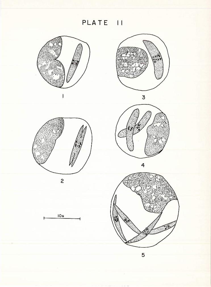
PLATE II
3
4
2
lOu
5

LITERATURE CITED
Brandt, B. B. 1936 . Parasites of Certain North Carolina Salientia . Eoologioal ~Ionographa, 6 ( 4 ) ~ 491 .. 532 .
Da Cunha , A. M. and Muniz , J . 1927 . Sobra 0 Cyole Endogene da Ha~mosregarina leptodactyli Lesage 1908 . Rio do Janeiro . Institudo Ocwaldo druz . Memor1as, 20: 307-314·
Diamond, L. s . and Herman, c. M. 1954. Incidence of Trypanosomas in the Canada Goose as Revealed by Bone Marrow Culture . Journal of Parasitology, 40: 195-202.
Hyland, K. E. 1950. The Life Cycle and Parasitic Habit of the Chigger Mite Hannemania dunn1 Sambon 1928, a Parasite of Amphibians . Journal of Parasitology, 36t 32-33·
Kudo, R. R. 19~~ · Protozoology . Springfield. Charles c. Thomas • 966 p .
Labbe~ A. 1894. Recherches Zoologiques et Biologiques sur lea Paraa1tes Endoglobulaires du Sang deo Vertebras . Thesis, Paris .
Lehmann, D. L. 1952. A New Species of Trypanoaome from the Pacific Coast Newt Triturus toroaa Rathke . Transactions American M!croacopioal Society, 71: 135•138 .
, 1959a. The Description of Hfemogresarina boyfi --n-.-sp. from the Yellow-legged Frog, Rana Soyli boy •
Journal of Parasitology, 45: 198-203.
--~~' 1959b. Kariolysu~ sonomae n . sp. , a Blood Parasite from the Yellow-legged Frog, Rana bo:rli ~oyli • Proceedings of the American Ph1losoph!081 Soo e y, 03 (4) z 545-553.
Reiohenow, E. 1921. Die Hamocooeidien der Eiderchren Vorbemerkungen und I Te11 . Die Entwioklungsgesoh1ckte von Karyolysus . Archiv fur Protistan, 62: 180·196.
Sanders, E. P. 1928 . Observations and Experiments on the Hemoparanitea of Oertain Amphibia . Journal of Parasitology, 14: 188-192 .
Scorza, J . v. , Pagert, c. and Arocha, L. 1956 . Estudo Sobre Hemoparaaitos de Bufo marinus L. de Venezuela . Rio de Janeiro . Inst!tu~swaldo aruz . Memor1as, 54 (2 ) s 373-391.

28
Stebbins, J . H. 1905. On the Occurrence of a Large Sized Parasite of tho Karyolysus Order in the Blood of Rana olamata. Zentralblatt fur Bakter1olog1e, Parasitenkllnde und Infektionskrankheiten. Abt. I . Mediz1nt1er1sohe, Hyg1enisohe, Dakteriologie und Tierisohe Parasitenkunde, 3~: 315-318.
Tobie, E. J . , von Brand, T. and Mohlman, B. 1950. Cultural and Physiological Observations on Trypanosoma rhodiesiense and Trypanosoma gamb1ense. Journal of Parasitology, 36: 48-54.
W6nyon, c. M. 1926 . Protozoology 2. New York , William Wood and Oo . 1563 p .