the biology of filamentous phage infection - the department of
TRANSCRIPT

Department of Immunotechnology
Lund University
THE BIOLOGY OF FILAMENTOUS PHAGE
INFECTION
IMPLICATIONS FOR DISPLAY TECHNOLOGY
Fredrik Karlsson
Lund 2004


THE BIOLOGY OF FILAMENTOUS PHAGE INFECTION_____________________________________________________________________________________
1
TABLE OF CONTENTSORIGINAL PAPERS 2ABBREVIATIONS 31 INTRODUCTION 42 BIOLOGY OF THE Ff PHAGE 8
2.1 Phage coat proteins 92.2 Infection mechanism 122.3 Genes, gene expression and replication 132.4 Assembly reactions 16
3 PHAGE DISPLAY TECHNOLOGY 193.1 Choice of coat protein and display format 203.2 Libraries for phage display 233.3 Principles of selection and analysis of selected clones 243.4 Alternative display systems 26
4 SELECTIVE INFECTION 284.1 Effect of fusion protein on selective infection 304.2 Factors affecting the outcome of selective infection 304.3 The future of selective infection 34
5 MOLECULAR DISSECTION OF THE Ff PHAGE INFECTION 355.1 pIII and TolA during infection 355.2 A novel model for the Ff phage infection mechanism 375.3 TolA function and importance 415.4 Separation of TolA functions 43
6 ALTERED TRANSCRIPTION OF PHAGE INFECTED CELLS 466.1 Global transcriptional analysis of phage infected E. coli 476.2 Regulation of host genes by Ff phage infection 47
7 CONCLUDING REMARKS 508 POPULÄRVETENSKAPLIG SAMMANFATTNING 529 ACKNOWLEDGEMENTS 5310 REFERENCES 55PAPERS I-IV

_____________________________________________________________________________________
2
ORIGINAL PAPERS
I Nilsson N, Karlsson F, Rakonjac J and Borrebaeck CAK (2002)Selective infection of E. coli as a function of a specific molecularinteraction J Mol Recognit 15, 27-32.
II Karlsson F, Nilsson N, Borrebaeck CAK and Malmborg A-C(2003) The mechanism of bacterial infection by filamentousphages involves molecular interactions between TolA and phageprotein 3 domains. J Bacteriol 185, 2628-2634.
III Karlsson F, Malmborg-Hager A-C and Borrebaeck CAK (2004)Identification of mutations that segregate membrane integrity andphage receptor function of the Escherichia coli TolA molecule.Submitted for publication.
IV Karlsson F, Malmborg-Hager A-C, Albrekt A-S and BorrebaeckCAK (2004) Genome-wide comparison of phage M13 infectedvs. uninfected Escherichia coli. Submitted for publication.
The publications are reproduced with written permission from the copyright holders.

THE BIOLOGY OF FILAMENTOUS PHAGE INFECTION_____________________________________________________________________________________
3
ABBREVIATIONS
aa amino acidBIA (real-time) biospecific interaction analysisCT C-terminal domain of phage minor coat protein pIIIcDNA complementary DNADNA deoxyribonucleic aciddsDNA double stranded DNAELISA enzyme-linked immunosorbent assayFf F-specific filamentousIG intergenicmRNA messenger RNAN1 the first N-terminal domain of phage minor coat protein pIIIN2 the second N-terminal domain of phage minor coat protein pIIIN1N2 both N-terminal domains of pIII, including flexible linkerORF open reading framePS packaging signalPCR polymerase chain reactionRF replicative formRNA ribonucleic acidSAP selection and amplification of phagescFv single-chain antibody fragment variableSIP selectively infective phageSPR surface plasmon resonancessDNA single stranded DNA

INTRODUCTION______________________________________________________________________________________
4
1 INTRODUCTION
There is a constant demand for molecules suitable for diagnosis and/ortreatment of illness. In order to devise the molecules that will carry out thesetasks, an array of biotechnological tools are available. One such tool is basedon the use of bacteria and a special kind of bacterial virus. If we learn moreabout the relationship between this virus and the bacteria, it will be feasible toimprove the design of the tool. Hopefully, this will ensure a more rapidprocess of finding the desired molecules.
Viruses that can infect bacteria are called bactoriophage and are harmlessto humans. Like all viruses, bacteriophage (phage) are metabolically inert intheir extra-cellular form and reproduce only by infecting and exploiting themetabolism of the host bacteria. Upon infection, the viral genomic material isintroduced into the host cell, where it directs the production of progeny phage.Most often, progeny phage are assembled in the cytoplasm of infected cells inlarge numbers, which cause the host cell to burst, killing it and then infectingmore bacteria (Calendar, 1988). There are innumerable types of phage, eachcapable of eradicating its host bacterial species. However, some special phageare assembled at the cell surface and co-ordinately exported by a secretorymechanism, a process that leaves the host cell fully viable. The secretedbacteriophage belong to the family of filamentous phage (called so due to theirmorphology) that are specific for the Gram-negative bacteria Escherichia coli
(E. coli) carrying F-pili (Hoffmann-Berling et al., 1963; Loeb, 1960; Marvinand Hoffmann-Berling, 1963; Zinder et al., 1963).
Some 25 years after filamentous phage were first isolated, it was reportedthat a genetic modification, the insertion of a foreign DNA fragment into thephage genome, yielded a phage particle displaying the foreign polypeptidesequence as a fusion to the coat protein on its surface (Smith, 1985).Furthermore, the displayed peptide was accessible to specific recognition byan antibody towards the peptide. This resulted in an enrichment of the peptide-displaying phage from a mixture of wild-type (non-binding) phage anddisplay-phage particles. In addition, the methodology ensured the directphysical link between phenotype (polypeptide) and genotype (DNA), meaningthat the displayed molecule had a tag, the DNA sequence encoding that
THE BIOLOGY OF FILAMENTOUS PHAGE INFECTION______________________________________________________________________________________
5
molecule, which could be addressed through DNA sequencing. Therefore, thereport by George Smith in 1985 is considered the first description of phagedisplay technology.
It was soon realised that it would be possible to build up large pools ofgenetic variants, i.e. DNA libraries, by combining several new technologies.Consequently, recombinant DNA technology, which includes the precisecutting (by restriction enzymes) and rejoining (by ligases) of DNA pieces(Linn and Arber, 1968), oligonucleotide-directed mutagenesis (Hutchison et
al., 1978) and the DNA amplification technique known as the polymerasechain reaction (PCR) (Saiki et al., 1985), were used to create DNA libraries.Only a few years after the publication by George Smith, the first phage-displayed random polypeptide libraries were assembled (Cwirla et al., 1990;Devlin et al., 1990; Scott and Smith, 1990). These reports were followed byothers, which demonstrated that properly folded and functional proteins couldalso be displayed on the surface of filamentous phage (Bass et al., 1990;McCafferty et al., 1990).
Many different types of molecules can be displayed on the surface ofphage. Most common is display of some sort of protein with an intrinsiccapacity of recognising and binding target molecules with a high degree ofspecificity, such as antibody fragments. The possibility to display antibodyfragments on the surface of phage is of particular interest, since thesemolecules have great potential as reagents in diverse settings like diagnostics,biological chemistry and as agents suitable for therapeutic applications(Borrebaeck, 2000; Borrebaeck and Carlsson, 2001; Bradbury et al., 2004;Hudson, 1998; Hudson and Souriau, 2001). Notably, up to 30 % of humanantibodies in clinical development have been isolated using phage displaytechnology (Kretzschmar and von Ruden, 2002). Because of these potentials,antibody fragments are the most common scaffolds diversified and used forselection processes in the field of phage display technology.
Proteins that bind to their target with a high binding strength (affinity)and a high degree of specificity are much desired for diagnostic andbiochemical purposes. Therefore, attention has been drawn to the creation ofprotocols that increase the chance of obtaining high affinity reagents, usingvarious display methods. One approach is to move to cell-free systems (Hanes

INTRODUCTION______________________________________________________________________________________
6
and Plückthun, 1997; Nord et al., 2003), allowing for larger libraries, whichtheoretically increases the likelihood of finding high affinity binding proteins.Another is to modify the selection procedure of the phage display technology,by studying the requirements for the infection process of filamentous phage, inhope of revealing key mechanistic events suitable to manipulation.
In this thesis, I will summarise and interpret data from independentinvestigations of the infection process of filamentous phage, as studied bydiffering approaches. In addition, I will present the results from my ownstudies in this area and how they may affect the development of phage displaytechnology. My experimental work has resulted in four original papers(reports) that deal with aspects of both the phage infection mechanism andphage display technology. The first report describes an evaluation of theparameters important for a special application of phage display, calledselective infection (PAPER I), the second and third present the results fromstudies of phage and host protein interactions during phage infection (PAPER IIand III) and the last is a description of the effects phage infection had on thetranscription of host genes (PAPER IV).
In experiments reported in PAPER I, a correlation between the affinity ofthe interacting pairs and infection efficiency of selective infection wasdetected, and a phage format that allows multiple display of antibodyfragments on each phage was found to be superior to one that does not allowsuch multiple display. PAPER II describes the molecular interactions betweenphage coat protein pIII and the bacterial co-receptor protein of phageinfection, TolA, as assayed by real-time bio-specific interaction analysis. Thebinding affinities between these proteins and their different domains werecharacterised and novel interactions were detected allowing us to make arefined hypothetical model of the infection mechanism of filamentous phage.In PAPER III, the bacterial co-receptor, TolA, which is also required for outermembrane integrity of E. coli, was studied. It was found that the membraneintegrity and phage receptor functions of TolA could be segregated. PAPER IVdescribes the changes in gene expression of phage infected E. coli, asmonitored by global transcription analysis and it was demonstrated thatseveral host genes were co-ordinately affected. I will also discuss how theresults of these studies have provided insights into several different aspects of

THE BIOLOGY OF FILAMENTOUS PHAGE INFECTION______________________________________________________________________________________
7
the phage infection process, as well as the possible implications of the resultsfor phage display technology. But first, the biology of filamentous phage andthe phage display technology will be introduced.

BIOLOGY OF THE Ff PHAGE_____________________________________________________________________________________
8
2 BIOLOGY OF THE Ff PHAGE
Filamentous bacteriophage appear as thin (6-10 nm) and long (900 nm),filamentous particles (Figure 1) containing circular single stranded (ss) DNA(Hoffmann-Berling et al., 1963; Marvin and Hoffmann-Berling, 1963;Newman et al., 1977). The actual length of the particle is determined by thelength of the DNA it has encapsulated (Marvin, 1990). The filamentous phagebelongs to a group of related viruses which only infect gram-negative bacteriavia specific adsorption to the tip of bacterial structures called F-pili (Loeb,1960). The F-pili (also called sex-pili) are normally involved in thetransmission of the F plasmid DNA, or chromosomal DNA containing theintegrated plasmid DNA, from one bacterium to another (Frost et al., 1994;Gaffney et al., 1983; Hayes, 1952). Most information about this type offilamentous phage derives from the very similar members of the same familyof F-specific filamentous (Ff) phage, classified as Inoviridae, of the genusInovirus, i.e. phage M13, f1 and fd, which infect Escherichia coli (E. coli)(Marvin and Hohn, 1969). The genomes of these three bacteriophage havebeen completely sequenced and are 98 % homologous (Beck et al., 1978;Beck and Zink, 1981; Hill and Petersen, 1982; van Wezenbeek et al., 1980).Because of their similarity and their dependence on the F-pilus for infection,the term Ff phage will be used throughout when referring to M13, fd and f1biology, unless otherwise stated.
Unlike other bacterial viruses, the Ff phage does not kill its hosts, butestablish a relationship in which new virions are continually released(Hoffmann-Berling and Mazé, 1964). Because of this non-lytic mode ofrelease it is possible to grow high-titre cultures of the virus. Also, the growthcurve of Ff phage shows a rapid increase of virus after a short latent period(Brown and Dowell, 1968; Hoffmann-Berling et al., 1963; Marvin and Hohn,1969). The first progeny phage particles are released about 15 minutes afterinfection (Hofschneider and Preuss, 1963), and the rate of production isexponential for the first 60 minutes, after which it becomes linear as thebacteria enter stationary phase (Brown and Dowell, 1968; Hoffmann-Berlinget al., 1963; Marvin and Hohn, 1969). This rate of production implies thatroughly 1000 phage per bacterium are produced within the first hour after

THE BIOLOGY OF FILAMENTOUS PHAGE INFECTION_____________________________________________________________________________________
9
infection. Although infected cells can continue to grow and divide indefinitely,the process causes the infected cells to continue growth at a rate significantlylower than uninfected cells (Brown and Dowell, 1968; Hoffmann-Berling et
al., 1963; Salivar et al., 1964), which is why plated cultures of Ff phage-infected cells yield turbid plaques. The Ff phage are relatively simplemolecular complexes and they have therefore been extensively characterised.At the Department of Immunotechnology, a derivative of Ff phage M13 is themost frequently used phage. Bacteriophage M13 was first isolated fromwastewater in Munich 1963 (Hofschneider, 1963). In the following sections,the life cycle, genetics and structural organisation of the Ff phage issummarised, with a focal point on factors of importance for phage displaytechnology.
FIGURE 1. An electron micrograph of a filamentous phage. The globular domains ofthe adsorption complex of the virion (the pIII) are visible at the right end of the viralparticle. The picture was taken by Dr. C. Brown and kind permission to reprint it wasgiven by Professor D. A. Marvin, University of Cambridge, UK.
2.1 PHAGE COAT PROTEINS
The most abundant coat protein of the virus is encoded by gene VIII. Theroughly 2700 copies of pVIII make it the major coat protein (Newman et al.,1977; Pratt et al., 1969). The minor coat proteins are located at the ends of thephage particle, and are present at only a few copies each, as can be seen inFigure 2. The distal end, which is assembled first, contains the pVII and pIX,and the proximal end, which enters the host first, contains pVI and pIII (Lopezand Webster, 1983). Estimations based on symmetry have proposed that there

BIOLOGY OF THE Ff PHAGE_____________________________________________________________________________________
10
are five copies of each of the minor coat proteins, although the distal endseems to have three copies each of pVII and pIX (Makowski, 1992; Simons et
al., 1981). These two proteins (33 and 32 residues) are required for phage tobe assembled and released, and in the absence of either no infectious particlesare formed (Lopez and Webster, 1983; Pratt et al., 1969). The two remainingminor coat proteins pVI and pIII, are necessary for particle stability andinfection (Gailus et al., 1994; Grant et al., 1981a; Grant et al., 1981b; Gray et
al., 1978; Lopez and Webster, 1983; Rakonjac and Model, 1998).
FIGURE 2. The localisation of the Ff phage coat proteins. At the left end, the minorcoat proteins pVII and pIX are located, while the major coat protein, pVIII, is spreadalong the entire length of the particle. At the right end, which penetrates the bacteriaupon infection, the minor coat proteins pVI and pIII are located.
Not much is known about the appearance of pVI on the surface of the phageparticle, but at least a portion of the protein seem to be surface exposed, andpVI and pIII are known to assemble in the membrane prior to incorporationinto phage particles (Gailus and Rasched, 1994; Rakonjac et al., 1999). Incontrast, the most commonly used coat protein for phage display, pIII (Kayand Hoess, 1996; Russel et al., 2004; Smith and Petrenko, 1997), has receiveda lot more attention, primarily because of its role in phage infection. Phageprotein pIII is synthesised with an 18-residue amino-terminal signal sequenceand requires a bacterial secretion and protein export system (Sec) for insertioninto the membrane (Rapoza and Webster, 1993). After removal of the signalsequence, mature pIII, as shown in Figure 3, is obtained. This molecule iscomposed of 406 residues and form three distinct, folded domains, which areseparated by glycine-rich, flexible, regions. The 149-residue carboxy-terminal
pIXpVII pVIII
pVIpIII

THE BIOLOGY OF FILAMENTOUS PHAGE INFECTION_____________________________________________________________________________________
11
(CT) domain (residues 256-406) anchors pIII to the phage particle and itsmembrane anchor region (residues 378-406) is probably buried within thephage particle (Marvin, 1998; Rakonjac et al., 1999), while the two remainingamino-terminal domains are surface exposed. The amino (N) terminal domains1 and 2 (N1 and N2) interact intramolecularly and form a N1N2 complex(residues 1-217), as shown by X-ray crystallography and NMR spectroscopy(Holliger et al., 1999; Lubkowski et al., 1998). The N1 and N2 domains ofpIII play important roles during the infection process of Ff phage, bymediating infection (Armstrong et al., 1981; Boeke et al., 1982; Crissman andSmith, 1984; Jakes et al., 1988; Rasched and Oberer, 1986; Riechmann andHolliger, 1997; Stengele et al., 1990).
FIGURE 3. The domain structure of the adsorption protein pIII. The amino acidscreating each domain are indicated by their number in the sequence. The N1 (blue)and N2 (light blue) domains are involved in inter-domain contacts, as determined byX-ray crystallography (Lubkowski et al., 1998). The N1 domain mediates binding tothe bacterial co-receptor protein TolA (Riechmann and Holliger, 1997). N2 binds toF-pilus via residues located on the outer rim of N2, and not in the central cavityformed in the interface between N1 and N2 (Deng and Perham, 2002). The CT(shaded gray) is required for virion assembly and its topology is unknown. Theflexible linker regions, containing tandem repeat sequences of GGGS and EGGGS
CT256-406
N287-217
N11-67

BIOLOGY OF THE Ff PHAGE_____________________________________________________________________________________
12
(van Wezenbeek and Schoenmakers, 1979), are indicated as black lines between thedomains.
2.2 INFECTION MECHANISM
Filamentous phage infection of E. coli starts with the adsorption to thebacterial structure called the F-pilus, which is of long and slenderconformation not unlike the one of phage. The infection process eventuallyleads to phage DNA translocation by a mechanism that remains uncertain.However, the events prior to this final step are well characterised and shown inFigure 4. The F-pilus extends from the cell envelope at contact sites betweeninner and outer membranes, called adhesion zones (Bayer, 1968a, b). Theinitial phage binding to the tip of the F-pilus is strong (Deng et al., 1999;Stengele et al., 1990; Tzagoloff and Pratt, 1964) and achieved by residues onthe outer rim of the N2 domain of pIII (Deng and Perham, 2002). The glycinerich linker region between N1 and N2 (residues 69-86) may also be involvedvia an unknown mechanism (Nilsson et al., 2000). After phage adsorption, thenatural process of pilus retraction brings the phage closer to the bacterialmembrane (Jacobson, 1972; Marvin and Hohn, 1969). It has been suggestedthat the binding to the F-pilus leads to a conformational change in N2 thatreleases N1 from the N1N2 complex (Deng and Perham, 2002; Riechmannand Holliger, 1997). Thus, after pilus retraction, the N1 domain is free tointeract with the bacterial co-receptor TolA as seen in Figure 4 (Riechmannand Holliger, 1997). The TolA protein is membrane bound and encoded by thetolA gene of E. coli, also located at adhesion zones (Guihard et al., 1994).TolA is believed to form a complex with two other membrane bound proteins,TolQ and TolR (Webster, 1991). Host cells that lack of any of the innermembrane proteins TolQ, TolR and TolA, are tolerant to phage infection(Click and Webster, 1997, 1998; Russel et al., 1988; Sun and Webster, 1986).From its anchor point in the cytoplasmic membrane, the TolA protein extendsinto the periplasm (Levengood et al., 1991), where it interacts with N1 duringphage infection. The putative complex formed by the three Tol proteins (Q, Rand A) mediates incorporation of phage coat proteins (pIII and pVIII) into thecytoplasmic membrane and the translocation of the ssDNA by an incompletelyunderstood mechanism (Armstrong et al., 1983; Boeke and Model, 1982;
THE BIOLOGY OF FILAMENTOUS PHAGE INFECTION_____________________________________________________________________________________
13
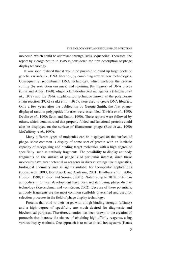
Russel et al., 1988; Smilowitz, 1974; Trenkner et al., 1967). Similarly, it is notknown if the TolA co-receptor molecules interact with each other during theinfection process. In a few of the following chapters some light will be shed onthese issues.
FIGURE 4. Schematic representation of the tip of an Ff phage binding to E. coli
receptor (F-pilus) and co-receptor (TolA) during the initial steps of the infectionprocess. The infection process is initiated by interaction of the N2 domain of pIII(blue) with an F-pilus projecting from the E. coli cell surface. This interactionreleases N1 from the inter domain interaction with N2, allowing it to bind to the C-terminal domain of the bacterial protein TolA. The upper and lower grey horizontalbars are outer and inner membranes of E. coli, respectively. The dotted line is thepeptidoglycan layer. For clarity, the F-pilin subunits of the F-pilus are only shownwhen they are part of the F-pilus.
2.3 GENES, GENE EXPRESSION AND REPLICATION
The infecting Ff phage contains a circular ssDNA molecule of 6407 (6408 forfd) nucleotides, which encodes 11 proteins from 9 open reading frames

BIOLOGY OF THE Ff PHAGE_____________________________________________________________________________________
14
(ORFs) (Beck et al., 1978; Beck and Zink, 1981; Day and Berkowitz, 1977;Guy-Caffey et al., 1992; Rapoza and Webster, 1995; Rasched and Oberer,1986; van Wezenbeek et al., 1980). All 11 proteins are necessary for DNAreplication and encapsulation (Figure 5) (Hohn et al., 1971; Rapoza andWebster, 1995). Five of the genes encode structural proteins (III, VI, VII, VIIIand IX), three are required for phage DNA replication (II, V and X) and theremaining encode products due for assembly and secretion reactions (I, IV andXI) (Rapoza and Webster, 1995; Rasched and Oberer, 1986). One large andone small non-coding intergenic (IG) region, containing signals for initiationof DNA synthesis and termination of RNA synthesis, as well as an imperfect32-bp hairpin containing the packaging signal (PS) for encapsulation of phageDNA (Dotto and Zinder, 1983; Schaller et al., 1969; Webster et al., 1981),complete the genome of the Ff bacteriophage.
FIGURE 5. Genetic map of the Ff phage. The nucleotide sequence of the Ff genomeis numbered counter clockwise from the unique HindII restriction site within gene II(start of inner circle arrow). Also, the direction of transcription is indicated by theinner circle arrow. The position of the common termination site or stem loop (T) andthe intergenic region (IG) (containing the packaging signal, PS) are also shown. Forthe two pairs of in-frame overlapping genes (II/X and I/XI), the positions are shown(broken lines) for the translation starts from which gene X and XI proteins areproduced (Guy-Caffey et al., 1992; Yen and Webster, 1981). The gene products aregrouped based on their known functions, as seen outside the brackets. The encoded
Coatproteins
Replication andDNA binding
Membraneproteinsrequired forassembly
IV
VI I
XI
IIX
VVII
IXVIII
T
III
IG

THE BIOLOGY OF FILAMENTOUS PHAGE INFECTION_____________________________________________________________________________________
15
proteins have differing sizes, which is indicated by the differing sizes of the “geneboxes” in this figure.
Once the viral ssDNA has entered the host, the RNA and DNA polymerases ofthe host convert it into a double stranded (ds) DNA molecule (Brutlag et al.,1971; Geider and Kornberg, 1974; Kaguni and Kornberg, 1982; Vicuna et al.,1977). The dsDNA is super-coiled and called replicative form (RF), which isthe template for phage gene expression leading to further replication (Lin andPratt, 1972; Mazur and Model, 1973; Tseng et al., 1972). Phage pII nicks the(+) strand of the RF, at a specific site within the IG region, and hostpolymerase III is involved in elongation of the strand, thus enabling viralstrand synthesis, and hence RF replication (Geider and Kornberg, 1974; Meyerand Geider, 1979; Model and Zinder, 1974). A host replicase, Rep, displacesthe (+) strand as the new (+) strand is synthesised by a rolling circlemechanism (Gilbert and Dressler, 1968; Meyer and Geider, 1982). The newlysynthesised viral strand is then re-circularised by pII (Meyer and Geider,1979). The (–) strand is synthesised via an RNA primer generated by the hostRNA polymerase, which initiates synthesis at a site within the IG region(Horiuchi et al., 1997).
During the period immediately after infection, newly synthesised singlestrands are instantaneously converted to RF, and both RF and phage proteinsincrease exponentially (Marvin and Hohn, 1969). When the concentration ofpV begins to build up, this ssDNA binding protein starts to bind to newlygenerated (+) strands by the formation of DNA-binding pV-dimers (Alberts et
al., 1972; Cavalieri et al., 1976). The binding of pV dimers to ssDNA preventspolymerase access and blocks the conversion to RF, by the formation of acomplex morphologically similar to the assembled phage (Pratt et al., 1974).At this stage, the pX protein, identical to the 111 C-terminal residues of pII(van Wezenbeek et al., 1980), ensures stable accumulation of single strands(Fulford and Model, 1984). The pV/ssDNA complex has an orientation thatallows the PS to protrude from one end of the complex, making it the substratefor phage assembly (Bauer and Smith, 1988).
Gene expression of phage proteins is carefully regulated by diversemechanisms that ensure an appropriate amount of each protein. Although all
BIOLOGY OF THE Ff PHAGE_____________________________________________________________________________________
16
phage proteins are generated simultaneously, there are e.g. differences inpromoter and ribosome binding strength or accessibility. Genes III, VI, I andIV are transcribed by a weak promoter (La Farina and Model, 1983) and inaddition there is a weak termination signal at the end of the gene preceding gI(La Farina and Vitale, 1984; Moses and Model, 1984), which limits gItranscription, and gI also contains a large number of infrequently used codons.Other examples of strategies to differentiate the gene expression levels of Ffphage, is to have overlapping transcripts from multiple promoters and multipleRNA processing events, which affect the abundance of RNAs (Goodrich andSteege, 1999). These measures ensure the required high levels of pV andpVIII. In addition excess pV bind gII and gX mRNAs (Oliver et al., 2000),thus repressing their translation. The net effect is a lower synthetic rate of pIIand (+) strand (Fulford and Model, 1988a, b), leading to a linear rate of phageproduction during the later stage of the proliferation process.
2.4 ASSEMBLY REACTIONS
One of the hallmarks of Ff phage proliferation is its mode of secretion, wherenascent phage are assembled and released through the cytoplasmic and outermembranes of infected cells, by a channel formed by phage encoded proteins(Lopez and Webster, 1985; Marciano et al., 1999, 2001). In total, eight phage-encoded proteins are involved in the assembly process, and all of them areintegral membrane proteins (Endemann and Model, 1995; Guy-Caffey et al.,1992; Ohkawa and Webster, 1981; Rapoza and Webster, 1995). Apart fromthe five coat proteins (III, VI, VII, VIII and IX), the assembly also involvesthe pI protein and its restart associate, pXI, and the outer membrane proteinpIV. The process of assembly starts with the formation of an assembly site(Lopez and Webster, 1985), where the cytoplasmic and outer membranesmake contact. The site is formed by the pI, pXI and pIV proteins, whichinteract via their periplasmic domains (Russel and Model, 1989; Russel andKazmierczak, 1993). Protein IV is a cylindrical protein that forms a largehomo-multimeric channel in the outer membrane for export of the phage(Marciano et al., 1999, 2001). The properties of the pIV channel may be oneof the factors that limit the size of polypeptides that can be displayed on pVIII(Greenwood et al., 1991; Malik et al., 1998).

THE BIOLOGY OF FILAMENTOUS PHAGE INFECTION_____________________________________________________________________________________
17
The initiation of assembly also requires the presence of the minor coatproteins pVII and pIX and the ssDNA/pV substrate, in addition to theassembly site formed by pI, pXI and pIV (Feng et al., 1999). The PS of thessDNA then associates to the cytoplasmic domain of pI to start the assemblyand release process, possibly by the removal of pV dimers from the ssDNA bythe action of pI and/or pXI (Rapoza and Webster, 1995; Russel, 1991). Theparticle is elongated by incorporation of membrane embedded pVIII, whichthen replaces the removed pV molecules on the ssDNA by an incompletelyunderstood process. The pVIII replacement reaction pushes the phage throughthe pIV-channel at the assembly site and thus translocates the DNA across themembrane. If either pIII or pVI are present in low amounts, very long phagethat contain multiple unit-length phage genomes are produced and remainattached to the cell surface (Rakonjac and Model, 1998; Rakonjac et al.,1999). During a normal infection cycle, such phage represent 5 % of the totalprogeny (Rakonjac and Model, 1998; Russel and Model, 1989).
The terminal release of the phage particle is achieved by the incorporationof the membrane embedded pIII-pVI complex at the end of the particle(Rakonjac and Model, 1998). The release process has been proposed to dependon a conformational change in the pIII-pVI complex that detaches the phagefrom the cytoplasmic membrane (Rakonjac et al., 1999). At least 132 of the149 C-terminal residues of pIII are required to form a stable virus particle;however, shorter variants of pIII are able to release nascent phage particles.The N-terminal domains of pIII can be replaced or removed without disturbingthe release and stability of the phage. This feature is used for display ofproteins fused to pIII. To close this chapter on the Ff phage biology, asummary of some of the major events in its proliferation cycle is described inFigure 6.

BIOLOGY OF THE Ff PHAGE_____________________________________________________________________________________
18
FIGURE 6. Schematic representation of the life cycle of Ff phage. Starting from theleft and moving to the right the major events, leading to the release of many progenyphage, are outlined. First the phage attach to the F-pilus of bacteria via the adsorptioncomplex (pIII), and are brought closer to the bacterial membrane, where the co-receptor molecule TolA is located in a protein complex. After DNA entry into thehost, the single stranded (ss) DNA is converted into double stranded (ds), replicativeform (RF) DNA, by the action of host polymerases. Gene expression is then initiatedand phage proteins start to accumulate. The subsequent step of assembly is initiatedby pV dimers binding to ssDNA, which prepares the ssDNA for incorporation intothe phage capsid. The pV/ssDNA complex is morphologically similar to intact phageparticles, and the packaging signal (PS) is exposed at one end. The assembly takesplace at adhesion zones of the cell, and is carried out by a phage encoded multimericprotein complex, formed by phage proteins pI, pXI and pIV. The E. coli outer andinner membranes are indicated as a thin black line and a thicker grey line,respectively.
F-pilus Assembly andsecretion ofnewlysynthesisedphage through achannel formedby a multimericprotein complex
Filamentousphage withpIII at the tip
E. coli
ssDNA
dsDNA(RF)
Geneexpression
PS
pV dimers
Innermembraneassembly ofcoat proteins
TolQRAcomplex
THE BIOLOGY OF FILAMENTOUS PHAGE INFECTION_____________________________________________________________________________
19
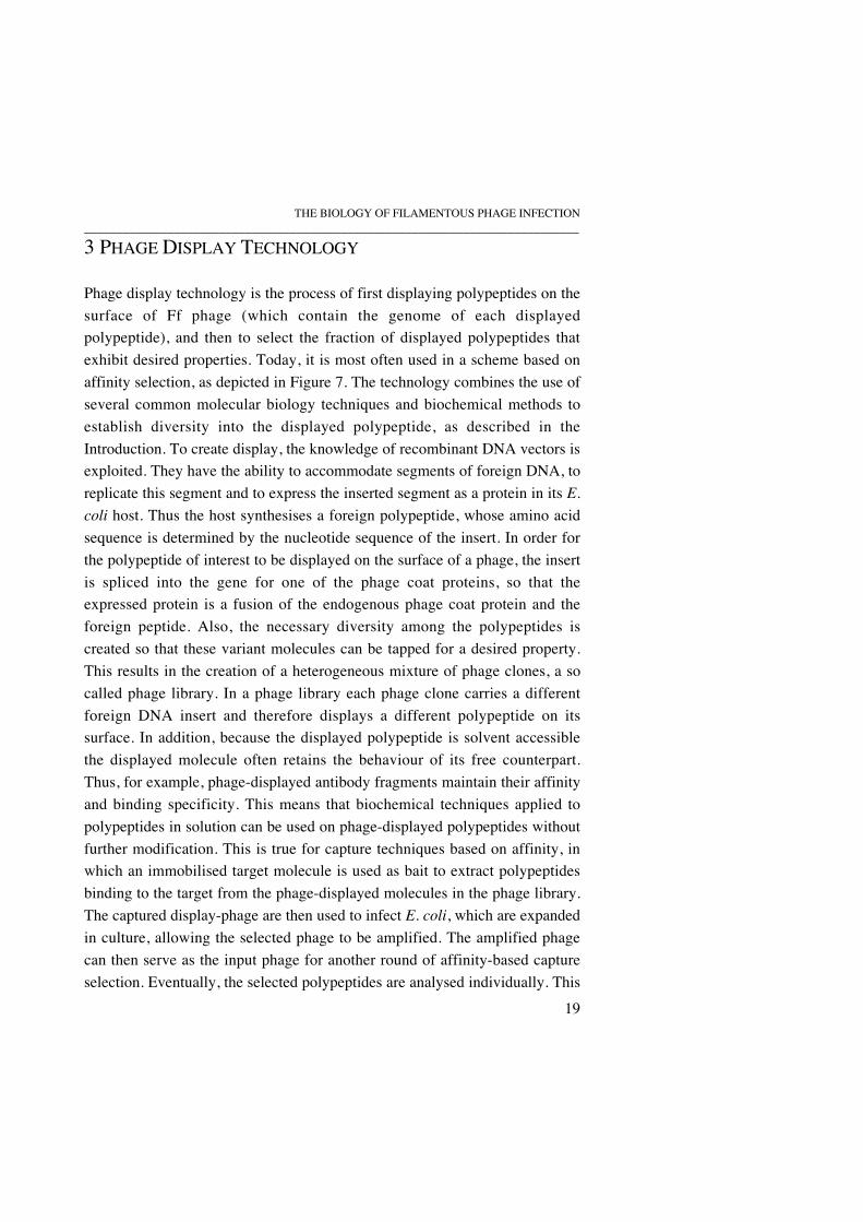
3 PHAGE DISPLAY TECHNOLOGY
Phage display technology is the process of first displaying polypeptides on thesurface of Ff phage (which contain the genome of each displayedpolypeptide), and then to select the fraction of displayed polypeptides thatexhibit desired properties. Today, it is most often used in a scheme based onaffinity selection, as depicted in Figure 7. The technology combines the use ofseveral common molecular biology techniques and biochemical methods toestablish diversity into the displayed polypeptide, as described in theIntroduction. To create display, the knowledge of recombinant DNA vectors isexploited. They have the ability to accommodate segments of foreign DNA, toreplicate this segment and to express the inserted segment as a protein in its E.
coli host. Thus the host synthesises a foreign polypeptide, whose amino acidsequence is determined by the nucleotide sequence of the insert. In order forthe polypeptide of interest to be displayed on the surface of a phage, the insertis spliced into the gene for one of the phage coat proteins, so that theexpressed protein is a fusion of the endogenous phage coat protein and theforeign peptide. Also, the necessary diversity among the polypeptides iscreated so that these variant molecules can be tapped for a desired property.This results in the creation of a heterogeneous mixture of phage clones, a socalled phage library. In a phage library each phage clone carries a differentforeign DNA insert and therefore displays a different polypeptide on itssurface. In addition, because the displayed polypeptide is solvent accessiblethe displayed molecule often retains the behaviour of its free counterpart.Thus, for example, phage-displayed antibody fragments maintain their affinityand binding specificity. This means that biochemical techniques applied topolypeptides in solution can be used on phage-displayed polypeptides withoutfurther modification. This is true for capture techniques based on affinity, inwhich an immobilised target molecule is used as bait to extract polypeptidesbinding to the target from the phage-displayed molecules in the phage library.The captured display-phage are then used to infect E. coli, which are expandedin culture, allowing the selected phage to be amplified. The amplified phagecan then serve as the input phage for another round of affinity-based captureselection. Eventually, the selected polypeptides are analysed individually. This

PHAGE DISPLAY TECHNOLOGY_____________________________________________________________________________
20
protocol of affinity selection is the leading example of how phage display isused to obtain polypeptides with desired properties.
FIGURE 7. A simplified version of the phage display cycle. First a library of variantDNA sequences is created in the format of recombinant vectors. These are thenintroduced into E. coli, which after introduction of a phage genome are converted intophage particles containing the gene for the DNA-variant and displaying the encodedpolypeptide on the surface of the phage, as a phage fusion protein (in this case topIII). After affinity selection of the displayed protein variants (the phage library),those binding to the target molecule are rescued. This selected population can theneither be analysed, by re-infecting E. coli and spreading the cells on plates to identifyindividual clones, or be used for another round of affinity-based selection.
3.1 CHOICE OF COAT PROTEIN AND DISPLAY FORMAT
Although all five capsid proteins have been used to display proteins orpeptides (Gao et al., 1999; Hufton et al., 1999; McCafferty et al., 1990), by farthe most commonly used proteins for phage display are pVIII and pIII (Smithand Petrenko, 1997; Webster, 1996). The major coat protein pVIII builds upthe tube of the phage particle and is present in thousands of copies. To achievedisplay on pVIII foreign sequences of interest are typically inserted at the N-
Transform E. coli withrecombinant vector library
Establishpopulation of DNAvariants (library)
Perform affinity selection ofphage-displayed proteinvariants
Re-infect E. coli cellsand amplify theselected phage-displayed population
Analyse theselected clonesindividually Generate
population ofphage-displayedprotein variants
Selected phage can be used as input foranother round of affinity selection
THE BIOLOGY OF FILAMENTOUS PHAGE INFECTION_____________________________________________________________________________
21
terminus, between the signal sequence and the beginning of the codingsequence for pVIII. As mentioned in a previous section (2.4), only shortpeptides (6-8 residues) allow efficient assembly of pVIII-display phage, due tosize restrictions of the pIV-channel during assembly. However, larger proteinslike antibody fragments have been displayed as fusions to pVIII (Kang et al.,1991; McCafferty et al., 1990). The limitation in the size of the displaymolecule is probably also related to the disruption of the interactions betweenpVIII molecules during assembly (Makowski, 1993).
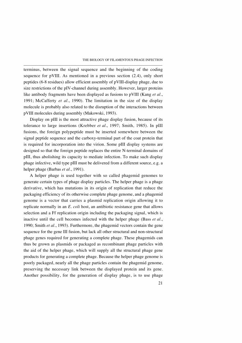
Display on pIII is the most attractive phage display fusion, because of itstolerance to large insertions (Krebber et al., 1997; Smith, 1985). In pIIIfusions, the foreign polypeptide must be inserted somewhere between thesignal peptide sequence and the carboxy-terminal part of the coat protein thatis required for incorporation into the virion. Some pIII display systems aredesigned so that the foreign peptide replaces the entire N-terminal domains ofpIII, thus abolishing its capacity to mediate infection. To make such displayphage infective, wild type pIII must be delivered from a different source, e.g. ahelper phage (Barbas et al., 1991).
A helper phage is used together with so called phagemid genomes togenerate certain types of phage display particles. The helper phage is a phagederivative, which has mutations in its origin of replication that reduce thepackaging efficiency of its otherwise complete phage genome, and a phagemidgenome is a vector that carries a plasmid replication origin allowing it toreplicate normally in an E. coli host, an antibiotic resistance gene that allowsselection and a Ff replication origin including the packaging signal, which isinactive until the cell becomes infected with the helper phage (Bass et al.,1990; Smith et al., 1993). Furthermore, the phagemid vectors contain the genesequence for the gene III fusion, but lack all other structural and non-structuralphage genes required for generating a complete phage. These phagemids canthus be grown as plasmids or packaged as recombinant phage particles withthe aid of the helper phage, which will supply all the structural phage geneproducts for generating a complete phage. Because the helper phage genome ispoorly packaged, nearly all the phage particles contain the phagemid genome,preserving the necessary link between the displayed protein and its gene.Another possibility, for the generation of display phage, is to use phage
PHAGE DISPLAY TECHNOLOGY_____________________________________________________________________________
22
vectors, in which the foreign sequence is fused to the single wild type gene forpIII, or to the gene III of an extra expression cassette, providing two sources ofpIII in the same phage DNA.
The different formats of pIII display achieved by these approaches areoutlined in Figure 8. First, the use of helper phage and phagemid genomes togenerate display phage particles is depicted. The result of using a combinationof wild type gene III from the helper phage genome and fusion gene III fromthe phagemid is a phage that displays a mixture of displayed molecules andwild type pIII on the phage coat (Figure 8A). The wild type pIII, delivered bythe helper phage, help this display phage to maintain infectivity, because therecombinant pIII encoded by a typical phagemid vector usually lacks one anda half N-terminal domain (residues 1-198) (Armstrong et al., 1996; Lowman et
al., 1991). Removal of this domain enhances the accessibility of the displayedpolypeptide (Lowman et al., 1991) and cells expressing this domain areresistant to superinfection by helper phage (Boeke et al., 1982). Furthermore,the hybrid format has the advantage of permitting monovalent display(Lowman et al., 1991). The valency of display is of importance principallybecause of its impact on the ability to discriminate between binding proteins ofdifferent affinities. Multivalency confers a high apparent affinity (avidity) onweak-binding clones, while monovalency will result in selection for trueaffinity. Polyvalency can be achieved by using a helper phage with a deletedgene III, and a phagemid that carries a fusion to an intact, full gene IIIsequence as shown in Figure 8B. The use of phage vectors to generatepolyvalent and monovalent display is shown in Figures 8C and 8D,respectively.

THE BIOLOGY OF FILAMENTOUS PHAGE INFECTION_____________________________________________________________________________
23
FIGURE 8. Different display formats for pIII-fusions. The helper phage (A and B) areshown in the upper end of the figure, while the display phage are shown in the lowerend. A comparison of polyvalent and monovalent display, as achieved by helperphage/phagemid systems (A and B) or phage vectors (C and D), is outlined.
3.2 LIBRARIES FOR PHAGE DISPLAY
As described in the introductory section of this chapter, phage displaytechnology is often used with a pool of polypeptide variants, called a phagelibrary, as starting material for the selection procedure. Prior to this, it isreasonable to confirm functional display of the polypeptide to be diversified, ifit has not previously been used for display (McCafferty and Johnson, 1996;Russel et al., 2004). Thereafter, a large variety of techniques can be exploitedto introduce diversity into the nucleotide sequence encoding the polypeptide tobe displayed. The most common approach involves the powerful and versatile
A B
C D
Gene III
Display gene sequence and short gene III
Phage structural genes
Phage ori of replication, PS
Display gene sequence and full gene III Protein III, with N1N2 and CT
Display proteinC-terminal of pIII
PHAGE DISPLAY TECHNOLOGY_____________________________________________________________________________
24
oligonucleotide-directed mutagenesis, using the polymerase chain reaction(PCR). This allows either complete randomisation of residues, partial (inwhich the wild-type residue is retained in some proportion) or tailoredrandomisation, in which only a defined subset of amino acids is specified, forintroduction of diversity. Other ways of obtaining diversity in your library isby way of in vitro recombination through DNA-shuffling (Stemmer, 1994a, b),which gives rise to new random combinations of gene segments, error pronePCR (Ling and Robinson, 1997; Zhou et al., 1991) or using mutator strains ofE. coli (Schaaper, 1988), which generates random changes throughout asequence. There are also specialised applications of phage display, in whichthe diversity of the library arises from a collection of genes rather than a singlegene. The leading example of this is antibody V-region libraries (Clackson et
al., 1991; Hoogenboom et al., 1992; McCafferty et al., 1990; Söderlind et al.,2000; Winter et al., 1994). Naturally, combinations of the methods describedabove can also be used.
Once diversity is obtained, the DNA is inserted into the phagemid forcloning by electroporation into the E. coli host. This allows the size of alibrary to be in the order of 109 – 1011 members (Söderlind et al., 2000;Vaughan et al., 1996). The host is then readily infected with a helper phage toobtain phage stocks, which finalises the creation of the phage display library.Once the phage library is obtained, the selection procedure follows.
3.3 PRINCIPLES OF SELECTION AND ANALYSIS OF SELECTED CLONES
The feasibility of obtaining large phage libraries have led to the developmentof a number of techniques for selecting the desired molecules from thelibraries (Clackson and Wells, 1994; Hoogenboom, 1997). The most commonmeans of doing this, is through an in vitro binding incubation, in which thespecific phage in the library are bound to a target tethered to a solid support,and then only retained phage are eluted, after washing away excess(unspecific) phage. This process is often referred to as sorting or panning andis based primarily on affinity for selection of binding polypeptides. Similarprinciples guide the use of antigen coated magnetic beads (Schier et al., 1996),immunotubes (Marks et al., 1991), immobilisation onto a sensor chip normallyused for kinetic analysis (Malmborg et al., 1996), microtitre plates

THE BIOLOGY OF FILAMENTOUS PHAGE INFECTION_____________________________________________________________________________
25
(Kretzschmar and Geiser, 1995) or targets naturally carried on a surface suchas a cell membrane (Fransson et al., 2004; Kupsch et al., 1999; Marks et al.,1993). Another common technique for capture of the target molecule is tocouple it to a chromatography column to perform affinity basedchromatography selection (Clackson et al., 1991; McCafferty et al., 1990).
In general, a successful selection implies that the initial population ofphage-displayed polypeptides have yielded a subpopulation typically enrichedfor polypeptides with improved or desired properties. This selectedsubpopulation can be used to re-infect fresh E. coli cells for preparation of anew phage stock, in which the specific phage that bound to the target areamplified, to allow further rounds of selection. During this enrichmentprocess, the number of binding clones over non-binding clones, from eachround of selection, is monitored by titration of the selected subpopulation onE. coli to determine the concentration of infectious particles. If the ratiobetween input and output phage has decreased, this is a good indication thatthe selection process has yielded enrichment of binding clones.
Selection parameters can often be manipulated in order to enhance theefficiency of selection, for example, the selection of antibody fragments withlow binding affinity is encouraged by fewer washes, multivalent display and ahigh coating level of antigen, whereas the isolation of those with a highaffinity for antigen is favoured by thorough washing, a low level of antigencoating and monovalent display (Barrett et al., 1992; Bass et al., 1990; Winteret al., 1994). The progress of affinity selection, through succeeding rounds, isordinarily reflected in increasing affinity of individual phage clones (or ofentire eluates) for the target molecule. Furthermore, the chances of findinghigh affinity antibody fragments from a phage library increases with the sizeof the library (Söderlind et al., 2000; Vaughan et al., 1996).
Analysis of the selected clones to determine their identity is carried out bysimply sequencing them and by carrying out an analysis of their bindingproperties. The most popular and versatile technique for this initialcharacterisation, is the phage ELISA (Mattheakis et al., 1994; Ohlin et al.,1996). In this method, the target of interest is immobilised in wells of a 96-well plate, individual phage supernatants prepared from selected clones areadded, and specific binding is detected by use of an anti-phage antibody. The

PHAGE DISPLAY TECHNOLOGY_____________________________________________________________________________
26
assay can also be used in a competition format to determine the specificity andrelative binding affinity of selected clones.
The next level of analysis often requires the production of soluble protein.Depending on which type of phagemid is being used, this can be accomplishedby expression in a non-supE E.coli (for phagemids that carry an amber stopcodon (TAG) between the displayed sequence and gene III) or by usingrestriction enzymes to excise to gene III sequence from the phagemid, to allowinduced expression of the remaining insert sequence (Lantto et al., 2002).
The display of polypeptides on Ff phage has evolved into a versatile andpowerful technology, as demonstrated by the diversity of molecules displayed(Griffiths et al., 1994; Lucic et al., 1998; Nissim et al., 1994; Saggio et al.,1995; Tanaka et al., 1999; Verhaert et al., 1999). Furthermore, the display ofe.g. antibody fragments have led to the identification of proteins capable ofrecognising a large diversity of targets, such as haptens, peptides,carbohydrates and proteins (Söderlind et al., 2000). In addition, the technologyis cheap, requires little or inexpensive equipment, and is easily accessible forindividual researchers or laboratories across the world. These factors havecontributed to the wide use of the technology.
3.4 ALTERNATIVE DISPLAY SYSTEMS
The alternatives to a traditional version of phage display technology are usedessentially as solutions to specific problems or limitations (e.g. the display orthe selection procedure) of the conventional use of the technology. As a firstexample, the problem of how to display cDNA encoded proteins on pIII wassolved by using the strong interaction of the Fos-Jun leucine zipper (Crameriand Suter, 1993). The Jun sequence is displayed as an N-terminal fusiondirectly onto intact pIII and anchored to the phage coat whereas the Fosprotein is co-secreted as a fusion protein with cDNA encoded gene products.Fos-Jun interaction and subsequent disulfide formation provides a system forC-terminal display of proteins, which has been used to identify novel genesinvolved in biological processes directly from cDNA libraries (Crameri et al.,1994; Crameri and Blaser, 1996). Alternatively, cDNA libraries may bedisplayed on pVI, which allows C-terminal cloning (Hufton et al., 1999;Jespers et al., 1995). A second example is the solution to problems of
THE BIOLOGY OF FILAMENTOUS PHAGE INFECTION_____________________________________________________________________________
27
displaying some proteins that are normally of intracellular location. For this,bacteriophage other than the Ff family have been used, including the lambda(Hoess, 2002), T4 (Ren et al., 1996) and T7 phage (Danner and Belasco,2001). These differ from Ff phage display in that assembly takes place entirelyin the cytoplasm prior to cell lysis, which means that display does not requiresecretion of the fusion polypeptide over the membrane before it is attached tothe phage particle.
The problems of the selection procedures of phage display are often relatedto establishing the proper conditions in terms of target molecule concentration,incubation times and the number of rounds needed for sufficient enrichment ofbinding proteins. Therefore, the alternatives seek to speed up the selectionprocedure, and to increase the chances of finding high affinity bindingproteins. One approach involves the use of display on cells rather than phage,because that will allow e.g. fluorescence activated cell-sorting (FACS) toselect display cells, which may be a more rapid selection procedure thanconventional panning techniques (Boder and Wittrup, 1997; Francisco et al.,1993). This approach has been applied on a number of different cell types(Georgiou, 2001; Wernérus et al., 2003; Wittrup, 2001) and has, incombination with FACS, yielded selection of high affinity scFv (Boder et al.,2000; Daugherty et al., 1998). Another approach, associated to the problemsof traditional affinity selection methods, is to directly link target recognition ofthe display phage to infectivity of the cell, which translates into a survivaladvantage if infection involves the transfer of a resistance marker to theinfected cell. This methodology was developed at the Department ofImmunotechnology under the name selection and amplification of phage(Duenas and Borrebaeck, 1994) (later also called selective infection) (PAPER
I).

SELECTIVE INFECTION_____________________________________________________________________________________
28
4 SELECTIVE INFECTION
Selective infection is a positive selection scheme for the identification ofinteracting partners, based on the direct link of target recognition to infectionof the cell. The principles of this method was first described in 1994 (Duenasand Borrebaeck, 1994; Gramatikoff et al., 1994; Krebber et al., 1995), and inthese reports, in essence, a fusion protein consisting of the N-terminal domainof pIII and a target molecule able to interact directly with a ligand (e.g. anantibody fragment) displayed on the N-terminal extremity of the truncated pIII(CT) of the phage is used. For this, helper phage carrying a deletion in gene IIIare employed, which generates a display phage that lack infectivity (Figure 9).Infectivity is then restored by the interaction with the fusion protein, whichrecreates a functional pIII, with F-pilus- and TolA-binding capacity, on thephage. Successful infection also means that the resistance marker carried bythe infecting phage is transferred to the cell, resulting in a growth advantagefor infected cells on selective media. Furthermore, because the techniqueinvolves protein-protein interaction in solution, selective infection can beviewed as a variant of affinity selection that does not rely on capture on a solidsupport. The technology has been coined both selection and amplification ofphage (SAP) (Duenas and Borrebaeck, 1994), selectively infective phage (SIP)(Krebber et al., 1997) and direct interaction rescue (DIRE) (Gramatikoff et al.,1994), but the term selective infection will be used throughout to describe thistechnology.

THE BIOLOGY OF FILAMENTOUS PHAGE INFECTION_____________________________________________________________________________________
29
FIGURE 9. The preparation of selectively infective phage and the principle ofselective infection. The helper phage genome carries a deletion in gene III, so in orderto generate the infective particles needed to introduce the phage DNA into the hostfor subsequent generation of selectively infective phage, the helper phage is producedin a host that is doubly transformed with the deleted helper phage genome and aplasmid containing the wild type gene III, but no packaging signal. Thus, only thedeleted helper phage genome should be packaged into the helper phage particles. Thedeleted helper phage is then used to infect e.g. an E. coli culture transformed with aphagemid library encoding variants of scFv. The phagemid genome carries a phagepackaging signal (black dot), why the non-infective phage extruded from such cellsshould typically contain the phagemid genome and display a truncated pIII-polypeptide fusion, to be used in selective infection experiments. The resulting non-infective phage carries a scFv capable of binding to soluble N1N2-fusion protein,which upon cognate interaction will be able to selectively infect E. coli. The rescuedscFv can then be identified by DNA sequencing.
wt pIII
E. coli
∆ geneIII
Helper phage genome
∆ geneIII
Helper phage genome
scFv CT
scFv CT
Phagemid librarywith Ff origin
E.coli
Infective helper phage
Non-infective selective phagedisplaying scFv
N1N2-fusion protein
E. coli Selectiveinfection of E.coli by scFv-displayingphage capableof binding to theN1N2-fusionprotein
scFv CT

SELECTIVE INFECTION_____________________________________________________________________________________
30
4.1 EFFECT OF FUSION PROTEIN ON SELECTIVE INFECTION
When selective infection of E. coli was first described, non-infective phagestocks displaying different antibody fragments were mixed and allowed tointeract with a target molecule fused to N1 alone (Duenas and Borrebaeck,1994). Despite that the F-pilus binding domain of pIII was missing, anenrichment of the cognate antibody-antigen pair was achieved. Later, amechanistic dissection of the influence of a large protein insertion at variouspositions between the different domains of pIII, demonstrated that thepresence of N2 enhanced the efficiency of selective infection, irrespective ofwhether N2 was on the phage or together with N1 as a soluble target fusion(Krebber et al., 1997). Therefore, the more recent versions of selectiveinfection use the N1N2-fusion to restore infectivity (Figure 9).
4.2 FACTORS AFFECTING THE OUTCOME OF SELECTIVE INFECTION
One factor that determines the outcome of selective infection is theconcentration of soluble fusion protein added to the mixture of non-infectivedisplay phage and E. coli cells during the selection process. It has beenreported that high concentrations of fusion protein have an inhibitory effect oninfectivity (Duenas and Borrebaeck, 1994; Krebber et al., 1997). Since then ithas been realised that the N1N2-fusion concentration needs to be adjusted toachieve optimal infection conditions (Krebber et al., 1997). The main reason isthat, at least for N1N2-fusions, the fusion protein binds to the pilus anddepolymerises it at a high fusion protein concentration. Another potentiallyimportant factor for successful rescue of interacting pairs is the display format.Several copies of the displayed antibody fragment, linked to the N1N2-fusions, may be required to achieve optimal conditions for selective infection.Furthermore, the binding strength (affinity) of the protein-ligand interaction issuspected to be of importance to the outcome of selective infectionexperiments. This notion comes from reports demonstrating a selection of highaffinity antibody fragments using the selective infection methodology,however, the results were inconclusive and could be shown to depend on e.g.advantages in folding (Duenas et al., 1996a; Pedrazzi et al., 1997; Spada et al.,1997).

THE BIOLOGY OF FILAMENTOUS PHAGE INFECTION_____________________________________________________________________________________
31
In the experiments described in PAPER I, we used two different gene III-deleted Ff phage; the R408d3 (Rakonjac et al., 1997) and the VCSM13 mutanthelper phage ∆g3N (Gao et al., 1997), for the creation of selectively infectivephage with different display format (Figure 10). The R408d3 helperphage/phagemid system gives rise to infective helper phage carrying pIII on itssurface, encapsulating a genome lacking the entire gene III. After infection ofan E. coli containing phagemid, the selectively infective phage produceddisplay antibody fragments on all copies of the truncated pIII molecule(residues 256-406), encoded by the phagemid genome. The ∆g3N helperphage carries a partial deletion of gene III, keeping the first 38 residues of N1and residues 252-406 of CT. During the production of the ∆g3N selectivelyinfective phage, the genome encoded CT molecules compete for incorporationinto the phage coat with the phagemid encoded CT-antibody fragment fusionprotein, thus giving particles where the majority of the displaying species aremonovalent (Lowman et al., 1991) (see section 3.1).
FIGURE 10. Schematic representations of the two different helper phage systemsused in PAPER I. The position of the deleted gene sequence within each helper phagegenome is indicated by a star (A). The R408d3 helper phage encapsulates the deletedhelper phage genome and will display wild type pIII if propagated in a host carrying
A. Phage gene sequence B. Appearance of helper phage C. Displaying phage(non-infective)
gVIII gVI
gVIII Partiallydeleted gIII
gVI
∆g3N
Carries ∆g3N genome
Carries R408d3 genome
R408d3 Wild type pIII
Truncated pIII (non-infective)
Only display fusions
Mixture of display fusions and truncated pIII
Phagemid
Phagemid
SELECTIVE INFECTION_____________________________________________________________________________________
32
pIII-complementing vectors. The ∆g3N helper phage will display a truncated pIII,lacking all of N2 and keeping the first 38 residues of N1, resulting in a non-infectiveparticle (B). The display-phage of R408d3 will only display CT-fused proteins, whilethe ∆g3N display-phage will carry a mixture of CT-fusions and truncated pIIIs (C).The picture is adapted from Paper I with written permission from the copyrightholders John Wiley & Sons Limited.
The size of the complex obtained after cognate interaction was also varied inthis study, since we chose to study three different interacting pairs ofmolecules, of which one pair was peptide-scFv, the second a non-peptidic ringstructure and scFv and the third was a protein-protein interaction.Furthermore, their respective binding affinity varied: Starting with the pair ofthe weakest affinity, we used a peptide (AD2) derived from glycoprotein B ofhuman cytomegalovirus (CMV) and a scFv (AE11F) binding to this peptidewith a KD = 6 × 10 -7 M (Lantto and Ohlin, unpublished; (Ohlin et al., 1993)),a fluorescein isothiocyanate (FITC) –specific scFv binding to FITC with a KD
= 3 × 10 -10 M (Vaughan et al., 1996) and a papain and cystatin C interactingpair having a KD = 1 × 10 -11 M (Pol et al., 1995).
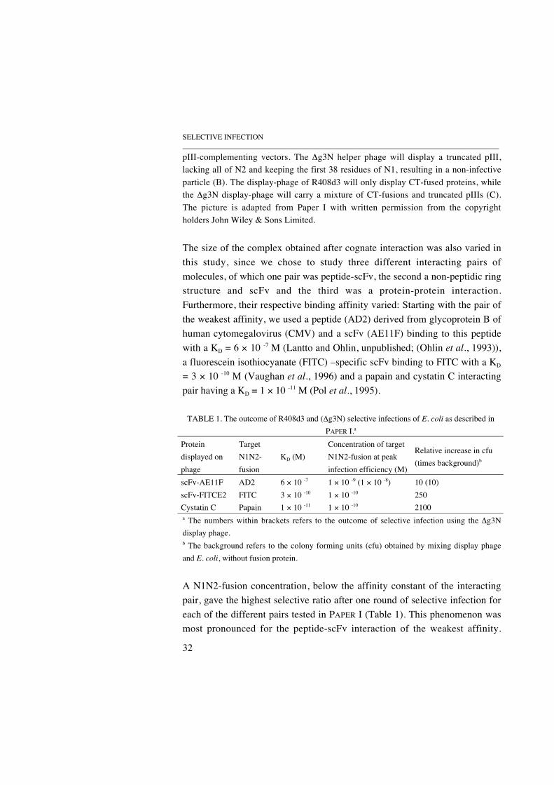
TABLE 1. The outcome of R408d3 and (∆g3N) selective infections of E. coli as described inPAPER I.a
Proteindisplayed onphage
TargetN1N2-fusion
KD (M)Concentration of targetN1N2-fusion at peakinfection efficiency (M)
Relative increase in cfu(times background)b
scFv-AE11F AD2 6 × 10 -7 1 × 10 -9 (1 × 10 -8) 10 (10)scFv-FITCE2 FITC 3 × 10 -10 1 × 10 -10 250Cystatin C Papain 1 × 10 -11 1 × 10 -10 2100a The numbers within brackets refers to the outcome of selective infection using the ∆g3Ndisplay phage.b The background refers to the colony forming units (cfu) obtained by mixing display phageand E. coli, without fusion protein.
A N1N2-fusion concentration, below the affinity constant of the interactingpair, gave the highest selective ratio after one round of selective infection foreach of the different pairs tested in PAPER I (Table 1). This phenomenon wasmost pronounced for the peptide-scFv interaction of the weakest affinity.

THE BIOLOGY OF FILAMENTOUS PHAGE INFECTION_____________________________________________________________________________________
33
Using higher concentrations of N1N2-fusion protein, the expected inhibitionof selective infection was observed (PAPER I; Figure 3). Using the ∆g3Ndisplay phage, selective infection was also achieved for the AE11F-AD2binding pair, at a fusion protein concentration below the affinity constant ofthe interacting pair. However, the concentration required for selectiveinfection was higher for this monovalent display system in comparison tousing the same interaction pair and the R408d3 multivalent display. Therefore,monovalent display requires a (undesirably) high concentration of fusionprotein, which was also demonstrated by the observation that no selectiveinfection could be achieved with the ∆g3N phage at concentrations below 10 -8
M (PAPER I). These findings are readily explained by effects of avidity on theinteraction between displayed protein and target, as the effective concentrationof e.g displayed scFv is higher with a multivalent display-phage when thesame concentration of mono- or multivalent phage are used. Other reportshave demonstrated that multivalent display on phage improve the selection forbinding proteins with low affinity (Crameri et al., 1994; Terskikh et al., 1997),and that an improved efficiency of display and antibody fragment selectioncan be achieved (O'Connell et al., 2002). Thus, in PAPER I we conclude that,for selective infection, a multivalent display format is of importance, becauseit allows the use of lower concentrations of fusion protein. Furthermore, wefound a direct correlation between the affinity constant and infectionefficiency of the interacting pairs, when using multivalent display (PAPER I).
The mechanism behind the observation that selective infection seems toselect for high affinity binding pairs, is about to become clear. Previousspeculations were based on the fact that N1N2-fusions could not be used athigher concentrations, otherwise it would saturate the pilus, thus only very lowamounts of N1N2-fusions are needed and the system must select for highaffinity pairs (Spada et al., 1997). Contrary to this, recent findings suggest thata strong interaction between the N-terminal domains and CT of pIII is ofimportance, to transfer a signal to the CT necessary for infection (Bennett andRakonjac, 2004). From their data, it was speculated that the link between theN-terminal domains and residues 253-323 of CT needs to be of a covalent-likecharacter to allow a conformational change in CT, which leads to thedissociation of pIII from the phage coat (J. Rakonjac, personal

SELECTIVE INFECTION_____________________________________________________________________________________
34
communication). Consequently, these findings can be used to explain theresults from the selective infection experiments presented in PAPER I, whichdemonstrated the highest rescue efficiency by the coupling of the interactingpair with the highest affinity.
4.3 THE FUTURE OF SELECTIVE INFECTION
Elaborate solid support protocols used in conventional phage displaytechnology may risk the permanent trapping of high affinity bindingpolypeptides during the passage of phage clones over e.g. an affinity column.The advantage of using selective infection is the simple selection step thatavoids the use of these solid support protocols. However, the simplifiedselection step of selective infection has been shown to require carefulevaluation of N1N2-fusion concentrations and incubation times in order todrive the selection process in the desired direction (Duenas et al., 1996b;Krebber et al., 1997) (PAPER I). Also, the in vitro selective infection approachhas demonstrated that there is a risk for obtaining a background of infectiousparticles (Pedrazzi et al., 1997) (PAPER I), which will have detrimental effectson library selections. Thus, there are a number of issues to resolve for thefuture use of the technology, before it can be a protocol of generalapplicability. Nevertheless, selective infection has been used to select highaffinity binding proteins (Duenas et al., 1996a; Duenas et al., 1996b; Pedrazziet al., 1997), and to be a powerful technique for the enrichment of proteinswith the best folding and binding characteristics from a library of similarmolecules (Hennecke et al., 1998; Spada et al., 1998). In addition, we haveshown that the technique demonstrates a potential to select for high affinitybinding proteins, when multivalent display is used (PAPER I). Thus, there canbe no doubt the technology is suitable for special applications like theselection of high affinity interaction pairs, but the methodological difficultiesdescribed above and the lack of understanding for the underlying mechanismof filamentous phage infection has limited its use.

THE BIOLOGY OF FILAMENTOUS PHAGE INFECTION_____________________________________________________________________________________
35
5 MOLECULAR DISSECTION OF THE Ff PHAGE INFECTION
The infection process of Ff phage depends on (exploits/parasitises) a numberof bacterial proteins for the phage DNA to pass the E. coli membranes. First,the specificity of the Ff phage is determined by the reaction, in which N2 ofpIII attaches to the tip of the F-pilus extending from the bacterial membrane(Jacobson, 1972; Marvin and Hohn, 1969). This step of the infection processalso brings the phage closer to the bacterial membrane, through the rapiddepolymerisation of the F-pilus. In the membrane, the products of the tolQRA
genes are required for Ff phage infection (Sun and Webster, 1986), in additionto being necessary for membrane integrity. Also, sensitivity towards bacterialtoxins specific for E. coli (colicins) is mediated through these proteins(Webster, 1991), which are preferentially associated with contact regionsbetween the inner and outer membranes, so called adhesion zones (Guihard et
al., 1994). Thus, the entry point of the Ff phage seems to be at a site, wherethe distance the phage DNA would have to travel to cross both membranes, isthe shortest. Once the phage has reached the bacterial outer membrane, the N1domain binds to the TolA receptor molecule (Click and Webster, 1997;Riechmann and Holliger, 1997). In order for N1 to bind TolA efficiently, theinitial interaction with the F-pilus must have taken place, since the binding siteof TolA and N2 for N1 overlap (Riechmann and Holliger, 1997). Thestoichiometry of the TolQRA complex required for phage DNA entry isunknown, but TolR dimerisation has been reported (Journet et al., 1999), andthe TolQ and R proteins have been shown to act as motor proteins, energisingthe TolA molecule to form a transmembrane link via binding to an outermembrane protein (Cascales et al., 2001). Following pIII and TolAinteraction, pIII crosses the outer membrane and inserts its CT in the innermembrane (Boeke and Model, 1982; Endemann and Model, 1995). Theinteractions between the pIII domains and TolA important for this processwere the focus of the study in PAPER II.
5.1 PIII AND TOLA DURING INFECTION
Not only pIII but also the 421 amino acid TolA has a sophisticated domainorganisation (Levengood et al., 1991). The first domain of TolA (TolAI) is the
MOLECULAR DISSECTION OF THE Ff PHAGE INFECTION_____________________________________________________________________________________
36
N-terminal membrane anchor domain, which spans the inner membrane once.The second domain (TolAII), consists of a segment of repetitive sequences,creating an extended helix, which is estimated to be of a length that wouldallow it to span the periplasm (Levengood et al., 1991). The C-terminal thirddomain of TolA (TolAIII) consists of a number of short helices and ß-strands,forming a globular fold (Lubkowski et al., 1999), which can bind to N1. Twoshort stretches of glycine residues are preceding TolA domains II and III,which might confer limited flexibility to the protein (Levengood et al., 1991).Each of these TolA domains and the N1, N2 and N1N2 domains of pIII wereprepared as purified polypeptides as described in PAPER II. The objective wasto measure the binding kinetics of the interactions between these polypeptidesin real time using the surface plasmon resonance (SPR) technology (Liedberget al., 1995). The principle of the analysis is that one of the interactingcounterparts is immobilised on a sensor chip surface , while the other passes ina continous flow in the BIAcore™ (Jönsson et al., 1991).
In PAPER II, Table 1, the affinities for the interactions between N-terminaldomains of pIII and bacterial TolA are presented. The affinity constant of theknown interaction between N1 and TolAIII was determined to be in the micro-molar range (KA = 1 × 10⁶ M-1). This is the first measurement of the kinetics ofthe interaction between TolAIII and N1 reported, although soluble TolAIII hasbeen shown to inhibit the interaction between N1-phage and TolAIII in vitro,at concentrations down to 0.7 micro-molar (Riechmann and Holliger, 1997). Apreviously unknown interaction between N2 and TolA was also detected. Ourresults showed that this interaction was dependent on TolAII, since no otherTolA domain was able to bind immobilised N2. In Table 2 of PAPER II, theaffinities of interactions between the different domains of TolA are presented.Intact TolA was found to bind to itself, as did each of the separate domains.Furthermore, TolAI and III, as well as TolAII and III had affinity for eachother, indicating the possibility of TolA-TolA functional interaction. Thus,using real-time bio-specific interaction analysis (BIAcore™), we demonstratedthat both N-terminal domains of pIII contribute to the overall affinity of thesedomains for TolA. Also, the extensive interactions between different domainsof TolA suggested that the TolA protein have the capability of interacting withitself in the bacterial membrane. In an attempt to weld the results from the

THE BIOLOGY OF FILAMENTOUS PHAGE INFECTION_____________________________________________________________________________________
37
BIAcore™ analysis with previously reported interactions between pIII and theTolQRA proteins, a refined infection mechanism model was hypothesised.
5.2 A NOVEL MODEL FOR THE Ff PHAGE INFECTION MECHANISM
In previous models of the Ff phage infection mechanism (Click and Webster,1998; Lazzaroni et al., 1999; Riechmann and Holliger, 1997; Webster, 1996),the knowledge that the TolQRA proteins interact with each other is taken intoaccount (Derouiche et al., 1995; Germon et al., 1998; Journet et al., 1999;Lazzaroni et al., 1995). In contrast, the knowledge of TolQ, TolR and TolAlocalisation to adhesion zones was not considered in these models. Also, mostof these previous models propose that this protein complex (and potentiallyalso pIII) is involved in the formation of a channel for phage DNA to cross thecytoplasmic membrane (Click and Webster, 1998; Lazzaroni et al., 1999;Riechmann and Holliger, 1997).
The new model, presented in Figure 11, tries to combine the knowledgefrom the above mentioned previous studies with the findings presented inPAPER II. It should be mentioned however, that the data from PAPER II wasgenerated by measuring polypeptide interaction in vitro, which may notdirectly translate into an in vivo situation. Nevertheless, in the new model forthe early events in Ff phage infection of E. coli, we depict the F-pilusretraction process at the adhesion zones of outer and inner membranes. Thislocalisation would allow a more compact state of assembly for the TolAprotein, thus keeping the two membranes closer together, in addition toallowing N2 to make contact with the TolAII at the adhesion zone. The modelalso suggests that the N2-TolAII interaction help orient pIII correctly into theinner membrane, and that the cooperation of more than one TolA moleculehelp achieve the insertion of pIII into the inner membrane. Furthermore, TolAmakes several interactions with the translocation domains of pIII duringinfection, similar to what has been seen for colicins during their TolAmediated insertion into the cytoplasmic membrane (Journet et al., 2001).
MOLECULAR DISSECTION OF THE Ff PHAGE INFECTION_____________________________________________________________________________________
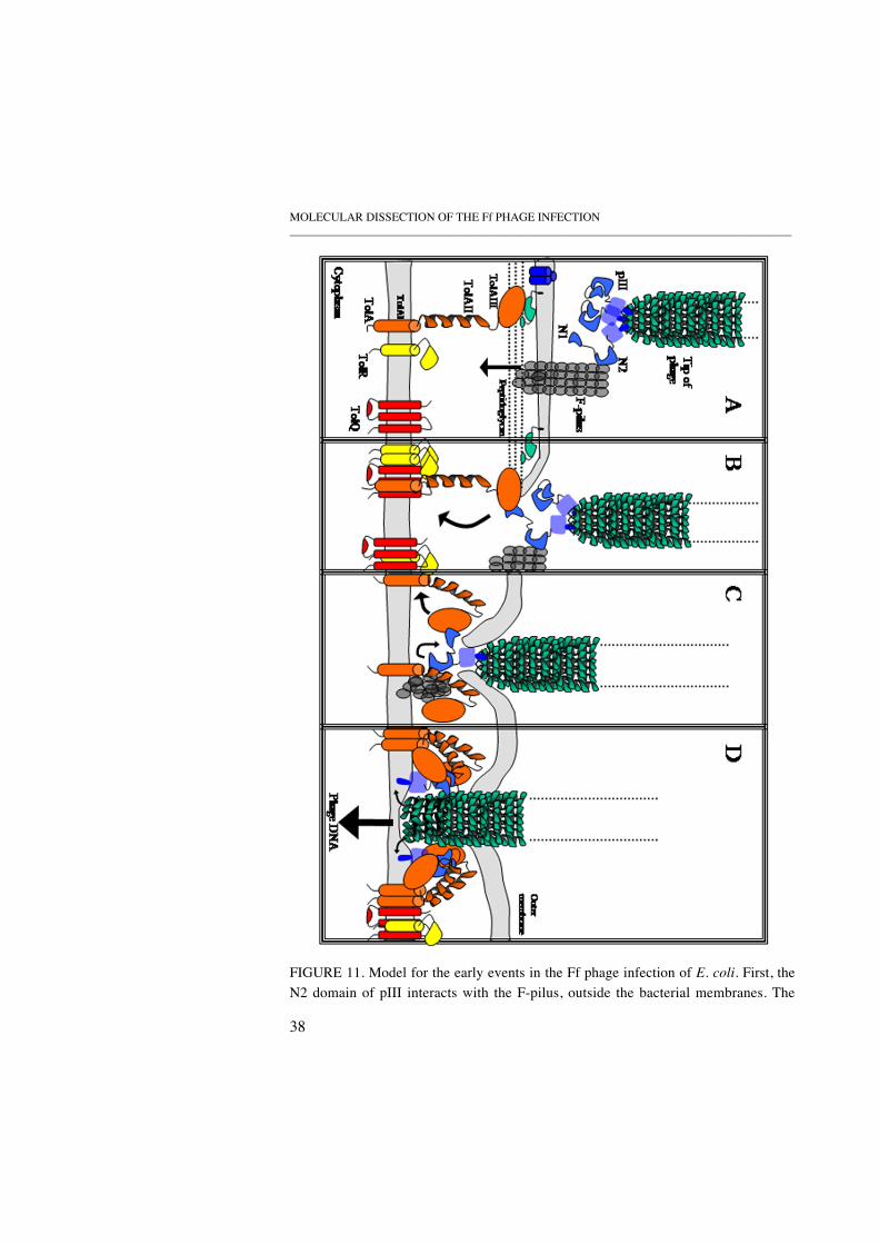
38
FIGURE 11. Model for the early events in the Ff phage infection of E. coli. First, theN2 domain of pIII interacts with the F-pilus, outside the bacterial membranes. The
THE BIOLOGY OF FILAMENTOUS PHAGE INFECTION_____________________________________________________________________________________
39
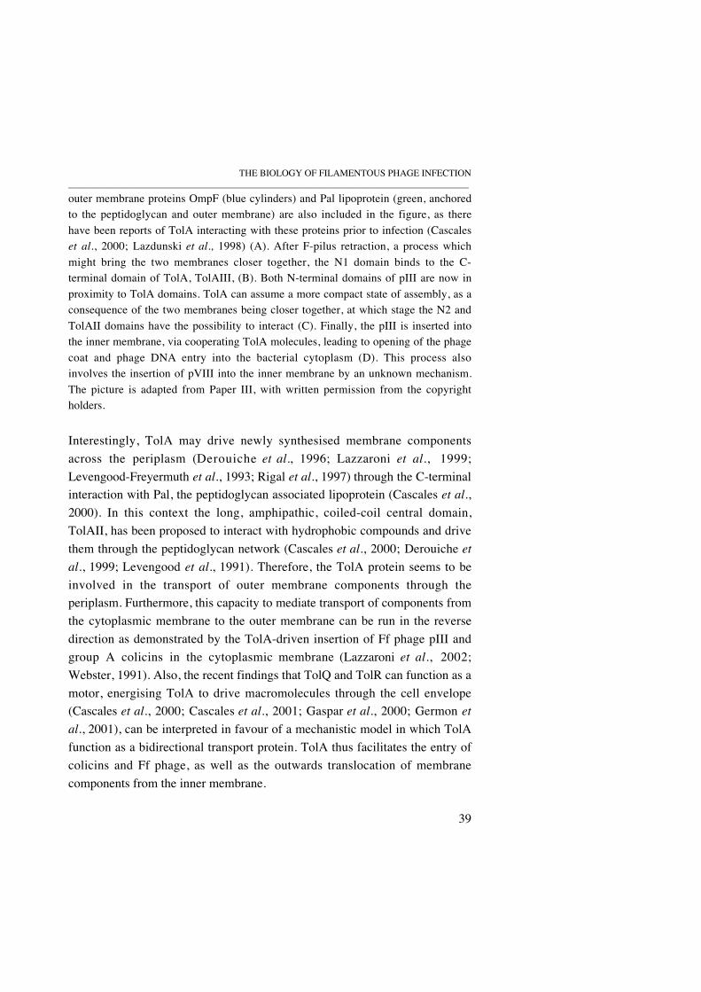
outer membrane proteins OmpF (blue cylinders) and Pal lipoprotein (green, anchoredto the peptidoglycan and outer membrane) are also included in the figure, as therehave been reports of TolA interacting with these proteins prior to infection (Cascaleset al., 2000; Lazdunski et al., 1998) (A). After F-pilus retraction, a process whichmight bring the two membranes closer together, the N1 domain binds to the C-terminal domain of TolA, TolAIII, (B). Both N-terminal domains of pIII are now inproximity to TolA domains. TolA can assume a more compact state of assembly, as aconsequence of the two membranes being closer together, at which stage the N2 andTolAII domains have the possibility to interact (C). Finally, the pIII is inserted intothe inner membrane, via cooperating TolA molecules, leading to opening of the phagecoat and phage DNA entry into the bacterial cytoplasm (D). This process alsoinvolves the insertion of pVIII into the inner membrane by an unknown mechanism.The picture is adapted from Paper III, with written permission from the copyrightholders.
Interestingly, TolA may drive newly synthesised membrane componentsacross the periplasm (Derouiche et al., 1996; Lazzaroni et al., 1999;Levengood-Freyermuth et al., 1993; Rigal et al., 1997) through the C-terminalinteraction with Pal, the peptidoglycan associated lipoprotein (Cascales et al.,2000). In this context the long, amphipathic, coiled-coil central domain,TolAII, has been proposed to interact with hydrophobic compounds and drivethem through the peptidoglycan network (Cascales et al., 2000; Derouiche et
al., 1999; Levengood et al., 1991). Therefore, the TolA protein seems to beinvolved in the transport of outer membrane components through theperiplasm. Furthermore, this capacity to mediate transport of components fromthe cytoplasmic membrane to the outer membrane can be run in the reversedirection as demonstrated by the TolA-driven insertion of Ff phage pIII andgroup A colicins in the cytoplasmic membrane (Lazzaroni et al., 2002;Webster, 1991). Also, the recent findings that TolQ and TolR can function as amotor, energising TolA to drive macromolecules through the cell envelope(Cascales et al., 2000; Cascales et al., 2001; Gaspar et al., 2000; Germon et
al., 2001), can be interpreted in favour of a mechanistic model in which TolAfunction as a bidirectional transport protein. TolA thus facilitates the entry ofcolicins and Ff phage, as well as the outwards translocation of membranecomponents from the inner membrane.
MOLECULAR DISSECTION OF THE Ff PHAGE INFECTION_____________________________________________________________________________________
40
Phage assembly involves the formation of a channel (Section 2.4), whichnewly synthesised phage pass through. It is not known whether phageinfection also requires the formation of a channel, but it has been proposed,both because of the channel formation during phage assembly and becausetranslocating colicins form channels (Lazzaroni et al., 2002; Riechmann andHolliger, 1997; Webster, 1991). In parallel, the infection process offilamentous phage can also be discussed in a comparison to F-pilusassembly/retraction mechanisms. The morphological similarities of phagefilaments and conjugative pili, when phage filaments are assembled withoutpIII, have led to the proposal that the two are likely to be evolutionarily related(Rakonjac and Model, 1998). Furthermore, in a recent study (Bidlack andSilverman, 2004), where it was found that F-pilus positive cells are moresensitive towards bile salts, the authors suggested that the sensitivity occurswhen F-pilin subunit secretion transiently opens the cell envelope to thesurrounding medium, allowing entry of the anionic detergents. Such transientopening may also occur during phage entry, when the pilus is retracted. Thiswould lend further support to a “channel theory”. However, the resultspresented by Bidlack and Silverman suggest that the pilus retraction processand the pilin subunits are more important in the formation of such a channel,than the Tol proteins and pIII, which were the previous prime suspects ofchannel forming proteins (Click and Webster, 1998; Lazzaroni et al., 1999;Riechmann and Holliger, 1997).
During phage assembly and infection, a stage can be envisaged in whichthe phage is attached to the cell surface, with the phage head (pIII/pVI) buriedin the membrane, thus resembling cell surface-attached F-pili. However, themovement of the phage proceeds in different directions (away from the cellsurface versus towards the cell surface) depending on whether the phage isbeing assembled or is infecting the cell. The driving forces of these opposingreactions can probably be found in the formation of the different membranecomplexes responsible for the separate reactions (the pIV-channel forassembly and the TolQRA and pilus proteins for infection), but also in thedifferences in membrane concentration of phage proteins (mainly pVIII). Thisbehaviour of the infecting and assembling Ff phage can be seen as an anologyto the process of F-pilus assembly/retraction. In F-pilus assembly, a membrane

THE BIOLOGY OF FILAMENTOUS PHAGE INFECTION_____________________________________________________________________________________
41
complex of assembly proteins (encoded by the F plasmid DNA), large enoughto span the bacterial envelope, has been proposed to be involved in the piluselongation process (Frost et al., 1994). This elongation process is achieved bythe incorporation of F-pilin subunits into the growing filament. The F-pilinsubunit share a similar size and fold (α-helical) with the phage pVIII molecule(Armstrong et al., 1980; Frost et al., 1994; Paranchych et al., 1978), and theassembly of F-pilin subunits into pilus and pVIII subunits into phage particlesappear from an inner membrane pool (Laine et al., 1985; Moore et al., 1981a,b; Rapoza and Webster, 1995; Russel, 1991). These similariteies of F-pilusand phage assembly mechanisms can be translated into mechanistic ideas ofrelevance for the infection process of Ff phage. Perhaps the F-pilus retractionsystem is used for the insertion of pVIII into the inner membrane, stripping offthe phage coat and releasing the naked ssDNA into the host. In such amechanism the function of TolA would be required for the uncapping of thephage coat, by multiple interaractions with the pIII (PAPER II), thus enablingpVIII to be accessible for membrane insertion. Therefore, future studies of theproposed F-pilus assembly/retraction complex may provide more leads thatwill help determine the last steps in the mechanism by which Ff phagepenetrate the bacterial membranes.
5.3 TOLA FUNCTION AND IMPORTANCE
In addition to its occasional role as the co-receptor for the Ff phage pIII duringphage infection, the normal function of TolA is to maintain membraneintegrity and probably to aid the transport of molecules through the cellenvelope (Lazzaroni et al., 1999). To carry out these multiple functions, TolAdepends on interactions with other proteins. It has been shown that mutationsin the tol-pal gene cluster, encoding TolQ, TolR, TolA, TolB (a periplasmicprotein) and Pal (a peptidoglycan associated lipoprotein), result in apleiotropic phenotype including hypersensitivity towards detergents and bilesalts, release of periplasmic content into the external milieu (Lazzaroni et al.,1989; Lazzaroni et al., 1999; Webster, 1991) and the formation of outermembrane vesicles at the cell surface (Bernadac et al., 1998). The TolQ, R andA and TolB and Pal form two complexes (TolQRA and TolBPal) that areconnected by the interaction between Pal and TolA, which requires the proton

MOLECULAR DISSECTION OF THE Ff PHAGE INFECTION_____________________________________________________________________________________
42
motive force, TolQ and R proteins (Cascales et al., 2000). Furthermore, theperiplasmic TolAIII domain interacts with the N-terminal domain of TolB(Dubuisson et al., 2002; Walburger et al., 2002). The exact role of the Tol-Palproteins (the Tol system) in maintaining outer membrane integrity andtransport functions are still largely unknown. Nevertheless, these proteins arebelieved to play an important role because tol-pal gene clusters have beenfound in many other Gram-negative bacteria (Sturgis, 2001). In a widerperspective, the function of TolA, and the Tol system, is of relevance to publichealth, since the Tol proteins have been found to be involved in the virulenceof Salmonella typhimurium (Bowe et al., 1998) and in the phage-mediatedimport of the cholera toxin gene into Vibrio cholerae (Heilpern and Waldor,2000).
To learn more about which epitopes on the TolA protein that arefunctionally important for phage infection and possibly also about the differentfunctions of TolA, we decided to introduce mutations in a part of TolA knownto be involved in contacts with N1 of phage pIII during phage infection(Lubkowski et al., 1999). We created four limited substitution libraries ofTolA, targeting six amino acid residues in the C-terminal domain of TolA(TolAIII) (Table 2). The residues at these positions (415-420) build up a β-strand of TolAIII, located in a concavity of the TolAIII molecule thataccommodates N1 during phage infection (Figure 12). The substitutionsintroduced in all four libraries were mainly restricted to related residues, inorder not to destabilise the structure and to maintain similar but modifiedinteractions, introducing the possibility to affect the binding of TolA to phageor outer membrane proteins. TolA-negative E. coli were then transformed withlibrary vectors, and the functionality of these different libraries was tested bygrowth on media containing detergent and sensitivity to M13 phage infection.

THE BIOLOGY OF FILAMENTOUS PHAGE INFECTION_____________________________________________________________________________________
43
FIGURE 12. Three different views of the TolAIII three-dimensional structure, withthe residues targeted by the study presented in PAPER III highlighted. In A) theTolAIII surface is shown, and residues 415-420 are highlighted by separatecolourings from left to right; Alanine 415 (red), Proline 416 (turquoise), Leucine 417(yellow), Aspartic acid 418 (light green), Phenyl alanine 419 (blue) and Lysine 420(purple). In B) a ribbon model is shown, with the side chains of residues 415-420exposed. (The TolAIII molecule is slightly trimmed.) In C) the binding of the Ffphage pIII N1 domain to TolA during phage infection is shown. The orientation ofTolA is the same as in B).
TABLE 2. The allowed sequence variation in the four different TolA libraries created.a
Position
Wtaminoacid
Allowed residuesin TolA-lib
Allowed residuesin lib-416
Allowed residuesin lib-417
Allowed residuesin lib-418
415 A D, V, G, E D, V, G, E D, V, G, E D, V, G, E416 P R, Q, L, H P R, Q, L, H R, Q, L, H417 L R, Q, P, H R, Q, P, H L R, Q, P, H418 D A, V, G, E A, V, G, E A, V, G, E D
419 F S, L S, L S, L S, L420 K T, N, I, M, R, S,
K
T, N, I, M, R, S,K
T, N, I, M, R, S,K
T, N, I, M, R, S, K
a The wild type TolA amino acids are shown in bold type.
5.4 SEPARATION OF TOLA FUNCTIONS
The results from PAPER III showed that few TolA mutants were tolerated whenthe TolA membrane integrity function was challenged (PAPER III; Table 2a).
A B C

MOLECULAR DISSECTION OF THE Ff PHAGE INFECTION_____________________________________________________________________________________
44
In contrast, considerable sequence alteration was tolerated by different TolA-variants still capable of mediating phage infection (PAPER III; Table 2a),leading to the identification of clones that displayed a segregation ofmembrane integrity and phage receptor function (PAPER III; Table 3). We alsoperformed a sequence alignment of TolA from closely related species, andidentified a conserved sequence, involving the amino acids at positions 415,417 and 419, that are crucial for TolA membrane integrity functionality(PAPER III; Figure 3). Furthermore, the dependence on L417 over the othertargeted residues was demonstrated by the finding that TolA moleculeslacking this residue were unable to sustain membrane integrity (PAPER III;Figure 2 and Table 2a).
Our results showed that the phage receptor function of TolA was moretolerant to the sequence variation introduced by the chosen mutations. Becausethe stability of the different TolA mutants is the same irrespective of whichfunction was challenged, this behaviour may be attributed to either differencesin the mechanisms of the two functions or to the influence of individual aminoacid residues for the binding to phage or membrane proteins. The influence ofindividual amino acids for the preservation of phage receptor function inTolA, can be discussed from the knowledge of phage pIII N1 contact residues,and compared to contact regions for outer membrane proteins on TolA. In thefirst case, we know from the crystal structure of N1 and TolAIII in complex,that the introduced mutations targeted the TolA residues normally in closecontact with the N1 molecule during phage infection (Lubkowski et al., 1999).The isolation of functional phage receptors, which were mutated at thesepositions, implies that either additional contacts (presumably in the α-helixformed by TolA residues 335-349) contribute enough to allow N1 binding, orthat proper formation of a beta-strand overrides the effect of individual N1contact residues in the 415-420 region of TolAIII. In comparison, the 415-420region of TolA has not been reported to be involved in any contacts with e.g.Pal or TolB, suggesting that the introduced mutations have not affected theTolA binding to outer membrane proteins. Therefore, the most plausibleexplanation for the observed segregation of TolA functions is the differencesin the mechanisms of phage receptor function and membrane integrityfunction. The phage infection process requires TolA interaction with the TolQ

THE BIOLOGY OF FILAMENTOUS PHAGE INFECTION_____________________________________________________________________________________
45
and R proteins (Click and Webster, 1998), and possibly TolA-TolAinteractions (PAPER II). In addition, there are only a few F-pili per bacterium(Brinton, 1965; Caro and Schnös, 1966), corresponding to the two or threeadsorption sites found for Ff phage (Hsu, 1968; Tzagoloff and Pratt, 1964).Consequently, only a few functional TolA molecules will be needed to carryout the phage receptor function. Contrary to this, the TolA membrane integrityfunction depends on a number of additional TolA interactions (with e.g. Paland TolB) (Cascales et al., 2000; Dubuisson et al., 2002), in which the TolAmolecules need to be evenly distributed along the entire E. coli cytoplasmicmembrane. Thus, if the introduced mutations affected the stability of the TolAmolecule, leading to fewer functional TolA in the cytoplasmic membrane, it isonly natural that the membrane integrity function would be most hampered.Therefore, the identification of mutants with segregated TolA functions isevidence of the mechanistic differences between the TolA membrane integrityand phage receptor functions. The technological and biological implications ofthe findings presented in PAPER III will be summarised in chapter 7,Concluding remarks.

ALTERED TRANSCRIPTION OF PHAGE INFECTED CELLS____________________________________________________________________________________
46
6 ALTERED TRANSCRIPTION OF PHAGE INFECTED CELLS
Bacterial cells protect themselves through their cell membrane, a formidablephysical barrier, but what are the cellular responses when this barrier has beenpenetrated by e. g. infecting Ff phage? The bacteria normally live in anenvironment where it is necessary to adapt rapidly to changes in e.g.temperature, pH and nutrient access (Gross, 1996; Hengge-Aronis, 1999;Slonczewski and Foster, 1996). The responses to such changes are calledstress responses, and are often aimed at adapting the metabolism to theenvironmental changes that may be the cause of the stress. During Ff phageinfection, a number of bacterial proteins are engaged and, as seen in theprevious sections of this thesis (2.2 and 2.3), not only the membrane proteinsinvolved in the infection process, but also host RNA and DNA polymerasesare used by the phage. For example, the processing of phage mRNA requiresthe activity of the host endonuclease RNase E (Goodrich and Steege, 1999;Kokoska and Steege, 1998). In addition to these proteins, Ff infection alsoresults in a stress response; the well known sensitisation of infected cells tobile salts among other agents (Boeke et al., 1982; Roy and Mitra, 1970).Furthermore, a deregulated phage gene expression (especially overexpressionof certain genes in the absence of assembly) may cause cell death (Model andRussel, 1988; Pratt et al., 1966; Schwartz and Zinder, 1968). Stressfulsituations like these are often triggers for stress responses; however, they aredifficult to measure because the conditions cause severe effects on cellsurvival. Nevertheless, the insertion of phage protein pIV into the cellmembrane during phage assembly has been shown to give rise to theexpression of an E. coli phage shock protein; PspA (Brissette et al., 1990;Brissette et al., 1991). This expression represents the only previously knownstress response of E. coli related to the expression of a single phage encodedgene. However, new technologies, such as oligo-nucleotide microarrays(Selinger et al., 2000), have made it possible to explore the entiretranscriptional profile of infected bacteria to look for host proteins affected bythe presence of filamentous phage. This was the aim of the study in PAPER IV.

THE BIOLOGY OF FILAMENTOUS PHAGE INFECTION_____________________________________________________________________________________
47
6.1 GLOBAL TRANSCRIPTIONAL ANALYSIS OF PHAGE INFECTED E. COLI
In the study presented in PAPER IV, commercially available oligo-nucleotidemicroarrays (genechips), containing sequences complementary to all knownopen reading frames of E. coli as well as those of intergenic regions, controlphage M13 and plasmid genes, were used. We chose to compare thetranscriptional profiles of bacteria infected with M13 bacteriophage for 2 or 20minutes to uninfected bacteria. The time points were carefully chosen to detecttranscriptional changes that arise early after infection. Phage transcripts arenormally detected after 2 minutes (Blumer and Steege, 1984; Steege, 2000)and the major phage mRNAs are generated sequentially over a 10 minuteperiod after infection (Blumer and Steege, 1984; Smits et al., 1980). The firstrelease of phage progeny appears 15-20 minutes after infection (Blumer andSteege, 1984; Hofschneider and Preuss, 1963). Thus, at this early phase postinfection one can expect the level of adaptation within the infected cells to bethe greatest.
In PAPER IV, Table 1, the up-regulation of phage transcripts, known to bepresent in vivo after 2 minutes (Blumer and Steege, 1984; Goodrich andSteege, 1999), is reported. After 20 minutes, the remaining phage mRNAswere detected (except gene VI mRNA), with many of the phage transcriptsbeing abundant, as can be seen from the level of expression (PAPER IV; Table1, Signal Intensity). Thus, the microarray technology was able to accuratelydetect transcriptional changes occurring in a fraction of the sampled cells.However, it was not until after 20 minutes of infection that we detected anysignificant regulation of the host genes.
6.2 REGULATION OF HOST GENES BY FF PHAGE INFECTION
Several host genes were found to be regulated by 20 minutes of phageinfection (PAPER IV, Table 1 and 2). Of the up-regulated genes (PAPER IV;Table 1), two (rraA and rof) are involved in transcription processing (Lee et
al., 2003; Pichoff et al., 1998). The products of these genes bind to twodifferent proteins, the host endonuclease RNase E and the transcriptiontermination factor Rho, respectively, that are both involved in the processingof phage transcripts. Thus, RraA and Rof may represent two novel proteins offunctional importance for phage transcription, although this remains to be

ALTERED TRANSCRIPTION OF PHAGE INFECTED CELLS____________________________________________________________________________________
48
tested. Phage infection also up-regulated two genes (nagE and dhaK) involvedin carbohydrate-dependent phosphotransfer of the high-energy carriermolecule phosphoenolpyruvate (PEP), during the initial steps of cellularenergy generation (Gutknecht et al., 2001; Plumbridge, 2001). This mightsuggest that the infected cells had an increased demand for energy comparedto uninfected cells. The pspA gene was not found to be up-regulated in thisstudy (PAPER IV), which would be expected from previous studies (Brissette et
al., 1990; Brissette et al., 1991). The reason for this disagreement is notknown, but could be related to different growth phases of the cells.Furthermore, two genes (kefA and b2431) were up-regulated, whose productsdo not have any previously known link to phage infection. MscK (encoded bykefA) is a mechanosensitive channel protein involved in cation efflux and theproduct of b2431 is of unknown function.
Naturally, a number of host genes were down-regulated after 20 minutes ofphage infection (PAPER IV; Table 2). All six of the down-regulated genesbelong to the same regulatory network of genes normally expressed to combatconditions of low pH; the acid resistance (AR) genes (Gajiwala and Burley,2000; Hersh et al., 1996; Pratt and Silhavy, 1998; Tucker et al., 2002). It is notclear how these results should be interpreted at present, but we decided toconfirm the down-regulation of these AR genes on protein level. Therefore itwas tested whether phage-infected cells were less tolerant to conditions of lowpH, by way of AR assays. These assays activate each of the three existing ARsystems separately (Castanie-Cornet et al., 1999). We found that two of thethree AR systems tested were severely affected by phage infection (PAPER IV;Fig 1), and that the six down-regulated genes are part of these two ARsystems. Thus, it seems like phage infection makes infected cells moresensitive to harmful conditions, in line with previous reports (Roy and Mitra,1970), but to determine which stage in the phage life cycle is causing theseeffects, further studies are required.
Taken together, the use of microarray technology identified a total oftwelve E. coli host genes potentially regulated by Ff phage infection, and theexpression of the major phage mRNA species could be correlated to the invivo pattern of expression. In addition, functional assays demonstrated thedown-regulation on protein level of the genes identified by the microarray

THE BIOLOGY OF FILAMENTOUS PHAGE INFECTION_____________________________________________________________________________________
49
analysis. However, it is currently difficult to evaluate the impact of thesefindings on display technology, until the functional relationships have beenthoroughly investigated.
CONDLUDING REMARKS_____________________________________________________________________________________
50
7 CONCLUDING REMARKS
In this thesis, I have provided the background information and rationales forthe studies presented in PAPER I-IV. In addition, I have aimed to place theresults of the investigations into the context of filamentous phage biology and(phage) display technology.
For the results presented in PAPER I, the possible evolution of thetechnology can be discussed. Selective infection have the capability of being ahighly competitive selection tool to identify interacting pairs of high affinity(PAPER I). However, in its current design, it is not permitting cloning andanalyses of cDNA, which is desirable for the cloning of full-length genes, toidentify genes of unknown function. In such a case, there are variants ofselective infection already existing that are compatible with cDNA display (onthe N1N2-fusion), called in vivo selective infection (Krebber et al., 1995) andDIRE (Gramatikoff et al., 1994), for optional use. Alternatively, the feasibilityto display peptide epitopes fused to the pilin subunits of the F-pilus(Malmborg et al., 1997; Rondot et al., 1998) suggests that a combination oftraditional polypeptide display on phage and cDNA encoded display on thebacteria can be achieved. Such an approach would have the benefit ofpreserving the link between phenotype and genotype after productive infectionof the cell (Malmborg et al., 1997). Similarly, one can envisage anotherapproach that resembles the yeast two-hybrid system (Chien et al., 1991;Fields and Song, 1989), by display on the other receptor molecule of phageinfection, TolA, and potentially in combination with non-infectious displayphage, in which N1 is replaced by the displayed polypeptide. However, asdemonstrated by the results in PAPER III, the TolA protein seems verysensitive to alterations in its sequence. Furthermore, conventional cell-displayon E. coli has so far mainly involved display on the flexible loops of outermembrane porins (Francisco and Georgiou, 1994; Samuelson et al., 2002).Therefore, care must be taken to avoid compromising the membrane integrityfunction of TolA in such a system.
Furthermore, the findings from PAPER II and III in combination withrecently published findings and reports concerning the roles of pilus assembly(Bidlack and Silverman, 2004; Frost et al., 1994) and the domain linking of

THE BIOLOGY OF FILAMENTOUS PHAGE INFECTION_____________________________________________________________________________________
51
phage pIII (Bennett and Rakonjac, 2004), have generated a discussionconcerning the mechanism of Ff phage infection. In PAPER II we discoveredinteractions that may be of mechanistic importance, and in PAPER III weobserved that certain residues within the TolA C-terminal region have a highfunctional impact, leading to the identification of TolA molecules lackingmembrane integrity function but preserving the phage receptor function. Theselatter results arise due to the different mechanisms involved in TolA-mediatedphage infection and preservation of membrane integrity, as discussed insection 5.4
The work presented in PAPER IV may have identified additional E. coli
proteins affected by phage infection. More specifically, the identification oftwo host candidates for phage transcription processing (RraA and Rof) couldprovide novel information about the regulation of phage gene expression.Additionally, the down-regulation of a number of general stress responsegenes are of interest to determine the cause for the decreased growth rate andincreased sensitivity of phage infected cells. To follow up the results fromPAPER IV, verification of up- or down-regulation by an independent methode.g. northern blotting or real-time PCR and subsequent analyses of thefunctional relationships by the study of rraA and rof mutants could beperformed.
In a more general view, one of the interesting aspects of phage biology isthat phage are known to carry key virulence factors (or beneficial factors),which result in the emergence of new bacterial pathogens (or fitness) as thephage infect new hosts (Brüssow et al., 2004). Examples of this are thetransfer of photosynthesis genes to marine living cyanobacteria (Lindell et al.,2004), accounting for the production of oxygen from the ocean, and thetransfer of toxin genes for cholera (Waldor and Mekalanos, 1996), shiga(Plunkett et al., 1999) and diphtheria (Matsuda and Barksdale, 1967), causinghealth problems and even death world wide. The transfer of the cholera toxingene from the CTXφ phage (a filamentous phage) to Vibrio cholerae, requiresthe protein products of the tolQRA genes (Heilpern and Waldor, 2000).Therefore, studies of the function of the TolA protein and its molecularinteractions during the process of phage infection may prove helpful in theunderstanding of how bacterial cell populations acquire pathogenicity.

_____________________________________________________________________________________
52
8 POPULÄRVETENSKAPLIG SAMMANFATTNING
Det finns ett stort behov av molekyler eller reagenser för korrekt och snabb diagnosoch/eller behandling av sjukdom. På laboratorier används särskilda verktyg för att tafram sådana reagenser. Verktygen utgörs ibland av levande organismer, som t exbakterier och virus, och kallas bioteknologiska verktyg. Ett av dessa verktyg baseraspå användandet av en speciell typ av trådlikt bakterievirus (fag). Om vi kan lära ossmer om samspelet mellan bakterie och fag, skulle detta verktyg kunna utvecklas så attprocessen att finna de eftersökta molekylerna för diagnos och terapi effektiviseras. Iden här avhandlingen, som bygger på fyra vetenskapliga originalartiklar, har jagbeskrivit den trådlika fagens biologi samt undersökt möjligheterna att utveckla denteknologi som heter “fag-display”. Detta har jag gjort dels genom att utvärdera envariant av fag-display-teknologin som benämns “selektiv infektion” och dels genomatt studera fagens mekanism för infektion av bakterien.
Mina resultat visar att verktyget “selektiv infektion” har egenskaper som gör detlämpligt för att hitta proteinlika molekyler som binder starkt till målmolekyler(exempelvis en markör för en sjukdom), något man med traditionell fag-display-teknologi inte alltid klarar av att göra. De övriga studierna har gällt fagensinfektionsförlopp, som jag har studerat m h a tre olika metoder. I en studiedissekerade jag de två proteiner som är inblandade i infektionsförloppet. Det enaproteinet (pIII) härrör från fagen och det andra (TolA) är ett bakterieprotein.Resultaten visade på flertalet interaktioner mellan olika delar av de två proteinerna,varav några varit hittills okända. I en annan studie introducerades mutationer ibakterieproteinet TolA, varefter den kvarvarande funktionen i mutanterna mättes. Deflesta mutanterna som isolerades hade behållit funktionen som fag-receptor menförlorat sin huvudsakliga funktion; att hålla samman cellmembranet. Därmed visadesatt TolA:s olika funktioner grundar sig i helt olika mekanismer. Slutligen studeradeshur bakteriecellens genreglering förändrades i samband med fag-infektion. För dettaanvändes mikromatriser som kan mäta genuttryck i hela organismer.Mikromatrisanalysen gav indikationer på att fagen påverkat uttrycket av vissabakteriella gener som dittills inte satts i samband med fag-infektion. Generellt kanstudier om fagens infektionsmekanism bidra till att öka förståelsen av de processerdär bakterier fångar upp nya genetiska element och omvandlas tillsjukdomsframkallande bakterier.

THE BIOLOGY OF FILAMENTOUS PHAGE INFECTION_____________________________________________________________________________________
53
9 ACKNOWLEDGEMENTS
Alla medarbetare, gamla såväl som nya, tackas för den goda stämningen påjobbet och för trevliga sammankomster. Jag vill därutöver särskilt tackaföljande:
Carl för att du var villig att låta mig påbörja en doktorandutbildning hos dig.Din entusiasm, uppmuntran och erfarenhet har varit välbehövlig, och jag trortill och med att jag har lärt mig ett och annat på vägen.
Till min likaledes uppmuntrande och optimistiska handledare Anki riktar jagett varmt tack för stödet, kritiken och expertisen, samt ett stort lycka till meddet nya tillskottet i familjen.
Ett speciellt tack går även till Nina för dina kunskaper om fagerna och för attdu förde in mig på den rätta vägen efter en vilsen start.
Jag vill även tacka Mats O för den uppskattade kritiken.Ann-Charlott och Lavinia, vars personer jag kommer att sakna på mina
framtida arbetsplatser. Ni båda har visat stort tålamod med min radio-konservatism och även varit en stor hjälp i mitt arbete.
Johan L tackas för tekniska tips och idéer, trevliga januarifester och för att dulärde mig hur en riktig portion pasta ska se ut. Peter E bör också ha enbrottarkram för goda idéer och hjälp mot slutet. Det blir en Schnitzel nästagång.
Mina labb-grannar (Yve, Conny, Helena, Jonas, Ulrika och Eva O) tackas föratt ni nästan alltid har lagt tillbaka sakerna ni lånat från min labbänk, och föratt ni nästan alltid struntat i att påpeka att jag tagit dom från er först.
Innebandyspelarna (Johan I, Johan F, Kristina La, Carl-Magnus, Anders,Christer, Helena (igen) plus några till) får ett speciellt tack för det svettigaavbrottet i vardagen.
Genchippare (Malin, Sara, Carl-Magnus och framförallt Ann-Sofie) tackar jagför medlemskapet i klubben och för all hjälpen.
Marianne för att du alltid fixar allting direkt.Jag ska också rikta några tack till BioInvent, SSF, TFR, Kungliga
Fysiografiska Sällskapet samt Anna och Edwin Bergers Stiftelse för detfinansiella stödet.
_____________________________________________________________________________________
54
LUGI p90 tackas för fyra fantastiska säsonger av förtvivlan, hopp och glädje,som utgjort ett stimulerande avbrott i jobb-vardagen.
Till försummade vänner och bekanta riktar jag ett stort tack för tålamodet.
Föräldrar och syskon som har givit mig sitt trygga stöd genom alla år.
Mina avslutande ord går till Kattis som jag alltid kommer att älska för dethelhjärtade engagemang och dom uppoffringar du gjort för min skull. Ditttålamod och din hjälpsamhet har varit särskilt värdefullt i slutfasen av mittarbete, vilket även vår Agnes varit. Ni betyder så mycket.

THE BIOLOGY OF FILAMENTOUS PHAGE INFECTION_____________________________________________________________________________________
55
10 REFERENCES
Alberts, B., Frey, L., and Delius, H. (1972) Isolation and characterization of gene 5 protein offilamentous bacterial viruses. J Mol Biol 68: 139-152.
Armstrong, G.D., Frost, L.S., Sastry, P.A., and Paranchych, W. (1980) Comparativebiochemical studies on F and EDP208 conjugative pili. J Bacteriol 141: 333-341.
Armstrong, J., Perham, R.N., and Walker, J.E. (1981) Domain structure of bacteriophage fdadsorption protein. FEBS Lett 135: 167-172.
Armstrong, J., Hewitt, J.A., and Perham, R.N. (1983) Chemical modification of the coatprotein in bacteriophage fd and orientation of the virion during assembly and disassembly.Embo J 2: 1641-1646.
Armstrong, N., Adey, N.B., McConnel, S.J., and Kay, B.K. (1996) Vectors for Phage Display.In Phage Display of Peptides and Proteins: a laboratory manual. Vol. 1. Kay, B.K., Winter,J. and McCafferty, J. (eds). San Diego, CA: Academic Press Inc., pp. 35-54.
Barbas, C.F., 3rd, Kang, A.S., Lerner, R.A., and Benkovic, S.J. (1991) Assembly ofcombinatorial antibody libraries on phage surfaces: the gene III site. Proc Natl Acad Sci U S
A 88: 7978-7982.Barrett, R.W., Cwirla, S.E., Ackerman, M.S., Olson, A.M., Peters, E.A., and Dower, W.J.
(1992) Selective enrichment and characterization of high affinity ligands from collections ofrandom peptides on filamentous phage. Anal Biochem 204: 357-364.
Bass, S., Greene, R., and Wells, J.A. (1990) Hormone phage: an enrichment method forvariant proteins with altered binding properties. Proteins 8: 309-314.
Bauer, M., and Smith, G.P. (1988) Filamentous phage morphogenetic signal sequence andorientation of DNA in the virion and gene-V protein complex. Virology 167: 166-175.
Bayer, M.E. (1968a) Areas of adhesion between wall and membrane of Escherichia coli. J
Gen Microbiol 53: 395-404.Bayer, M.E. (1968b) Adsorption of bacteriophages to adhesions between wall and membrane
of Escherichia coli. J Virol 2: 346-356.Beck, E., Sommer, R., Auerswald, E.A., Kurz, C., Zink, B., Osterburg, G., Schaller, H.,
Sugimoto, K., Sugisaki, H., Okamoto, T., and Takanami, M. (1978) Nucleotide sequence ofbacteriophage fd DNA. Nucleic Acids Res 5: 4495-4503.
Beck, E., and Zink, B. (1981) Nucleotide sequence and genome organisation of filamentousbacteriophages fl and fd. Gene 16: 35-58.
Bennett, N.J., and Rakonjac, J. (2004) Role of the C domain of pIII in filamentous phageinfection. In The New Phage Biology. Young, R. (ed.) Miami, FL: ASM, pp. 65.
Bernadac, A., Gavioli, M., Lazzaroni, J.C., Raina, S., and Lloubes, R. (1998) Escherichia coli
tol-pal mutants form outer membrane vesicles. J Bacteriol 180: 4872-4878.Bidlack, J.E., and Silverman, P.M. (2004) An active type IV secretion system encoded by the
F plasmid sensitizes Escherichia coli to bile salts. J Bacteriol 186: 5202-5209.
_____________________________________________________________________________________
56
Blumer, K.J., and Steege, D.A. (1984) mRNA processing in Escherichia coli: an activityencoded by the host processes bacteriophage f1 mRNAs. Nucleic Acids Res 12: 1847-1861.
Boder, E.T., and Wittrup, K.D. (1997) Yeast surface display for screening combinatorialpolypeptide libraries. Nat Biotechnol 15: 553-557.
Boder, E.T., Midelfort, K.S., and Wittrup, K.D. (2000) Directed evolution of antibodyfragments with monovalent femtomolar antigen-binding affinity. Proc Natl Acad Sci U S A
97: 10701-10705.Boeke, J.D., and Model, P. (1982) A prokaryotic membrane anchor sequence: carboxyl
terminus of bacteriophage f1 gene III protein retains it in the membrane. Proc Natl Acad Sci
U S A 79: 5200-5204.Boeke, J.D., Model, P., and Zinder, N.D. (1982) Effects of bacteriophage f1 gene III protein
on the host cell membrane. Mol Gen Genet 186: 185-192.Borrebaeck, C.A.K. (2000) Antibodies in diagnostics - from immunoassays to protein chips.
Immunol Today 21: 379-382.Borrebaeck, C.A.K., and Carlsson, R. (2001) Human therapeutic antibodies. Curr Opin
Pharmacol 1: 404-408.Bowe, F., Lipps, C.J., Tsolis, R.M., Groisman, E., Heffron, F., and Kusters, J.G. (1998) At
least four percent of the Salmonella typhimurium genome is required for fatal infection ofmice. Infect Immun 66: 3372-3377.
Bradbury, A.R., Velappan, N., Verzillo, V., Ovecka, M., Marzari, R., Sblattero, D., Chasteen,L., Siegel, R., and Pavlik, P. (2004) Antibodies in proteomics. Methods Mol Biol 248: 519-546.
Brinton, C.C., Jr. (1965) The structure, function, synthesis and genetic control of bacterial piliand a molecular model for DNA and RNA transport in gram negative bacteria. Trans N Y
Acad Sci 27: 1003-1054.Brissette, J.L., Russel, M., Weiner, L., and Model, P. (1990) Phage shock protein, a stress
protein of Escherichia coli. Proc Natl Acad Sci U S A 87: 862-866.Brissette, J.L., Weiner, L., Ripmaster, T.L., and Model, P. (1991) Characterization and
sequence of the Escherichia coli stress-induced psp operon. J Mol Biol 220: 35-48.Brown, L.R., and Dowell, C.E. (1968) Replication of coliphage M-13. I. Effects on host cells
after synchronized infection. J Virol 2: 1290-1295.Brüssow, H., Canchaya, C., and Hardt, W.D. (2004) Phages and the evolution of bacterial
pathogens: from genomic rearrangements to lysogenic conversion. Microbiol Mol Biol Rev
68: 560-602.Brutlag, D., Schekman, R., and Kornberg, A. (1971) A possible role for RNA polymerase in
the initiation of M13 DNA synthesis. Proc Natl Acad Sci U S A 68: 2826-2829.Calendar, R. (1988) The Bacteriophages. New York, NY: Plenum Press.Caro, L.G., and Schnös, M. (1966) The attachment of the male-specific bacteriophage f1 to
sensitive strains of Escherichia coli. Proc Natl Acad Sci U S A 56: 126-132.

THE BIOLOGY OF FILAMENTOUS PHAGE INFECTION_____________________________________________________________________________________
57
Cascales, E., Gavioli, M., Sturgis, J.N., and Lloubes, R. (2000) Proton motive force drives theinteraction of the inner membrane TolA and outer membrane Pal proteins in Escherichia
coli. Mol Microbiol 38: 904-915.Cascales, E., Lloubes, R., and Sturgis, J.N. (2001) The TolQ-TolR proteins energize TolA and
share homologies with the flagellar motor proteins MotA-MotB. Mol Microbiol 42: 795-807.Castanie-Cornet, M.P., Penfound, T.A., Smith, D., Elliott, J.F., and Foster, J.W. (1999)
Control of acid resistance in Escherichia coli. J Bacteriol 181: 3525-3535.Cavalieri, S.J., Neet, K.E., and Goldthwait, D.A. (1976) Gene 5 protein of bacteriophage fd: a
dimer which interacts co-operatively with DNA. J Mol Biol 102: 697-711.Chien, C.T., Bartel, P.L., Sternglanz, R., and Fields, S. (1991) The two-hybrid system: a
method to identify and clone genes for proteins that interact with a protein of interest. Proc
Natl Acad Sci U S A 88: 9578-9582.Clackson, T., Hoogenboom, H.R., Griffiths, A.D., and Winter, G. (1991) Making antibody
fragments using phage display libraries. Nature 352: 624-628.Clackson, T., and Wells, J.A. (1994) In vitro selection from protein and peptide libraries.
Trends Biotechnol 12: 173-184.Click, E.M., and Webster, R.E. (1997) Filamentous phage infection: required interactions with
the TolA protein. J Bacteriol 179: 6464-6471.Click, E.M., and Webster, R.E. (1998) The TolQRA proteins are required for membrane
insertion of the major capsid protein of the filamentous phage f1 during infection. J Bacteriol
180: 1723-1728.Crameri, R., and Suter, M. (1993) Display of biologically active proteins on the surface of
filamentous phages: a cDNA cloning system for selection of functional gene products linkedto the genetic information responsible for their production. Gene 137: 69-75.
Crameri, R., Jaussi, R., Menz, G., and Blaser, K. (1994) Display of expression products ofcDNA libraries on phage surfaces. A versatile screening system for selective isolation ofgenes by specific gene-product/ligand interaction. Eur J Biochem 226: 53-58.
Crameri, R., and Blaser, K. (1996) Cloning Aspergillus fumigatus allergens by the pJuFofilamentous phage display system. Int Arch Allergy Immunol 110: 41-45.
Crissman, J.W., and Smith, G.P. (1984) Gene-III protein of filamentous phages: evidence for acarboxyl-terminal domain with a role in morphogenesis. Virology 132: 445-455.
Cwirla, S.E., Peters, E.A., Barrett, R.W., and Dower, W.J. (1990) Peptides on phage: a vastlibrary of peptides for identifying ligands. Proc Natl Acad Sci U S A 87: 6378-6382.
Danner, S., and Belasco, J.G. (2001) T7 phage display: a novel genetic selection system forcloning RNA-binding proteins from cDNA libraries. Proc Natl Acad Sci U S A 98: 12954-12959. Epub 12001 Oct 12923.
Daugherty, P.S., Chen, G., Olsen, M.J., Iverson, B.L., and Georgiou, G. (1998) Antibodyaffinity maturation using bacterial surface display. Protein Eng 11: 825-832.

_____________________________________________________________________________________
58
Day, L.A., and Berkowitz, S.A. (1977) The number of nucleotides and the density andrefractive index increments of fd virus DNA. J Mol Biol 116: 603-606.
Deng, L.W., Malik, P., and Perham, R.N. (1999) Interaction of the globular domains of pIIIprotein of filamentous bacteriophage fd with the F-pilus of Escherichia coli. Virology 253:271-277.
Deng, L.W., and Perham, R.N. (2002) Delineating the site of interaction on the pIII protein offilamentous bacteriophage fd with the F-pilus of Escherichia coli. J Mol Biol 319: 603-614.
Derouiche, R., Benedetti, H., Lazzaroni, J.C., Lazdunski, C., and Lloubes, R. (1995) Proteincomplex within Escherichia coli inner membrane. TolA N-terminal domain interacts withTolQ and TolR proteins. J Biol Chem 270: 11078-11084.
Derouiche, R., Gavioli, M., Benedetti, H., Prilipov, A., Lazdunski, C., and Lloubes, R. (1996)TolA central domain interacts with Escherichia coli porins. Embo J 15: 6408-6415.
Derouiche, R., Lloubes, R., Sasso, S., Bouteille, H., Oughideni, R., Lazdunski, C., and Loret,E. (1999) Circular dichroism and molecular modeling of the E. coli TolA periplasmicdomains. Biospectroscopy 5: 189-198.
Devlin, J.J., Panganiban, L.C., and Devlin, P.E. (1990) Random peptide libraries: a source ofspecific protein binding molecules. Science 249: 404-406.
Dotto, G.P., and Zinder, N.D. (1983) The morphogenetic signal of bacteriophage f1. Virology
130: 252-256.Dubuisson, J.F., Vianney, A., and Lazzaroni, J.C. (2002) Mutational analysis of the TolA C-
terminal domain of Escherichia coli and genetic evidence for an interaction between TolAand TolB. J Bacteriol 184: 4620-4625.
Duenas, M., and Borrebaeck, C.A.K. (1994) Clonal selection and amplification of phagedisplayed antibodies by linking antigen recognition and phage replication. Biotechnology (N
Y) 12: 999-1002.Duenas, M., Chin, L.T., Malmborg, A.C., Casalvilla, R., Ohlin, M., and Borrebaeck, C.A.K.
(1996a) In vitro immunization of naive human B cells yields high affinity immunoglobulin Gantibodies as illustrated by phage display. Immunology 89: 1-7.
Duenas, M., Malmborg, A.C., Casalvilla, R., Ohlin, M., and Borrebaeck, C.A.K. (1996b)Selection of phage displayed antibodies based on kinetic constants. Mol Immunol 33: 279-285.
Endemann, H., and Model, P. (1995) Location of filamentous phage minor coat proteins inphage and in infected cells. J Mol Biol 250: 496-506.
Feng, J.N., Model, P., and Russel, M. (1999) A trans-envelope protein complex needed forfilamentous phage assembly and export. Mol Microbiol 34: 745-755.
Fields, S., and Song, O. (1989) A novel genetic system to detect protein-protein interactions.Nature 340: 245-246.

THE BIOLOGY OF FILAMENTOUS PHAGE INFECTION_____________________________________________________________________________________
59
Francisco, J.A., Campbell, R., Iverson, B.L., and Georgiou, G. (1993) Production andfluorescence-activated cell sorting of Escherichia coli expressing a functional antibodyfragment on the external surface. Proc Natl Acad Sci U S A 90: 10444-10448.
Francisco, J.A., and Georgiou, G. (1994) The expression of recombinant proteins on theexternal surface of Escherichia coli. Biotechnological applications. Ann N Y Acad Sci 745:372-382.
Fransson, J., Ek, S., Ellmark, P., Söderlind, E., Borrebaeck, C.A.K., and Furebring, C. (2004)Profiling of internalizing tumor-associated antigens on breast and pancreatic cancer cells byreversed genomics. Cancer Lett 208: 235-242.
Frost, L.S., Ippen-Ihler, K., and Skurray, R.A. (1994) Analysis of the sequence and geneproducts of the transfer region of the F sex factor. Microbiol Rev 58: 162-210.
Fulford, W., and Model, P. (1984) Gene X of bacteriophage f1 is required for phage DNAsynthesis. Mutagenesis of in-frame overlapping genes. J Mol Biol 178: 137-153.
Fulford, W., and Model, P. (1988a) Regulation of bacteriophage f1 DNA replication. I. Newfunctions for genes II and X. J Mol Biol 203: 49-62.
Fulford, W., and Model, P. (1988b) Bacteriophage f1 DNA replication genes. II. The roles ofgene V protein and gene II protein in complementary strand synthesis. J Mol Biol 203: 39-48.
Gaffney, D., Skurray, R., and Willetts, N. (1983) Regulation of the F conjugation genesstudied by hybridization and tra-lacZ fusion. J Mol Biol 168: 103-122.
Gailus, V., Ramsperger, U., Johner, C., Kramer, H., and Rasched, I. (1994) The role of theadsorption complex in the termination of filamentous phage assembly. Res Microbiol 145:699-709.
Gailus, V., and Rasched, I. (1994) The adsorption protein of bacteriophage fd and itsneighbour minor coat protein build a structural entity. Eur J Biochem 222: 927-931.
Gajiwala, K.S., and Burley, S.K. (2000) HDEA, a periplasmic protein that supports acidresistance in pathogenic enteric bacteria. J Mol Biol 295: 605-612.
Gao, C., Lin, C.H., Lo, C.H., Mao, S., Wirsching, P., Lerner, R.A., and Janda, K.D. (1997)Making chemistry selectable by linking it to infectivity. Proc Natl Acad Sci U S A 94: 11777-11782.
Gao, C., Mao, S., Lo, C.H., Wirsching, P., Lerner, R.A., and Janda, K.D. (1999) Makingartificial antibodies: a format for phage display of combinatorial heterodimeric arrays. Proc
Natl Acad Sci U S A 96: 6025-6030.Gaspar, J.A., Thomas, J.A., Marolda, C.L., and Valvano, M.A. (2000) Surface expression of
O-specific lipopolysaccharide in Escherichia coli requires the function of the TolA protein.Mol Microbiol 38: 262-275.
Geider, K., and Kornberg, A. (1974) Conversion of the M13 viral single strand to the double-stranded replicative forms by purified proteins. J Biol Chem 249: 3999-4005.
_____________________________________________________________________________________
60
Georgiou, G. (2001) Analysis of large libraries of protein mutants using flow cytometry. Adv
Prot Chem 55: 293-315.Germon, P., Clavel, T., Vianney, A., Portalier, R., and Lazzaroni, J.C. (1998) Mutational
analysis of the Escherichia coli K-12 TolA N-terminal region and characterization of itsTolQ-interacting domain by genetic suppression. J Bacteriol 180: 6433-6439.
Germon, P., Ray, M.C., Vianney, A., and Lazzaroni, J.C. (2001) Energy-dependentconformational change in the TolA protein of Escherichia coli involves its N-terminaldomain, TolQ, and TolR. J Bacteriol 183: 4110-4114.
Gilbert, W., and Dressler, D. (1968) DNA replication: the rolling circle model. Cold Spring
Harb Symp Quant Biol 33: 473-484.Goodrich, A.F., and Steege, D.A. (1999) Roles of polyadenylation and nucleolytic cleavage in
the filamentous phage mRNA processing and decay pathways in Escherichia coli. Rna 5:972-985.
Gramatikoff, K., Georgiev, O., and Schaffner, W. (1994) Direct interaction rescue, a novelfilamentous phage technique to study protein-protein interactions. Nucleic Acids Res 22 :5761-5762.
Grant, R.A., Lin, T.C., Konigsberg, W., and Webster, R.E. (1981a) Structure of thefilamentous bacteriophage fl. Location of the A, C, and D minor coat proteins. J Biol Chem
256: 539-546.Grant, R.A., Lin, T.C., Webster, R.E., and Konigsberg, W. (1981b) Structure of filamentous
bacteriophage: isolation, characterization, and localization of the minor coat proteins andorientation of the DNA. Prog Clin Biol Res 64: 413-428.
Gray, C.P., Sommer, R., Polke, C., Beck, E., and Schaller, H. (1978) Structure of the orgin ofDNA replication of bacteriophage fd. Proc Natl Acad Sci U S A 75: 50-53.
Greenwood, J., Willis, A.E., and Perham, R.N. (1991) Multiple display of foreign peptides ona filamentous bacteriophage. Peptides from Plasmodium falciparum circumsporozoiteprotein as antigens. J Mol Biol 220: 821-827.
Griffiths, A.D., Williams, S.C., Hartley, O., Tomlinson, I.M., Waterhouse, P., Crosby, W.L.,Kontermann, R.E., Jones, P.T., Low, N.M., Allison, T.J., and et al. (1994) Isolation of highaffinity human antibodies directly from large synthetic repertoires. Embo J 13: 3245-3260.
Gross, C.A. (1996) Function and regulation of the heat shock proteins. In Escherichia coli and
Salmonella: Cellular and Molecular Biology. Vol. 1. Neidhart, F.C., Curtiss, R., 3rd,Ingraham, J.L., Lin, E.C.C., Low, K.B., Magasanik, B., Reznikoff, W.S., Schaechter, M. andUmbarger, H.E. (eds). Washington, DC: ASM Press, pp. 1382-1399.
Guihard, G., Boulanger, P., Benedetti, H., Lloubes, R., Besnard, M., and Letellier, L. (1994)Colicin A and the Tol proteins involved in its translocation are preferentially located in thecontact sites between the inner and outer membranes of Escherichia coli cells. J Biol Chem
269: 5874-5880.

THE BIOLOGY OF FILAMENTOUS PHAGE INFECTION_____________________________________________________________________________________
61
Gutknecht, R., Beutler, R., Garcia-Alles, L.F., Baumann, U., and Erni, B. (2001) Thedihydroxyacetone kinase of Escherichia coli utilizes a phosphoprotein instead of ATP asphosphoryl donor. Embo J 20: 2480-2486.
Guy-Caffey, J.K., Rapoza, M.P., Jolley, K.A., and Webster, R.E. (1992) Membranelocalization and topology of a viral assembly protein. J Bacteriol 174: 2460-2465.
Hanes, J., and Plückthun, A. (1997) In vitro selection and evolution of functional proteins byusing ribosome display. Proc Natl Acad Sci U S A 94: 4937-4942.
Hayes, W. (1952) Genetic recombination in Bact. coli K12; analysis of the stimulating effectof ultra-violet light. Nature 169: 1017-1018.
Heilpern, A.J., and Waldor, M.K. (2000) CTXphi infection of Vibrio cholerae requires thetolQRA gene products. J Bacteriol 182: 1739-1747.
Hengge-Aronis, R. (1999) Interplay of global regulators and cell physiology in the generalstress response of Escherichia coli. Curr Opin Microbiol 2: 148-152.
Hennecke, F., Krebber, C., and Plückthun, A. (1998) Non-repetitive single-chain Fv linkersselected by selectively infective phage (SIP) technology. Protein Eng 11: 405-410.
Hersh, B.M., Farooq, F.T., Barstad, D.N., Blankenhorn, D.L., and Slonczewski, J.L. (1996) Aglutamate-dependent acid resistance gene in Escherichia coli. J Bacteriol 178: 3978-3981.
Hill, D.F., and Petersen, G.B. (1982) Nucleotide sequence of bacteriophage f1 DNA. J Virol
44: 32-46.Hoess, R.H. (2002) Bacteriophage lambda as a vehicle for peptide and protein display. Curr
Pharm Biotechnol 3: 23-28.Hoffmann-Berling, H., Marvin, D.A., and Duerwald, H. (1963) Ein fädiger DNS-Phage (fd)
und ein sphärisher RNS-Phage (fr), wirtsspezifisch für mannliche stamme von E. coli.1.Präparation und chemische eigenschaften von fd und fr. Z Naturforsch B 18: 876-883.
Hoffmann-Berling, H., and Mazé, R. (1964) Release of Male-Specific Bacteriophages fromSurviving Host Bacteria. Virology 22: 305-313.
Hofschneider, P.H. (1963) Untersuchungen über "kleine" E. coli K 12 bakteriophagen. 1. und2. Mitteilung. Z Naturforsch B 18: 203-210.
Hofschneider, P.H., and Preuss, A. (1963) M 13 Bacteriophage Liberation from IntactBacteria as Revealed by Electron Microscopy. J Mol Biol 20: 450-451.
Hohn, B., von Schutz, H., and Marvin, D.A. (1971) Filamentous bacterial viruses. II. Killingof bacteria by abortive infection with fd. J Mol Biol 56: 155-165.
Holliger, P., Riechmann, L., and Williams, R.L. (1999) Crystal structure of the two N-terminaldomains of g3p from filamentous phage fd at 1.9 Å: evidence for conformational lability. JMol Biol 288: 649-657.
Hoogenboom, H.R., Marks, J.D., Griffiths, A.D., and Winter, G. (1992) Building antibodiesfrom their genes. Immunol Rev 130: 41-68.
Hoogenboom, H.R. (1997) Designing and optimizing library selection strategies forgenerating high-affinity antibodies. Trends Biotechnol 15: 62-70.

_____________________________________________________________________________________
62
Horiuchi, K., Higashitani, A., and Higashitani, N. (1997) Initiation mechanisms in replicationof filamentous phage DNA Minus-strand origin of filamentous phage versus transcriptionalpromoters in recognition of RNA polymerase. Genes Cells 2: 425-432.
Hsu, Y.C. (1968) Propagation or elimination of viral infection in carrier cells. Bacteriol Rev
32: 387-399.Hudson, P.J. (1998) Recombinant antibody fragments. Curr Opin Biotechnol 9: 395-402.Hudson, P.J., and Souriau, C. (2001) Recombinant antibodies for cancer diagnosis and
therapy. Expert Opin Biol Ther 1: 845-855.Hufton, S.E., Moerkerk, P.T., Meulemans, E.V., de Bruine, A., Arends, J.W., and
Hoogenboom, H.R. (1999) Phage display of cDNA repertoires: the pVI display system andits applications for the selection of immunogenic ligands. J Immunol Methods 231: 39-51.
Hutchison, C.A., 3rd, Phillips, S., Edgell, M.H., Gillam, S., Jahnke, P., and Smith, M. (1978)Mutagenesis at a specific position in a DNA sequence. J Biol Chem 253: 6551-6560.
Jacobson, A. (1972) Role of F pili in the penetration of bacteriophage fl. J Virol 10: 835-843.Jakes, K.S., Davis, N.G., and Zinder, N.D. (1988) A hybrid toxin from bacteriophage f1
attachment protein and colicin E3 has altered cell receptor specificity. J Bacteriol 170: 4231-4238.
Jespers, L.S., Messens, J.H., De Keyser, A., Eeckhout, D., Van den Brande, I., Gansemans,Y.G., Lauwereys, M.J., Vlasuk, G.P., and Stanssens, P.E. (1995) Surface expression andligand-based selection of cDNAs fused to filamentous phage gene VI. Biotechnology (N Y)
13: 378-382.Journet, L., Rigal, A., Lazdunski, C., and Benedetti, H. (1999) Role of TolR N-terminal,
central, and C-terminal domains in dimerization and interaction with TolA and TolQ. J
Bacteriol 181: 4476-4484.Journet, L., Bouveret, E., Rigal, A., Lloubes, R., Lazdunski, C., and Benedetti, H. (2001)
Import of colicins across the outer membrane of Escherichia coli involves multiple proteininteractions in the periplasm. Mol Microbiol 42: 331-344.
Jönsson, U., Fagerstam, L., Ivarsson, B., Johnsson, B., Karlsson, R., Lundh, K., Löfås, S.,Persson, B., Roos, H., and Rönnberg, I. (1991) Real-time biospecific interaction analysisusing surface plasmon resonance and a sensor chip technology. Biotechniques 11: 620-627.
Kaguni, J.M., and Kornberg, A. (1982) The rho subunit of RNA polymerase holoenzymeconfers specificity in priming M13 viral DNA replication. J Biol Chem 257: 5437-5443.
Kang, A.S., Barbas, C.F., Janda, K.D., Benkovic, S.J., and Lerner, R.A. (1991) Linkage ofrecognition and replication functions by assembling combinatorial antibody Fab librariesalong phage surfaces. Proc Natl Acad Sci U S A 88: 4363-4366.
Kay, B.K., and Hoess, R.H. (1996) Principles and Applications of Phage Display. In Phage
Display of Peptides and Proteins: a laboratory manual. Vol. 1. Kay, B.K., Winter, J. andMcCafferty, J. (eds). San Diego, CA: Academic Press Inc., pp. 21-34.

THE BIOLOGY OF FILAMENTOUS PHAGE INFECTION_____________________________________________________________________________________
63
Kokoska, R.J., and Steege, D.A. (1998) Appropriate expression of filamentous phage f1 DNAreplication genes II and X requires RNase E-dependent processing and separate mRNAs. JBacteriol 180: 3245-3249.
Krebber, C., Spada, S., Desplancq, D., and Plückthun, A. (1995) Co-selection of cognateantibody-antigen pairs by selectively-infective phages. FEBS Lett 377: 227-231.
Krebber, C., Spada, S., Desplancq, D., Krebber, A., Ge, L., and Plückthun, A. (1997)Selectively-infective phage (SIP): a mechanistic dissection of a novel in vivo selection forprotein-ligand interactions. J Mol Biol 268: 607-618.
Kretzschmar, T., and Geiser, M. (1995) Evaluation of antibodies fused to minor coat proteinIII and major coat protein VIII of bacteriophage M13. Gene 155: 61-65.
Kretzschmar, T., and von Ruden, T. (2002) Antibody discovery: phage display. Curr Opin
Biotechnol 13: 598-602.Kupsch, J.M., Tidman, N.H., Kang, N.V., Truman, H., Hamilton, S., Patel, N., Newton
Bishop, J.A., Leigh, I.M., and Crowe, J.S. (1999) Isolation of human tumor-specificantibodies by selection of an antibody phage library on melanoma cells. Clin Cancer Res 5:925-931.
La Farina, M., and Model, P. (1983) Transcription in bacteriophage f1-infected Escherichiacoli. Messenger populations in the infected cell. J Mol Biol 164: 377-393.
La Farina, M., and Vitale, M. (1984) Rho-dependence of the terminator active at the end of theI region of transcription of bacteriophage f1. Mol Gen Genet 195: 5-9.
Laine, S., Moore, D., Kathir, P., and Ippen-Ihler, K. (1985) Genes and gene products involvedin the synthesis of F-pili. Basic Life Sci 30: 535-553.
Lantto, J., Jirholt, P., Barrios, Y., and Ohlin, M. (2002) Chain shuffling to modify propertiesof recombinant immunoglobulins. Methods Mol Biol 178: 303-316.
Lazdunski, C.J., Bouveret, E., Rigal, A., Journet, L., Lloubes, R., and Benedetti, H. (1998)Colicin import into Escherichia coli cells. J Bacteriol 180: 4993-5002.
Lazzaroni, J.C., Fognini-Lefebvre, N., and Portalier, R. (1989) Cloning of the excC and excD
genes involved in the release of periplasmic proteins by Escherichia coli K12. Mol Gen
Genet 218: 460-464.Lazzaroni, J.C., Vianney, A., Popot, J.L., Benedetti, H., Samatey, F., Lazdunski, C., Portalier,
R., and Geli, V. (1995) Transmembrane alpha-helix interactions are required for thefunctional assembly of the Escherichia coli Tol complex. J Mol Biol 246: 1-7.
Lazzaroni, J.C., Germon, P., Ray, M.C., and Vianney, A. (1999) The Tol proteins ofEscherichia coli and their involvement in the uptake of biomolecules and outer membranestability. FEMS Microbiol Lett 177: 191-197.
Lazzaroni, J.C., Dubuisson, J.F., and Vianney, A. (2002) The Tol proteins of Escherichia coli
and their involvement in the translocation of group A colicins. Biochimie 84: 391-397.

_____________________________________________________________________________________
64
Lee, K., Zhan, X., Gao, J., Qiu, J., Feng, Y., Meganathan, R., Cohen, S.N., and Georgiou, G.(2003) RraA: a Protein Inhibitor of RNase E Activity that Globally Modulates RNAAbundance in E. coli. Cell 114: 623-634.
Levengood, S.K., Beyer, W.F., Jr., and Webster, R.E. (1991) TolA: a membrane proteininvolved in colicin uptake contains an extended helical region. Proc Natl Acad Sci U S A 88:5939-5943.
Levengood-Freyermuth, S.K., Click, E.M., and Webster, R.E. (1993) Role of the carboxyl-terminal domain of TolA in protein import and integrity of the outer membrane. J Bacteriol
175: 222-228.Liedberg, B., Nylander, C., and Lundström, I. (1995) Biosensing with surface plasmon
resonance--how it all started. Biosens Bioelectron 10: i-ix.Lin, N.S., and Pratt, D. (1972) Role of bacteriophage M13 gene 2 in viral DNA replication. J
Mol Biol 72: 37-49.Lindell, D., Sullivan, M.B., Johnson, Z.I., Tolonen, A.C., Rohwer, F., and Chisholm, S.W.
(2004) Transfer of photosynthesis genes to and from Prochlorococcus viruses. Proc Natl
Acad Sci U S A 101: 11013-11018. Epub 12004 Jul 11015.Ling, M.M., and Robinson, B.H. (1997) Approaches to DNA mutagenesis: an overview. Anal
Biochem 254: 157-178.Linn, S., and Arber, W. (1968) Host specificity of DNA produced by Escherichia coli, X. In
vitro restriction of phage fd replicative form. Proc Natl Acad Sci U S A 59: 1300-1306.Loeb, T. (1960) Isolation of a bacteriophage specific for the F plus and Hfr mating types of
Escherichia coli K-12. Science 131: 932-933.Lopez, J., and Webster, R.E. (1983) Morphogenesis of filamentous bacteriophage f1:
orientation of extrusion and production of polyphage. Virology 127: 177-193.Lopez, J., and Webster, R.E. (1985) Assembly site of bacteriophage f1 corresponds to
adhesion zones between the inner and outer membranes of the host cell. J Bacteriol 163:1270-1274.
Lowman, H.B., Bass, S.H., Simpson, N., and Wells, J.A. (1991) Selecting high-affinitybinding proteins by monovalent phage display. Biochemistry 30: 10832-10838.
Lubkowski, J., Hennecke, F., Plückthun, A., and Wlodawer, A. (1998) The structural basis ofphage display elucidated by the crystal structure of the N-terminal domains of g3p. Nat
Struct Biol 5: 140-147.Lubkowski, J., Hennecke, F., Plückthun, A., and Wlodawer, A. (1999) Filamentous phage
infection: crystal structure of g3p in complex with its coreceptor, the C-terminal domain ofTolA. Structure Fold Des 7: 711-722.
Lucic, M.R., Forbes, B.E., Grosvenor, S.E., Carr, J.M., Wallace, J.C., and Forsberg, G. (1998)Secretion in Escherichia coli and phage-display of recombinant insulin-like growth factorbinding protein-2. J Biotechnol 61: 95-108.

THE BIOLOGY OF FILAMENTOUS PHAGE INFECTION_____________________________________________________________________________________
65
Makowski, L. (1992) Terminating a macromolecular helix. Structural model for the minorproteins of bacteriophage M13. J Mol Biol 228: 885-892.
Makowski, L. (1993) Structural constraints on the display of foreign peptides on filamentousbacteriophages. Gene 128: 5-11.
Malik, P., Terry, T.D., Bellintani, F., and Perham, R.N. (1998) Factors limiting display offoreign peptides on the major coat protein of filamentous bacteriophage capsids and apotential role for leader peptidase. FEBS Lett 436: 263-266.
Malmborg, A.C., Duenas, M., Ohlin, M., Söderlind, E., and Borrebaeck, C.A.K. (1996)Selection of binders from phage displayed antibody libraries using the BIAcore biosensor. JImmunol Methods 198: 51-57.
Malmborg, A.C., Söderlind, E., Frost, L., and Borrebaeck, C.A.K. (1997) Selective phageinfection mediated by epitope expression on F pilus. J Mol Biol 273: 544-551.
Marciano, D.K., Russel, M., and Simon, S.M. (1999) An aqueous channel for filamentousphage export. Science 284: 1516-1519.
Marciano, D.K., Russel, M., and Simon, S.M. (2001) Assembling filamentous phage occludepIV channels. Proc Natl Acad Sci U S A 98: 9359-9364.
Marks, J.D., Hoogenboom, H.R., Bonnert, T.P., McCafferty, J., Griffiths, A.D., and Winter,G. (1991) By-passing immunization. Human antibodies from V-gene libraries displayed onphage. J Mol Biol 222: 581-597.
Marks, J.D., Ouwehand, W.H., Bye, J.M., Finnern, R., Gorick, B.D., Voak, D., Thorpe, S.J.,Hughes-Jones, N.C., and Winter, G. (1993) Human antibody fragments specific for humanblood group antigens from a phage display library. Biotechnology (N Y) 11: 1145-1149.
Marvin, D.A., and Hoffmann-Berling, H. (1963) A Fibrous DNA Phage (Fd) and a SphericalRna Phage (Fr) Specific for Male Strains of E Coli. II. Physical Characteristics. Z
Naturforsch B 18: 884-893.Marvin, D.A., and Hohn, B. (1969) Filamentous bacterial viruses. Bacteriol Rev 33: 172-209.Marvin, D.A. (1990) Model-building studies of Inovirus: genetic variations on a geometric
theme. Int J Biol Macromol 12: 125-138.Marvin, D.A. (1998) Filamentous phage structure, infection and assembly. Curr Opin Struct
Biol 8: 150-158.Matsuda, M., and Barksdale, L. (1967) System for the investigation of the bacteriophage-
directed synthesis of diphtherial toxin. J Bacteriol 93: 722-730.Mattheakis, L.C., Bhatt, R.R., and Dower, W.J. (1994) An in vitro polysome display system
for identifying ligands from very large peptide libraries. Proc Natl Acad Sci U S A 91: 9022-9026.
Mazur, B.J., and Model, P. (1973) Regulation of coliphage f1 single-stranded DNA synthesisby a DNA-binding protein. J Mol Biol 78: 285-300.
McCafferty, J., Griffiths, A.D., Winter, G., and Chiswell, D.J. (1990) Phage antibodies:filamentous phage displaying antibody variable domains. Nature 348: 552-554.

_____________________________________________________________________________________
66
McCafferty, J., and Johnson, K.S. (1996) Construction and Screening of Antibody DisplayLiraries. In Phage Display of Peptides and Proteins: a laboratory manual. Vol. 1. Kay, B.K.,Winter, J. and McCafferty, J. (eds). San Diego, CA: Academic Press Inc., pp. 79-112.
Meyer, T.F., and Geider, K. (1979) Bacteriophage fd gene II-protein. II. Specific cleavage andrelaxation of supercoiled RF from filamentous phages. J Biol Chem 254: 12642-12646.
Meyer, T.F., and Geider, K. (1982) Enzymatic synthesis of bacteriophage fd viral DNA.Nature 296: 828-832.
Model, P., and Zinder, N.D. (1974) In vitro synthesis of bacteriophage f1 proteins. J Mol Biol
83: 231-251.Model, P., and Russel, M. (1988) Filamentous Bacteriophage. In The Bacteriophages. Vol. 2.
Calendar, R. (ed). New York, NY: Plenum, pp. 375-456.Moore, D., Sowa, B.A., and Ippen-Ihler, K. (1981a) The effect of tra mutations on the
synthesis of the F-pilin membrane polypeptide. Mol Gen Genet 184: 260-264.Moore, D., Sowa, B.A., and Ippen-Ihler, K. (1981b) Location of an F-pilin pool in the inner
membrane. J Bacteriol 146: 251-259.Moses, P.B., and Model, P. (1984) A rho-dependent transcription termination signal in
bacteriophage f1. J Mol Biol 172: 1-22.Newman, J., Swinney, H.L., and Day, L.A. (1977) Hydrodynamic properties and structure of
fd virus. J Mol Biol 116: 593-603.Nilsson, N., Malmborg, A.C., and Borrebaeck, C.A.K. (2000) The phage infection process: a
functional role for the distal linker region of bacteriophage protein 3. J Virol 74: 4229-4235.Nissim, A., Hoogenboom, H.R., Tomlinson, I.M., Flynn, G., Midgley, C., Lane, D., and
Winter, G. (1994) Antibody fragments from a 'single pot' phage display library asimmunochemical reagents. Embo J 13: 692-698.
Nord, O., Uhlén, M., and Nygren, P.Å. (2003) Microbead display of proteins by cell-freeexpression of anchored DNA. J Biotechnol 106: 1-13.
O'Connell, D., Becerril, B., Roy-Burman, A., Daws, M., and Marks, J.D. (2002) Phage versus
phagemid libraries for generation of human monoclonal antibodies. J Mol Biol 321: 49-56.Ohkawa, I., and Webster, R.E. (1981) The orientation of the major coat protein of
bacteriophage f1 in the cytoplasmic membrane of Escherichia coli. J Biol Chem 256: 9951-9958.
Ohlin, M., Sundqvist, V.A., Mach, M., Wahrén, B., and Borrebaeck, C.A.K. (1993) Finespecificity of the human immune response to the major neutralization epitopes expressed oncytomegalovirus gp58/116 (gB), as determined with human monoclonal antibodies. J Virol
67: 703-710.Ohlin, M., Owman, H., Mach, M., and Borrebaeck, C.A.K. (1996) Light chain shuffling of a
high affinity antibody results in a drift in epitope recognition. Mol Immunol 33: 47-56.Oliver, A.W., Bogdarina, I., Schroeder, E., Taylor, I.A., and Kneale, G.G. (2000) Preferential
binding of fd gene 5 protein to tetraplex nucleic acid structures. J Mol Biol 301: 575-584.

THE BIOLOGY OF FILAMENTOUS PHAGE INFECTION_____________________________________________________________________________________
67
Paranchych, W., Frost, L.S., and Carpenter, M. (1978) N-Terminal amino acid sequence ofpilin isolated from Pseudomonas aeruginosa. J Bacteriol 134: 1179-1180.
Pedrazzi, G., Schwesinger, F., Honegger, A., Krebber, C., and Plückthun, A. (1997) Affinityand folding properties both influence the selection of antibodies with the selectively infectivephage (SIP) methodology. FEBS Lett 415: 289-293.
Pichoff, S., Alibaud, L., Guedant, A., Castanie, M.P., and Bouche, J.P. (1998) An Escherichia
col i gene (yaeO) suppresses temperature-sensitive mutations in essential genes bymodulating Rho-dependent transcription termination. Mol Microbiol 29: 859-869.
Plumbridge, J. (2001) Regulation of PTS gene expression by the homologous transcriptionalregulators, Mlc and NagC, in Escherichia coli (or how two similar repressors can behavedifferently). J Mol Microbiol Biotechnol 3: 371-380.
Plunkett, G., 3rd, Rose, D.J., Durfee, T.J., and Blattner, F.R. (1999) Sequence of Shiga toxin 2phage 933W from Escherichia coli O157:H7: Shiga toxin as a phage late-gene product. JBacteriol 181: 1767-1778.
Pol, E., Olsson, S.L., Estrada, S., Prasthofer, T.W., and Björk, I. (1995) Characterization byspectroscopic, kinetic and equilibrium methods of the interaction between recombinanthuman cystatin A (stefin A) and cysteine proteinases. Biochem J 311: 275-282.
Pratt, D., Tzagoloff, H., and Erdahl, W.S. (1966) Conditional lethal mutants of the smallfilamentous coliphage M13. I. Isolation, complementation, cell killing, time of cistron action.Virology 30: 397-410.
Pratt, D., Tzagoloff, H., and Beaudoin, J. (1969) Conditional lethal mutants of the smallfilamentous coliphage M13. II. Two genes for coat proteins. Virology 39: 42-53.
Pratt, D., Laws, P., and Griffith, J. (1974) Complex of bacteriophage M13 single-strandedDNA and gene 5 protein. J Mol Biol 82: 425-439.
Pratt, L.A., and Silhavy, T.J. (1998) Crl stimulates RpoS activity during stationary phase. Mol
Microbiol 29: 1225-1236.Rakonjac, J., Jovanovic, G., and Model, P. (1997) Filamentous phage infection-mediated gene
expression: construction and propagation of the gIII deletion mutant helper phage R408d3.Gene 198: 99-103.
Rakonjac, J., and Model, P. (1998) Roles of pIII in filamentous phage assembly. J Mol Biol
282: 25-41.Rakonjac, J., Feng, J., and Model, P. (1999) Filamentous phage are released from the bacterial
membrane by a two- step mechanism involving a short C-terminal fragment of pIII. J Mol
Biol 289: 1253-1265.Rapoza, M.P., and Webster, R.E. (1993) The filamentous bacteriophage assembly proteins
require the bacterial SecA protein for correct localization to the membrane. J Bacteriol 175:1856-1859.
Rapoza, M.P., and Webster, R.E. (1995) The products of gene I and the overlapping in-framegene XI are required for filamentous phage assembly. J Mol Biol 248: 627-638.
_____________________________________________________________________________________
68
Rasched, I., and Oberer, E. (1986) Ff coliphages: structural and functional relationships.Microbiol Rev 50: 401-427.
Ren, Z.J., Lewis, G.K., Wingfield, P.T., Locke, E.G., Steven, A.C., and Black, L.W. (1996)Phage display of intact domains at high copy number: a system based on SOC, the smallouter capsid protein of bacteriophage T4. Protein Sci 5: 1833-1843.
Riechmann, L., and Holliger, P. (1997) The C-terminal domain of TolA is the coreceptor forfilamentous phage infection of E. coli. Cell 90: 351-360.
Rigal, A., Bouveret, E., Lloubes, R., Lazdunski, C., and Benedetti, H. (1997) The TolBprotein interacts with the porins of Escherichia coli. J Bacteriol 179: 7274-7279.
Rondot, S., Anthony, K.G., Dubel, S., Ida, N., Wiemann, S., Beyreuther, K., Frost, L.S., Little,M., and Breitling, F. (1998) Epitopes fused to F-pilin are incorporated into functionalrecombinant pili. J Mol Biol 279: 589-603.
Roy, A., and Mitra, S. (1970) Increased fragility of Escherichia coli after infection withbacteriophage M13. J Virol 6: 333-339.
Russel, M., Whirlow, H., Sun, T.P., and Webster, R.E. (1988) Low-frequency infection of F-bacteria by transducing particles of filamentous bacteriophages. J Bacteriol 170: 5312-5316.
Russel, M., and Model, P. (1989) Genetic analysis of the filamentous bacteriophage packagingsignal and of the proteins that interact with it. J Virol 63: 3284-3295.
Russel, M. (1991) Filamentous phage assembly. Mol Microbiol 5: 1607-1613.Russel, M., and Kazmierczak, B. (1993) Analysis of the structure and subcellular location of
filamentous phage pIV. J Bacteriol 175: 3998-4007.Russel, M., Lowman, H.B., and Clackson, T. (2004) Introduction to phage biology and phage
display. In Phage Display – A Practical Approach. Clackson, T. and Lowman, H.B. (eds).Oxford: Oxford University Press, pp. 1-26.
Saggio, I., Gloaguen, I., and Laufer, R. (1995) Functional phage display of ciliaryneurotrophic factor. Gene 152: 35-39.
Saiki, R.K., Scharf, S., Faloona, F., Mullis, K.B., Horn, G.T., Erlich, H.A., and Arnheim, N.(1985) Enzymatic amplification of beta-globin genomic sequences and restriction siteanalysis for diagnosis of sickle cell anemia. Science 230: 1350-1354.
Salivar, W.O., Tzagoloff, H., and Pratt, D. (1964) Some Physical-Chemical and BiologicalProperties of the Rod-Shaped Coliphage M13. Virology 24: 359-371.
Samuelson, P., Gunneriusson, E., Nygren, P.-Å., and Ståhl, S. (2002) Display of proteins onbacteria. J. Biotechnol. 96: 129-154.
Schaaper, R.M. (1988) Mechanisms of mutagenesis in the Escherichia coli mutator mutD5:role of DNA mismatch repair. Proc Natl Acad Sci U S A 85: 8126-8130.
Schaller, H., Voss, H., and Gucker, S. (1969) Structure of the DNA of bacteriophage fd. II.Isolation and characterization of a DNA fraction with double strand-like properties. J Mol
Biol 44: 445-458.
THE BIOLOGY OF FILAMENTOUS PHAGE INFECTION_____________________________________________________________________________________
69
Schier, R., Bye, J., Apell, G., McCall, A., Adams, G.P., Malmqvist, M., Weiner, L.M., andMarks, J.D. (1996) Isolation of high-affinity monomeric human anti-c-erbB-2 single chainFv using affinity-driven selection. J Mol Biol 255: 28-43.
Schwartz, F.M., and Zinder, N.D. (1968) Morphological changes in Escherichia coli infectedwith the DNA bacteriophage fl. Virology 34: 352-355.
Scott, J.K., and Smith, G.P. (1990) Searching for peptide ligands with an epitope library.Science 249: 386-390.
Selinger, D.W., Cheung, K.J., Mei, R., Johansson, E.M., Richmond, C.S., Blattner, F.R.,Lockhart, D.J., and Church, G.M. (2000) RNA expression analysis using a 30 base pairresolution Escherichia coli genome array. Nat Biotechnol 18: 1262-1268.
Simons, G.F., Beintema, J., Duisterwinkel, F.J., Konings, R.N., and Schoenmakers, J.G.(1981) Genes VI, VII and IX of bacteriophage M13: identification of their products as minorcapsid proteins. Prog Clin Biol Res 64: 401-411.
Slonczewski, J.L., and Foster, J.W. (1996) pH-regulated genes and survival at extreme pH. InEscherichia coli and Salmonella: Cellular and Molecular Biology. Vol. 1. Neidhart, F.C.,Curtiss, R., 3rd, Ingraham, J.L., Lin, E.C.C., Low, K.B., Magasanik, B., Reznikoff, W.S.,Schaechter, M. and Umbarger, H.E. (eds). Washington, DC: ASM Press, pp. 1539-1549.
Smilowitz, H. (1974) Bacteriophage f1 infection: fate of the parental major coat protein. JVirol 13: 94-99.
Smith, G.P. (1985) Filamentous fusion phage: novel expression vectors that display clonedantigens on the virion surface. Science 228: 1315-1317.
Smith, G.P., Schultz, D.A., and Ladbury, J.E. (1993) A ribonuclease S-peptide antagonistdiscovered with a bacteriophage display library. Gene 128: 37-42.
Smith, G.P., and Petrenko, V.A. (1997) Phage Display. Chem Rev 97: 391-410.Smits, M.A., Schoenmakers, J.G., and Konings, R.N. (1980) Expression of bacteriophage
M13 DNA in vivo. Isolation, identification and characterization of phage-specific mRNAspecies. Eur J Biochem 112: 309-321.
Spada, S., Krebber, C., and Plückthun, A. (1997) Selectively infective phages (SIP). Biol
Chem 378: 445-456.Spada, S., Honegger, A., and Plückthun, A. (1998) Reproducing the natural evolution of
protein structural features with the selectively infective phage (SIP) technology. The kink inthe first strand of antibody kappa domains. J Mol Biol 283: 395-407.
Steege, D.A. (2000) Emerging features of mRNA decay in bacteria. Rna 6: 1079-1090.Stemmer, W.P. (1994a) Rapid evolution of a protein in vitro by DNA shuffling. Nature 370:
389-391.Stemmer, W.P. (1994b) DNA shuffling by random fragmentation and reassembly: in vitro
recombination for molecular evolution. Proc Natl Acad Sci U S A 91: 10747-10751.

_____________________________________________________________________________________
70
Stengele, I., Bross, P., Garces, X., Giray, J., and Rasched, I. (1990) Dissection of functionaldomains in phage fd adsorption protein. Discrimination between attachment and penetrationsites. J Mol Biol 212: 143-149.
Sturgis, J.N. (2001) Organisation and evolution of the tol-pal gene cluster. J Mol Microbiol
Biotechnol 3: 113-122.Sun, T.P., and Webster, R.E. (1986) fii, a bacterial locus required for filamentous phage
infection and its relation to colicin-tolerant tolA and tolB. J Bacteriol 165: 107-115.Söderlind, E., Strandberg, L., Jirholt, P., Kobayashi, N., Alexeiva, V., Aberg, A.M., Nilsson,
A., Jansson, B., Ohlin, M., Wingren, C., Danielsson, L., Carlsson, R., and Borrebaeck,C.A.K. (2000) Recombining germline-derived CDR sequences for creating diverse single-framework antibody libraries. Nat Biotechnol 18: 852-856.
Tanaka, A.S., Silva, M.M., Torquato, R.J., Noguti, M.A., Sampaio, C.A., Fritz, H., andAuerswald, E.A. (1999) Functional phage display of leech-derived tryptase inhibitor (LDTI):construction of a library and selection of thrombin inhibitors. FEBS Lett 458: 11-16.
Terskikh, A.V., Le Doussal, J.M., Crameri, R., Fisch, I., Mach, J.P., and Kajava, A.V. (1997)"Peptabody": a new type of high avidity binding protein. Proc Natl Acad Sci U S A 94: 1663-1668.
Trenkner, E., Bonhoeffer, F., and Gierer, A. (1967) The fate of the protein component ofbacteriophage fd during infection. Biochem Biophys Res Commun 28: 932-939.
Tseng, B.Y., Hohn, B., and Marvin, D.A. (1972) Filamentous bacterial viruses. IV. Fate of theinfecting parental single-stranded deoxyribonucleic acid. J Virol 10: 362-370.
Tucker, D.L., Tucker, N., and Conway, T. (2002) Gene Expression Profiling of the pHResponse in Escherichia coli. J Bacteriol 184: 6551-6558.
Tzagoloff, H., and Pratt, D. (1964) The Initial Steps in Infection with Coliphage M13.Virology 24: 372-380.
van Wezenbeek, P., and Schoenmakers, J.G. (1979) Nucleotide sequence of the genes III, VIand I of bacteriophage M13. Nucleic Acids Res 6: 2799-2818.
van Wezenbeek, P.M., Hulsebos, T.J., and Schoenmakers, J.G. (1980) Nucleotide sequence ofthe filamentous bacteriophage M13 DNA genome: comparison with phage fd. Gene 11: 129-148.
Vaughan, T.J., Williams, A.J., Pritchard, K., Osbourn, J.K., Pope, A.R., Earnshaw, J.C.,McCafferty, J., Hodits, R.A., Wilton, J., and Johnson, K.S. (1996) Human antibodies withsub-nanomolar affinities isolated from a large non-immunized phage display library. Nat
Biotechnol 14: 309-314.Verhaert, R.M., Van Duin, J., and Quax, W.J. (1999) Processing and functional display of the
86 kDa heterodimeric penicillin G acylase on the surface of phage fd. Biochem J 342: 415-422.

THE BIOLOGY OF FILAMENTOUS PHAGE INFECTION_____________________________________________________________________________________
71
Vicuna, R., Ikeda, J.E., and Hurwitz, J. (1977) Selective inhibition of phiX RFII comparedwith fd RFII DNA synthesis in vitro. II. Resolution of discrimination reaction into multiplesteps. J Biol Chem 252: 2534-2544.
Walburger, A., Lazdunski, C., and Corda, Y. (2002) The Tol/Pal system function requires aninteraction between the C-terminal domain of TolA and the N-terminal domain of TolB. Mol
Microbiol 44: 695-708.Waldor, M.K., and Mekalanos, J.J. (1996) Lysogenic conversion by a filamentous phage
encoding cholera toxin. Science 272: 1910-1914.Webster, R.E., Grant, R.A., and Hamilton, L.A. (1981) Orientation of the DNA in the
filamentous bacteriophage f1. J Mol Biol 152: 357-374.Webster, R.E. (1991) The tol gene products and the import of macromolecules into
Escherichia coli. Mol Microbiol 5: 1005-1011.Webster, R.E. (1996) Biology of the Filamentous Bacteriophage. In Phage Display of
Peptides and Proteins: a laboratory manual. Vol. 1. Kay, B.K., Winter, J. and McCafferty, J.(eds). San Diego, CA: Academic Press Inc., pp. 1-16.
Wernérus, H., Samuelson, P., and Ståhl, S. (2003) Flourescence-Activated cell sorting ofspecific affibody-displaying staphylococci. Appl. Environ. Microbiol. 69:5328-5335.
Winter, G., Griffiths, A.D., Hawkins, R.E., and Hoogenboom, H.R. (1994) Making antibodiesby phage display technology. Annu Rev Immunol 12: 433-455.
Wittrup, K.D. (2001) Protein engineering by cell-surface display. Curr Opin Biotechnol 12:395-399.
Yen, T.S., and Webster, R.E. (1981) Bacteriophage f1 gene II and X proteins. Isolation andcharacterization of the products of two overlapping genes. J Biol Chem 256: 11259-11265.
Zhou, Y.H., Zhang, X.P., and Ebright, R.H. (1991) Random mutagenesis of gene-sized DNAmolecules by use of PCR with Taq DNA polymerase. Nucleic Acids Res 19: 6052.
Zinder, N.D., Valentine, R.C., Roger, M., and Stoeckenius, W. (1963) F1, a Rod-ShapedMale-Specific Bacteriophage That Contains DNA. Virology 20: 638-640.