the biology of cancer december 12, 2006. cancer: a cellular disease
TRANSCRIPT
The Biology of Cancer
December 12, 2006

Cancer: A Cellular Disease

Principles of Cellular Growth• Ability to produce exact replica
– essential component of life
• Normal cellular regulation– Balance between division and death (apoptosis)– Limits on proliferation
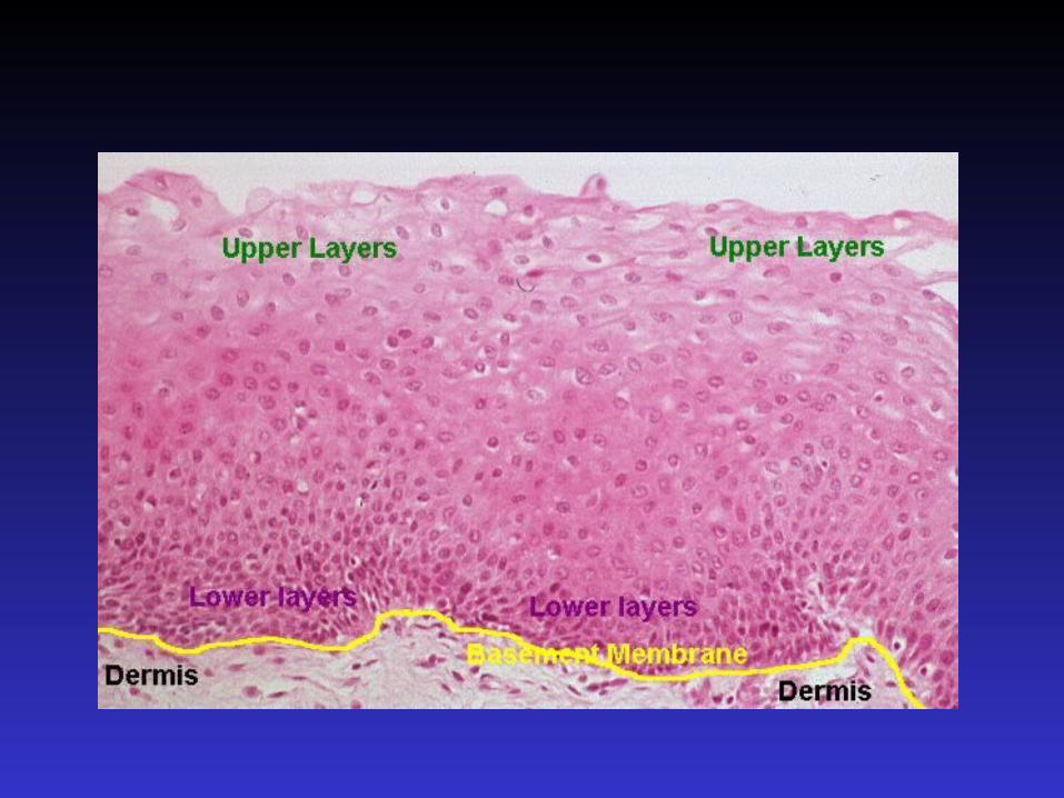
• Physical boundaries (e.g. basement membrane)• Tissue pressure contact inhibition
– Cell cycle regulation
• Error correction– Lack of fidelity in cellular reproduction genetic
instability– Repair genes– Immune mechanisms: removal of non-self cells– Apoptosis

G2: Preparation for Mitosis
G1: Preparation
for Synthesis
G0: Rest phase
M phase:
Functional phases
Preparatory phases

Cell Cycle Check Points
• Events of cell cycle highly ordered:– different extra cellular/intracellular events
• Progression through cell cycle controlled by:– regulation of gene products – checkpoints genes

Normal cellular stop signals
• Cellular hypoxia (outgrowth of blood supply)
• Decreased availability of nutrients
• Alternation in cytokine/hormonal milieu
• Accumulation in toxic metabolites
• Inhibitition of cell-cell contact

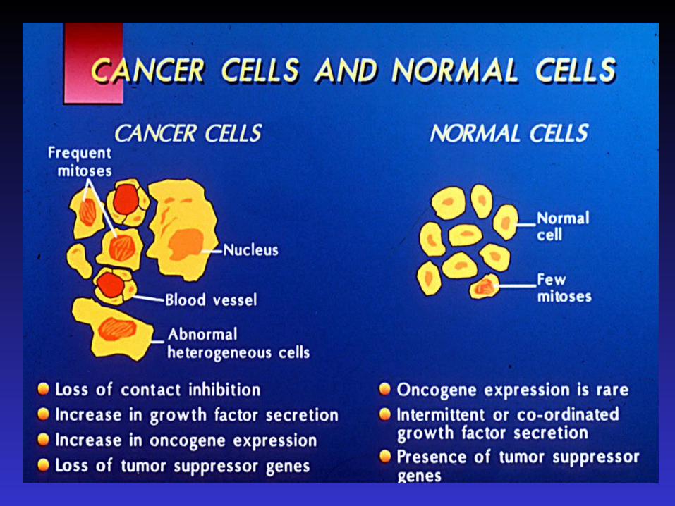
Cancel: Cellular Derangements
• In-exact replica– Genetic instability– Loss of certain function, gain of others
• Abnormal cellular regulation: Loss of Balance– Enhanced proliferation – Disruption of Physical boundaries– Increased tissue pressure, loss of contact inhibition– Inhibition of apoptosis (programmed cell death)
• Loss of error correction– Lack of fidelity in cellular reproduction genetic instability– Loss of Repair genes– Immune inhibition (anergy)– Inhibition of apoptosis– Selective advantage certain clones

Cell CycleExtra cellular Signals
• Complex regulation and division not in a vacuum• Cell integrate signals into control mechanisms:
– Nutrient status– Cell to cell contact– Extra cellular peptides
• Growth factors cause cells in G0 phase through cell cycle• Continued growth factor exposure • Cytokines:
– soluble mediators of cell to cell communication – interleukins, interferon, CSF– bind to receptors on surface of cells– cascade of biochemical signals activation/suppressing of genes

CARCINOGENESISSummary of the carcinogenic process.
Initiation Promotion ProgressionInvasiveness
Metastasis (eg Vogelstein model for colon cancer)
Normal adenoma I adenoma II adenoma III carcinomaAPC gene
Chr 5qtransformation
to hyper-proliferation
Ras mutation
proliferation signal left on
DCC gene8q21 allelic losstumour suppressor
involved in differentiation
P53Chr 17p loss of tumour suppressor and
apoptosis

Causes of CancerFactor or Class of Factors Percent of all
Cancer Deaths
Tobacco 30%
Diet 35%
Reproductive and sexual behaviour 7%
Occupation 4%
Alcohol 3%
Pollution 2%
Geophysical factors 3%
Industrial products 1%
Medicines and medical procedures 1%
Inherited <5%

Life-cycle
• 1cm3 -> 1g tumor ( 109) cells– 1 cm the limit of clinical detection– 30 doublings occurred prior to clinical detection
• Only 10 more doublings (3 logs)– 1kg of tumor– terminal disease
• Pre-clinical phase 75% of “life of tumor”
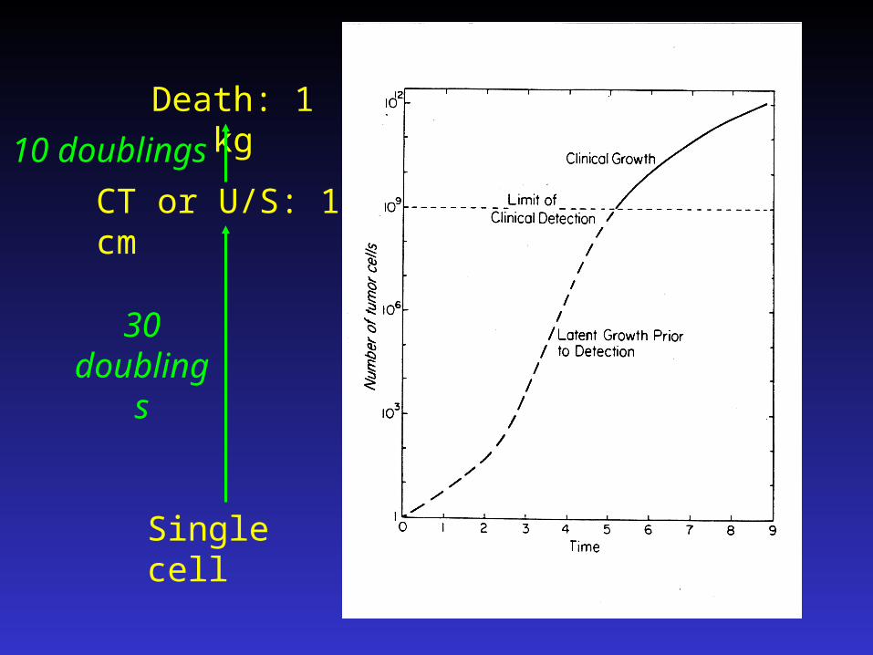
Single cell
CT or U/S: 1 cm
30 doublings
Death: 1 kg
10 doublings
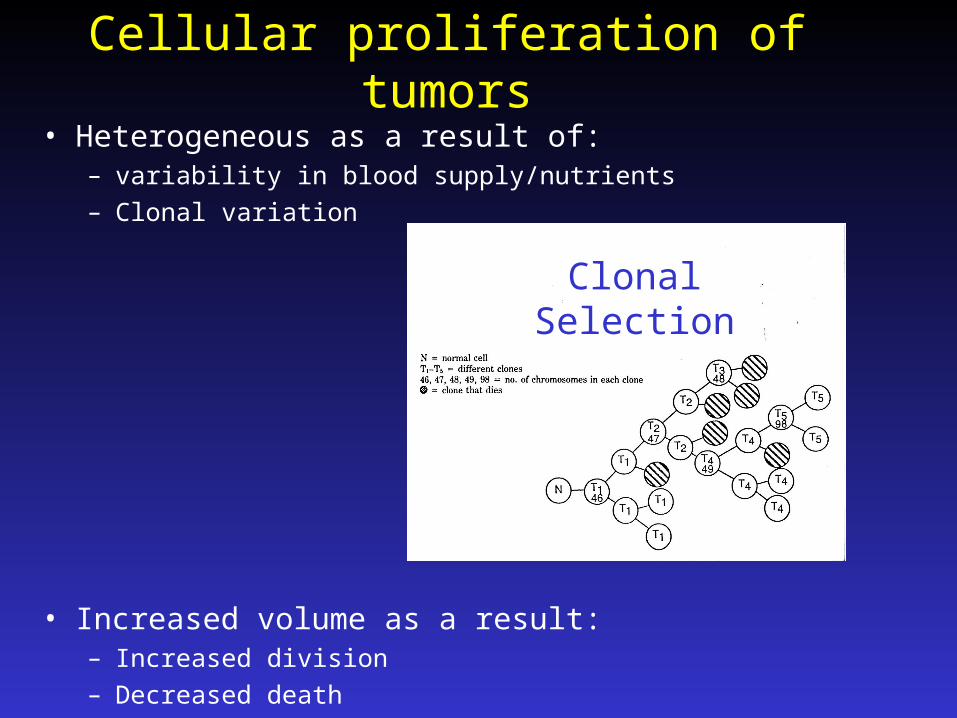
Cellular proliferation of tumors
• Heterogeneous as a result of:– variability in blood supply/nutrients– Clonal variation
• Increased volume as a result:– Increased division– Decreased death
Clonal Selection

Principles of Metastases
• Principle cause of death• Mainly routes of dissemination:
– via blood steam– lymphatic
• Are flow and organ specific• Establishment of metastases is inefficient:
– subpopulation/clone have the abilities to metastases– generally most malignant/aggressive

Steps in Metastatic Cascade
• Escape
• Travel through the blood/lymphatic system
• Arrest/attachment
• Establishment of clone

Metastases: Escape
• May be biologically facilitated by:
– ability to commit vascular invasion
– cell necrosis
– molecules of the cell surface
– protease ( enzyme) secretion by tumor

Metastases: Travel
• Blood supply ( angiogenesis) must be adequate
• Adequate lymphatic drainage
• Special circulatory circumstances


Angiogenesis
• Concept first put forward by Folkman• Tumour produces factors to induce / generate its
own blood supply• VEGF one of the most important mediators• Interacts with endothelial cell receptors :
– VEGFR-1 and VEGFR-2
• Essential for normal embryonic vasculogenesis• VEGF upregulated in many cancer types
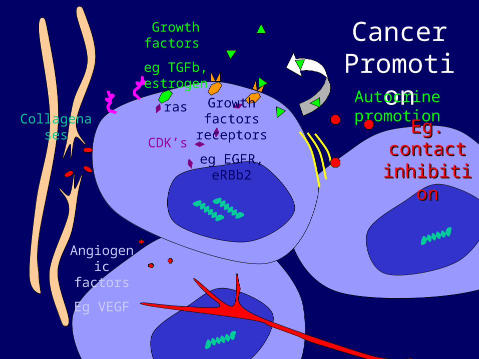
Cancer Promotion
Growth factors
eg TGFb, estrogen
Autocrine promotion
CDK’s
Collagenases
Angiogenic factors
Eg VEGF
Growth factors receptors
eg EGFR, eRBb2
ras
Eg. contact Eg. contact inhibitioninhibition

Chemotherapy

Principles of Chemotherapy
• Exponential relationship between dose and kill– small decrease in drug dose results in large
increase in cell survival
• Cycling cells at greatest risk
• Multiple courses of therapy– each treatment kills same proportion
(not number) of cells– e.g.: 3 log killed 1010 to 107
1 log regrowth between cycles


Mechanisms of resistance
• Tumor sanctuaries
• Drug exposure/Selection pressure– chemotherapeutic agents selects for resistant cells
• Resistance within a tumor a function of:– inherent genetic instability of a tumor– size of tumor ( # cells) Goldie-Coldman hypothesis
(chance resistance size)


ChemotherapyStimulate immune system
Block growth factor
receptors
Turn off the renegade
“grow” signal
Stop destruction of
barriers
•Interferon
•MoAb’s
•Herceptin
•Rituxan
•Tamoxifen
•Farnesyl transferase inhibitors
•Matrix metalloproteinase inhibitors
Stop new blood vessel
formation
•Endostatin
•Angiostatin
•COX2 inhibitors

Blood supply as the TargetVEGF
VEGFR - 2
Cell membrane
Tyrosine Kinase
Signal Transduction
X e.g. bevacizumab / Avastin®
X
X
X VEGF trap

Blood supply as target:Bevacizumab in colon cancer

Generalized Staging Principles: TNM
• Stage I– Organ confinement
• Stage II– Locally advanced / larger / penetration
• Stage III– Nodal involvement
• Stage IV– Metastatic
Look for: Molecular staging elements

Lymph Nodes• Prognosticator
– E.g. colon N0 25% recurrence(less than 4 negative nodes 50%)
N1 (1-3 nodes) 60% recurrence N2 (4+ nodes) 70% recurrence
• Source of disease– Axillary dissection as a therapeutic intervention– TME
• Techniques for analysis– Toluene fat dissolving techniques (yield)– Immunohistochemistry for cytokeratins
• The sentinel node– Extensive focused analysis

Pattern Recognition• Colon
– Mesenteric nodes– Drains through portal vein: first stop liver– Of those stage IV, 90 have liver mets, 70 only liver– Other sites peritoneal, nodes > lung >> [bone/brain]
• Lung– Mediastinal nodes– Pleural effusions– Lung, Liver, adrenal, bone, brain
• Breast– Axillary nodes– Lung, liver, bone, brain
• Kidney or Melanoma anywhere!

Unknown primary
• Peritoneal + ovarian masses– Ovarian, PPC, Stomach, colon
• Axillary nodes– Breast, lymphoma
• Brain metastases– Lung, breast>> kidney..
• Bone metastases– “Buy The Kid Long Pants”

The curable metastasis
• Surgery Colon cancer– 35% 5yOS after complete resection of liver, lung or
splenic metastases
• Chemotherapy for testicular cancer or lymphoma

Thank-you for your attention!