the biology of acute lymphoblastic leukemia
TRANSCRIPT
29G.H. Reaman and F.O. Smith (eds.), Childhood Leukemia, DOI: 10.1007/978-3-642-13781-5_2, © Springer-Verlag Berlin Heidelberg 2011
2.1 Introduction
Discoveries of the underlying biological pathways that drive leukemogenesis in children have taken place at an astonishing pace. These findings have resulted in large part because of the evolution of technical developments in analyzing chromosome structure, the development of monoclonal antibodies capable of recognizing dis-crete cell surface proteins that correlate with cell lin-eage and differentiation state, recombinant DNA technology, and engineered mouse models (e.g., trans-genic and “knock out” models). More recently, advances in high-throughput genomics and progress in stem cell biology have transformed the field of cancer biology in general and perhaps more so in hematological malig-nancies. A cohesive view of the stepwise process of transformation and the cellular heterogeneity of the leukemic clone is emerging and, importantly, leukemia-specific targets have been identified and novel thera-peutic approaches have been directed at these lesions.
The Biology of Acute Lymphoblastic Leukemia
William L. Carroll, Mignon Loh, Andrea Biondi, and Cheryl Willman
W.L. Carroll (*) NYU Medical Center Smilow Research Building, 522 1st Avenue, SML 1201, New York, NY 10016, USA e-mail: [email protected]
M. Loh Clinical Pediatrics, Hematology-Oncology, University of California, San Francisco, e-mail: [email protected]
A. Biondi Universita Milano, Ospedale San Geraldo, Monza
C. Willman UMN Cancer Research Facility, School of Medicine, University of New Mexico, MSC 08-4630, USA e-mail: [email protected]
2
Contents
2.1 Introduction ................................................................. 29
2.2 The Cellular Biology of Acute Lymphoblastic Leukemia (ALL) .......................................................... 30
2.2.1 Lymphoid Development and Immunophenotype of Acute Lymphoblastic Leukemia ............................... 30
2.2.2 Antigen Receptor Genes and Clonality ......................... 332.2.3 Leukemia-Initiating Cells in ALL ................................ 35
2.3 The Molecular Biology of ALL .................................. 362.3.1 Introduction to Cancer Genomics
and New Technology .................................................... 362.3.2 Host Susceptibility to ALL ........................................... 372.3.3 Somatic Genetic Changes in ALL ................................ 38
2.4 Signaling Pathways in Childhood ALL ..................... 502.4.1 BCR-ABL Tyrosine Kinase .......................................... 502.4.2 FLT-3 Receptor Tyrosine Kinase .................................. 502.4.3 JAK Tyrosine Kinase .................................................... 512.4.4 Pre-B Cell Receptor ...................................................... 512.4.5 RAS Pathway ................................................................ 512.4.6 NOTCH1 Pathway ........................................................ 522.4.7 Therapy Targeted to Signaling Pathways ...................... 52
2.5 The Apoptotic Pathway and ALL .............................. 52
2.6 The Biology of Relapsed ALL .................................... 54
2.7 Summary ...................................................................... 57
References .............................................................................. 57

30 W.L. Carroll et al.
2.2 The Cellular Biology of Acute Lymphoblastic Leukemia (ALL)
2.2.1 Lymphoid Development and Immunophenotype of Acute Lymphoblastic Leukemia
2.2.1.1 Lymphoid Development
The immune system in mammals includes three main lymphoid cell populations: B-cells, T-cells, and Natural Killer (NK) cells. They arise from progenitors located in central lymphoid organs such as fetal liver, bone marrow, and thymus. Lymphocyte subpopulations can be recognized by the expression of surface and intrac-ellular markers, which can allow the dissection of dif-ferent lineage-restricted maturation stages and/or functional subtypes. Blood cells, including lympho-cytes, originate from a small number of self-renewing hematopoietic stem cells (HSCs) capable of producing all cell types in the blood (Bryder et al. 2006). Amplifications in cell numbers in concert with pro-gressive restrictions in lineage potential lead HSCs to generate terminally differentiated cell progeny.
The understanding of lymphoid differentiation has arisen primarily from studying mouse models. The majority of the multipotent progenitor cells in the mouse bone marrow (BM) do not express high levels of classical lineage markers, but express SCA1 and KIT (Lin–/SCA+/Kit+ (LSK) cells) (Fig. 2.1). A primary event in HSC differentiation is loss of self-renewing potential, while retaining the capacity for multilineage differentiation to give rise to multipotent progenitors (MPPs). Subsequent dif-ferentiation processes can be demonstrated by the identification of lympho-myeloid-restricted multi-potent progenitors (LMPP), with the capacity to produce granulocytes, macrophages (GM), and all the defined lymphoid cell types such as B-cells, T-cells, and NK-cells (Adolfsson et al. 2005). LMPP can differentiate into early lymphoid progenitors (ELPs) that start to express recombination activat-ing gene 1 (Rag1) and Rag2 and initiate rearrange-ment at the immunoglobulin heavy chain (IGH) locus. ELPs can further differentiate into thymic precursors of the T-cell lineage (early T- cell-lineage progenitors, ETPs) or into bone-marrow common lymphoid progenitors (CLPs), which are lymphoid restricted.
Pro-T-cell ImmatureB-cell
SmallPre-BII-cell
LargePre-BII-cell
Pre-BI-cell
Pro-B-cell
Pro-NK-cell
CLP
DC
LSK
ELP
CMP
MPP
Self-renewing
HSC
Erythroid lineage
Thy
mus
Bon
e m
arro
wP
erip
hera
lbl
ood
Myeloid lineage
ETP
Fig. 2.1 Mouse B-cell development from early hematopoietic progenitors. HSC, Hematopoietic stem cell; MPP, Multipotential progenitor; ELP, Early lymphoid progenitor; CMP, Common myeloid progenitor; CLP, Common lymphoid progenitor; DC, Dendritic cell; LSK, Lin-/SCA+/Kit+; ETP, Early T-cell-lineage progenitor; NK, Natural killer. (Modified from Czerny and Busslinger 1995)

31 2 The Biology of Acute Lymphoblastic Leukemia
2.2.1.2 B-Cell Development
Expression of the B-cell marker B220 by a subset of CLPs (known as pro-B cells) coincides with their entry into the B-cell-differentiation pathway. The next step can be identified by expression of CD19 and completion of IGH diversity (DH)-to-joining (JH) gene segment rearrangement by pre-BI cells. The IGH locus then con-tinues to rearrange its variable (V)-region gene segments until productive VH–DJH alleles are generated in large pre-BII cells. These cells cease to express Rag1 and Rag2, and they display the product of the rearranged IGH gene at the cell surface, where it assembles with the surrogate immunoglobulin light chains (IgLs), Vpre-B, and l5, together with the signaling molecules IgA (which is encoded by the MB-1 gene) and Igb (which is encoded by the B29 gene) to form the pre-B-cell receptor (pre-BCR). Expression of the pre-BCR is a crucial check-point in early B-cell development, at which the functionality of the heavy chain is monitored. Signaling through the pre-BCR allows for allelic exclu-sion of the IGH locus and stimulates a burst of prolifera-tive clonal expansion of large pre-BII cells, which is followed by reexpression of RAGs and rearrangement at the IGL locus in small pre-BII cells. During normal development, appearance of the assembled BCR at the cell surface defines the immature B-cell stage (Fig. 2.1) (Czerny and Busslinger 1995; Busslinger 2004).
Stages of human B-cell development seem to follow mechanisms similar to mouse, confirming the previous studies (reviewed in Ghia et al. (1998)). Although some differences in surface marker expression as well as dif-ferences in growth requirements remain, the strong resemblance of B-cell development in mouse to that in man allows for a comparison of the two systems. For the early multipotent progenitor to proceed in development into a lymphoid-restricted stage, an interplay between several concurrent mechanisms, including external sig-nals, internal transcription factor networks, and epige-netic changes, have to take place (Bryder and Sigvardsson 2010; Ramirez et al. 2010). Differentiation processes of multipotent LSK cells into lymphoid-restricted progeni-tors is correlated with the expression of FLT3, the recep-tor for the FLT3 ligand (FL) and IL7R (Ramirez et al. 2010). The transcription factors essential for priming lineage-associated genes and restricting fates to the B-lineage within CLP compartment are Ikaros, Purine
box factor 1 (PU.1), and E2A. Ikaros is encoded by Ikaros Family Zinc Finger 1 (IKZF1) and contains vari-able numbers of Kruppel-like zinc fingers in two domains that mediate DNA binding and formation of dimers and multimeric complexes (Yoshida et al. 2006). Following the expression of E2A and EBF1, Ikaros mediates chromatin accessibility necessary for V(D)J recombination, and it also modulates the expression of early B-cell-specific genes, including IgLL1 (l5) (Thompson et al. 2007).
2.2.1.3 T-Cell Development
Mammalian T-cells originate from pluripotent precur-sors in the bone marrow or fetal liver that migrate to the thymus, where T-cell differentiation is initiated and sustained. T-cell progenitors migrate into the thymus and then respond to the surrounding environment by proliferating extensively, and initiate the T-cell differ-entiation transcriptional program, gradually turning off genes that allow differentiation to non-T-cell lin-eages (Hayday and Pennington 2007). They then undergo T-cell receptor (TCR) gene rearrangements and assemble TcR complexes. These cells can mature into different T-cell lineages, including gd T- and ab T-cells. The ab T-cells further diverge into different sublineages, such as CD4+ T-cells, CD8+ T-cells, nat-ural killer T- (NKT) cells, and regulatory T-cells (TReg cells). The T-cell-lineage commitment process consists of a progression of distinct developmental stages, and particular regulatory changes that drive the cells from one stage to the next (Rothenberg et al. 2008) (Fig. 2.2). Clear changes in gene expression and developmental potential mark these transitions. Once they migrate from the thymus to the periphery, each of these cell subsets will have different functions. Genetic evidence, from germline and conditional knockout mouse mod-els, emphasizes the requirements for a stable core group of transcription factors that act repeatedly at successive stages.
One major progenitor source in adult mice consists of LMPPs in the bone marrow or blood, which can give rise to macrophages, dendritic cells (DCs), NK-cells, B-cells, and T-cells, but not erythrocytes or megakaryo-cytes (Adolfsson et al. 2005; Yoshida et al. 2006). Within the mouse LMPP population, cells with the

32 W.L. Carroll et al.
capacity to migrate to the thymus are probably distin-guished by their expression of the CC-chemokine recep-tor 9 (CCR9), in addition to the stem- and progenitor-cell markers KIT, stem-cell antigen 1 (SCA1), and the growth-factor-receptor tyrosine kinase FLT3 (Schwarz et al. 2007). Development from the early T-cell-lineage progenitor (ETP) stage to the double-negative 3 (DN3) stage is independent of the TcR and is coordinated with migration through distinct thymic microenvironments. ETPs and DN2 cells proliferate extensively while acquiring their first T-cell characteristics. As the T-cells reach the DN3 stage, they stop proliferating, greatly increase TCR gene rearrangement, and generate the first fully rearranged TCR loci. DN3 T-cells that succeed in making in-frame TCR gene rearrangements become activated by TcR-dependent selection (these are referred to as DN3b cells); this distinguishes them from DN3 cells that are not yet selected (referred to as DN3a cells). Expression of TcRb qualifies the cells to undergo b selection, turning on expression of CD4 and CD8 to become double positive (DP) cells, and eventually acquiring cell-surface TcRab complexes. This prepares them for positive selection and negative selection to generate mature CD4+ or CD8+ TcRab+ T-cells.
Alternatively, DN3 T-cells that successfully rearrange TCR g- and d-chains instead of b-chains are selected as gd T-cells (Rothenberg et al. 2008).
Several transcription factors are involved in expres-sion during the progression of T-cell precursors from the ETP to the later stages, including most of the T-cell factors known to be essential for early T-cell develop-ment as well as most factors implicated in cell-lineage plasticity, those implicated in the regulation of TCR and other T-cell-lineage gene expression (reviewed in Rothenberg et al. (2008)).
2.2.1.4 Immunophenotype of Acute Leukemia
Cellular immunophenotype can be defined as the expression of leukocyte antigens (proteins or glycopro-teins) either on the cell surface or in the cytoplasm, detectable by applying immunologic methods with the use of monoclonal antibodies. Many monoclonal anti-bodies available for such purposes have been grouped into Clusters of Differentiation (CD) based on their reactivity with identical antigens (Mason et al. 2002). Precursor cells and their malignant counterparts can be
ETP
DN2a
DN2b
DN3aDN3b
DN4ISP
DP
TCR+DP
Cell death
CD4+ SP CD8+ SP
TCR gene rearrangement
γδ T-cell
Blood vessel
Immigrantprecursor
Commitmentcomplete
β γδselection
Adult thymic lobule(cross section)
precursor
Commitmentcomplete
β γδselection
CCR9+LMPP
or
Fig. 2.2 Stages in early T-cell development. LMPP, lymphoid primed multipotent progenitor; ETP, early T-cell-lineage progenitor; DN2a, double-negative 2a; DN2b, double-negative 2b; DN3a, double-negative 3a; TCR, T-cell receptor; DN3b, double-negative 3b; DN4 ISP, double negative 4 immature single positive; DP, double positive; SP, single positive. (see text). (Modified from Rothemberg VE et al., Nature Reviews Immunology 2008, 8:9–21)

33 2 The Biology of Acute Lymphoblastic Leukemia
recognized on the basis of morphological and cytochem-ical characteristics. However, a more accurate charac-terization of the leukemic clone can be assessed by immunophenotyping (van Dongen et al. 1988). Modern immunophenotyping approaches are based on the use of flow cytometric technique (Carter and Meyer 1994). A typical flow cytometry consists of one or more laser-based light sources that provide monochromatic light beams (generally at 488 nm and at 635 nm). The cells, flowing through the laser beam, refract the light and, if present, fluorochromes are excited and emit fluores-cence. Signals obtained by interaction of the cell with the light provide information about the cells including volume (Forward Side Scatter, FSC), nucleo-cytoplas-matic complexity (Orthogonal side light scatter, SSC), and presence of antigens due to their cross-link with fluorochrome-conjugated specific antibodies. By flow cytometry, it is possible to assess many biological fea-tures with potential impact on the diagnosis and man-agement of acute leukemia including the detection of minimal residual disease (MRD). Systematic guide-lines for immunological classification of acute lympho-blastic leukemias (ALLs) have been proposed by the European Group for the Immunological Characterization of Leukemia (EGIL) (Bene et al. 1995) (Table 2.1). More recently, correlation of immunophenotype with cytogenetic and molecular genetic characteristics has identified new biologically and clinically distinct sub-groups of ALL. Details related to the immunological classification of ALL are provided in chapters dedi-cated to classification and treatment of ALL.
2.2.2 Antigen Receptor Genes and Clonality
2.2.2.1 Immunoglobulin (Ig) and T-Cell Receptor (TCR) Gene Rearrangements in ALL
Somatic rearrangement of Ig and TCR gene loci occurs during early differentiation of any B- and T-cell, by joining the germline variable (V), diversity (D), and joining (J) gene segments (reviewed in Janeway et al. (2001)). By this process, each lympho-cyte gets a specific combination of V-(D-) J segments that encode the variable domains of Ig or TcR mole-cules. The uniqueness of each rearrangement further depends on random insertion and deletion of nucle-otides at the junction sites of V, (D), and J gene seg-ments, making the junctional regions of Ig and TCR genes “fingerprint-like” sequences for that particular clone. Due to the clonal origin of the neoplasm, each malignant lymphoid disease will represent the expan-sion of a clonal population with a specific Ig/TCR signature.
The frequencies and patterns of Ig and TCR gene rearrangements in ALL can be analyzed by Southern blot- and PCR-based methods (reviewed in Szczepanski et al. (2001)). Currently, PCR-based methodologies are more easily and frequently applied to the detection of clonal Ig and TCR gene rearrangements. Virtually all B-lineage ALL patients have rearranged Immuno-globulin heavy chain (IGH) genes (van Dongen and Wolvers-Tettero 1991). In addition, rearrangements of the Ig Kappa deleting element (Kde) occur at a rela-tively high frequency (approximately 60%) (Beishuizen et al. 1997). Cross-lineage incomplete TcR Delta (TCRD) rearrangements occur in more than 40% of all patients (40% Vd2-Dd3, 19% Dd2-Dd3, and 13% showed both) (van der Velden et al. 2003). Complete TCRD rearrangements (Vd-Jd) are very rare in B-lineage ALL. Detection of TcR Gamma (TCRG) rearrange-ments occurs in more than 50% of the B-lineage ALL patients (van der Velden et al. 2003). Cross-lineage TcR Beta (TCRB) recombination occurs in a small percent-age (15–20%) of B-lineage ALL patients.
Most T-ALL patients have rearranged TCRB, TCRG and/or TCRD genes (van Dongen and Wolvers-Tettero 1991). Frequency analysis of the patterns of recombi-nation in T-ALL patients showed that TCRG rearrange-ments represent the most frequent ones (identifiable in
Table 2.1 Immunological classification of acute lymphoblastic leukemia according to EGIL proposal (Bene et al. 1995)
B-lineage ALL (CD19+ and/or CD79a+ and/or CD22+)
Pro-B-ALL (B-I) No expression of other differentiation of B-cell antigens
Common ALL (B-II) CD10+
Pre-B- ALL (B-III) Cytoplasmic IgM+
Mature B-ALL (B-IV) Cytoplasmic or surface kappa or lambda+
T-lineage ALL (cytoplasmic/membrane CD3+)
Pro-T-ALL (T-I) CD7+
Pre-T-ALL (T-II) CD2+ and/or CD5+ and/or CD8+
Cortical-T-ALL (T-III) CD1a+
Mature T-ALL (T-IV) Membrane CD3+, CD1a-

34 W.L. Carroll et al.
84% of patients), followed by complete and incomplete TCRD joinings. In practice, TCRG and/or TCRD gene rearrangements occur in >95% of childhood T-ALL patients (Pongers-Willemse et al. 1999; van der Velden et al. 2003). Incomplete IGH rearrangements (DH-JH) could be identified in 12% of T-ALL cases, consistent with the finding that cross-lineage Ig gene rearrange-ments occurred at relatively low frequency in T-ALL (10–20%) and are virtually restricted to incomplete IGH rearrangements (Pongers-Willemse et al. 1999).
2.2.2.2 Assessment of Clonality by PCR Amplification
The assessment of clonality by Ig and TCR gene relies on the PCR amplification of the different target gene recombinations. Primers and protocols have been stan-dardized (van der Velden and van Dongen 2009). After PCR identification of Ig/TCR targets, the clonal origin of PCR products must be assessed by heteroduplex analysis or by gene scanning, to confirm their origin from the malignant cells and not from contaminating normal cells with similar Ig or TCR gene rearr-angements. The homo-heteroduplex analysis takes advantage of the different migration properties in poly-acrylamide gel of V-(D-) J rearrangements containing a few mismatches (heteroduplex) compared with fully matched V-(D-) J junctions (homoduplex). Fingerprint analysis consists of PCR amplification with a fluores-cent primer and an electrophoretic run in polyacrylam-ide gels, where clonal amplification results in a single peak within a background of polyclonal, constitutional amplification products. After the clonal rearrange-ments are recognized, several methods can be applied to specifically detect the leukemia-derived PCR prod-ucts, for example, during the follow-up of patients who have undergone therapy. The major variable lies in the sensitivity of the test, which can significantly interfere with interpretation of the assay results.
2.2.2.3 Use of Ig and TCR Gene Rearrangements for the Detection of MRD
Sequential monitoring of MRD with specific and sen-sitive methods (capable of recognizing one leukemic cell among 10−4 or more normal BM cells, at least 100-fold more sensitive than morphologic examination),
recently compelled the redefinition of complete remis-sion in patients with ALL, and further improved the clinical utility of risk assessment. Several techniques have been developed over the past 10 to 15 years to complement morphology in assessing response to treatment, including immunologic and molecular methods, fluorescent in situ hybridization (FISH), in vitro drug response, and colony assays (reviewed in Szczepanski et al. (2001)). Ig and TCR genes are the most widely applicable genes and therefore can be considered a universal target for MRD detection in childhood ALL (Cazzaniga and Biondi 2005; van der Velden and van Dongen 2009). Its feasibility has been proved in a multicenter ALL trial (Flohr et al. 2008).
In the most sensitive assay so far available, clonal PCR products from homo-heteroduplex analysis are directly sequenced. V, D, and J gene segments are then identified, and randomly inserted nucleotides are rec-ognized by comparison with germline sequences in databases (http://imgt.cines.fr; http://www.ncbi.nlm.nih.gov/igblast). The sequence information allows the design of junctional region-specific oligonucleotides, which can be used to detect malignant cells among normal lymphoid cells during follow-up of patients in two different ways. One uses the oligonucleotides as patient-specific junctional region probes in semi-quan-titative hybridization experiments (“dot blot”) to detect PCR products derived from the malignant cells. Alternatively, the junctional region-specific oligonu-cleotide can be used as a primer to quantitatively amplify the rearrangements of the malignant clone.
The applicability of the allele-specific oligonuc-leotide (ASO) primer approach depends on its sensi-tivity and specificity. The specificity of detection is checked for each probe on at least three different polyclonal samples. The sensitivity of each probe is assessed by testing serial dilutions of the patient’s blasts in a mixture of polyclonal marrow mono-nuclear cells. In this way, PCR-based MRD detec - tion via clone-specific junctional regions generally reaches a sensitivity of 10−4 to 10−5. A less-sensitive assay consists of a modified fingerprint analysis, in which the patient- and clone-specific peak corre-sponding to PCR amplification from residual leu-kemic cells can be discriminated from normal back ground. Polyclonal background levels vary, but usually limit the sensitivity of this approach to the detection of one leukemic cell among 10−2 to 10−3 normal cells.

35 2 The Biology of Acute Lymphoblastic Leukemia
IGH rearrangements represented the most sensitive group of targets and usually reached sensitivities £10−4. However, despite excellent RT-PCR sensitivi-ties, IGH gene loci are prone to oligoclonality in 30–40% of B-lineage ALL (for example, multiple rearrangements (subclones) within the same clone) owing to continuing and secondary rearrangement processes (Szczepanski et al. 2001). Therefore, the use of oligoclonal Ig/TCR targets in MRD PCR analy-sis can lead to an underestimation of the leukemic tumor load, because they might occur in a subclone of low frequency, hence leading to potentially false-neg-ative results. In consequence, IGH targets should routinely be used in combination with IGK-Kde rear-rangements (especially Vk-Kde), since these targets represent highly stable ‘end-point rearrangements’ suitable for sensitive MRD detection. Using incom-plete TCRD rearrangements as a third priority further increases the number of applicable MRD targets as Vd2-Dd3 and Dd2-Dd3 recombinations show little clonal instability and also comprise a group of mark-ers with sensitivity comparable to DH-JH rearrange-ments. In contrast, TCRG rearrangements in precursor B-lineage ALL have proven to be less applicable in MRD detection since their sensitivity is more fre-quently limited (>10−4) due to small junctional regions and nonspecific amplification of TCR gene rearrange-ments in normal T-lymphocytes. Taking the published results on target availability and sensitivity into account, the following priority order using antigen receptor gene rearrangements for MRD PCR targets in B-lineage ALL can be deduced: IGH > IGK (Vk-Kde) > TCRD > TCRG and IGK (intron-Kde).
The success rate of detecting appropriate MRD-PCR targets is lower in T-ALL compared to precursor B-lineage cell ALL. The addition of TCRB gene rear-rangements to MRD-PCR target identification increa-ses the availability of targets in T-ALL. Moreover, the junctional regions of (complete) TCRD rearrange-ments, similar to IGH in B-lineage ALL, frequently include extensive N-nucleotide insertions, thus enabl-ing the design of highly specific ASO primers. In con-trast, the junctions of TCRG and incomplete IGH rearrangements are commonly smaller resulting in a significantly lower ratio of sensitive targets (about 75%). Taking results on target sensitivity in T-ALL together, the following conclusion on the preferential use of MRD PCR targets can be drawn: TCRD/TCRB > IGH (DH-JH) > TCRG.
2.2.3 Leukemia-Initiating Cells in ALL
Recent evidence supports the hypothesis that specific subsets of tumor cells retain features similar to stem cells and are capable of propagating clonal cancer cells. The presence of leukemia-initiating cells has important biologic and therapeutic implications. While there is generally broad acceptance about the identification of such a cell population in myeloid malignancies, which was first elegantly demonstrated by Dick and col-leagues (Lapidot et al. 1994), the identification of a uniform lymphoblastic leukemia-initiating cell has been much more evasive. Indeed, it is also well recog-nized that murine xenograft modeling systems and dif-ferent experimental methodologies can yield disparate results with respect to the engraftment of leukemia and the minimum number of cells required to propagate disease. These findings have undoubtedly made the field of ALL stem cell biology even more challenging.
While subtle differences in definitions have led to some confusion, for the purposes of this discussion, a can-cer stem cell is a tumor-initiating cell. The definition of a cancer stem cell needs to be distinguished from a normal stem cell; while both share critical features of self-renewal and differentiation, it is important to realize that a tumor-initiating cell may in fact reflect a reprogrammed progeni-tor cell that acquires stem cell-like features (Krivtsov et al. 2006). There is general agreement, however, that one essential experimental property of any unique subpopula-tion of cancer initiating cells is its ability to produce leu-kemia in an immunocompromised mouse (Clarke et al. 2006). Recent advances in the identification of primitive stem cell markers have facilitated sorting methods to achieve very pure populations of normal stem cells, but it is not clear that cancer-initiating stem cells uniformly dis-play only one set of these markers, and this is true for investigations of cancer stem cells in ALL.
In addition, the type and age of immunocompro-mised mouse, the level of radiation, and the mode of delivering the purified cancer initiating cells appear to greatly affect experimental results. In the earliest stud-ies, Lapidot used intravenous injection of acute myeloid leukemia (AML) cells into severe combined immuno-deficient (SCID) mice and determined that the cells required to confer leukemia were contained within the CD34+/CD38− cell fraction (Lapidot et al. 1994). Limiting dilution analysis in this system demonstrated that 1/250,000 cells were required. Since this seminal work was published, additional mouse models with

36 W.L. Carroll et al.
progressive degrees of immunodeficiency have become available for study. Some of the more recent of these, the NK cell-depleted Non-Obese Diabetic/SCID (NOD/SCID) and the NOD/SCID gamma (NSG) mice have been recently used by le Viseur and colleagues to model ALL (le Viseur et al. 2008). Interestingly, these progres-sively more immunocompromised mouse models result in fewer and fewer cancer-initiating cell requirements. Intrafemoral injection has also been used by a number of investigators to maximize the “homing” of cancer ini-tiating cells and limit the number of cells that get trapped in pulmonary capillaries, a potential requirement for engraftment of myeloid diseases, but ALL cells do not seem to absolutely require this additional step.
Identifying a single cancer initiating cell population for ALL has been elusive, not only in part due to the various strains of mice employed and the methodologies to engraft them, but also likely due to the genetic hetero-geneity of human ALL. Recent genomic studies have firmly established that ALL is a disease requiring mul-tiple genetic hits for full transformation (Mullighan et al. 2007a). Greaves and colleagues have further shown in elegant FISH studies that there is not always a linear hierarchy to acquiring these multiple mutations, but that, in fact, there is considerable complexity to the acquisi-tion of these lesions that more resembles “a branching pattern,” akin to Darwin’s theories of evolution (Greaves 2009). In this manner, one can appreciate how difficult it would be to a priori identify a single population of leu-kemia-initiating cells for all subtypes of ALL.
Earlier data supported the ability of CD34+/CD38− or CD34+/CD19− human leukemia cells to engraft NOD-SCID mice (Cobaleda et al. 2000), while other studies have demonstrated the engraftment capability of several different lineages, including CD19+ cells. Indeed, it has been shown by several groups that TEL/AML1 (ETV6/RunX1)-positive leukemia cells able to confer disease are restricted to the CD34+/CD19+ pop-ulation (Hotfilder et al. 2002; Castor et al. 2005; Hong et al. 2008) and are conspicuously absent from the CD19− fraction. Hong and colleagues were able to study a set of monochorionic twins, one of whom was diagnosed at the age of 2 years with ETV6/RUNX1-positive ALL, while the other remained healthy at the time of their report (Hong et al. 2008). A population of CD34+CD38−/lowCD19+ cells that was detected in the patient’s bone marrow was also detected at extremely low levels in the healthy twin (0.002%) and harbored the identical fusion gene. However, further analysis of these cells revealed that a DJ recombination event had
occurred in the healthy twin while a VDJ and DJ recom-bination event had occurred in the affected twin, sug-gesting that there was further clonal evolution from the same basic cell population shared by both siblings. Engineering healthy human cord blood to express ETV6/RUNX1 alone resulted in a population of CD34+CD38−/
lowCD19+ cells with significant self-renewal and differ-ential pot ential, but did not confer acute leukemia, sup-porting that these cells exhibited at least some of the hallmark features of stem cells, but that additional events are required for full transformation. Indeed, the vast maj ority of children with TEL/AML-positive leukemia frequently display additional events at diagnosis, includ-ing loss of the normal TEL allele.
More recently, le Viseur and colleagues have reported that lymphoblasts displaying a wide spectrum of differ-entiation markers were able to engraft primary as well as successive immunocompromised recipients, using an intrafemoral injection strategy in NOD/SCID mice (le Viseur et al. 2008). One fascinating observation was that leukemia cells from patients with high-risk disease were more likely to yield multiple fractions (CD34+CD19−, CD34+CD19+, and CD34−CD19−) with stem cell potential and that only CD19+ cells from standard-risk patients were able to confer disease. While high-density single nucleotide polymorphism data was not available for sorted cell fractions, based on Greaves recent work showing the genetic heterogeneity within ALL cells by FISH analysis (Greaves 2009), one could hypothesize that these populations of high-risk leukemia cells might very well harbor a more complete compendium of genetic lesions required for full transformation as opposed to standard-risk leukemia cells.
In summary, recent data have yielded heteroge-neous results about the identification of a unique leu-kemia-initiating cell population in B-lineage ALL, consistent with some variation in the phenotype within particular biological subtypes of ALL.
2.3 The Molecular Biology of ALL
2.3.1 Introduction to Cancer Genomics and New Technology
The development of new tools for high throughput eval-uation of human genomes and detailed direct sequencing has ushered in a new field of personalized medicine.

37 2 The Biology of Acute Lymphoblastic Leukemia
This approach integrates clinical, genetic, and environ-mental information for treatment decisions on individual patients. “Genomic Medicine,” defined as the use of information from the genome or its derivatives (mRNA, micro-RNA, protein, and metabolites), is already impact-ing the way childhood ALL is classified and treated.
The sequencing of the human genome was com-pleted in 2003, 50 years after the landmark discovery by Drs. Watson and Crick of the structural basis of DNA. The human genome contains some 3.2 billion base pairs and it is estimated that only 2% of the genome encodes the 20,000 to 25,000 genes. The non-protein coding portion of the human genome contains regulatory sequences including regions that generate small noncoding RNAs that regulate transcription and translation of protein-coding elements. While the base sequence of individuals from different racial and eth-nic backgrounds is 99.9% identical to one another, more subtle genomic variation exists between individ-uals with the most frequent difference being single nucleotide polymorphisms (SNPs). By definition, a SNP means that the frequency of the minor allele exceeds 1% in at least one population. In addition, many insertions and deletions occur and all such changes may lead to a change in protein structure, thereby influencing function and/or changes in regula-tory regions that impact on expression.
The ability to analyze chromosome structure with new standard karyotyping techniques led to a mole-cular classification that correlated with outcome. However, karyotyping is limited by the need for fresh tumor, overgrowth of cultures by nonneoplastic cells, and generally low resolution. Many of these difficul-ties are overcome by FISH, which relies on the use of DNA probes that are hybridized to cells. While this technique requires preselection of possible lesions, it has refined the mapping of chromosome structure and led to the identification of new lesions such as the prognostically important ETV6-RUNX1 (TEL-AML1), seen in 20% of B-precursor ALL that is not detected using standard karyotyping. Many subtypes of child-hood ALL are characterized by sentinel transloca-tions such as t(9;22)(q34;q11) that results in the chimeric BCR-ABL1 fusion or the t(12;21)(p13;q21) ETV6-RUNX1. The well-defined structural elements of these fusions lend themselves to detection using PCR-based methods. The added value of these approaches is the greater sensitivity (1 × 10⁵–106) compared to FISH (1 in 100) and the fact that they can be quantitative.
In recent years, high throughput hybridization array-based methods that are capable of detecting global mRNA and miRNA levels as well as DNA copy number changes (e.g., deletions and amplifications) have surfaced. In addition, the “SNP” arrays can sur-vey the genome for the inheritance of SNPs that can also be used to discover predisposition loci (so-called genome-wide association studies (GWAS)).
2.3.2 Host Susceptibility to ALL
2.3.2.1 Genetic Syndromes and Down Syndrome ALL
A number of genetic syndromes are linked to an increased incidence of leukemia. Many chromosomal breakage syndromes such as ataxia telangiectasia, Bloom syndrome, Fanconi anemia, and Shwachman syndrome are well known to be associated with ALL, but Down syndrome (DS) accounts for the overwhelm-ing majority of ALL cases linked to a genetic condi-tion (Malinge et al. 2009). The incidence of ALL and acute megakaryocytic leukemia in patients with DS is 20-fold and 500-fold, respectively, greater than the general population (Lange 2000). In fact, 97% of all cases of cancer in patients with DS are leukemia. What accounts for this predisposition is unknown, but the fact that trisomy 21 is a common acquired abnormality in ALL seems to indicate that a gene(s) on chromo-some 21 influences hematopoiesis and predisposition to malignant transformation. Experiments using fetal liver cells show an increased number of erythroid, megakaryocytic, and other hematopoietic progenitors in culture. Analysis of children with partial trisomy 21 has led to definition of a “Down Syndrome Critical Region” that includes attractive candidates including RUNX1, ERG, and ETS2 (Korenberg et al. 1994).
The biological features of ALL in DS are unique from those that occur in non DS children. While chil-dren with DS who develop ALL do so at an age simi-lar to other children with ALL, there are fewer cases of T-ALL, and among B-cell precursor cases (BCP), there are far fewer hyperdiploid (HD) and ETV6-RUNX1 cases. In fact, the absence of HD and ETV6-RUNX1 low-risk genotypes explains the relatively poor prognosis of ALL in DS (Whitlock et al. 2005).
A critical question is whether ALL in DS represents a unique biological subtype. Indeed, approximately 20%

38 W.L. Carroll et al.
of DS ALL samples harbor mutations in JAK2 compared to 10% of very high risk non-DS ALL cases (Malinge et al. 2007). However, global gene expression profiling and copy number analysis using an unsupervised app roach where samples are compared based on global genetic analysis indicates that ALL in DS is a very het-erogeneous disease (Rabin et al. 2009). While molecular subtypes such as BCR-ABL1 and MLL rearranged ALL cluster in relatively discrete subgroups indicating modu-lation of shared biological pathways, no such clustering is observed for ALL in DS. However, a more detailed supervised analysis to discovering unique changes shared by DS samples that are distinct from non-DS ALL reveals that approximately two-thirds of cases show increased expression of the cytokine receptor, cytokine receptor-like factor 2 (CRLF2) (Rabin et al. 2009). Deregulated expression of CRLF2 seen in 5% of ALL can be caused by translocations into the IGH locus (Hertzberg et al. 2010). More commonly in ALL in DS, an interstitial deletion involving the pseudoautosomal region 1 of Xp22.3/Yp11.3 creates a fusion between the first noncoding exon of P2RY8 with the coding region of CRLF2 (Mullighan et al. 2009a). While P2RY8-CRLF2 fusions are seen in 7% of patients with ALL, these lesions are observed in 53% of samples from patients with ALL in DS. CRLF2 is known to dimerize with IL7RA to form a receptor for thymic stromal lymphopoietin (TSLP) and this pathway has an important role in T-cell and dendritic cell development as well as B-cell proliferation. There is a strong association between activating JAK2 mutations and the P2RY8-CRLF2 fusion. In preclinical models, the two cooperate in transformation, while each alone fails to induce cytokine independence in Ba/F3 mouse B-progenitor cells. Since most cases of ALL with CRLF2 overexpression lack JAK2 mutations, it is suspected that other mutations in the JAK-STAT pathway exist. These findings are discussed in more detail below.
2.3.2.2 Germline Genetic Variation and ALL
The causes of most cases of ALL remain elusive and while a number of environmental exposures including infections have been proposed to be associated with ALL, many have not been substantiated in follow-up studies (Belson et al. 2007). Certain individuals may be predis-posed to the multistep process of leukemogenesis based on differences in germline genetic variation possibly trig-gered by environmental exposures. The premise is that
genetic variation in genes that affect the metabolism of environmental triggers, thereby heightening exposure and/or the response to infectious challenges, might prime lymphoid cells to second step molecular lesions (Greaves 2006). Indeed, studies in candidate genes like DNA mismatch repair, glutathione-S-transferase, cytochrome P450, and HLA genes have supported this hypothesis (Krajinovic et al. 2002a, b; Chen et al. 1997).
As mentioned, the sequencing of the human genome has identified individual variation such as SNPs and this variation can be used to determine the location of genes whose variation might be linked to the development of cancer (Dutt and Beroukhim 2007). These studies broadly compare variation between cancer and control populations in normal cells of the host (e.g., germline non cancer cells) and are called genome-wide association studies (GWAS). Two groups have now reported germline genetic variants that are associated with the risk of ALL. In one study, 10 SNPs representing three loci on 7p12.2, 10q21.2, and 14q11.2 were associated with the risk of ALL, while in the second study, 18 SNPS annotated to 12 unique genes were detected (Papaemmanuil et al. 2009; Trevino et al. 2009). In both studies, polymorphisms associated with ARID5B, a member of the AT-rich interaction domain of transcrip-tion factors, were linked to childhood ALL. This associa-tion was highly significant for B-cell hyperdiploid ALL. While the mechanism of the risk induced by SNPs located in ARID5B is unknown, homozygous knockout mice of ARID5B display disrupted B-cell development, indicating a role in differentiation of the B-cell lineage. A strong association with SNPs in IKZF1 encoding the IKAROS transcription factor that plays a key role in B-cell differen-tiation was also observed. This finding is provocative since IKZF1 deletions are observed in B-cell ALL and are asso-ciated with a poor prognosis (Papaemmanuil et al. 2009). It is noteworthy then that the risk alleles indentified are associated with decreased expression, thereby suggesting that the germline risk might be associated with less effi-cient B-cell differentiation.
2.3.3 Somatic Genetic Changes in ALL
2.3.3.1 Chromosomal Lesions and Karyotype
Successful treatment of ALL in the current era incorpo-rates measures of modern risk classification that allow clinicians to allocate patients to specific therapies. Intensity of therapy has led to improvements in

39 2 The Biology of Acute Lymphoblastic Leukemia
outcome; yet, it comes with added toxicity, so risk-based therapy optimally improves outcome and mini-mizes toxicity. One key determinant for allocating appropriate therapy includes the identification of spe-cific somatic genetic risk factors that have been studied in cohorts of patients for prognostic significance and biological relevance. Indeed, the detection of somatic genetic events in human cancer remains a keen area of research as technologies have evolved to rapidly sequence the human genome with finer resolution.
The first insights into cancer genetics emerged with the ability to perform cytogenetic analysis on cultured bone marrow and peripheral blood (Tjio and Whang 1962), with subsequent mapping of chromosomes based on successful techniques to detect unique band-ing patterns. In 1960, Nowell and Hungerford first identified the Ph+ chromosome in human leukemias, characterized by a translocation between chromosome 9 and 22 detected by metaphase karyotype (Nowell and Hungerford 1960). Further molecular analysis in the 1970s revealed that the product of this balanced translocation was the BCR/ABL fusion gene, present in most cases of chronic myeloid leukemia, and in appr-oximately 2–3% of childhood ALL. This seminal observation of cancer arising from a single clonal event is the subject of intense scrutiny today, and the com-plexity and heterogeneity of these somatic events across human disease is now astonishing.
Further refinement of karyotypic analysis has con-tinued to evolve, and FISH allows clinicians to more accurately identify relatively large regions of gain, loss, disruption, and translocation, depending on the specific fluorescently labeled DNA probes utilized. Indeed, both traditional karyotype and FISH analysis are still able to more accurately identify translocations that are not detected by newer approaches such as high density SNP arrays. However, one complementary benefit to the copy number gain or loss information provided by SNP arrays is the identification of regions of copy neutral loss-of-heterozygosity, or acquired iso-disomy, an increasingly frequent event detected in human cancer (Dutt and Beroukhim 2007). The spec-trum of somatic abnormalities that can be detected using karyotypic or FISH analysis of ALL alone is powerful, but then, also somewhat limited in this era of high throughput genomic technologies. However, a discussion about ALL risk classification cannot pro-ceed without a basic review of the most common and powerful cytogenetic predictors of outcome.
There are two main types of chromosomal lesions that can be detected using conventional karyotype: chromosomal number and structural abnormalities. Numerical abnormalities resulting in ploidy changes can be associated with dramatically different clinical outcomes. For instance, high hyperdiploidy (51–65 chromosomes) is generally associated with a favorable outcome in childhood ALL, especially when accompa-nied by trisomies of chromosomes 4, 10, and 17, while hypodiploidy (<44 chromosomes) is associated with inferior outcomes (Trueworthy et al. 1992; Heerema et al. 2000; Harrison et al. 2004; Nachman et al. 2007). Indeed, some of the most dismal outcomes with mod-ern therapy occur amongst patients whose leukemia cells approach near haploidy (23–29 chromosomes) (Harrison et al. 2004).
Some of the most common structural abnormalities, such as balanced or unbalanced translocations, that carry variable prognostic significance in childhood ALL can be readily detected by metaphase analysis or FISH, including the t(4;11) (MLL/AF4), the t(12;21) (TEL/AML1 or ETV6/RunX1), the t(1;19) (E2A/PBX1), and the afore mentioned t(9;22) (BCR/ABL) fusion genes. These alte rations differ in incidence between children and adults with ALL. A brief discussion of each of the most significant translocations will ensue.
The t(4;11) or MLL/AF4 translocation is most fre-quently found in infants with B-lineage ALL. Up to 93% of infants under the age of 90 days harbor MLL rearrangements (48% t(4;11), 32% (11;19), and 4% t(1;11)), and most of these children will die with cur-rently available therapy (van der Linden et al. 2009). Beyond 90 days, the prognosis of infants with MLL rearrangements (alternative partners are myriad, but commonly include AF9 or ENL) is not as dismal, but event-free survival (EFS) remains at approximately 50% (Silverman et al. 1997; Pui et al. 2002; Silverman 2007; van der Linden et al. 2009). In older children with MLL rearrangements, early response to therapy seems to be an essential component to determine out-come, with patients exhibiting EFS ranging from 33 ± 16% (poor response to prednisone) to 80 ± 18% (favorable response to prednisone) (Pui et al. 2003).
Some controversy exists over the treatment of patients with high-risk MLL rearrangements; in infants, collective data does not support the automatic use of hematopoietic stem cell transplant (HSCT) for these children in first complete remission (CR1); and recent strategies in the Children’s Oncology Group (COG)

40 W.L. Carroll et al.
support this approach (Pui et al. 2003). However, novel approaches to therapy, including FLT3 inhibition, are being tested in these children based on data to suggest that overexpression (but not mutation) of FLT3 is com-mon in these children (Armstrong et al. 2001). Future analysis will determine if such targeted agents will be valuable in this disease.
The t(12;21) or TEL/AML1 (ETV6/RUNX1) fusion gene was initially identified using FISH strategies (Romana et al. 1995); it is a cryptic translocation that is not readily detectable by conventional karyotype. It is the most common translocation in childhood ALL, detected in up to 28% of B-lineage ALL patients (reviewed in Loh and Rubnitz (2002)). In multiple studies, it has been identified to confer a favorable prognosis, independent of presenting white blood cell count, age, or sex. It is most frequently found in younger children and is known to occur as a prenatal event in utero, leading to the hypoth-esis that it represents an initial somatic event, required but not sufficient for full leukemic transformation (Ford et al. 1998). Up to 1% of newborns harbor this fusion gene, but only a fraction of these children will subse-quently develop ALL, supporting this hypothesis (Mori et al. 2002). At diagnosis, deletion of the wildtype TEL allele is found in approximately 80% of children with TEL/AML1-positive ALL, also supporting the idea that additional genetic “hits” are required (Romana et al. 1995). Experimentally, multiple established investigators have had difficulty modeling TEL/AML1-positive leuke-mia in transgenic mice. Taken together, this supports the multistep pathogenesis required for full transformation to leukemia.
The t(1;19) translocation results from a translocation of the E2A gene on chromosome 1 with the PBX1 gene on chromosome 19. There are both balanced and unbal-anced translocations, and the early data indicated that only the balanced translocation conferred an independent poor prognosis. However, this is a key example of the important component of therapy as a prognostic variable. Because of improvements in outcome related to the intensification of systemic chemotherapy over the years, most investigators no longer consider the t(1;19) as a higher risk leukemia. Indeed, through successive clinical trials, the St. Jude Children’s Research Hospital studies have moved the t(1;19)-positive patients into a group with some of the best outcomes (Raimondi et al. 1990).
The t(9;22) or BCR/ABL translocation is also a very recent modern success story with the advent of tyrosine kinase inhibitor (TKI) therapy. Prior to the introduction
of imatinib, cure rates for children with Ph+ ALL were 35% in the absence of HSCT (Arico et al. 2000; Schultz et al. 2007). The COG recently completed a clinical trial that combined TKI therapy with intensive chemo-therapy (AALL0031) and reported EFS in patients continuously treated with imatinib and chemotherapy is 80% at a median follow-up time of 3 years (Schultz et al. 2009). Newer trials will test the contribution of dasatinib, a second generation of TKI, in combination with chemotherapy.
Additional, less common lesions with either prognos-tic significance or biological relevance that can be detected with karyotype or by FISH include deletion of 7p/monosomy 7, iamp21, or CRLF2 translocations/dele-tions. The presence of monosomy 7 is frequently associ-ated with a dismal prognosis in myeloid malignancies. Thus, Heerema and colleagues identified 75 children among 1880 (4%) who were treated on legacy Children’s Cancer Group (CCG) clinical trials with a loss in chro-mosome 7, defined as either monosomy 7, del 7p, or del 7q (Heerema et al. 2004). Both monosomy 7 and del 7p were independently associated with a poorer EFS when adjusted for the presence of the Ph+ chromosome, National Cancer Institute (NCI) risk status, and ploidy. However, overall survival (OS) was not significant for monosomy 7 when Ph+ status was taken into account.
In children treated on Medical Research Council (MRC) UKALL trials, the presence of iAMP21 was sig-nificantly associated with a poorer EFS and OS at 5 years (Moorman et al. 2007). However, additional analyses published by the Berlin-Franfurt-Münster group (BFM) indicated that response to Induction therapy measured by minimal residual disease assays might allow further stratification of those patients with iAMP21 who require more intensive therapy (Attarbaschi et al. 2008). Indeed, in their series, only those patients with intermediate or high-risk MRD levels at the end of Induction experi-enced a relapse event (n=8), while those with low levels did not (n = 9) (p = 0.02).
2.3.3.2 Copy Number Abnormalities
Over the past 3 years, using comprehensive genomic platforms and technologies such as gene expression pro-filing, genome-wide assessment of copy number varia-tions in normal and leukemic DNA, targeted DNA resequencing of potential candidate genes, and the use of next generation sequencing methods that either sequence

41 2 The Biology of Acute Lymphoblastic Leukemia
transcriptomes or whole genomes, many new genetic abnormalities have been discovered in pediatric ALL. Through these studies, a more comprehensive picture of the full spectrum and the unexpected and striking com-plexity of the cooperating somatic genetic lesions that promote lymphoid leukemogenesis has begun to emerge. New discoveries have emerged in particular through the work of several research teams around the world who have applied genomic approaches to the study of chil-dren with “high-risk” ALL, a clinical risk category largely defined by pretreatment clinical characteristics (age >10 years and presenting WBC count >50,000/mL) and the absence of genetic abnormalities associated with “low-risk” (hyperdiploidy, t(12;21)(ETV6-RUNX1)) or “very high-risk” (hypodiploidy, t(9;22)(BCR-ABL1)) disease. Over 25% of children diagnosed with ALL are initially classified as “high-risk,” a risk category in which outcomes remain poor with high rates of relapse and relapse-free survivals (RFSs) of only 45–60%. As the underlying genetic features and recurring genetic muta-tions associated with this form of ALL had not been pre-viously identified or characterized, this risk category was particularly ripe for discovery. As discussed herein, comprehensive molecular technologies focused on high-risk ALL have identified new genes and genetic differ-ences that impact treatment response and molecular classifiers that are being rapidly translated to the clinical setting for improved risk classification. Therapeutic agents targeted to new genetic mutations are beginning to be tested in early phase clinical trials.
A landmark study published in 2007 (Mullighan et al. 2007c) first reported on the spectrum of genome-wide genetic abnormalities in pediatric ALL, focusing on DNA copy number variations and targeted sequenc-ing of candidate genes in regions of copy number varia-tion. Using relatively high resolution (500K) SNP arr ays to detect copy number variations, these investi-gators studied a selected series of 242 pediatric ALL cases from St. Jude that represented a spectrum of B-precursor and T-cell ALL. They discovered that over 40% of pediatric B-precursor ALL cases had copy number variations (primarily deletions, but also regions of amplification), structural rearrangements, and point mutations in genes that primarily serve as transcrip-tional regulators of the B-cell development pathway. Strikingly, many of these copy number variations, dele-tions, and mutations occurred in concert with the estab-lished, frequently recurring cytogenetic abnormalities long known to be associated with ALL, such as t(12;21)
(ETV6-RUNX1), t(1;19)(TCF3-PBX1), t(9;22)(BCR-ABL1), or translocations involving 11q23(MLL), high-lighting the previously unappreciated genetic complexity of pediatric ALL (Table 2.2). CDNK2 and PAX5 deletions or mutations were the most frequent genetic abnormalities seen in the St. Jude ALL cohort, each occurring in approximately 32% of all cases stud-ied. However the frequency of these mutations varied in specific cytogenetic subgroups. While PAX5 mutations were detected in 100% of ALL cases with hypodip-loidy, only 50% of ALL cases with t(1;19)(TCF3-PBX1) or t(9;22)(BCR-ABL1), and only 33% of ALL cases with t(12;21)(ETV6-RUNX1) had PAX5 muta-tions. These studies suggest that PAX5 mutations, which result in reduced levels of the PAX5 protein or hypo-morphic alleles, are an important secondary or cooper-ating mutation in the development of pediatric ALL. PAX5 mutations were seen more rarely in ALL cases with 11q23 (MLL) abnormalities (18%), in hyperdip-loid ALL (11%), and in T-cell ALL (10%) (Table 2.2) (Mullighan et al. 2007c).
Gene/copy number abnormality (deletion)
COG 9906 B-ALL case cohort (N = 221)
St. Jude B-ALL case cohort (N = 232)
P (Fisher exact)
N (%) N (%)
CDKN2A 101 (45.7) 77 (33.2) 0.007
PAX5 70 (31.7) 72 (31.0) NS
IKZF1 55 (24.9) 40 (17.2) 0.05
ETV6 28 (12.7) 52 (22.4) 0.007
RB1 25 (11.3) 14 (6.0) 0.06
BTG1 23 (10.4) 17 (7.3) NS
13q14.2 (miRNA)
21 (9.5) 16 (6.9) NS
C20orf94 19 (8.6) 18 (7.8) NS
EBF 18 (8.1) 11 (4.7) NS
IL3RA 15 (6.8) 15 (6.5) NS
DMD 15 (6.8) 9 (3.9) NS
FHIT 2 (0.9) 12 (5.2) 0.012
B-Development Pathway Lesions
111 (50.2) 98 (42.2) 0.09
Table 2.2 DNA copy number abnormalities detected in B-precur-sor ALL cases in a cohort from St. Jude Children’s Research Hospital and the Children’s Oncology Group 9906 Trial

42 W.L. Carroll et al.
Other critical transcription factors regulating B-cell development were also found to be deleted in the St. Jude ALL cohort, including ETV6 (in 22% of cases), EBF1 (in 4% of cases), IKZF1/IKAROS (in 17% of cases), and IKZF3 (AIOLOS). Deletions of IKZF1/IKAROS were noted to be particularly frequent in pediatric (75%) and adult (>90%) ALL cases with t(9;22)(BCR-ABL1). In a subsequent study, St. Jude investigators determined that IKZF1/IKAROS deletions were very frequently acquired with the transformation of chronic phase chronic myel-ogenous leukemia (CML) to ALL blast crisis in both children and adults (Mullighan et al. 2008a). These highly significant studies clearly demonstrated that, in addition to well-known recurring translocations, pediat-ric ALL is associated with a wide spectrum of cooperat-ing mutations that arise by DNA deletion, amplification, or point mutation in transcription factors controlling B-cell development, implying that disruption of these development pathways is critical for the promotion of B-cell leu kemogenesis.
As these studies by Charles Mullighan and James Downing were underway at St. Jude, other investiga-tors, including Cheryl Willman and colleagues at the University of New Mexico Cancer Center, William L. Carroll at New York University Cancer Institute, Monique de Boer and Richard Pieters at Erasmus University, and Ursula Kees and colleagues in Australia were also focusing on the use of gene expression profil-ing platforms and computational and statistical model-ing tools to identify genes and develop molecular classifiers for improved outcome prediction and risk classification in pediatric leukemias. A second, but equally important goal of many of these studies was to use these gene expression profiles to discover new ther-apeutic targets for ALL. Through the development of a collaboration with the COG, the Willman group focused on gene expression profiling in a uniformly treated group of approximately 200 high risk B-precursor ALL patients registered to COG trial P9906 testing an aug-mented BFM regimen. (Kang et al. 2010). Study of this high-risk ALL cohort by gene expression profiling, described in detail below, was ideal, as the majority of cases had no known recurring genetic abnormalities and had experienced a poor outcome to current thera-pies. As these studies progressed, it became clear that detailed investigation of this high-risk ALL cohort using multiple different comprehensive genomic plat-forms would be particularly fruitful for the identifica-tion of novel genetic abnormalities in leukemic cells
and for the identification of germline genetic polymor-phisms associated with risk, therapeutic response, and toxicity. Thus, a collaboration was born between the COG and investigators at the University of New Mexico Cancer Center, St. Jude Children’s Research Hospital, the NCI, and the cancer genome sequencing efforts of the NCI Cancer Genome Atlas Project, and the first National Cancer Institute TARGET (Therapeutically Applicable Research to Generate Effective Treatments; www.target.cancer.gov) was launched. The goal of this project was to use multiple comprehensive genomic platforms (gene expression, copy number variation, and germline genetic polymorphisms) to derive large genomic data sets, and, to integrate the analysis of these datasets to identify candidate genes for targeted seque-n cing (and ultimately next generation sequencing) to identify new ALL-associated genetic abnormalities that could be exploited for the development of more effective therapies.
Using DNA samples from this same COG P9906 cohort of high-risk ALL cases, Mullighan and col-leagues again assessed copy number variations in leu-kemic DNA with SNP arrays and found significant chromosome gains and losses (Mullighan et al. 2009b, c). In contrast to their initial studies in the St. Jude cohort, in which the majority of ALL cases were either low or standard/intermediate-risk, the spectrum and frequency of DNA deletions and amplifications were different in the COG high-risk ALL cohort (Table 2.2). In the high-risk cohort, in which the majority of cases lacked known recurring cytogenetic abnormalities, over 50% of the cases had deletions or amplifications in genes that serve as regulators of B cell development, with frequent deletions in CDKN2A (in 45% of cases), PAX5 (in 32% of cases), and IKZF1/IKAROS (in 25% of cases). Though not statistically significant, deletions in RB1 (11% of cases), BTG1 (10%), the 13q14 region containing micro RNAs (9.5%), and EBF (8.1%) were also seen at a higher frequency in the high-risk ALL cohort when compared to the earlier St. Jude case series. However, as the COG P9906 high-risk ALL cases had been uniformly treated, the prognostic sig-nificance of these copy number variations and muta-tions could be more readily determined. Strikingly, despite their frequency, PAX5 mutations were not found to have any prognostic significance in either the St. Jude or the COG ALL cohorts. In contrast, deletion of IKZFI/IKAROS, BTLA, and EBF1 were each indi-vidually associated with a significantly higher risk of

43 2 The Biology of Acute Lymphoblastic Leukemia
relapse (Fig. 2.3). Interestingly, all children whose ALL blasts contained a BTLA deletion experienced relapse. Yet, in multivariate analyses, only deletion of IKZF1/IKAROS was determined to have indepen - dent prognostic significance. In the COG high-risk cohort, 70% of the ALL cases containing IKZF1/IKAROS deletions relapsed, compared to only 26% in cases lacking IKZF1/IKAROS deletions (p = 0.002) (Mullig han et al. 2009b).
Through the integrated analysis of the copy number variation data and the gene expression profiling data performed on the same cohort, a second novel discov-ery was made: a significant fraction of the high-risk ALL cases containing IKZF1/IKAROS deletions shared a gene expression profile similar to or reflective of “activated” tyrosine kinase signaling pathways; these cases clustered similarly to, but distinct from, ALL cases containing a t(9;22)(BCR-ABL1) (Mullighan et al. 2009c; Harvey et al. 2010). Gene set enrichment anal-yses clearly demonstrated the similarity of these high-risk ALL cases that lacked a t(9;22)(BCR-ABL1) to true ALL cases with t(9;22)(BCR-ABL1), leading to the speculation that these cases might have an underly-ing mutation in a gene encoding a tyrosine kinase (Mullighan et al. 2009c). Den Boer and colleagues from the Netherlands published very similar results on an independent cohort of 190 newly diagnosed ALL cases (Den Boer et al. 2009). They reported that approximately 15% of cases had a gene expression signature that they termed “BCR-ABL1-like” and found that these cases were associated with a very poor out-come with a 5 year disease-free survival of 59.5%
(95% CI: 37.1–81.9%); such cases were found to be particularly resistant to l-asparaginase (p = 0.001) and daunorubicin (p = 0.017). Interestingly, like Mullighan, Willman and colleagues, they reported that these “BCR-ABL1-like” ALL cases had frequent deletions of genes involved in the B-cell development pathway, including IKAROS, E2A, EBF1, PAX5, and VPREB1. Thus, parallel studies by these two teams of investiga-tors not only demonstrated that pediatric ALL is a more genetically complex disease than previously appreciated, but also identified new genetic subtypes of ALL with prognostically important deletions of IKZF1/IKAROS and associated gene expression pro-files reflective of activated or mutated tyrosine kinases. As discussed in subsequent sections, these and other studies laid the foundation for the discovery of novel tyrosine kinase mutations in pediatric ALL.
2.3.3.3 Gene Expression Profiling
Over the past 7 years since the technologic platform was first introduced, gene expression profiling microar-rays have been used by several groups to identify gene expression “signatures” or profiles associated with recurrent cytogenetic abnormalities (Yeoh et al. 2002; Ross et al. 2003) and in vitro drug responsiveness in the acute leukemias (Cheok et al. 2003; Holleman et al. 2004; Lugthart et al. 2005; Sorich et al. 2008). Fewer studies have developed and reported gene expression signatures or have developed and modeled gene ex pre-s sion classifiers predictive of survival that could be
Fig. 2.3 Likelihood of relapse in high-risk B-precursor ALL patients from the COG 9906 cohort in children whose leukemic blasts contained (dotted line) or lacked (solid line) deletions of
IKZF1 (left panel), BTLA (middle panel), or EBF1 (right panel). y-axis: probability of relapse; x-axis: days

44 W.L. Carroll et al.
validated on independent case cohorts or datasets gen-erated by other laboratories. Using a selected cohort of approximately 90 children with high-risk ALL (a matched case: control series of failure vs. continuous complete remission), Bhowjani, Carroll, and colleagues developed a 24 probe set signature that predicted day 7 marrow status (p = 0.0061) and a 47 probe set signature predictive of long-term response (Bhojwani et al. 2006; Bhojwani et al. 2008). While these gene expression classifiers could be validated on other independent ALL cohorts, and while interesting candidate genes that are now being pursued as novel therapeutic targets (SURVIVIN) were identified, in multivariate analysis, these predictors did not retain independent prognostic significance beyond traditional prognostic features rou-tinely used in risk classification, including age, WBC, and recurring cytogenetic abnormalities. Similarly, Hoffmann, Kees, and colleagues from the University of Western Australia profiled 55 ALL cases and identified 3 genes (GLUL, AZIN, and IGJ) whose signatures together were predictive of outcome in an independent test set; a multivariate analysis to determine whether these genes retained independent prognostic signifi-cance beyond traditional prognostic factors was not reported (Hoffmann et al. 2008).
Under the auspices of the NCI TARGET Project, using samples from the same COG P9990 high-risk ALL cohort used to discover IKZF1/IKAROS deletions and the activated tyrosine kinase signature or novel “BCR-ABL1-like” subset of ALL, Kang, Willman and colleagues performed gene expression profiling and employed supervised learning methods to develop a gene expression classifier highly predictive of outcome in high-risk ALL (Kang et al. 2010). From the gene expression profiles obtained using Affymetrix U133-Plus2 gene expression arrays with pretreatment leuke-mic samples from 207 uniformly treated children with high-risk ALL, supervised learning algorithms and extensive cross-validation techniques were used to build a 42 probe-set (38 gene) expression classifier predictive of RFS. This gene expression classifier was able to dis-tinguish two groups with differing relapse risks at pre-treatment: low (4 year RFS: 81%, n = 109) vs. high (4 year RFS: 50%, n = 98) (p < 0.0001). In multivariate analyses, only the gene expression classifier (p = 0.001) and flow cytometric measures of MRD (p = 0.001) retained prognostic significance and each provided independent prognostic information. Together, these measures could be used to classify children with
high-risk ALL into low (87% RFS), intermediate (62% RFS), or high-risk (29% RFS) groups (p < 0.0001) (Fig. 2.4). A 21-gene expression classifier predictive of end-Induction MRD effectively substituted for flow cytometric measures of MRD, yielding a combined classifier that could distinguish these three risk groups at diagnosis (P < 0.0001). These classifiers were further validated on the independent high-risk ALL cohort (P = 0.006) studied by Carroll and colleagues (Bhojwani et al. 2008) and retained independent prognostic signifi-cance (P < 0.0001) in the presence of other recently described poor prognostic factors for high-risk ALL (IKAROS/IKZF1 deletions, JAK mutations (discussed below), and the activated tyrosine kinase signature or novel “BCR-ABL1-like” signature). These studies thus demonstrated that gene expression classifiers could be used to improve ALL risk classification and for prospec-tive identification of children who will respond to, or fail, current treatment regimens. The classifier devel-oped by Kang and colleagues particularly identified a group of children most likely to fail current therapeutic approaches (whose 5 year RFS rate was essentially 0%). The ability to identify children at diagnosis who are likely to receive little to no benefit from therapeutic intensification allows one to prospectively target these children to alternative treatment regimens.
Low Risk
Intermediate Risk
High Risk
YEARS
% S
UR
VIV
AL
0
0.0
0.2
0.4
0.6
0.8
1.0
1 2 3 4 5
Fig. 2.4 Striking differences in relapse-free survival in the low, intermediate, and high-risk groups defined by the combined gene expression classifier for relapse-free survival and flow cytometric measures of minimal residual disease at end-Induc-tion in a cohort of high-risk ALL patients from COG Trial 9906 (Modified from Kang et al. 2010)

45 2 The Biology of Acute Lymphoblastic Leukemia
Unexpectedly, 72/207 (38%) of the “high-risk” ALL patients studied in the COG 9906 ALL cohort were found by the combined gene expression classifier for RFS and flow MRD to have a significantly better sur-vival (87% RFS at 4 years) when compared with the entire cohort (66% survival at 4 years). This group of patients, which included all 20 cases with t(1;19)(TCF3-PBX1) and an additional 52 cases whose underlying genetic abnormalities remain to be discovered, was characterized by high expression of the tumor suppres-sor genes and signaling proteins RGS2, NFKBIB, NR4A3, DDX21, and BTG3. Application of the com-bined classifier also identified 38/207 (20%) patients in the COG 9906 cohort who had a dismal 4 year RFS of 29% (approaching 0% at 5 years), as discussed above. Highly expressed in this group of patients with the worst outcome were genes (BMPR1B, CRLF2, CTGF (CCN2), TTYH2, IGJ, PON2, CD73, CDC42EP3, TSPAN7, SEMA6A) involved in adaptive cell signaling responses to TGFb, stem cell function, B-cell development and differentiation, and the regulation of tumor growth. Not surprisingly, given that all cases with a “BCR-ABL1-like” or “activated tyrosine kinase” signature were assigned to the highest risk group with the combined classifier, six of the genes associated with the kinase signature (BMPR1B, ECM1, IGJ, PON2, SEMA6A, and TSPAN7), also found by Den Boer and colleagues (Den Boer et al. 2009), were contained within the gene expression classifier for RFS.
Perhaps most important among these findings, par-ticularly in terms of the potential clinical utility of gene expression-based classifiers for risk classification, was the demonstration that the gene expression classifier for RFS and/or the combined classifier retained inde-pendent prognostic significance for outcome predic-tion in the presence of new genetic abnormalities associated with a poor outcome in pediatric ALL (IKAROS/IKZF1 deletions, JAK mutations, and kinase signatures). Kang and colleagues found that the com-bined classifier further refined outcome prediction in the presence of each of these mutations or signatures, distinguishing which cases with JAK mutations, acti-vated tyrosine kinase signatures, or IKAROS/IKZF1 deletions would have a good (“low-risk”), intermedi-ate, or poor (“high-risk”) outcome. Thus, as discussed below, while IKZF1 deletions and JAK mutations are exciting new targets for the development of novel ther-apeutic approaches in pediatric ALL, assessment of these genetic abnormalities alone may not be fully
sufficient for risk classification or to predict overall outcome. As gene expression profiles reflect the full constellation and consequence of the multiple genetic abnormalities seen in each ALL patient and as mea-sures of minimal residual disease are a functional bio-logic measure of residual or resistant leukemic cells, they may have an enhanced clinical utility for refine-ment of risk classification and outcome prediction.
Taking an alternative approach, Harvey, Willman, Mullighan, and colleagues also studied the gene expres-sion profiles derived from the COG high-risk ALL cohort using unsupervised learning methods for “class discovery”: the identification of distinct cluster groups of patients who shared common patterns of gene expression (Harvey et al. 2010a). Expression profiles were correlated with DNA copy number abnormalities and clinical and outcome features. Unsupervised clus-tering revealed eight unique patient cluster groups in the high-risk ALL cohort, two of which were associ-ated with known chromosomal translocations (t(1;19)(TCF3-PBX1) or MLL), and six of which were novel, lacking known cytogenetic abnormalities. Harvey and colleagues developed a novel statistical method, termed ROSE (Recognition of Outliers by Sampling Ends), similar to COPA (Cancer Outlier Profile Analysis) (Tomlins et al. 2005) to define the “outlier” genes asso-ciated with each unique cluster group. Such “outlier” genes, frequently expressed several logs above or below the median in a subset of cases compared to lev-els of expression across all the samples, are often either directly involved in genetic lesions (e.g., transloca-tions, deletions, insertions) or are present in pathways downstream of these activating events. Thus, methods such as COPA and ROSE allow one to potentially mine gene expression profiling data sets to identify potential target genes disrupted through novel genetic lesions. One of the unique clusters (termed R6) discovered by Harvey and colleagues was characterized by high expression of AGAP1, CCNJ, CHST2/7, CLEC12A/B, and PTPRM; ERG DNA deletions; and a 4-year RFS of 94.7±5.1%, compared to 63.5±3.7% for the remain-ing cohort (p = 0.002). A second unique cluster, termed R8, was characterized by high expression of distinct outlier genes BMPR1B, CRLF2, GPR110, and MUC4; frequent deletion of EBF1, IKZF1, RAG1-2, and IL3RA-CSF2RA; an activated tyrosine kinase or BCR-ABL1-like signature; Hispanic race/ethnicity (p < .001); and a very poor 4-year RFS (21.0 ± 9.5%; p < .001) (Harvey 2010b Fig. 2.5). These studies further revealed

46 W.L. Carroll et al.
the striking clinical and genetic heterogeneity within high-risk B-precursor ALL and pointed to novel genes which may serve as new targets for diagnosis, risk clas-sification, and therapy.
2.3.3.4 Discovery of Novel Therapeutic Targets Through DNA Sequencing and Genomic Studies
The discovery of the subset of high-risk ALL cases with an “activated tyrosine kinase” or “BCR-ABL1-like” gene expression signature provided an important clue for targeted DNA resequencing. As part of the COG NCI TARGET Project, investigators selected 125 genes (based on recurrent copy number variations, gene expression profiles, genes involved in tyrosine kinase signaling pathways, and known cancer genes) and sequenced them in 187 cases in the COG high-risk B-precursor ALL cohort from COG P9906 (Zhang et al. 2009). The entire coding region and untranslated regions (UTRs) of each gene were sequenced. Somatic mutations were frequently found in genes that encode for proteins involved in signal transduction, B-cell development, and p53/RB signaling. A notable finding was the presence of somatic mutations resulting in con-stitutive activation of RAS signaling in at least 39% of
the high-risk ALL cases. Seventy-three cases had at least one mutation in NRAS (30), KRAS (28), PTPN11 (9), FLT3 (7), and NF1 (6), including seven patients with multiple mutations (KRAS and NRAS (3), FLT3 and NF1 (1), PTPN11 and FLT3 (1), PTPN11 and FLT3 (1), PTPN11 and NRAS (1), PTPN11 and KRAS (1)). While RAS may represent an important and previously unappreciated target in this form of ALL, RAS muta-tions were not predictive of event-free survival or relapse in this cohort. Notably, RAS pathway mutations occurred most frequently in ALL cases lacking known sentinel cytogenetic lesions (68/145 cases, 47%, p < 0.0001). Sequence mutations that are known or pre-dicted to impair normal B-cell development were observed in at least 14% of the cohort (PAX5 (21), IKZF1 (7)), while sequence mutations disrupting TP53/RB1 signaling ((TP53 (10), RB1 (4), CDKN2A (4)) occurred in 10% of cases (Zhang et al. 2009).
In addition to the discovery of a high frequency of RAS pathway mutations in high-risk ALL, activating sequence mutations in members of the JAK family of tyrosine kinases were discovered in approximately 11% of the high-risk ALL cases (Mullighan et al. 2009c). Not unexpectedly, cases with JAK mutations were found nearly exclusively in the R8 gene expres-sion cluster group, described above, where they were strongly associated with IKZF1/IKAROS deletions, the BCR-ABL1-like or activated tyrosine kinase gene expression signature, and the worst overall outcome. Activating mutations were found in JAK1 (n = 3 cases), JAK2 (n = 16, where the R683G mutation predomi-nated), and JAK3 (n = 1) in 20 of 187 (10.7%) cases (Fig. 2.6). Only two of the ALL cases in this high-risk
60 1 2 3 4 5
0.0
0.2
0.4
0.6
0.8
1.0
Fig. 2.5 Relapse-free survival in all high-risk B-precursor ALL patients accrued to COG Trial P9906 (dotted line) and in gene expression cluster group R8 (solid line), with the poorest overall survival. y-axis: survival; x-axis: years. (Modified from Harvey et al., submitted for publication)
Fig. 2.6 The spectrum of mutations in the JAK family of tyrosine kinases in the COG 9006 High-Risk ALL cohort. Mutations in individual patients are shown in solid triangles. (From Mullighan et al. 2009c)

47 2 The Biology of Acute Lymphoblastic Leukemia
cohort occurred in children with Down Syndrome. The JAK1 and JAK2 mutations involved highly conserved residues in the kinase and pseudokinase domains and resulted in constitutive JAK-STAT activation. These results first suggested that inhibition of JAK signaling is a logical target for therapeutic intervention in ALL cases harboring JAK mutations, and early phase clini-cal trials testing JAK inhibitors in pediatric malignan-cies are now underway in the COG. Interestingly, as these data developed, Bercovich, Izraeli, and col-leagues also first reported the presence of JAK2 muta-tions in nearly 20% of ALL cases arising in children with Down syndrome, with five different mutant alleles each affecting R683G (Bercovich et al. 2008), as men-tioned in preceding sections. Thus, both ALL arising in Down Syndrome and cases of high-risk ALL were discovered to harbor novel mutations in the JAK fam-ily of kinases yielding a promising new target for ther-apeutic intervention in ALL.
While the discovery of JAK mutations in ALL was tantalizing and an important advance, the story contin-ued to rapidly advance through parallel investigations by several teams of investigators. Examination of whole genome copy number variations in the COG high-risk ALL cohort, particularly focusing on cases with vari-ous gene mutations reported previously, led to the dis-covery of a subset of high-risk ALL cases, often with JAK mutations, who also had genomic deletions within the pseudoautosomal region (PAR1) of Xp22.3/Yp11.3, near the IL3-RA and a novel gene termed CRLF2 (encoding cytokine receptor-like factor 2, or, the thy-mic stromal lymphopoietin receptor) (Mullighan et al. 2009a). Even more tantalizing was the finding that cases with these copy number variations frequently had very high levels of expression of CRLF2, which was previously discovered as an “outlier” gene associated with the gene expression cluster group that had the poorest overall outcome (R8), discussed above. Reexamination of the gene expression profiles within the COG high-risk ALL cohort revealed 29/207 cases (14%) with markedly elevated CRLF2 expression. As these studies were underway, Christine Harrison and colleagues first reported two different CRLF2 genomic rearrangements in ALL: a translocation of the immuno-globulin heavy chain locus (IGH) to CRLF2 at Xp22.3 or Yp11.3, or deletion upstream of CRLF2 in approxi-mately 5% of patients with B-progenitor ALL (Russell et al. 2009). Investigators in the NCI TARGET Project then determined that the PAR1 deletion upstream (cen-tromeric) of CRLF2 juxtaposes the first noncoding exon
of P2RY8 to the entire coding region of CRLF2, result-ing in a P2RY8-CRLF2 fusion, and that this fusion occurs in over 50% of cases of childhood ALL associ-ated with Down syndrome (DS) (Mullighan et al. 2009a). Similar results were rapidly reported by Izraeli and colleagues (Hertzberg et al. 2010). In each series of cases studied by each of these groups across the world, CRLF2 rearrangements were also shown to be associ-ated with concomitant mutation of JAK1 and JAK2 and these lesions together were shown to be transforming in vitro (Mullig han et al. 2009a; Harvey et al. 2010b; Hertzberg et al. 2010). Weinstock and colleagues also first reported interesting activating mutations of CRLF2 in B-precursor ALL, particularly the Phe232Cys gain of function mutation (Yoda et al. 2010). CRLF2 genetic abnormalities are particularly frequent in children with high-risk B-progenitor ALL, where they are very sig-nificantly associated with Hispanic/Latino ethnicity, JAK1 or JAK2 mutations, IKAROS/IKZF1 deletions, and a very poor outcome (Fisher exact test P < .0001 for each) (Harvey et al. 2010b). Within this cohort, ALL patients with CRLF2 rearrangements had extremely poor treatment outcomes when compared to those without CRLF2 rearrangements (35.3% vs. 71.3% relapse-free survival at 4 years; p = .0001). Together, these observations suggest that activation of CRLF2 expression, mutation of JAK kinases, and alterations of IKZF1 cooperate to promote B-cell leukemogenesis and identify these pathways as important new therapeu-tic targets in this disease (Fig. 2.7).
Continued discovery of novel genetic mutations in ALL are also resulting from the use of next generation sequencing techniques. As a complementary approach to identify novel genomic alterations, the NCI TARGET group of investigators is currently using Illumina next generation sequencing technology to sequence the tumor transcriptome of representative cases from the COG P9906 high-risk ALL cohort (Mulligan et al. 2009d). cDNA libraries were generated from poly-A enriched RNA, and 36–50 base paired-end sequencing was performed using the Illumina Genome Analyzer. Sequence alignment, variant detection, and fusion transcript identification were performed using custom scripts and multiple published reference alignment and de novo assembly algorithms. Interestingly, putative novel fusion transcripts were identified in several cases, including a novel transcript with an in-frame fusion of exon 9 of the striatin gene STRN3 to exon 18 of JAK2 (STRN3-JAK2) and a fusion of NUP214 to ABL1. Both of these fusions occurred in ALLs with a

48 W.L. Carroll et al.
BCR-ABL1-like gene expression signature that lacked known JAK mutations, suggesting that additional novel activating kinase mutations can be discovered via detailed sequence analysis of the 50% of BCR-ABL1-like ALLs that have high levels of CRLF2 expression and CRLF2 genomic rearrangements but lack JAK mutations. In addition, these data identified over 400 candidate nonsynonymous single nucleotide and inser-tion/deletion variations in each patient. Whole genome sequencing of matched normal DNA is underway to remove germline variation from the putative variants, and transcriptomic sequencing of additional cases of high-risk childhood ALL are being performed. Together, these data indicate that transcriptomic sequencing is a powerful method to identify novel genetic alterations in ALL and may be used to identify novel targets for therapeutic intervention.
2.3.3.5 Epigenetic Modifications and Posttranscriptional Regulation by microRNAs
The structure of the genome, as it is packaged in the cell nucleus, comprises the helical DNA associated with multiple proteins to form a complex structure called chromatin. DNA strands are wrapped around histone proteins, forming disc-shaped nucleosomes that are arranged in arrays, like beads on a string. The tails of histone proteins protrude from the nucleosomes and can be chemically modified. Epigenetics is the study of biochemical modifications of chromatin. These modifi-cations do not alter the primary sequence of DNA, but have an impact on gene expression regulation, most fre-quently gene suppression. The field of epigenetics is
rapidly expanding from DNA methylation to include histone modifications and, more recently, to the discov-ery of microRNAs as having a role in DNA methylation control (Robertson and Wolffe 2000; Jones and Baylin 2007). Another relevant reason for the increased inter-est in epigenetic lesions in cancer lies in the possibility to reverse the epigenetic marks to a physiological gene expression by using several chemotherapeutics agents, including DNA hypomethylating agents and histone deacetylase inhibitors (Garcia-Manero and Issa 2005; Muller and Florek 2010).
Most current techniques use bisulfite treatment of DNA to detect regions of methylation (Clark et al. 1994). Sodium bisulfite is used to convert cytosine residues to uracil residues in single-stranded DNA, under conditions in which 5-methylcytosine remains nonreactive. The converted DNA is amplified with specific primers and sequenced. All the cytosine resi-dues remaining in the sequence represent previously methylated cytosines in the genome. These concepts have been further developed by the combination of bisulfite PCR and pyrosequencing that allows asse-ssment of allele-specific DNA methylation. In this method, DNA is first treated with sodium bisulfite and then genes of interest are subsequently amplified using PCR. Allele-specific methylation can then be deter-mined by pyrosequencing each allele individually using sequencing primers that incorporate SNPs that allow differentiation between the two parental alleles (Wong et al. 2006). More recently, in order to over-come the limitations of analyses restricted to specific loci, a genome-scale and entire methylomes can now be performed and can be characterized at single- base-pair resolution. Several methods of DNA methy-lation profiling techniques are currently available with
TSLP
JAK2
STAT5
ACTIVATING MUTATIONS
LEUKEMIA
CRLF2IL-7Rα
Fig. 2.7 Overexpression of CRLF2, which heterodi-merizes with the IL-7a receptor, through genomic rearrangements or activating mutations in concert with activating mutations of the JAK kinase (blue circle) promote B-cell leukemogenesis

49 2 The Biology of Acute Lymphoblastic Leukemia
different relative merits and limitations, especially for data analysis and comparison (Laird 2010).
Aberrant DNA methylation of multiple promoter CpG islands is a common feature of adult ALL (reviewed in Garcia-Manero et al. (2009)). The results in pediatric ALL are far more limited. When selected genes were analyzed (i.e., the estrogen receptor gene (ER), multidrug resistance gene 1 (MDR1), p15, c-ABL, CD10, p16, and p73), most of the pediatric ALL patients had methylation of >/= 1 gene, and 4 patients (25%) had methylation of 3–4 genes. By contrast, methylation of the same genes was <2% (or methyla-tion-specific polymerase chain reaction negative) in nonneoplastic tissues (Garcia-Manero et al. 2003). When aberrant methylation of multiple genes was cor-related with standard prognostic factors, including immunophenotype, age, sex, WBC count, and presence of specific translocations (ETV6/RunX1, BCR-ABL, E2A-PBX1, or MLL-AF4), only age (³10 years) and WBC count at diagnosis (³50 × 109/L) were found associated with a higher frequency of methylation (Garcia-Manero et al. 2003). T-ALLs have a lower fre-quency of methylation than B-precursor ALLs. Among the different molecular subgroups, MLL-positive ALLs demonstrate the highest frequency of methylation, while ALLs carrying the t(1;19) have the lowest (Gutierrez et al. 2003). The most common epigenetic lesion in childhood ALL is the methylation of E-cadherin (72%) independent of the molecular sub-type or other clinicopathological factors. Distinct pro-moter CpG island methylation patterns separate different genetic subtypes of MLL-rearranged ALL in infants. MLL translocations t(4;11) and t(11;19) char-acterize extensively hypermethylated leukemia, whereas t(9;11)-positive infant ALL and infant ALL carrying wild-type MLL genes epigenetically resemble normal bone marrow (Stumpel et al. 2009). The combi-nation of gene expression and the analysis of methyla-tion of the 5¢CpG region of selected genes can provide new insights into leukemogenesis. In 100% of the infant MLL cases, methylation of the FHIT 5¢CpG region occurs, resulting in strongly reduced mRNA and protein expression. FHIT expression can be restored upon exposing leukemic cells to the demethylating agent decitabine, which induces apoptosis. Likewise and more specifically, leukemic cell death is induced by transfecting MLL rearranged leukemic cells with expression vectors encoding wild-type FHIT, confirm-ing tumor suppressor activity of this gene (Stam et al.
2006). The degree of aberrant methylation may portend a poor prognosis (Roman-Gomez et al. 2007). Among infant ALL patients carrying t(4;11) or t(11;19) trans-locations, methylation influences relapse-free survival, with patients displaying accentuated methylation being at high relapse risk (Stam et al. 2006).
Finally, whether inhibition of aberrant DNA methy-lation may be an important novel therapeutic strategy for childhood ALL is still uncertain. DNA methyl-transferase inhibitors prevent hypermethylation of pro-moter CpG islands and rescue normal expression of tumor suppressor genes. Promising preclinical data in MLL-rearranged leukemia showed that demethylating agent zebularine reverses aberrant DNA methylation and effectively induces apoptosis (Stam et al. 2006). Clinical trials of the DNA methyltransferase inhibitor 5-azacytadine (azacitidine) has been assessed in adult malignancies (Muller and Florek 2010), but not in pediatric ALL. The more potent 5-aza-2¢-deoxycytidine (decitabine) is currently being evaluated in several early phase studies in pediatric ALL, and has been associated with successful remission status in a case report of a pediatric patient with multiply relapsed ALL (Yanez et al. 2009).
MicroRNAs are small (19 to 22 nucleotide) RNA molecules that are capable of regulating genes at the posttranscriptional level (Iorio and Croce 2009). It is estimated that 1% of the genome is made up of miRNA genes and that up to 30% of genes may be regulated by miRNAs (Bartel 2004). MicroRNAs are first produced in the nucleus as longer transcripts with hairpin regions. They are subsequently processed by RNAase III Drosha into 70 to 100 nucleotide (nts) precursor mol-ecules that are further processed in the cytoplasm by the RNAase III Dicer to generate a mature double stranded miRNA. miRNAs usually bind to the 3¢ untranslated regions of transcripts where they lead to degradation and/or inhibition of translation.
Not surprisingly, miRNA dysregulation has been implicated in cancer development and progression. Indeed, two miRNAs, miR-15a and miR-16-1, were first shown to reside in an area of frequent deletion in chronic lymphocytic leukemia and subsequent experi-ments established that underexpression of these miR-NAs resulted in downstream up-regulation of Bcl-2 and Mcl-1 (see below) (Calin et al. 2005). Many addi-tional examples of how altered miRNA expression alters tumor suppressors and oncogenes have been discovered.

50 W.L. Carroll et al.
The contribution of miRNAs in the pathogenesis of childhood ALL is just being elucidated. Investigators have demonstrated that miRNA expression signatures correlate with biological subtype in childhood ALL (Fulci et al. 2009; Schotte et al. 2009). Kotani et al showed that miR – 128b and miR-221 are down regu-lated in MLL-AF4 ALL and that restoration of levels restores glucocorticoid sensitivity (Kotani et al. 2009). On the other hand, the miR-17–92 cluster may be ampli-fied and overexpression has been shown to increase proliferation and replating capacity of normal bone marrow cells, thereby underscoring their importance as oncogenes (Mi et al. 2007). These observations under-score the therapeutic potential of such approaches. Antisense oligonucleotides to oncogenic miRNAs and the reintroduction of tumor suppressor miRNAs can reverse some aspects of the leukemia phenotype.
2.4 Signaling Pathways in Childhood ALL
Although the majority of patients with ALL respond well to current therapies, some patients necessitate intensified chemotherapy regimens because of high-risk features. These patients are unlikely to benefit from further adjustments to the dosing or timing of the same chemotherapy. Research in the past years clearly dem-onstrated that childhood ALL is a heterogeneous group of cancers, containing different genotypic and pheno-typic signatures. This progress has implicated signaling pathways in the pathogenesis of the disease, to which novel therapies can be directed, such as treating MLL-rearranged leukemia with FLT-3 inhibitors, or Notch1-mutated T-cell ALL with a gamma-secretase inhibitor. High throughput sequencing and array technology will continue to discover aberrant signaling pathways in childhood ALL that will lead to more refined targeting of leukemia-specific signaling pathways.
In lymphoid leukemias, several signaling pathways are pathologically altered to provide a survival advan-tage for uncontrolled malignant growth, and thus poten-tially serve as excellent targets for cancer treatment. Although tyrosine kinases represented the initial model of targeted therapy in CML, it is now known that a wide variety of biochemical intracellular pathways, gene expression patterns, and cell surface markers might be deregulated and thus contribute to a cell’s malignant
phenotype, and additional potential targets are likely to be discovered in the near future by new technologies. A number of relevant signaling pathways aberrantly regu-lated in pediatric ALL represent targets for novel thera-peutic approaches.
2.4.1 BCR-ABL Tyrosine Kinase
The t(9;22) translocation, occurring in about 2–3% of childhood ALL, generates the Philadelphia chromo-some (Ph+). As a consequence, the cytoplasmic tyrosine kinase ABL on chromosome 9 is linked with the BCR gene on chromosome 22, resulting in a constitutively active kinase protein (reviewed in Quintas-Cardama and Cortes (2009)). The dysregulated ABL tyrosine kinase (TK) leads to cellular proliferation by activating the phosphoinositide 3-kinase (PI3K) and the downstream prosurvival proteins AKT and mTOR, therefore induc-ing the transformation process. A small molecule, ima-tinib mesylate, has been developed to compete for the BCR-ABL tyrosine kinase ATP binding site, stabilizing it in its inactive conformation. In clinical trials in CML, it successfully halted the aberrant TK constitutive activ-ity, leading to sustained clinical remissions (Druker et al. 2001). More recently, imatinib has been used in combination with chemotherapy in a small subset of pediatric patients with Ph+ ALL, with excellent results without any additional toxicity (Schultz et al. 2009) although long-term survival data are not yet known.
Despite initial successes with imatinib, de novo mutations involving the ATP binding pocket have been found in cases resistant to this first generation of ABL inhibitor. New generations of ABL TK inhibitors (i.e., dasatinib) have been developed in an attempt to over-come this resistance (reviewed in Quintas-Cardama and Cortes (2009)).
2.4.2 FLT-3 Receptor Tyrosine Kinase
Point mutations, overexpression or internal tandem duplications (ITD) of the Fms-like tyrosine kinase (FLT-3) are found in MLL-rearranged (MLL-R) ALL, some T-cell ALLs, and high hyperdiploid ALL, as well as acute myelogenous leukemia and other malignan-cies (reviewed in Meshinchi and Appelbaum (2009)).

51 2 The Biology of Acute Lymphoblastic Leukemia
These mutations constitutively activate the FLT-3 tyro-sine kinase, which in turn activates the RAS/RAF/ERK, PI3K/AKT/mTOR and signal transducer and activator of transcription-5 (STAT5) pathways, result-ing in uncontrolled cell proliferation and loss of nor-mal apoptotic control. Therefore, the presence of FLT-3 aberrancies is associated with a prosurvival phenotype, high resistance to multiple chemothera-peutic agents, and poor prognosis. Several drugs have been developed to target this signaling pathway, includ-ing lestaurtinib (CEP-701), midostaurin (PKC-412), and several others, and are currently being tested in clinical studies to improve selectivity and efficacy of targeting this receptor (reviewed in Meshinchi and Appelbaum (2009)).
2.4.3 JAK Tyrosine Kinase
The Janus kinase (JAK) family of tyrosine kinases is activated by cytokine binding to a Type I cytokine receptor. Activation of JAK leads to phosphorylation of STAT, and subsequent activation of both the RAS/RAF and PI3K/AKT pathways, ultimately leading to the leukemic phenotype. As noted previously, sev-eral activating JAK mutations have been identified, freq -uently associated with other gene abnormali-ties, inclu ding deletion or mutation of IKZF1 and overexpression the CRLF2 gene as a consequence of genomic rearrangements, both of which confer poor prognosis (Bercovich et al. 2008; Mullighan et al. 2009b,c; Harvey et al. 2010; Hertzberg et al. 2010b). Interestingly, two mutations affecting the same domain of the JAK2 gene are associated with two completely different hematological diseases, polycythemia vera (617 mutation) and ALL (683 mutation). An explanation for this unusual geno-type-phenotype association may be due to differ-ences in protein binding with crucial lineage-specific signaling molecules mediated by the two sites.
The IKZF1 gene codes for the IKAROS transcrip-tion factor, necessary for normal lymphocyte develop-ment. IKAROS deletion is present in up to 30% of ALL cases depending on the clinical and biological subtype (for example, it is more frequent in Ph+ ALL) and is associated with poor prognosis (Mullighan et al. 2008a; Mullighan et al. 2009b). CRLF2 is a subunit of the type I cytokine receptor, which forms a heterodimer with
IL7R; cytokine binding to this receptor is known to stimulate B-cell proliferation. Rearrangements invol-ving CRLF2 have been found to cause constitutive dimerization with IL7R, resulting in cytokine-indepen-dent activation of JAK2 and STAT5, B-cell prolifera-tion and cell transformation, especially in the presence of a constitutively activated JAK mutation. Several JAK inhibitors are being clinically tested in adult trials, and in the future, they might lead to improved prognosis for pediatric patients with IKAROS mutations and CRLF2 overexpression, particularly in Down syndrome ALL cases and Latino/Hispanic patients in whom those rear-rangements are more prevalent (Harvey et al. 2010b).
2.4.4 Pre-B Cell Receptor
The pre-B cell receptor in normal early B cell develop-ment has the dual function to promote survival and proliferation of pre-B cells and subsequently to induce differentiation. It consists of an immunoglobulin m heavy chain (IGHM) coupled to the surrogate light chain with its two components VpreB (VPREB1) and l5 (IGLL1) (Nahar and Muschen 2009). B cell precur-sor-ALL is characterized by cells arrested at early stages of B cell development. Interestingly, a defective expression of IGLL1, CD79B, IGHM, and SLP65 was shown as a frequent feature in Ph+ ALL; in addition, recent genomic studies in various subtypes of ALL identified multiple genetic lesions within the pre-B-cell receptor signaling pathway (Mullighan et al. 2007), indicating that the developmental arrest in B-cell lin-eage ALL may predominantly reflect aberrant pre-B cell receptor function, although this hypothesis needs to be functionally tested.
2.4.5 RAS Pathway
Activating mutations of the RAS gene have been observed in several pediatric leukemias. The intracellular protein RAS is associated with prosurvival cytokine receptor signaling via RAF, MEK, and ERK 1/2 (Case et al. 2008). Because the addition of a farnesyl isoprene group by farnesyltransferase is a posttranslational modification of the RAS protein required for its localization to the cellular membrane and subsequent cell transformation,

52 W.L. Carroll et al.
farnesyltransferase inhibitors are currently tested as tar-get therapies for multiple intercellular proteins, includ-ing RAS, especially in T-ALL, which seems to be more sensitive to this drug than precursor B-cell leukemias (Goemans et al. 2005).
2.4.6 NOTCH1 Pathway
NOTCH is a transmembrane heterodimeric receptor that, after activation by ligands and cleavage by the g-secretase, releases the intracellular domain Notch1, which translocates to the nucleus and acts as a tran-scription factor regulating T-cell development in nor-mal cells. The fundamental components of the NOTCH pathway include the Delta and Serrate family of ligands, four distinct NOTCH receptors (NOTCH1-4), and the RBPJ/CSL (CBF1/Su(H)/LAG-1) DNA-binding pro-tein (reviewed in Ferrando (2009)). Mutations in the NOTCH receptor have been found in more than 50% of pediatric T-cell ALL; they result in ligand-independent cleavage and activation of Notch1, and are leukemo-genic in in vivo studies (Ferrando 2009). The prognos-tic significance of NOTCH activation in T-ALL is still uncertain, because differences in therapy seem to influ-ence the effect of NOTCH1 mutations on prognosis (Ferrando 2009). Currently, g-secretase inhibitors are under testing, with the aim to prevent release of Notch1 from the transmembrane receptor, thereby decreasing viability of T-cell ALL. Although severe gastrointesti-nal toxicity has been observed, this could be prevented by concomitant use of glucocorticoids, which also seem to increase their antileukemic effects. Moreover, second generation g- secretase inhibitors, with decrea-sed toxicity, are being evaluated (Real and Ferrando 2009).
2.4.7 Therapy Targeted to Signaling Pathways
Although a better comprehension of the signaling pathways can direct several selected and promising therapies, many challenges still need to be overcome, including definition of resistance pathways, either intrinsic to the leukemic cell or induced by the treat-ment. In addition, considering the complexity of the
biological system, inhibition of a single protein might be compensated by related pathways. Moreover, all therapies have a certain degree of potential side effects due to their relative activity on normal somatic cells. In the future, increasing recognition that childhood ALL is a heterogeneous group of cancers, composed by dif-ferent genotypic and phenotypic signatures, each of which requires development of novel specific treat-ments based on the exact specifications of the disease, such as treating MLL-rearranged leukemia with a FLT-3 inhibitor, or treating T-cell ALL with a gamma-secretase inhibitor is likely. Moreover, this specificity will require ongoing editing based on new knowledge coming from high throughput sequencing and array technologies. The overarching goal is that application of targeted therapy will allow improvement and/or maintenance of the cure rate by reducing or eliminat-ing the use of pan-cytotoxic chemotherapeutic agents, thereby decreasing both short and long-term side effects of current ALL treatment.
2.5 The Apoptotic Pathway and ALL
Apoptosis is an evolutionarily conserved intrinsic cell death mechanism required for the maintenance of cell and tissue homeostasis. In contrast to necrosis, apop-totic cells display a distinct morphology characterized by membrane blebbing, cell shrinkage, nuclear conden-sation, DNA cleavage, and phagocytosis by neighbor-ing mononuclear cells (Fulda 2009a). Progress in this field greatly accelerated with the first discovery of the antiapoptotic Bcl-2 protein family, whose expression is upregulated as a result of the t(14; 18) in follicular lym-phoma (Reed and Pellecchia 2005). Many agents used in anticancer therapy eradicate cancer cells by initiating apoptosis. Apoptosis in hematological malignancies is initiated by two major pathways (Fulda 2009b); both pathways converge terminally to activate “effector” caspases including caspases -6, -7, and -3 (Schimmer et al. 2001). These cysteine proteases mediate the dis-mantling of essential structural and biochemical ele-ments of the cell. The intrinsic pathway is activated by DNA damage, among other stimuli, that leads to changes in mitochondrial permeability, usually through elevation of p53. This leads to the release of proapop-totic factors from the intermembrane space into the cytoplasm: cytochrome c, second mitochondria-derived
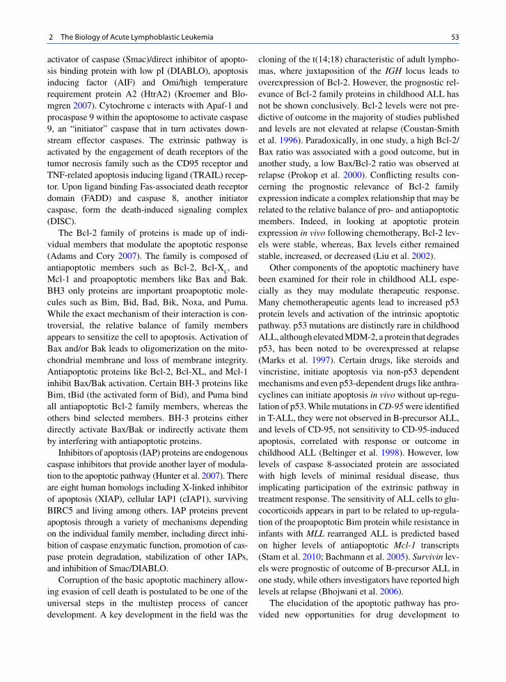
53 2 The Biology of Acute Lymphoblastic Leukemia
activator of caspase (Smac)/direct inhibitor of apopto-sis binding protein with low pI (DIABLO), apoptosis inducing factor (AIF) and Omi/high temperature requirement protein A2 (HtrA2) (Kroemer and Blo-mgren 2007). Cytochrome c interacts with Apaf-1 and procaspase 9 within the apoptosome to activate caspase 9, an “initiator” caspase that in turn activates down-stream effector caspases. The extrinsic pathway is activated by the engagement of death receptors of the tumor necrosis family such as the CD95 receptor and TNF-related apoptosis inducing ligand (TRAIL) recep-tor. Upon ligand binding Fas-associated death receptor domain (FADD) and caspase 8, another initiator caspase, form the death-induced signaling complex (DISC).
The Bcl-2 family of proteins is made up of indi-vidual members that modulate the apoptotic response (Adams and Cory 2007). The family is composed of antiapoptotic members such as Bcl-2, Bcl-X
L, and
Mcl-1 and proapoptotic members like Bax and Bak. BH3 only proteins are important proapoptotic mole-cules such as Bim, Bid, Bad, Bik, Noxa, and Puma. While the exact mechanism of their interaction is con-troversial, the relative balance of family members appears to sensitize the cell to apoptosis. Activation of Bax and/or Bak leads to oligomerization on the mito-chondrial membrane and loss of membrane integrity. Antiapoptotic proteins like Bcl-2, Bcl-XL, and Mcl-1 inhibit Bax/Bak activation. Certain BH-3 proteins like Bim, tBid (the activated form of Bid), and Puma bind all antiapoptotic Bcl-2 family members, whereas the others bind selected members. BH-3 proteins either directly activate Bax/Bak or indirectly activate them by interfering with antiapoptotic proteins.
Inhibitors of apoptosis (IAP) proteins are endogenous caspase inhibitors that provide another layer of modula-tion to the apoptotic pathway (Hunter et al. 2007). There are eight human homologs including X-linked inhibitor of apoptosis (XIAP), cellular IAP1 (cIAP1), surviving BIRC5 and living among others. IAP proteins prevent apoptosis through a variety of mechanisms depending on the individual family member, including direct inhi-bition of caspase enzymatic function, promotion of cas-pase protein degradation, stabilization of other IAPs, and inhibition of Smac/DIABLO.
Corruption of the basic apoptotic machinery allow-ing evasion of cell death is postulated to be one of the universal steps in the multistep process of cancer development. A key development in the field was the
cloning of the t(14;18) characteristic of adult lympho-mas, where juxtaposition of the IGH locus leads to overexpression of Bcl-2. However, the prognostic rel-evance of Bcl-2 family proteins in childhood ALL has not be shown conclusively. Bcl-2 levels were not pre-dictive of outcome in the majority of studies published and levels are not elevated at relapse (Coustan-Smith et al. 1996). Paradoxically, in one study, a high Bcl-2/Bax ratio was associated with a good outcome, but in another study, a low Bax/Bcl-2 ratio was observed at relapse (Prokop et al. 2000). Conflicting results con-cerning the prognostic relevance of Bcl-2 family expression indicate a complex relationship that may be related to the relative balance of pro- and antiapoptotic members. Indeed, in looking at apoptotic protein expression in vivo following chemotherapy, Bcl-2 lev-els were stable, whereas, Bax levels either remained stable, increased, or decreased (Liu et al. 2002).
Other components of the apoptotic machinery have been examined for their role in childhood ALL espe-cially as they may modulate therapeutic response. Many chemotherapeutic agents lead to increased p53 protein levels and activation of the intrinsic apoptotic pathway. p53 mutations are distinctly rare in childhood ALL, although elevated MDM-2, a protein that degrades p53, has been noted to be overexpressed at relapse (Marks et al. 1997). Certain drugs, like steroids and vincristine, initiate apoptosis via non-p53 dependent mechanisms and even p53-dependent drugs like anthra-cyclines can initiate apoptosis in vivo without up-regu-lation of p53. While mutations in CD-95 were identified in T-ALL, they were not observed in B-precursor ALL, and levels of CD-95, not sensitivity to CD-95-induced apoptosis, correlated with response or outcome in childhood ALL (Beltinger et al. 1998). However, low levels of caspase 8-associated protein are associated with high levels of minimal residual disease, thus implicating participation of the extrinsic pathway in treatment response. The sensitivity of ALL cells to glu-cocorticoids appears in part to be related to up-regula-tion of the proapoptotic Bim protein while resistance in infants with MLL rearranged ALL is predicted based on higher levels of antiapoptotic Mcl-1 transcripts (Stam et al. 2010; Bachmann et al. 2005). Survivin lev-els were prognostic of outcome of B-precursor ALL in one study, while others investigators have reported high levels at relapse (Bhojwani et al. 2006).
The elucidation of the apoptotic pathway has pro-vided new opportunities for drug development to

54 W.L. Carroll et al.
promote apoptosis in cancer cells with new agents alone or in combination with conventional chemother-apy and irradiation. The first agent targeting the apop-totic pathway focused on Bcl-2, since overexpression confers chemoresistance. Oblimersen sodium (G3139, Genasense) is a BCL-2 antisense oligodeoxynucleotide that leads to degradation of BCL-2 mRNA (O’Brien et al. 2005). Preclinical models show that oblimersen can induce apoptosis alone and is synergistic with con-ventional agents. There is data to indicate that its mechanism of action may also be due to nonantisense effects mediated by the CpG motifs present in the mol-ecule (Kim et al. 2007). Oblimersen has been evalu-ated in a number of clinical trials including those involving patients with chronic lymphocytic leukemia, acute myelogenous leukemia, multiple myeloma, and small cell lung cancer among others (Kang and Reynolds 2009). While there was a suggestion in some of these trials that oblimersen might have added a sur-vival advantage, the drug is yet to gain FDA approval.
More recently, a series of small molecules has been developed that interact directly with Bcl-2 proteins (e.g., Bcl-2, Bcl-XL, Mcl-1, and Bcl-w) at their hydrophobic binding groove in the place of BH3 only proteins. These “BH3 mimetics” include gossypol, ABT-737, ABT-263 (an oral version of ABT-737), GX15-070 (Obatoclax), and others. Each has differing affinities for Bcl-2 pro-tein family members, and preclinical evaluation docu-ments increased response to conventional radiation and chemotherapy. These agents are actively being explored in clinical trials for a wide variety of malignancies including ALL (Kang and Reynolds 2009).
Finally, since increased expression of IAPs has cor-related with outcome in some hematological malignan-cies, multiple strategies have been developed to negate the antiapoptotic effect of these proteins (Fulda 2009b). Based on the structure of Smac binding to XIAP (Bir3) as well as cIAP 1 and cIAP2, small molecule antago-nists of IAPs have been developed. Since the BIR2 domain of XIAP interacts with caspase-3, XIAP BIR2 antagonists have also been developed. Survivin is a particularly attractive target since expression is enhanced in tumor cells compared to normal cells. Gene expression profiling has also shown that survivin is upregulated in ALL blasts at relapse (Bhojwani et al. 2006). Many Phase II trials are now evaluating LY2813008, a survivin antisense oligonucleotide, and agents to repress survivin transcription (YM155 and EM-1421) are also in early phase protocols.
2.6 The Biology of Relapsed ALL
A central question related to the biology of relapse is what is the origin of the relapsed clone? Does it emerge with therapy or was it present at diagnosis? Is relapse a completely new clone or did it surface from a reservoir of premalignant cancer stem cells? Many studies have confirmed that relapsed blasts demonstrate biological differences from those noted at diagnosis. (Lilleyman et al. 1995; Guglielmi et al. 1997) However, analysis of antigen receptor rearrangements confirms that, in almost all cases, relapsed blasts are clonally related to the original disease. As mentioned, rearrangements of the T-cell receptor (TCR) and immunoglobulin (Ig) genes are clonal markers that can be detected in 90% of B-precursor ALL and 95% of T-cell ALL cases. While 40–50% of all relapse samples display a new rearrangement, at least one stable clonal Ig/TCR rear-rangement is almost always observed (>95%) at relapse. (Szczepanski et al. 2002; Germano et al. 2003) Furthermore, studies show that the relapsed sample almost always contains the same dominant karyotypic features observed at initial diagnosis (Heerema et al. 1992). Thus studies prove, with rare exceptions, that relapse is clonally related to the disease at diagnosis. Despite this, the only consistent genetic change char-acteristic of relapse involves frequent deletions involv-ing 9p, consistent with p16INK4a deletion/inactivation (Germano et al. 2003).
A number of studies examining antigen receptor rearrangements on a broader range of diagnosis/relapse samples have now shown that, in many cases, the relapsed clone existed at diagnosis, albeit making up a minor subset of the bulk leukemia population (Guggemos et al. 2003). These resistant cells showed a much lower rate of regression after application of ini-tial chemotherapy and the greater their numerical con-tribution to the bulk leukemia at diagnosis, the shorter the duration of first remission (Choi et al. 2007). However, while the relapse clone could be detected at diagnosis in many cases, this was not a uniform find-ing, still suggesting that some relapses may be due to the genesis of additional mutations during therapy (Henderson et al. 2008). Finally, in a select number of relapse cases defined by the presence of the t(12;21) ETV6/RUNX1 (TEL-AML1) fusion, there is evidence of a fetal preleukemic stem cell that acts as a reservoir for the re-emergence of a clonally related second leukemia (Seeger et al. 2001; Pine et al. 2003; Zuna et al. 2004).

55 2 The Biology of Acute Lymphoblastic Leukemia
The development of chemoresistance is another key issue in relapsed ALL, as evidenced by the lower remission re-Induction rate and event-free survival. Ex vivo analysis of chemosensitivity to individual agents demonstrates that relapsed samples are significantly more resistant to 6-thiogaunine, vincristine, predni-sone, dexamethasone, cytarabine, doxorubicin, idaru-bicin, and steroids (Hongo and Fujii 1991; Klumper et al. 1995). Resistance to steroids is the most dramatic difference noted in relapsed blasts.
Global gene expression analysis correlating gene expression signatures with drug resistance provides for a nonbiased approach to identifying biological pathways responsible for drug resistance and relapse of the disease. Recent analysis of in vitro resistant vs. sensitive samples led to the definition of genes whose protein products play a role in drug resistance (Holleman et al. 2004). Differentially expressed genes were noted for cells resistant to four drugs commonly used in therapy. Notably, 121 of these 124 genes had previously never been implicated in resistance to the four agents studied. Furthermore, resistance genes were unique to each agent and no single cross-resis-tance gene was identified. In a follow-up study, 45 genes were identified for which expression correlated with cross-resistance to all four agents, as well as an unanticipated signature that was associated with sensi-tivity to asparaginase and re sistance to vincristine (Lugthart et al. 2005).
Gene expression profiles of 35 matched diagnosis/relapse pairs (32 B-precursor cases) revealed 126 probe sets (48 high at diagnosis, 78 high at relapse) that were significantly different at relapse compared to diagnosis in B-precursor ALL (pair wise analysis: false discovery rate <10%). The most striking differ-ence was the much greater representation of genes involved in proliferation, cell cycle control, and cellu-lar metabolism in samples at early relapse (Bhojwani et al. 2006). Previous studies have also noted that relapsed blasts are in a higher proliferative state (Staber et al. 2004; Beesley et al. 2005). Genes that were iden-tified, including those involved in DNA repair (e.g., PTTG1, RAD51, POLE2) and those that play a role in inhibiting apoptosis (e.g., survivin (BIRC5), AATF, API5, AVEN), may aid the cells in overcoming the toxic effects of DNA damaging chemotherapeutic agents. Importantly, many of these genes such as sur-vivin (BIRC5) are attractive targets for therapeutic intervention.
While gene expression studies are informative, they are incapable of distinguishing “driver” vs. “passenger” pathways. To discover genes and path-ways fundamentally involved in drug resistance, many investigators have performed copy number analysis, reasoning that the identification of unique copy number abnormalities (CNAs) at relapse could be associated with outgrowth of a clone that escaped chemotherapy. In a pilot cohort of 20 diagnosis/relapse leukemia pairs from B-precursor ALL patients, genome-wide copy number profiles were surveyed using Affymetrix 500K SNP arrays. (Yang et al. 2008) This analysis revealed a total of 758 somatic genetic lesions. The number of genetic lesions varied significantly among patients, ranging from 3 to 84 per sample. These CNAs included gross copy number changes indicated by conventional cytogenetic analysis, but were mostly cryptic. Thus, the median size of CNAs identified in this study was 353 Kb, with 22.7% < 100 Kb, and 66.4% < 1 Mb. The median copy number loss per sample was 9 at diagnosis and 9.5 at relapse. Copy number gains were less common (p < 0.001), with a median of only 3.5 amplification events per sample at diagnosis and 4 at relapse. Across patients, there was a modest increase of CNAs at relapse (p = 0.035). Systematic enumeration of CNA events in the matched diagnosis and relapse samples revealed features that are com-mon and those that differ at these two time points. Of the 74 copy number gains observed at diagnosis, 71 (94.7%) persisted in the relapse sample from the same individual. Likewise, 256 of 288 (88.9%) copy number loss events at diagnosis remained at relapse. Conversely, 24 novel amplifications and 45 novel deletions arose at relapse, accounting for 25.0% and 14.9% of total copy number gains and losses at relapse, respectively. It should also be noted that the majority of the diagnosis or relapse-specific CNAs were focal, with a median size of 537 Kb. There was a significant correlation between the change in DNA copy number and change in gene expression from diagnosis to relapse (p = 2.2 × 10⁻¹⁶).
Although all 44 autosome arms showed one or more CNAs, a number of regions appeared to be affected more frequently. The most common CNA events were deletions at 9p21.3, occurring in 12 of 20 (60.0%) cases and persisting from diagnosis to relapse. Of these 12 cases, 11 exhibited deletion of both CDKN2A and CDKN2B, consistent with prior reports (Maloney et al.

56 W.L. Carroll et al.
1999; Graf Einsiedel et al. 2002). CNAs involving sev-eral transcription regulators essential in early lymphoid specification and B-lineage commitment (Nutt and Kee 2007) (PAX5, EBF1, and IKZF1) were also com-mon in this relapsed ALL cohort. Somatically acquired deletions at these four loci (CDKN2A/B, PAX5, EBF1, and IKZF1) have been previously reported in child-hood ALL. (Maloney et al. 1999; Graf Einsiedel et al. 2002; Mullighan et al. 2007; French et al. 2009) However, the frequencies of these lesions (except PAX5) appeared to be higher in the relapsed cases ana-lyzed here relative to newly diagnosed B-precursor ALL (Mullighan et al. 2007) CDKN2A, 60.0% vs. 33.9% (p = 0.038); EBF1, 25.0% vs. 4.2% (p = 0.0013); and IKZF1: 35.0% vs. 8.9% (p = 0.0016). Over-representation of these genetic aberrations, especially IKZF1 and EBF1 in relapsed ALL cases indicate their potential prognostic value at diagnosis. The fact that numerous examples were identified in which IKZF1 and EBF1 deletions were seen only in the relapse clone indicates that such deletions may not be required for full transformation, but that they endow a subclone with drug resistance properties. Other investigators have published similar findings in 47 matched pairs of
B-precursor ALL (Mullighan et al. 2008b). While many regions of interest were identical in the two stud-ies, there were clear differences. Whether these differ-ences are due to variations in therapeutic regimens can be answered only with a larger data set.
These results show clear differences in the genetic profile of blasts from patients who relapse early vs. late, consistent with the better (but still suboptimal) salvage rates for those patients who relapse off therapy. (Fig. 2.8) shows a model that incorporates findings from many laboratories. In this model, most cases of early relapse occur because an intrinsically drug- resistant (IDR) clone exists at diagnosis before therapy is initiated. Under the selective pressure of chemother-apy the clone emerges relatively soon in treatment. In another scenario, a subclone that may or may not repre-sent a leukemic stem cell (LSC) and is relatively resis-tant to standard therapy undergoes additional genetic events that lead to drug resistance (acquired drug resis-tance, ADR). A version of this second model is repre-sented by late relapsing ETV6/RunX1 cases described to date, which represent a “new” but clonally related clone that is sensitive to retreatment since it emanates from a LSC. However, more commonly, the low salvage rates
Fig. 2.8 Hypothetical Model of Relapsed ALL. A pre-leukemic stem cell (LSC, blue) gives rise to frank ALL (light blue cells). Intrinsically drug resistant (IDR) clones (dark blue) may be present at diagnosis. LSCs and other subclones may survive ini-
tial treatment and acquire additional lesions that result in acquired drug resistance (ADR). IDR clones are more likely to account for early relapse whereas more cases of late relapse are associated with ADR clones

57 2 The Biology of Acute Lymphoblastic Leukemia
seen in most cases of late relapse indicate that acquired drug resistance is operative (for example, not a “new leukemia”). In both models (IDR and ADR clones), residual blasts at end-Induction signify a greater likeli-hood of relapse. However, the remaining clones in these two cases are qualitatively different insofar as in the second scenario, additional mutations and or epigenetic lesions are needed to result in the drug-resistant pheno-type. Early Intensification is capable of eradicating such clones, but failure to apply augmented therapy early allows a window for such changes to occur. Finally, in a small number of cases, relapse represents a true non-clonally related second malignancy.
2.7 Summary
ALL is a biologically heterogeneous disease repre-sented by distinct clinical and biological subtypes. Recent data using genome-wide approaches indicates that certain individuals may be predisposed to the development of ALL and host differences are likely to account for some differences in response to therapy also. ALL is a multistep process requiring the acquisi-tion of multiple somatic lesions, and the definition of such pathways are being elucidated, including those lesions directly associated with drug resistance. These pathways are now being used for treatment assignment and serve as targets for novel therapy.
References
Adams JM, Cory S (2007) The Bcl-2 apoptotic switch in cancer development and therapy. Oncogene 26(9):1324–1337
Adolfsson J, Mansson R et al (2005) Identification of Flt3+ lympho-myeloid stem cells lacking erythro-megakaryocytic potential a revised road map for adult blood lineage commit-ment. Cell 121(2):295–306
Arico M, Valsecchi MG et al (2000) Outcome of treatment in children with Philadelphia chromosome-positive acute lym-phoblastic leukemia. N Engl J Med 342(14):998–1006
Armstrong SA, Staunton JE et al (2001) MLL translocations specify a distinct gene expression profile that distinguishes a unique leukemia. Nat Genet 30(1):41–47
Attarbaschi A, Mann G et al (2008) Minimal residual disease values discriminate between low and high relapse risk in children with B-cell precursor acute lymphoblastic leukemia and an intrachromosomal amplification of chromosome 21: the Austrian and German acute lymphoblastic leukemia
Berlin-Frankfurt-Munster (ALL-BFM) trials. J Clin Oncol 26(18):3046–3050
Bachmann PS, Gorman R et al (2005) Dexamethasone resis-tance in B-cell precursor childhood acute lymphoblastic leukemia occurs downstream of ligand-induced nuclear translocation of the glucocorticoid receptor. Blood 105(6): 2519–2526
Bartel DP (2004) MicroRNAs: genomics, biogenesis, mecha-nism, and function. Cell 116(2):281–297
Beesley AH, Cummings AJ et al (2005) The gene expression signature of relapse in paediatric acute lymphoblastic leu-kaemia: implications for mechanisms of therapy failure. Br J Haematol 131(4):447–456
Beishuizen A, de Bruijn MA et al (1997) Heterogeneity in junc-tional regions of immunoglobulin kappa deleting element rearrangements in B cell leukemias: a new molecular target for detection of minimal residual disease. Leukemia 11(12): 2200–2207
Belson M, Kingsley B et al (2007) Risk factors for acute leuke-mia in children: a review. Environ Health Perspect 115(1): 138–145
Beltinger C, Bohler T et al (1998) Mutation analysis of CD95 (APO-1/Fas) in childhood B-lineage acute lymphoblastic leukaemia. Br J Haematol 102(3):722–728
Bene MC, Castoldi G et al (1995) Proposals for the immuno-logical classification of acute leukemias. European Group for the Immunological Characterization of Leukemias (EGIL). Leukemia 9(10):1783–1786
Bercovich D, Ganmore I et al (2008) Mutations of JAK2 in acute lymphoblastic leukaemias associated with Down’s syn-drome. Lancet 372(9648):1484–1492
Bhojwani D, Kang H et al (2008) Gene expression signatures predictive of early response and outcome in high-risk childhood acute lymphoblastic leukemia: A Children’s Oncology Group Study [corrected]. J Clin Oncol 26(27): 4376–4384
Bhojwani D, Kang H et al (2006) Biologic pathways associ-ated with relapse in childhood acute lymphoblastic leuke-mia: a Children’s Oncology Group study. Blood 108(2): 711–717
Bryder D, Rossi DJ et al (2006) Hematopoietic stem cells: the paradigmatic tissue-specific stem cell. Am J Pathol 169: 338–346
Bryder D, Sigvardsson M (2010) Shaping up a lineage – lessons from B lymphopoesis. Curr Opin Immunol 22(2):148–153
Busslinger M (2004) Transcriptional control of early B cell development. Annu Rev Immunol 22:55–79
Calin GA, Ferracin M et al (2005) A MicroRNA signature asso-ciated with prognosis and progression in chronic lympho-cytic leukemia. N Engl J Med 353(17):1793–1801
Carter NP, Meyer EW (1994) Introduction to the principles of flow cytometry. In: Ormerod MG (ed) Flow cytometry: a practical approach. Oxford University Press, New York
Case M, Matheson E et al (2008) Mutation of genes affecting the RAS pathway is common in childhood acute lymphoblastic leukemia. Cancer Res 68(16):6803–6809
Castor A, Nilsson L et al (2005) Distinct patterns of hematopoi-etic stem cell involvement in acute lymphoblastic leukemia. Nat Med 11(6):630–637
Cazzaniga G, Biondi A (2005) Molecular monitoring of child-hood acute lymphoblastic leukemia using antigen receptor

58 W.L. Carroll et al.
gene rearrangements and quantitative polymerase chain re action technology. Haematologica 90(3):382–390
Chen CL, Liu Q et al (1997) Higher frequency of glutathione S-transferase deletions in black children with acute lympho-blastic leukemia. Blood 89(5):1701–1707
Cheok MH, Yang W et al (2003) Treatment-specific changes in gene expression discriminate in vivo drug response in human leukemia cells. Nat Genet 34(1):85–90
Choi S, Henderson MJ et al (2007) Relapse in children with acute lymphoblastic leukemia involving selection of a preex-isting drug-resistant subclone. Blood 110(2):632–639
Clark SJ, Harrison J et al (1994) High sensitivity mapping of methylated cytosines. Nucleic Acids Res 22(15): 2990–2997
Clarke MF, Dick JE et al (2006) Cancer stem cells–perspectives on current status and future directions: AACR Workshop on cancer stem cells. Cancer Res 66(19):9339–9344
Cobaleda C, Gutierrez-Cianca N et al (2000) A primitive hematopoietic cell is the target for the leukemic transforma-tion in human philadelphia-positive acute lymphoblastic leukemia. Blood 95(3):1007–1013
Coustan-Smith E, Kitanaka A et al (1996) Clinical relevance of BCL-2 overexpression in childhood acute lymphoblastic leukemia. Blood 87(3):1140–1146
Czerny T, Busslinger M (1995) DNA-binding and transactivation properties of Pax-6: three amino acids in the paired domain are responsible for the different sequence recognition of Pax-6 and BSAP (Pax-5). Mol Cell Biol 15(5): 2858–2871
Den Boer ML, van Slegtenhorst M et al (2009) A subtype of childhood acute lymphoblastic leukaemia with poor treat-ment outcome: a genome-wide classification study. Lancet Oncol 10(2):125–134
Druker BJ, Sawyers CL et al (2001) Activity of a specific inhibi-tor of the BCR-ABL tyrosine kinase in the blast crisis of chronic myeloid leukemia and acute lymphoblastic leuke-mia with the Philadelphia chromosome. N Engl J Med 344(14): 1038–1042
Dutt A, Beroukhim R (2007) Single nucleotide polymorphism array analysis of cancer. Curr Opin Oncol 19(1):43–49
Ferrando AA (2009) The role of NOTCH1 signaling in T-ALL. Hematology Am Soc Hematol Educ Program 353–361
Flohr T, Schrauder A et al (2008) Minimal residual disease-directed risk stratification using real-time quantitative PCR analysis of immunoglobulin and T-cell receptor gene rear-rangements in the international multicenter trial AIEOP-BFM ALL 2000 for childhood acute lymphoblastic leukemia. Leukemia 22(4):771–782
Ford AM, Bennett CA et al (1998) Fetal origins of the TEL-AML1 fusion gene in identical twins with leukemia. Proc Natl Acad Sci USA 95(8):4584–4588
French D, Yang W et al (2009) Acquired variation outweighs inherited variation in whole genome analysis of methotrex-ate polyglutamate accumulation in leukemia. Blood 113(19): 4512–4520
Fulci V, Colombo T et al (2009) Characterization of B- and T-lineage acute lymphoblastic leukemia by integrated analy-sis of MicroRNA and mRNA expression profiles. Genes Chromosomes Cancer 48(12):1069–1082
Fulda S (2009a) Apoptosis pathways and their therapeutic exploi-tation in pancreatic cancer. J Cell Mol Med 13(7): 1221–1227
Fulda S (2009b) Therapeutic opportunities for counteracting apoptosis resistance in childhood leukaemia. Br J Haematol 145(4):441–454
Garcia-Manero G, Issa JP (2005) Histone deacetylase inhibitors: a review of their clinical status as antineoplastic agents. Cancer Invest 23(7):635–642
Garcia-Manero G, Jeha S et al (2003) Aberrant DNA methyla-tion in pediatric patients with acute lymphocytic leukemia. Cancer 97(3):695–702
Garcia-Manero G, Yang H et al (2009) Epigenetics of acute lym-phocytic leukemia. Semin Hematol 46(1):24–32
Germano G, del Giudice L et al (2003) Clonality profile in relapsed precursor-B-ALL children by GeneScan and sequencing analyses. Consequences on minimal residual disease monitoring. Leukemia 17(8):1573–1582
Ghia P, ten Boekel E et al (1998) B-cell development: a comparison between mouse and man. Immunol Today 19(10):480–485
Goemans BF, Zwaan CM et al (2005) In vitro profiling of the sensitivity of pediatric leukemia cells to tipifarnib: identifi-cation of T-cell ALL and FAB M5 AML as the most sensi-tive subsets. Blood 106(10):3532–3537
Graf Einsiedel H, Taube T et al (2002) Deletion analysis of p16(INKa) and p15(INKb) in relapsed childhood acute lym-phoblastic leukemia. Blood 99(12):4629–4631
Greaves M (2006) Infection, immune responses and the aetiol-ogy of childhood leukaemia. Nat Rev Cancer 6(3):193–203
Greaves M (2009) Darwin and evolutionary tales in leukemia. American Society of Hematology, New Orleans
Guggemos A, Eckert C et al (2003) Assessment of clonal stabil-ity of minimal residual disease targets between 1st and 2nd relapse of childhood precursor B-cell acute lymphoblastic leukemia. Haematologica 88(7):737–746
Guglielmi C, Cordone I et al (1997) Immunophenotype of adult and childhood acute lymphoblastic leukemia: changes at first relapse and clinico-prognostic implications. Leukemia 11(9):1501–1507
Gutierrez MI, Siraj AK et al (2003) Concurrent methylation of multiple genes in childhood ALL: Correlation with phenotype and molecular subgroup. Leukemia 17(9): 1845–1850
Harrison CJ, Moorman AV et al (2004) Three distinct subgroups of hypodiploidy in acute lymphoblastic leukaemia. Br J Haematol 125(5):552–559
Harvey R, Mulligan RC et al (2010a) Identification of novel cluster groups in pediatric high-risk B-precursor acute lym-phoblastic leukemia with gene expression profiling: correla-tion with genomw wide copy mumber alterations, clinical charactersitics, and outcome
Harvey RC, Mullighan CG et al (2010b) Rearrangement of CRLF2 is associated with mutation of JAK kinases, altera-tion of IKZF1, Hispanic/Latino ethnicity, and a poor out-come in pediatric B-progenitor acute lymphoblastic leu - kemia. Blood 115 (26): 5312–5321
Hayday AC, Pennington DJ (2007) Key factors in the organized chaos of early T cell development. Nat Immunol 8(2): 137–144
Heerema NA, Nachman JB et al (2004) Deletion of 7p or mono-somy 7 in pediatric acute lymphoblastic leukemia is an adverse prognostic factor: a report from the Children’s Cancer Group. Leukemia 18(5):939–947
Heerema NA, Palmer CG et al (1992) Cytogenetic analysis in relapsed childhood acute lymphoblastic leukemia. Leukemia 6(3):185–192
Heerema NA, Sather HN et al (2000) Clinical significance of deletions of chromosome arm 6q in childhood acute

59 2 The Biology of Acute Lymphoblastic Leukemia
lymphoblastic leukemia: a report from the Children’s Cancer Group. Leuk Lymphoma 36(5–6):467–478
Henderson MJ, Choi S et al (2008) Mechanism of relapse in pediatric acute lymphoblastic leukemia. Cell Cycle 7(10): 1315–1320
Hertzberg L, Vendramini E et al (2010) Down syndrome acute lymphoblastic leukemia, a highly heterogeneous disease in which aberrant expression of CRLF2 is associated with mutated JAK2: a report from the International BFM Study Group. Blood 115(5):1006–1017
Hoffmann K, Firth MJ et al (2008) Prediction of relapse in pae-diatric pre-B acute lymphoblastic leukaemia using a three-gene risk index. Br J Haematol 140(6):656–664
Holleman A, Cheok MH et al (2004) Gene-expression patterns in drug-resistant acute lymphoblastic leukemia cells and response to treatment. N Engl J Med 351(6):533–542
Hong D, Gupta R et al (2008) Initiating and cancer-propagating cells in TEL-AML1-associated childhood leukemia. Science 319(5861):336–339
Hongo T, Fujii Y (1991) In vitro chemosensitivity of lympho-blasts at relapse in childhood leukemia using the MTT assay. Int J Hematol 54(3):219–230
Hotfilder M, Rottgers S et al (2002) Immature CD34+CD19– progenitor/stem cells in TEL/AML1-positive acute lympho-blastic leukemia are genetically and functionally normal. Blood 100(2):640–646
Hunter AM, LaCasse EC et al (2007) The inhibitors of apoptosis (IAPs) as cancer targets. Apoptosis 12(9):1543–1568
Iorio MV, Croce CM (2009) MicroRNAs in cancer: small mol-ecules with a huge impact. J Clin Oncol 27(34):5848–5856
Janeway CA, Travers P et al (2001) Immunobiology. Garland, New York, London
Jones PA, Baylin SB (2007) The epigenomics of cancer. Cell 128(4):683–692
Kang H, Chen IM et al (2010) Gene expression classifiers for relapse-free survival and minimal residual disease improve risk classification and outcome prediction in pediatric B-precursor acute lymphoblastic leukemia. Blood 115(7):1394–1405
Kang MH, Reynolds CP (2009) Bcl-2 inhibitors: targeting mito-chondrial apoptotic pathways in cancer therapy. Clin Cancer Res 15(4):1126–1132
Kim R, Emi M et al (2007) Antisense and nonantisense effects of antisense Bcl-2 on multiple roles of Bcl-2 as a chemosen-sitizer in cancer therapy. Cancer Gene Ther 14(1):1–11
Klumper E, Pieters R et al (1995) In vitro cellular drug resis-tance in children with relapsed/refractory acute lymphoblas-tic leukemia. Blood 86(10):3861–3868
Korenberg JR, Chen XN et al (1994) Down syndrome pheno-types: the consequences of chromosomal imbalance. Proc Natl Acad Sci USA 91(11):4997–5001
Kotani A, Ha D et al (2009) miR-128b is a potent glucocorticoid sensitizer in MLL-AF4 acute lymphocytic leukemia cells and exerts cooperative effects with miR-221. Blood 114(19):4169–4178
Krajinovic M, Labuda D et al (2002a) Polymorphisms in genes encoding drugs and xenobiotic metabolizing enzymes, DNA repair enzymes, and response to treatment of childhood acute lymphoblastic leukemia. Clin Cancer Res 8(3):802–810
Krajinovic M, Sinnett H et al (2002b) Role of NQO1, MPO and CYP2E1 genetic polymorphisms in the susceptibility to childhood acute lymphoblastic leukemia. Int J Cancer 97(2):230–236
Krivtsov AV, Twomey D et al (2006) Transformation from com-mitted progenitor to leukaemia stem cell initiated by MLL-AF9. Nature 442(7104):818–822
Kroemer G, Blomgren K (2007) Mitochondrial cell death con-trol in familial Parkinson disease. PLoS Biol 5(7):e206
Laird PW (2010) Principles and challenges of genome-wide DNA methylation analysis. Nat Rev Genet 11(3):191–203
Lange B (2000) The management of neoplastic disorders of haematopoiesis in children with Down’s syndrome. Br J Haematol 110(3):512–524
Lapidot T, Sirard C et al (1994) A cell initiating human acute myeloid leukaemia after transplantation into SCID mice. Nature 367(6464):645–648
le Viseur C, Hotfilder M et al (2008) In childhood acute lympho-blastic leukemia, blasts at different stages of immunopheno-typic maturation have stem cell properties. Cancer Cell 14(1): 47–58
Lilleyman JS, Stevens RF et al (1995) Changes in cytomorphol-ogy of childhood lymphoblastic leukaemia at the time of disease relapse. Childhood Leukaemia Working Party of the United Kingdom Medical Research Council. J Clin Pathol 48(11):1051–1053
Liu T, Raetz E et al (2002) Diversity of the apoptotic response to chemotherapy in childhood leukemia. Leukemia 16(2): 223–232
Loh ML, Rubnitz JE (2002) TEL/AML1-positive pediatric leu-kemia: prognostic significance and therapeutic approaches. Curr Opin Hematol 9(4):345–352
Lugthart S, Cheok MH et al (2005) Identification of genes asso-ciated with chemotherapy crossresistance and treatment response in childhood acute lymphoblastic leukemia. Cancer Cell 7(4):375–386
Malinge S, Ben-Abdelali R et al (2007) Novel activating JAK2 mutation in a patient with Down syndrome and B-cell pre-cursor acute lymphoblastic leukemia. Blood 109(5): 2202–2204
Malinge S, Izraeli S et al (2009) Insights into the manifestations, outcomes, and mechanisms of leukemogenesis in Down syndrome. Blood 113(12):2619–2628
Maloney KW, McGavran L et al (1999) Acquisition of p16(INK4A) and p15(INK4B) gene abnormalities between initial diagnosis and relapse in children with acute lympho-blastic leukemia. Blood 93(7):2380–2385
Marks DI, Kurz BW et al (1997) Altered expression of p53 and mdm-2 proteins at diagnosis is associated with early treat-ment failure in childhood acute lymphoblastic leukemia. J Clin Oncol 15(3):1158–1162
Mason D, Andre P et al (2002) Leucocyte Typoing VII. Oxford University Press, Oxford
Meshinchi S, Appelbaum FR (2009) Structural and functional alterations of FLT3 in acute myeloid leukemia. Clin Cancer Res 15(13):4263–4269
Mi S, Lu J et al (2007) MicroRNA expression signatures accu-rately discriminate acute lymphoblastic leukemia from acute myeloid leukemia. Proc Natl Acad Sci USA 104(50): 19971–19976
Moorman AV, Richards SM et al (2007) Prognosis of children with acute lymphoblastic leukemia (ALL) and intrachromo-somal amplification of chromosome 21 (iAMP21). Blood 109(6):2327–2330
Mori H, Colman SM et al (2002) Chromosome translocations and covert leukemic clones are generated during normal

60 W.L. Carroll et al.
fetal development. Proc Natl Acad Sci USA 99(12): 8242–8247
Muller A, Florek M (2010) 5-Azacytidine/Azacitidine. Recent Results Cancer Res 184:159–170
Mullighan CG, Goorha S et al (2007a) Genome-wide analysis of genetic alterations in acute lymphoblastic leukaemia. Nature 446(7137):758–764
Mullighan CG, Miller CB et al (2008a) BCR-ABL1 lympho-blastic leukaemia is characterized by the deletion of Ikaros. Nature 453(7191):110–114
Mullighan CG, Phillips LA et al (2008b) Genomic analysis of the clonal origins of relapsed acute lymphoblastic leukemia. Science 322(5906):1377–1380
Mullighan CG, Collins-Underwood JR et al (2009a) Rearrange- ment of CRLF2 in B-progenitor- and Down syndrome- associated acute lymphoblastic leukemia. Nat Genet 41: 1243–1246
Mullighan CG, Su X et al (2009b) Deletion of IKZF1 and prog-nosis in acute lymphoblastic leukemia. N Engl J Med 360(5):470–480
Mullighan CG, Zhang J et al (2009c) JAK mutations in high-risk childhood acute lymphoblastic leukemia. Proc Natl Acad Sci USA 106(23):9414–9418
Mullighan RC, Morin RD et al (2009d) Next generation tran-scriptomic resequencing identifies novel genetic alterations in high-risk (HR) childhood acute lymphoblastic leukemia (ALL): a report from the Children’s Oncology group HR ALL TARGET Project. Blood 114:704
Nachman JB, Heerema NA et al (2007) Outcome of treatment in children with hypodiploid acute lymphoblastic leukemia. Blood 110(4):1112–1115
Nahar R, Muschen M (2009) Pre-B cell receptor signaling in acute lymphoblastic leukemia. Cell Cycle 8(23): 3874–3877
Nowell PC, Hungerford DA (1960) Chromosome studies on normal and leukemic human leukocytes. J Natl Cancer Inst 25:85–109
Nutt SL, Kee BL (2007) The transcriptional regulation of B cell lineage commitment. Immunity 26(6):715–725
O’Brien SM, Cunningham CC et al (2005) Phase I to II multi-center study of oblimersen sodium, a Bcl-2 antisense oligo-nucleotide, in patients with advanced chronic lymphocytic leukemia. J Clin Oncol 23(30):7697–7702
Papaemmanuil E, Hosking FJ et al (2009) Loci on 7p12.2, 10q21.2 and 14q11.2 are associated with risk of childhood acute lymphoblastic leukemia. Nat Genet 41(9):1006–1010
Pine SR, Wiemels JL et al (2003) TEL-AML1 fusion precedes differentiation to pre-B cells in childhood acute lymphoblas-tic leukemia. Leuk Res 27(2):155–164
Pongers-Willemse MJ, Seriu T et al (1999) Primers and proto-cols for standardized detection of minimal residual disease in acute lymphoblastic leukemia using immunoglobulin and T cell receptor gene rearrangements and TAL1 deletions as PCR targets: report of the BIOMED-1 CONCERTED ACTION: investigation of minimal residual disease in acute leukemia. Leukemia 13(1):110–118
Prokop A, Wieder T et al (2000) Relapse in childhood acute lymphoblastic leukemia is associated with a decrease of the Bax/Bcl-2 ratio and loss of spontaneous caspase-3 process-ing in vivo. Leukemia 14(9):1606–1613
Pui CH, Chessells JM et al (2003) Clinical heterogeneity in childhood acute lymphoblastic leukemia with 11q23 rear-rangements. Leukemia 17(4):700–706
Pui CH, Gaynon PS et al (2002) Outcome of treatment in child-hood acute lymphoblastic leukaemia with rearrangements of the 11q23 chromosomal region. Lancet 359(9321): 1909–1915
Quintas-Cardama A, Cortes J (2009) Molecular biology of bcr-abl1-positive chronic myeloid leukemia. Blood 113(8): 1619–1630
Rabin KR, Wang J et al (2009) Gene expression profiling in Down Syndrome acute lymphoblastic leukemia identifies distinct profiles assocaited with CRLF2 expression. Blood 114:2389
Raimondi SC, Behm FG et al (1990) Cytogenetics of pre-B-cell acute lymphoblastic leukemia with emphasis on prognostic implications of the t(1;19). J Clin Oncol 8(8):1380–1388
Ramirez J, Lukin K et al (2010) From hematopoietic progenitors to B cells: mechanisms of lineage restriction and commit-ment. Curr Opin Immunol 22(2):177–184
Real PJ, Ferrando AA (2009) NOTCH inhibition and glucocor-ticoid therapy in T-cell acute lymphoblastic leukemia. Leukemia 23(8):1374–1377
Reed JC, Pellecchia M (2005) Apoptosis-based therapies for hematologic malignancies. Blood 106(2):408–418
Robertson KD, Wolffe AP (2000) DNA methylation in health and disease. Nat Rev Genet 1(1):11–19
Roman-Gomez J, Jimenez-Velasco A et al (2007) Poor progno-sis in acute lymphoblastic leukemia may relate to promoter hypermethylation of cancer-related genes. Leuk Lymphoma 48(7):1269–1282
Romana SP, Mauchauffe M et al (1995) The t(12;21) of acute lymphoblastic leukemia results in a tel-AML1 gene fusion. Blood 85(12):3662–3670
Ross ME, Zhou X et al (2003) Classification of pediatric acute lymphoblastic leukemia by gene expression profiling. Blood 102(8):2951–2959
Rothenberg EV, Moore JE et al (2008) Launching the T-cell-lineage developmental programme. Nat Rev Immunol 8(1): 9–21
Russell LJ, Capasso M et al (2009) Deregulated expression of cytokine receptor gene, CRLF2, is involved in lymphoid transformation in B-cell precursor acute lymphoblastic leu-kemia. Blood 114(13):2688–2698
Schimmer AD, Hedley DW et al (2001) Receptor- and mito-chondrial-mediated apoptosis in acute leukemia: a transla-tional view. Blood 98(13):3541–3553
Schotte D, Chau JC et al (2009) Identification of new microRNA genes and aberrant microRNA profiles in childhood acute lymphoblastic leukemia. Leukemia 23(2):313–322
Schultz KR, Bowman WP et al (2009) Improved early event-free survival with imatinib in Philadelphia chromosome-positive acute lymphoblastic leukemia: a children’s oncology group study. J Clin Oncol 27(31):5175–5181
Schultz KR, Pullen DJ et al (2007) Risk- and response-based classification of childhood B-precursor acute lymphoblastic leukemia: a combined analysis of prognostic markers from the Pediatric Oncology Group (POG) and Children’s Cancer Group (CCG). Blood 109(3):926–935
Schwarz BA, Sambandam A et al (2007) Selective thymus set-tling regulated by cytokine and chemokine receptors. J Immunol 178(4):2008–2017
Seeger K, von Stackelberg A et al (2001) Relapse of TEL-AML1–positive acute lymphoblastic leukemia in childhood: a matched-pair analysis. J Clin Oncol 19(13):3188–3193

61 2 The Biology of Acute Lymphoblastic Leukemia
Silverman LB (2007) Acute lymphoblastic leukemia in infancy. Pediatr Blood Cancer 49(7 Suppl):1070–1073
Silverman LB, McLean TW et al (1997) Intensified therapy for infants with acute lymphoblastic leukemia: results from the Dana-Farber Cancer Institute Consortium. Cancer 80(12): 2285–2295
Sorich MJ, Pottier N et al (2008) In vivo response to metho-trexate forecasts outcome of acute lymphoblastic leukemia and has a distinct gene expression profile. PLoS Med 5(4):e83
Staber PB, Linkesch W et al (2004) Common alterations in gene expression and increased proliferation in recurrent acute myeloid leukemia. Oncogene 23(4):894–904
Stam RW, den Boer ML et al (2006) Silencing of the tumor sup-pressor gene FHIT is highly characteristic for MLL gene rearranged infant acute lymphoblastic leukemia. Leukemia 20(2):264–271
Stam RW, Den Boer ML et al (2010) Association of high-level MCL-1 expression with in vitro and in vivo prednisone resis-tance in MLL-rearranged infant acute lymphoblastic leuke-mia. Blood 115(5):1018–1025
Stumpel DJ, Schneider P et al (2009) Specific promoter methy-lation identifies different subgroups of MLL-rearranged infant acute lymphoblastic leukemia, influences clinical out-come, and provides therapeutic options. Blood 114(27): 5490–5498
Szczepanski T, Orfao A et al (2001) Minimal residual disease in leukaemia patients. Lancet Oncol 2(7):409–417
Szczepanski T, Willemse MJ et al (2002) Comparative analysis of Ig and TCR gene rearrangements at diagnosis and at relapse of childhood precursor-B-ALL provides improved strategies for selection of stable PCR targets for monitoring of minimal residual disease. Blood 99(7):2315–2323
Thompson EC, Cobb BS et al (2007) Ikaros DNA-binding pro-teins as integral components of B cell developmental-stage-specific regulatory circuits. Immunity 26(3):335–344
Tjio JH, Whang J (1962) Chromosome preparations of bone marrow cells without prior in vitro culture or in vitro colchi-cine administration. Stain Technol 37:17–20
Tomlins SA, Rhodes DR et al (2005) Recurrent fusion of TMPRSS2 and ETS transcription factor genes in prostate cancer. Science 310(5748):644–648
Trevino LR, Yang W et al (2009) Germline genomic variants associated with childhood acute lymphoblastic leukemia. Nat Genet 41(9):1001–1005
Trueworthy R, Shuster J et al (1992) Ploidy of lymphoblasts is the strongest predictor of treatment outcome in B-progenitor cell acute lymphoblastic leukemia of childhood: a Pediatric Oncology Group study. J Clin Oncol 10(4):606–613
van der Linden MH, Valsecchi MG et al (2009) Outcome of congenital acute lymphoblastic leukemia treated on the Interfant-99 protocol. Blood 114(18):3764–3768
van der Velden VH, Szczepanski T et al (2003) Age-related pat-terns of immunoglobulin and T-cell receptor gene rearrange-ments in precursor-B-ALL: implications for detection of minimal residual disease. Leukemia 17(9):1834–1844
van der Velden VH, van Dongen JJ (2009) MRD detection in acute lymphoblastic leukemia patients using Ig/TCR gene rearrangements as targets for real-time quantitative PCR. Methods Mol Biol 538:115–150
van Dongen JJ, Adriaansen HJ et al (1988) Immunophenotyping of leukaemias and non-Hodgkin’s lymphomas. Immuno-logical markers and their CD codes. Neth J Med 33(5–6): 298–314
van Dongen JJ, Wolvers-Tettero IL (1991) Analysis of immuno-globulin and T cell receptor genes. Part I: Basic and techni-cal aspects. Clin Chim Acta 198(1–2):1–91
Whitlock JA, Sather HN et al (2005) Clinical characteristics and outcome of children with Down syndrome and acute lymphoblastic leukemia: a Children’s Cancer Group study. Blood 106(13):4043–4049
Wong HL, Byun HM et al (2006) Rapid and quantitative method of allele-specific DNA methylation analysis. Biotechniques 41(6):734–739
Yanez L, Bermudez A et al (2009) Successful induction therapy with decitabine in refractory childhood acute lymphoblastic leukemia. Leukemia 23(7):1342–1343
Yang JJ, Bhojwani D et al (2008) Genome-wide copy number profiling reveals molecular evolution from diagnosis to relapse in childhood acute lymphoblastic leukemia. Blood 112(10):4178–4183
Yeoh EJ, Ross ME et al (2002) Classification, subtype discov-ery, and prediction of outcome in pediatric acute lympho-blastic leukemia by gene expression profiling. Cancer Cell 1(2):133–143
Yoda A, Yoda Y et al (2010) Functional screening identifies CRLF2 in precursor B-cell acute lymphoblastic leukemia. Proc Natl Acad Sci USA 107(1):252–257
Yoshida T, Ng SY et al (2006) Early hematopoietic line - age restrictions directed by Ikaros. Nat Immunol 7(4): 382–391
Zhang J, Mulligan RC et al (2009) Mutations in the RAS Signaling, B-cell development, TP53/RB1, and JAK signal-ing pathways are common in high risk B-precursor child-hood acute lymphoblastic leukemia (ALl): A report from the Children’s Oncology group High Risk TARGET Project. Blood 114:85
Zuna J, Ford AM et al (2004) TEL deletion analysis supports a novel view of relapse in childhood acute lymphoblastic leukemia. Clin Cancer Res 10(16):5355–5360

http://www.springer.com/978-3-642-13780-8