the biology and future treatment options
TRANSCRIPT

Remodeling the failing heart: : the biology and future treatment options
J-L Balligand (UCL-Brussels, BE)

• phenotypic plasticity : remodeling is characterized by changes in myocardial structure that happen in response to either mechanical overload or a loss of substance such as that occurring after myocardial infarction.
• Myocardial remodeling is an essential mechanism that allows for cardiac output to be maintained in the presence of chronically abnormal loading conditions or depressed contractility.
Myocardial remodeling: definitions

Jessup et al N Engl J Med, 2003 (review)

Jessup et al N Engl J Med, 2003 (review)
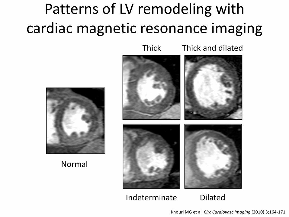
Thick Thick and dilated
Dilated Indeterminate
Normal
Patterns of LV remodeling with cardiac magnetic resonance imaging
Khouri MG et al. Circ Cardiovasc Imaging (2010) 3;164-171

Harding JD et al Circulation 2001
Reversal of electrical remodeling with LVAD

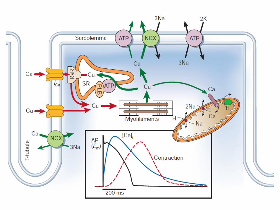
Myocyte changes
Extracellular matrix changes
Microvascular changes
Myocardial regeneration
Remodeling involves:

Hirsch E et al on behalf of WG Myocardial Function ESC Eur J Heart Fail (2013)
Cell-cell cross-talk

Cardiac myocyte

Physiological hypertrophy
Concentric hypertrophy
Eccentric hypertrophy
Increased expression of embryonic genes
Normal muscle cell
Growth stimuli
Apoptosis
Autophagy
Patterns of myocyte hypertrophy in myocardial remodeling

dilated cardiomyopathy
Beuckelmann D et al Circulation 1992


Serca2a
• Gene therapy (AAV1-Serca2a) • Antagomir (miR-25) • SUMO-ylation

Del Monte et al Circulation 1999
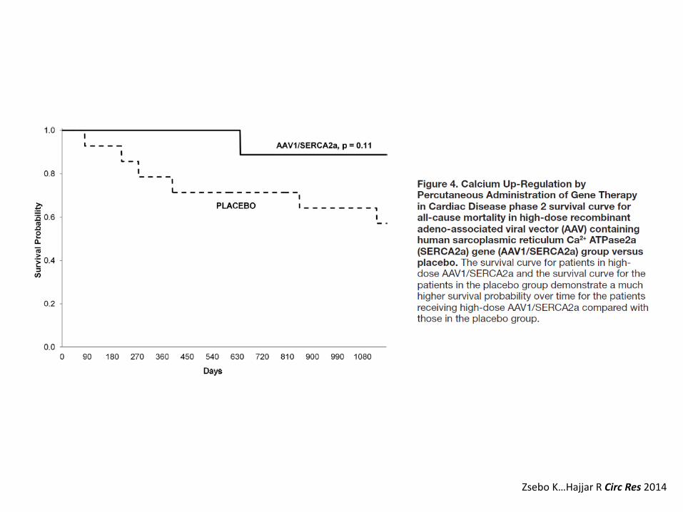
Zsebo K…Hajjar R Circ Res 2014


Myofilaments
• Myosin ATPase: omecantiv mecarbil

Malik F et al Science 2011
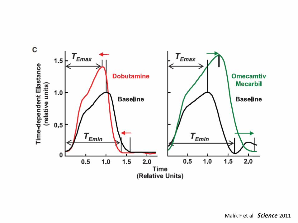
Malik F et al Science 2011

Cleland JGF et al Lancet 2011

Circ Heart Fail. 2015 May 29. pii: CIRCHEARTFAILURE.114.002152. [Epub ahead of print]


Mitochondria
• Cardiolipin protection by SS peptides

SS peptides protect mitochondria from oxidative damage


Membrane receptors
• The beta3-adrenergic receptor

Balligand et al. Proc Nat Acad Sci USA(1993)
β-adrenergic stimulation of NO Synthase attenuates the inotropic
effect
β1+2-adrenergic receptor blockade unveils a negative inotropic effect of
isoproterenol, a non-specific β-adrenergic agonist
Which β-adrenergic receptor ? By what mechanism ?
Gauthier et al. J Clin Invest (1996)

β3 AR coupling in mammalian heart
Gauthier et al J Clin Invest 1998:102 (7): 1377 Moniotte et al Circulation 2001: 103 (12); 1649 Dessy et al Circulation 2004;110(8):948 Moniotte et al Eur J Heart Failure 2007; 9 (12): 1163 Hammond J, Balligand JL, JMCC 2012 ; 52(2): 330-340 Belge C et al. Circulation 2014; 129: 451
β3AR
Gαi
NOS
NO
sGC
cGMP
β1AR
Gαs
Effect on remodeling ? 1. hypertrophy 2. coronary vasodilatation 3. fibrosis 4. metabolism ?

β3AR protects against cardiac hypertrophy PBS PE In vitro
1000
1200
1400
1600
1800
NRC
M a
era
(µm
2 )
AdV:GFP
*
Belge C*, Hammond J*, Dubois-Deruy E* et al, Circulation 2014 129, 451-62 AdV:hβ3AR
#
AdV:hβ3AR AdV:GFP
hβ3AR TG
#
hβ3AR TG
#
hβ3AR TG
#
hβ3AR TG
#
In vivo
Myo
cyte
are
a (µ
m2)
0 100 200 300 400 500 600 700
WT
*
saline Iso 50mg/kg/d
0 100 200 300 400 500 600 700
WT AngII
*
0 100 200 300 400 500 600 700
WT Iso 30mg/kg/d
*
0
250
500
750
WT
*
TAC 9weeks
Belge C et al. Circulation 2014;129:451-462

β3AR
Gαi
NOS
NO
sGC
cGMP
Targets downstream NOS/cGMP
PKG
Hypertrophy
Titin-P Troponin-I-P
Myocyte/tissue elasticity Myofilament Ca++ sensitivity
The cardiac fibroblast and myocyte-fibroblast cross-talk
$
*
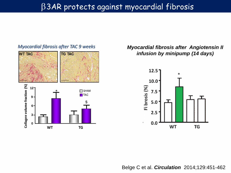
Myocardial fibrosis after TAC 9 weeks
WT TG 0
3
9
6
12
β3AR protects against myocardial fibrosis
Myocardial fibrosis after Angiotensin II infusion by minipump (14 days)
Belge C et al. Circulation 2014;129:451-462
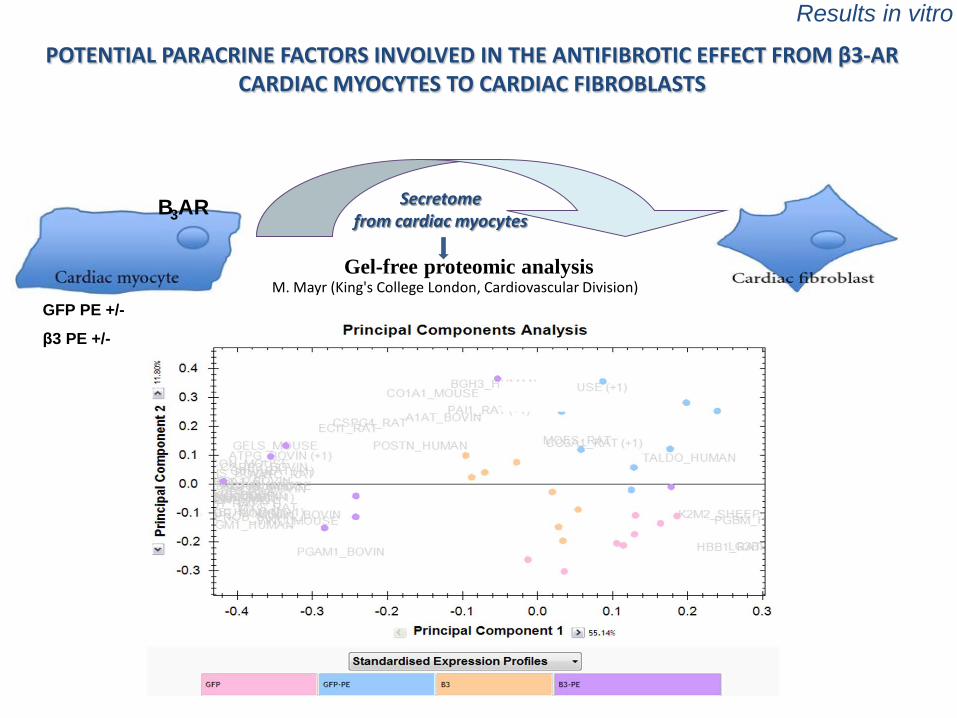
Results in vitro
POTENTIAL PARACRINE FACTORS INVOLVED IN THE ANTIFIBROTIC EFFECT FROM β3-AR CARDIAC MYOCYTES TO CARDIAC FIBROBLASTS
Secretome from cardiac myocytes
B3AR
GFP PE +/-
β3 PE +/-
M. Mayr (King's College London, Cardiovascular Division) Gel-free proteomic analysis

This project has received funding from the European Union’s Horizon 2020 research and innovation programme under grant agreement N° 634559

Membrane receptors
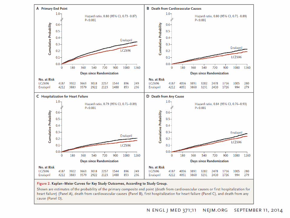
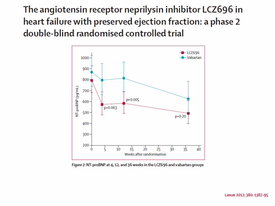
• Combined AT1-R antagonist and neprilysin inhibitor: LCZ 696


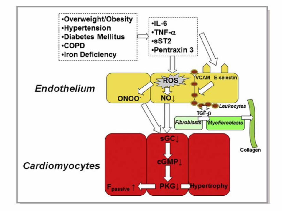
The endothelial cell and endothelial-myocyte cross-talk

RBC
EC
CM
Close apposition of cardiac myocytes and capillary endothelial cells

Lim SL et al Eur Heart J 2015


That’all
• Questions