the bap test and the global assessment - · pdf filethe bap test and the global assessment ......
TRANSCRIPT

Eugenio Luigi Iorio AA A
ss shh h
oo orr rtt t rr r
ee evv v
ii i ee eww w
The BAP test and the global assessment
of oxidative stress in clinical practice
RR Ree ell l ee eaa ass s ee e 44 4.. . 11 1 –– – 22 200 011 100 0

www.fras4evolvo.it 2

www.fras4evolvo.it 3
Table of Contents
Preface and acknowledgments P. 4 1 Introduction p. 5 2 Principle p. 6 3 Validation and comparative studies p. 8 4 Procedure p. 9 5 Analytical performances p. 10 6 Interpretation of results p. 12 7 Clinical studies on Humans p. 13 8 Clinical studies on Animals p. 16 9 Experimental studies in vitro p. 17 10 Indications and clinical usefulness p. 17 11 Concluding remarks p. 18 References P. 19
Info P. 24

www.fras4evolvo.it 4
Antioxidants play a relevant role in the biochemical homeostasis of our body by defending
our cells and tissues against potentially toxic chemical oxidants species, including free radicals and
reactive oxygen species (ROS).
Therefore, in the light to prevent/to treat the possible unwanted effects of oxidative stress,
many peoples currently take antioxidant supplements with the aim to restore and/or to maintain
an optimal level of antioxidant defense effectiveness.
However recent studies and clinical experience suggest that any antioxidant supplement
should be taken only after a biochemical evidence of its deficiency. Indeed high doses of some
antioxidants may result pro-oxidant.
On these basis, in order to establish the clinical indications for antioxidant treatment and to
avoid concomitantly the unwanted side effects of unneeded supplements, some years ago the
Italian researcher Mauro Carratelli developed a new test, i. e. the BAP test, where the initials mean
Biological Antioxidant Potential. This test is based on the ability of a plasma blood sample to
reduce ferric ions to ferrous ions as a FRAP assay (ferric reducing ability of plasma). However
compared to the classical FRAP assay firstly described by Benzie, BAP test appears quicker and
simpler to perform. Moreover according to the novel concept of global oxidative stress assessment,
the BAP test showed able to integrate the results of the d-ROMs test, which allows the assessment
of serum/plasma total oxidant capacity.
The aim of the present book was to describe the principle, the validation and the clinical
application of BAP test in Human and Veterinary Medicine.
We acknowledge the help of all Authors of clinical and experimental studies reported in this
book and, particularly, Mauro Carratelli, the “inventor” of BAP test.
Dr. Eugenio Luigi Iorio, MD, PhD
The President of International Observatory of Oxidative Stress

www.fras4evolvo.it 5
1. Introduction
The blood of Vertebrates contains several compounds/activities which are able, taken together, to oppose the oxidant potential of oxidant chemical species, like the reactive oxygen species (ROS) (1).
Virtually, any “endogenous” compound (i. e. albumin, transferrin, ceruloplasmin, bilirubin, uric acid, reduced glutathione, etc.) or “exogenous” biomolecule (i. e. tocopherols, carotens, ubiquinol, ascorbate, methionine, flavonoids, polyphenols, etc.) which is able to give equivalent reducing units (e. g. electrons) is able also to block the potential damage of ROS. Indeed, the reactivity of a free radical is just the result of a lack of electrons (2).
Of course, any injury to such “blood barrier to oxidation” – e. g. exposure either to radiations, or xenobiotics or infectious agents – can result in oxidative tissue damage and, therefore, in early aging and in the so-called oxidative stress related diseases (e. g. atherosclerosis, arterial hypertension, stroke, myocardial infarction, diabetes, arthritis, dementia, colitis, pancreatitis, respiratory diseases, cancer, infections, etc.) (3).
The velocity of these processes can be slowed down not only by specific pharmacotherapies (e. g. antihypertensive or lipid-lowering drugs), but also by modifying some lifestyles (e. g. diet, exercise, etc.) and, when indicated, by the intake of compounds with antioxidant activity (4).
However, before any medical intervention, the antioxidant effectiveness of plasma barrier to oxidation should be assessed, in order to limit the eventual intake of antioxidants only to patients showing a reduced antioxidant plasma activity (5). In fact, an uncontrolled intake of antioxidants has been shown to induce a paradoxical unwanted pro-oxidant action (6, 7).
Therefore, the assessment of antioxidant activity of plasma/serum blood is the first step of any medical intervention aimed to redress the oxidative balance in oxidative stress related conditions.
Over the last few years, a number of tests based on different chemical principles has been
proposed in order to assess the whole antioxidant activity of plasma or serum blood (5, 8-18).
In this subject, one of the most know examples of oxidation in Nature is the change of iron from ferrous to ferric form, as it happen in the generation of the rust. Therefore one can consider as antioxidant a solution, such the serum/blood plasma, which is able to bring back the iron from its ferric form to the ferrous form.
In other words from the merely analytical point of view, the effectiveness of antioxidant plasma barrier can be evaluated by testing its ability to reduce a specific substrate, i. e. by assessing its ability to give oxidant agents (e. g. free radicals) to one or more electrons.
For this purpose, several chemical reducing-oxidizing couples are available. For instance, transition metals (i e. iron) exhibit the property to receive one electron thus shifting from the oxidized state (Fe3+) to reduced state (Fe2+). Such agents can work as “meters” in tests aimed to assess antioxidant power of biological systems. Indeed, the so-called “plasma antioxidant power” is ultimately a measure of the reducing or “electron-giving” activity of blood plasma.
On the other hand, some compounds share the ability to change their absorbance just when bound to compounds able to switch from the oxidized to reduced state. For instance, some thiocyanates are able to reversibly shift from colored to uncolored derivatives, in the presence of ferric or ferrous salts, respectively (19). Such “chromogens” can work as excellent “detectors” when coupled with adequate “oxidizing/reducing meters” in a test designed to assess antioxidant activity of biological samples.
Indeed, when a ferric salt is dissolved in a uncolored solution containing a particular thiocyanate derivative, the resulting solution becomes red, as a function of the ferric ions concentration. This phenomenon is ascribable to the formation of a complex between ferric salt and thiocyanate. Further adding of a small amount of blood plasma will reduce ferric ions

www.fras4evolvo.it 6
to ferrous ions thus making uncolored the initial red solution. Such a chromatic change – may be due to the release of ferrous ions by the colored thiocyanate complex – can be read by means of a photometer, previously set at the wavelength of chromogen.
Therefore, the entity of absorbance change will directly correlate with the antioxidant “potential” of blood plasma against the specific substrate which has been used as oxidant/detector (ferric ions). In other words, the ability of the tested plasma to reduce ferric to ferrous ions may provide a direct measure of the ability of a sample of such plasma to give reducing equivalents and then neutralize chemical species lacking of electrons, like ROS
– obviously in the reduction-oxidation potential range of chosen oxidant-reducing couple (Fe3+/Fe2+).
On the basis of this background, a novel test, aimed to assess the antioxidant plasma activity, has been recently developed by the Italian Chemist Mauro Carratelli and first described by Dohi et al. (20).
This test has been called “BAP” test which initials underline that the assay is designed to assess the Biological Antioxidant Potential of blood plasma.
The aim of this short review is to present the principle, the validation, the procedure, the indications and clinical usefulness of the BAP test in the light of the available literature.
2. Principle
BAP test is based on the ability of a colored solution, containing a source of ferric (Fe3+) ions adequately bound to a special chromogenic substrate, to decolour when Fe3+ ions are reduced to ferrous ions (Fe2+), as it occurs by adding a reducing/anti-oxidant system, i. e. a blood plasma sample (20).
Therefore, in the BAP test, a small amount of blood plasma (10 µL) to be tested is dissolved in a colored solution, which has been previously obtained by mixing a source of ferric ions (i. e. ferric chloride, FeCl3, the R2 reagent) with a special chromogenic substrate (i. e. a thiocyanate derivative, the R1 reagent) (19).
After a short incubation (5 min), at 37 °C, such solution will decolor and the intensity of this chromatic change will be directly proportional to the ability of plasma to reduce, during the incubation, ferric ions (initially responsible for the color of solution) to ferrous ions, according to these reactions: 1. FeCl3 + AT(uncolored) → FeCl3 – AT(colored) 2. FeCl3–AT(colored)+ BP(e-) → FeCl2 + AT(uncolored) + BP
where: • FeCl3 is ferric chloride; • AT(uncolored) is a thiocyanate derivative (uncolored); • FeCl3 – AT(colored) is the colored complex of ferric
chloride with the thiocyanate derivative;
• BP(e-) is a molecule of blood plasma barrier with reducing/electron giving/antioxidant activity against ferric ions;
• BP is the oxidized form of BP(e-); • FeCl2 is the ferrous chloride obtained by the
reducing activity of BP(e-).
By assessing photometrically the intensity of decoloration, the amount of reduced ferric ions can be adequately evaluated thus allowing an effective measurement of reducing ability or antioxidant potential of tested blood plasma (20).
Such a “potential” is obviously not absolute but relative to the tested substrate, i. e. ferric ions. Considering that such ions are naturally occurring components of the body, BAP test, as its initials suggest (Biological Antioxidant Potential), provides a reliable measure of antioxidant power of the fraction of plasma barrier to oxidation which is directly involved – due to the implicated reducing-oxidant potentials – against the attack of reactive chemical species in “physiological” or “biological” conditions.
However, since the main aim of BAP test is to provide a global assessment of antioxidant defenses of our body, no studies has been performed in order to establish the exact contribution to the assay of each serum/plasma blood antioxidants, which amount should be detected by means of

www.fras4evolvo.it 7
different analytical approach (e. g. mass spectrometry, chromatography and so on).
In any case by considering that BAP test exploits the same principle of FRAP assay (21) i. e. the reduction of ferric to ferrous ions, it is likely that BAP test measures the same antioxidants of any assay based on iron reduction (Table 1).
Table 1. Relative activity of individual plasma antioxidants and their estimated contributions to total ferric reducing activity of plasma (modified by Benzie et al, 1996) (21).
Plasma antioxidants
Relative Activity1
Expected plasma level2
Estimated contribution3
Uric acid 2.0 (2.0-2.4) 150 – 450 60 Ascorbic acid 2.0 (1.9-2.1) 30 – 100 15
Proteins 0.1(0.1-0.15) 800 – 1100 10 α-Tocopherol 2.0 (1.7-2.1) 15 – 40 5
Bilirubin 4.0 (4.2-4.6) < 20 5 Others – – 5
1Measured range. 2Fasting concrentrations (µmol/L). 3To total iron-reducing plasma activity.
Indirect evidence that BAP test measures
at least some of these antioxidants has been provided by some experiments performed either on standard solutions or blood plasma samples.
For instance BAP test was able to detect in vitro the antioxidant potency of uric acid (UA) as ferric reducing capacity in solutions containing increasing amounts of UA (22) (Figure 1).
Figure 1. Effects of aqueous solutions of uric acid (UA) on BAP test in vitro. For each concentration UA in aqueous solution, BAP was measured three times, and values are presented as means ± SD. All differences among solutions were statistically significant (P<0.05) (modified by Nakayama et al, 2007) (22).
Similar results were obtained in analogous
experiments by using ascorbic acid (AsA) as antioxidant (22) (Figure 2).
Figure 2. Effects of aqueous solutions of ascorbic acid (AsA) on BAP test in vitro. For each concentration AsA in aqueous solution, BAP was measured three times, and values are presented as means ± SD. All differences among solutions were statistically significant (P<0.05) (modified by Nakayama et al, 2007) (22).
However, when UA and/or AsA were added ex vivo to a plasma samples from patients who underwent haemodialysis no further increase of BAP was shown (22) (Figure 3). Figure 3. Effects of the addition of uric acid (UA) and ascorbic acid (AsA) to serum on BAP ex vivo. In samples drawn at the post-haemodialysis time point from eight patients, each sample was evaluated for BAP, with a 5-mg/dL increment of UA (+UA) and 5-µg/mL of AsA (+AsA), and a combination of the two (+UA+AsA). Values are presented as means ± SD. No significant differences wee found among the solutions (modified by Nakayama et al, 2007) (22).
This datum may indicate that BAP test provides a reliable measure of all reducing compounds/activities of a blood plasma sample rather than a specific antioxidant. This should be an advantage of BAP test. In fact a minor criticism against BAP test is that BAP test results seems to be too closely related to UA concentration, so that a determination of UA should be done concomitantly to BAP test. On the contrary the herein shown data suggest that BAP test is able to detect many major ferric reducing compounds/activities including UA and AsA but each reducing agent like UA alone or combined (UA+AsA) seems to don’t influence significantly the whole plasma iron reducing activity as measured by BAP test.
0
400
800
1200
1600
µE
q/L
0 10 20 30mg/dL
0
400
800
1200
1600
µE
q/L
0 10 20 30mg/dL
0
400
800
1200
1600
µE
q/L
0 10 50 100µµµµg/mL
0
400
800
1200
1600
µE
q/L
0 10 50 100µµµµg/mL
1400
1800
µE
q/L
P-HD serum +UA +ASA +UA+ASA
2200
2600
1400
1800
µE
q/L
P-HD serum +UA +ASA +UA+ASA
2200
2600

www.fras4evolvo.it 8
The result of a clinical study on hyperbaric therapy seems to confirm this hypothesis (23) (see below, clinical studies on Humans and Figure 11 ). In any case BAP test was able to detect in vitro increased amounts of bilirubin a minor iron-reducing agent (20) (Figure 4).
Figure 4. Effects of solutions of bilirubin on BAP test in vitro (modified by Dohi et Al, 2005) (20).
Finally, experiments ex-vivo performed on human plasma showed a direct and significant correlation between BAP test values and α-tocopherol levels (24) (Figure 5).
Figure 5. Correlation between BAP test values and α-tocopherol concentration in a sample of rat blood plasma (modified by Dohi et al, 2007) (24).
Taken together the evidence now available indicates that BAP test provides a reliable measure of all the antioxidant plasma compounds/activities able to reduce ferric ions to ferrous ions including UA, AsA, bilirubin and α−tocopherol, as previously described for FRAP assay (21) (see also below, paragraph 3).
3. Validation and comparative studies
The validation of BAP test has been achieved by means of experiments performed with the Electron Spin/Paramagnetic Resonance Spectrometry (ESR/EPR) that is considered the “golden standard” technique to study free radicals ex vivo.
According to its principle, BAP test provides an evaluation of the whole antioxidant capacity of blood plasma, measured as reducing potential against ferric ions.
Several compounds may contribute to this “biological antioxidant potential” of whom some – like bilirubin (see Table 1) – exhibits a “scavenger” activity against free radicals, having the ability to neutralize directly free radicals.
On these basis, an ESR/EPR study performed showed that the bilirubin exhibits “in vitro” direct scavenging activity against either the hydroxyl radical (HO•), the most potentially dangerous oxygen free radicals in living organisms, or the 1,1-diphenylpicrylhydrazyl radical already at physiological conditions (20).
In experiments performed in parallel with the BAP test it was shown that the results of both assays (as conducted either by ESR/EPR
or photometry) overlapped (20) (Figure 6).
Figure 6. Comparison between photometric BAP assay (histograms) and Electron Spin Resonance Spectrometry (ESR/EPR) (box): increasing concentrations of bilirubin are able to increase BAP test values (because bilirubin acts an antioxidant/iron reducing compound) and to reduce concomitantly ESR/EPR signal (because bilirubin acts as a scavenger against hydroxyl radical) (modified by Dohi et al, 2005) (20).
By considering that ESR/EPR is the reference technique to study the free radicals, BAP test must be considered, thanks to this authoritative validation, a test really able to detect and quantify in a specific and suitable manner scavenging/antioxidant activities, as a confirmation of the above proposed principle of its mechanism.
0
2000
4000
6000
8000
10000
µE
q/L
Control 0.1 0.5 1.0 2.0 5.0
Bilirubin (mg/dL)
0
2000
4000
6000
8000
10000
µE
q/L
Control 0.1 0.5 1.0 2.0 5.0
Bilirubin (mg/dL)
0
2000
3000
4000
5000
0 2
4
6
8
10
12
µµ µµEq/L
14
R=0.7
µµµµM

www.fras4evolvo.it 9
On the other hand, in experimental trials, it has been shown that the BAP results satisfyingly correlate with those of FRAP, which is considered the comparison method the most similar to BAP test, in the actual panorama of all the assays proposed to evaluate the plasma antioxidant capacity.
Indeed, the FRAP assay is based on the reduction of ferric tripyridyltriazine (FeIII-TPTZ) to ferrous tripyridyltriazine (FeII-TPTZ), which develops an intense blue color, phometrically measurable by setting the wavelength to 593 nm (21). The calibration may be performed by means of solutions with known concentrations of FeII (FeSO4
.7H2O). Analogously, the BAP test is based on the use of a colored solution which is obtained by adding a source of ferric ions to a chromogenic mixture.
The entity of decoloration is then assessed by measuring the absorbance at 505 nm, which is proportional to the reducing activity of the sample.
According to these observations, Vassalle (unpublished data) recently assessed comparatively analytical performance of BAP and FRAP test on 18 samples of human blood plasma.
According to the result of this study, mean value (± SD) of BAP test was 3163±770 µEq/L, while the mean value (± SD) of FRAP test was 521±132 µmol/L.
The correlation between test was shown to be acceptable in 16/18 tested sample (88.89%).
The remaining two samples were shown to not correlate, maybe to the higher level of plasma lipids, which can interfere with the photometric readings of every photometric test (Figure 7).
Figure 7. Comparative evaluation of BAP test and FRAP assay (modified by Vassalle et al, 2006) (Unpublished data).
4. Procedure
BAP test can be performed either on
whole blood or heparinised blood plasma or serum. Such samples should be fresh, in order to avoid an eventual underestimation of the results due to possible auto-oxidation phenomena.
Therefore, while waiting further data by studies in progress, it is suggested to centrifuge as soon as possible (within 1 hour) the blood and to recover plasma or serum. If the sample (plasma/serum) cannot be immediately processed it should be stored at +4 °C and tested within 1-to-3 days or according to the guidelines of laboratory good practice.
However very recent evidence suggests that BAP test maintains its analytical reliability even on previous frozen samples (at -80°C)
The BAP test can be performed with a common photometer, according to the exact standardized work conditions (Table 2).
Table 2. Work conditions of BAP test. Wavelenght Optical path Temperature Mode
505 nm 1 cm 37 °C Differential
However, due to some practical reasons,
the test can be performed also with dedicated analytical devices, like FREE system (DIACRON International s. r. l., Grosseto, Italy) (24), for blood plasma or serum, or FRAS 4 system (H&D s. r. l., Parma, Italy) (20) by starting form whole (capillary) blood. Both systems, which require dedicated kits, substantially limit manual intervention to a few steps compared to common photometers, thus reducing a possible source of analytical imprecision.
Moreover, either FREE or FRAS system allows a global assessment of oxidative stress, due to the possibility to perform not only the BAP test (antioxidant status assessment) but also the d-ROMs test (oxidant status assessment) (20, 24).
0
100
200
300
400
500
600
700
800
1500 2000 2500 3000 3500 4000 4500
FR
AP
(µµ µµ
mo
l/l)
FRAP t
est
(µm
ol/L)
BAP test (µEq/L)

www.fras4evolvo.it 10
The FREE system is an “open” system consisting of a photometer and a dry thermostating block. Both the tools are managed by a computerized system able to receive, to elaborate and to export the data. The initials FREE means Free Radical Elective Evaluator, i. e. system for the elective study of free radicals.
The dedicated kit of the BAP test for FREE contains cuvettes ready-to-use with a solution of a thiocyanate derivative (R1 reagent) and a vial of ferric chloride (R2 reagent).
According to the procedure for the FREE system, at least 10 min before the analytical session, it is preferable to seat the cuvettes, ready to use for analysis, in the thermostated housing of instrument. It is also very important to bring the reagents to working temperature before the use.
When ready, the operator must select, through the keyboard of instrument, the pre-set channel.
For each analytical series, the procedure involves, before testing plasma or serum, the zeroing and the subsequent automatic subtraction of reagent blank absorbance.
Practically, after having adjusted the zero against distilled water in the photometer, 50 µL of R2 reagent (ferric chloride) is transferred into the cuvette containing the R1 reagent (a thiocyanate derivative). The resulting colored solution, after gently mixing by inversion, undergoes photometric reading. Then, 10 µL of sample is added in the same cuvette and the obtained solution is gently mixed and incubated into the thermostatic block for 5 min at 37 °C. After incubation, the sample can be read for absorbance.
FRAS 4 is a new integrated analytical system consisting of a dedicated photometer
with incorporate centrifuge designed to allow for the global assessment of oxidative stress on whole blood, by means of two tests, i. e. d-ROMs test and BAP test. The initials FRAS means Free Radical Analytical System, i. e. system for the analytical study of free radicals.
The most innovative technological feature of FRAS 4 is the integration of centrifuge with the analytical module. This system enables the operator to perform not only the centrifugation but also the photometric analysis.
The dedicated kit of BAP test for FRAS 4 is similar to that one of FREE system; moreover, it contains also the tools for taking the whole blood from a finger.
Practically, a small amount of plasma (10 µL), which has been obtained after centrifuging the whole blood sample (100-150 µL), is dissolved in the colored solution, previously prepared by mixing the R2 reagent (FeCl3) with R1 reagent (a thiocyanate derivative). After 5 min of incubation at 37 °C, the solution undergoes photometric reading.
To periodically calibrate FREE and FRAS 4 systems, a lyophilized human control serum is available with a known antioxidant title expressed as µmol/L.
Both devices directly provide the results of the BAP test – that is the “biological antioxidant potential” of plasma – as µEquivalents/L (µEq/L) (by using ascorbate as iron reducing agent).
Data of Producer from healthy individuals indicated initially that the optimal value of BAP test should be higher than 2.200 µEq/L. The studies performed in the last 3 years substantially confirmed this statement (see below), although every laboratory should determine its own reference value.
5. Analytical performances
Analytical performances were initially evaluated with a first set of experiments on standard solutions of ascorbic acid (AsA) with known values, by using a FREE equipment (Diacron International, Grosseto, Italy), in order to establish the lowest limits of detection and quantification, respectively, and linearity.
The lowest detection limit, i. e. the lowest concentration of a signal that can be distinguished by the blank (as calculated by multiplying 3 times the standard deviation, as obtained by the average of 10 replicated analyses of the blank) was 118 (mean value). The lowest quantification limit, i. e. the lowest

www.fras4evolvo.it 11
concentration by which a quantitative measure can be performed (as calculated by multiplying 10 times the standard deviation, as obtained by the average of 10 replicated analyses of the blank) was 393 µEq/L (mean value).
The BAP test showed linearity within a wide range of absorbance and the highest linearity limit, i. e. the concentration able to produce a signal lower than 3.0-3.5% compared to that predicted by regression (straight) line, was 20.000 µEq/L (Table 3 and Figure 8).
Table 3. Linearity data from BAP test.
µEq/L Measured ∆Abs Predicted ∆Abs %
40000 1,307 2,649 50,660236
20000 1,339 1,349 -0,739745
16000 1,114 1,089 2,2781006
13400 0,938 0,920 2,0111852
10000 0,678 0,699 -2,961637
8000 0,566 0,569 -0,463358
4000 0,310 0,309 0,4503123
Figure 8. Linearity of BAP test (data from human plasma).
A second set of experiments was
performed on a control human serum with know value, by using a FRAS equipment (H&D srl, Parma, Italy) in order to establish the coefficient of variation (CV).
To reach this goal a human-derived lyophilised control serum (know value 2100 µM) was repeatedly (n=10) tested after dilution in distilled water for BAP test in two different days.
The within-run CV was 2.72% and 2.91%, hence lower than 5%, in both the experiments done in two different days, respectively (Table 4).
Table 4. Determination of BAP test repeatability. First experiment Second Experiment
Sample (n) BAP test* Sample (n) BAP test*
1 2168 1 2105 2 2112 2 2095 3 2190 3 2136 4 2064 4 2193 5 2103 5 2004 6 2231 6 2057 7 2182 7 2037 8 2081 8 2100 9 2125 9 2090
10 2063 10 2192
MEAN 2132 MEAN 2101 SD 57,9 SD 61,1 CV (%) 2,72 CV (%) 2,91
*Results as µEq/L.
The above findings demonstrate that the
BAP test exhibits an acceptably low intra-assay coefficient of variation. Moreover, the very similar values obtained in two different days suggest that between-run CV of BAP test should be acceptably low. Noticeably, the concordance between expected (2100 µEq/L) and real results (2132±58 µEq/L and 2101±61 µEq/L) is evident.
A third set of experiments was done in order to establish CV on blood plasma either freshly keep or after frozen. With this aim, Benedetti (unpublished data) performed the BAP test on 12 samples of heparinised fresh plasma from apparently healthy volunteers (blood donors from the Transfusion Centre of Urbino’s Hospital, Urbino, Italy). After repeating the test three times in the same day, the BAP test showed a mean value 2169 ± 105 µM (mean ± SD) and within run coefficient of variation (CV) 4.8%. Two days later the Author repeated the test on the same samples stored at +4°C. The BAP test showed a mean value 2155 ± 118 µEq (mean ± SD) and a between-run CV 5.4%. Moreover, the BAP test as performed on frozen plasma samples (-20°C) showed overlapping results when compared to the results from fresh samples, thus suggesting the possibility to perform the assay also on previously stored biological materials.
More recently analytical performance of the BAP test – linearity, precision, repeatability,– was also evaluated on animals (see also below, Clinical Studies on Animals).
0
0,4
0,8
1,2
1,6
0 10000 20000 30000 40000
∆∆ ∆∆Abs
µEq/L
y= 0,000130x + 0,048979 R2 = 0,998927

www.fras4evolvo.it 12
In particular, 140 Labrador Retriever dogs, 75 females and 55 males, between 1 and 5 years old, fed with the same super-premium dry food and with the same lifestyle, were included in a study (25). All the subjects were in good health conditions at the physical and laboratory examination (haemogram, biochemical profile, urine analysis and serum protein electrophoresis) (25).
The BAP test showed linear in the tested interval (Figure 9), with a 0,95 “r” coefficient.
Figure 9. Linearity of BAP test (data from canine plasma) (modified by Pasquini et al, 2008) (25).
The recovery values of accuracy turned out
106,41%. The within-run CV WAS 6,4 %, while the between-run CV was 8,1% (25). These differences, compared to the lower data shown
in Humans, can be at least partially ascribable to pre-analytical errors. In fact, preliminary reports on rats indicate that the intake of food and/or antioxidants significantly increases the basal value of the BAP test. Therefore, it is mandatory to perform the BAP test in the same pre-analytical conditions of other biochemical tests, like blood glucose (the morning, at rest). This should significantly decrease any variability of the BAP test even in the same individual.
Taken together, these findings indicate that the BAP test shows good to excellent analytical performances as a reliable tool to assess antioxidant status in blood samples either from Humans or Animals.
These performances were shown improved recently by automating the test on a multiple analyzer (OLYMPUS 450) (data submitted for publication).
Interestingly, BAP test showed excellent repeatability on other biological matrices, like water-soluble antioxidant formulas, with an intra-assay coefficient of variation 1.9% (see below, Studies in Vitro).
At the moment, the only described interfering factor as well as for all photometric assays is a very high blood level of lipids (see below).
6. Interpretation of results
According to the data of the Producer (Diacron International, Grosseto, Italy), the “optimal value” of BAP test in healthy peoples should be at least 2.200 µEq/L.
Benedetti et al. confirmed this finding, by showing values 2169 ± 105 µEq/L on plasma blood samples (n=12) from apparently healthy peoples, selected among a more large population of healthy blood donors (unpublished data).
Noticeably these values are very similar to these very recently reported by Nagata et al. in 50 healthy volunteers (2137 ± 228 µEq/L) (26).
Therefore, the normal range proposed by the producer is substantially confirmed by these preliminary data (see also below, Clinical
Studies on Humans). In other words, values lower than 2.200
µEq/L indicate a reduced “biological potential” and hence a decreased effectiveness of the antioxidant plasma barrier, according to an arbitrary scale of severity (Table 5).
Table 5. BAP test results and antioxidant barrier impairment.
BAP test results (µEq/L)
Impairment degree of plasma antioxidant barrier
2.200 – 2.000 Border line condition 2.000 – 1.800 Slight reduction 1.800 – 1.600 Moderate reduction 1.600 – 1.400 Strong reduction
< 1.400 Very strong reduction
Optimal value > 2.200 µEq/L
0.00
500.00
1000.00
1500.00
2000.00
2500.00
0 20 40 60 80 100 120
µµ µµEq/L
%
R2=0.95
www.fras4evolvo.it 13
Preliminary data also suggests that elderly peoples have results of the BAP test lower than healthy adults (unpublished data).
Interestingly, dogs showed a normal range very similar to that one of Humans (2446 ± 585 µEq/L) with a normal-like distribution of all values, ranging from 1440 to 3260 µEql/L (25) (Table 6).
Table 6. Referral range of BAP test in dogs (modified by Pasquini et al, 2008) (25).
Units µmol/L Kurtosis – 1.4
Skewness – 0.2 Media 2466
Mediana 2610 SD 585
Reference range 1440 – 3260
7. Clinical studies on Humans
The clinical usefulness of the BAP test in Humans bas been demonstrated in several studies, either in physiological or pathological conditions. Most of these papers were performed by coupling d-ROMs test with BAP test. However, because this short review is focused on BAP test we will describe particularly clinical studies on this test with a reference to d-ROMs test when indicated.
Due to the close relationships between oxygen pressure and oxidative stress preliminary studies evaluated BAP test in conditions mimicking hyperoxia.
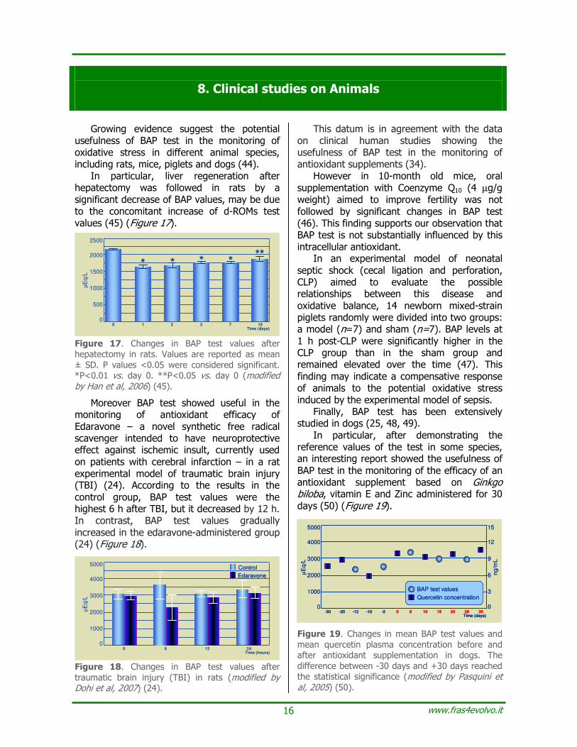
It has been suggested that hyperbaric oxygen (HBO) causes oxidative stress through the production of reactive oxygen metabolites, as also detected by d-ROMs test (27). However some patients with decompression illness (DCI) need repeated HBO treatments which can lead to concerns about an accumulation of oxidative stress (28). Therefore 9 DCI patients were exposed to two session of U. S. Navy Treatment Table 6 at an interval of 3 days (23). BAP significantly increased after the 1st and after the 2nd HBO compared with before 1st HBO (23) (Figure 10). Figure 10. Significant increase of BAP test values after two session of hyperbaric oxygen therapy (n=9, means ± SE, one-way repeated-measures ANOVA followed by Dunnett’s test) (modified by Kongoji et al, 2007) (23).
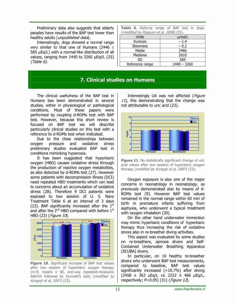
Interestingly UA was not affected (Figure 11), this demonstrating that the change was not attributable to uric acid (23).
Figure 11. No statistically significant change of uric acid values after two session of hyperbaric oxygen therapy (modified by Kongoji et al, 2007) (23).
Oxygen exposure is also one of the major
concerns in neonatology in neonatology, as previously demonstrated also by means of d-ROMs test (9). However BAP test values remained in the normal range within 60 min of birth in premature infants suffering from asphyxia, who underwent a typical treatment with oxygen inhalation (30).
On the other hand underwater immersion may mimic hyperbaric conditions of hyperbaric therapy thus increasing the risk of oxidative stress also in re-breather diving activities.
This aspect was evaluated by some studies on re-breathers, apnoea divers and Self-Contained Underwater Breathing Apparatus (SCUBA) divers.
In particular, on 10 healthy re-breather divers who underwent BAP test measurements, compared to baseline, BAP test values significantly increased (+10.7%) after diving (2458 ± 363 µEq/L vs. 2222 ± 466 µEq/L, respectively; P<0.05) (31) (Figure 12).
0
500
1000
2500
µE
q/L
First Session
Before
After
Second Session
1500
2000
P<0.05P<0.05
0
500
1000
2500
µE
q/L
First Session
Before
After
Second Session
1500
2000
P<0.05P<0.05
0
500
1000
2500
µE
q/L
First Session
Before
After
Second Session
1500
2000
P<0.05P<0.05P<0.05P<0.05
0
2
4
10
mg/d
L
First Session
Before
After
Second Session
6
8
0
2
4
10
mg/d
L
First Session
Before
After
Second Session
6
8
0
2
4
10
mg/d
L
First Session
Before
After
Second Session
6
8

www.fras4evolvo.it 14
Figure 12. Significant increase of BAP test values after diving (n=10, means ± SE, one-way repeated-measures ANOVA followed by Dunnett’s test) (modified by Yamami et al, 2007) (31).
On these basis, 4 apparently healthy
professional divers, one apnea diver and three Self-Contained Underwater Breathing Apparatus (SCUBA) divers were recruited in a preliminary study (32). When the results of the tests before immersion were compared with that after immersion, in the apnea diver total oxidative balance did not change. In the SCUBA divers BAP test results significantly increased from 2217±288 µEq/L to 2414±363 µEq/L (P=0.009) (32). Interestingly these values were very similar to those reported in the previous study on re-breathing divers (31).
Taken together the above data indicate that the potential oxidative stress related to hyperbaric condition seems to be compensate by an early increase of plasma antioxidant barrier to oxidation as reducing iron capacity measured by BAP test.
The possibility to evaluated blood plasma antioxidants in SCUBA divers may spread the potential usefulness of BAP test in some sports of whom is kickboxing, a martial art having fighting style and technique derived by the traditional Thai martial art, in which the use of punching, kicking, and, under some rules, kneeing, and elbowing are permitted, with possible injury (33).
Thus in a preliminary study 10 apparently healthy male individuals, half-professional players of martial arts, mainly kickboxing, 27.5 years aged (range 20 to 30 yrs) were recruited for an observational period of two months (34). At the time of the inclusion in the study (T0), after the first month (T1) and at the end of the second month (T2) all the subjects – after giving their informed consensus – undergo oxidative stress evaluation by means of d-ROMs test and BAP test (34). During the second month only, all the subjects were
supplemented with Cucumis melo concentrated juice (36 mg/day), vitamin C (240 mg/day) and vitamin E (24 mg/day) according to a commercially available formula (34).
The oxidant status, at the time T0, as measured by means of the d-ROMs test increased from 292.00±50.26 CARR U (before the training) to 362.20±38.00 CARR U (24 hours after the training) and decreased at the end of study, T2, to 234.60±27.76 CARR U. All the changes were statistically significant (T0, before vs. after training, P=0.0000011770; T0, before training, vs. T2, P=0,0000408367; T0, after training, vs. T2, P=0,0000000035) (34).
The antioxidant status, as measured by means of BAP test, increased from 2759.00±556.53 µEq/L to 2833.30±524.75 µEq/L although the change was not statistically significant (P=0,6516738232) (34) (Figure 13).
Figure 13. Changes in pro-oxidant status (d-ROMs test) and antioxidant status (BAP test) in kickboxing athletes who underwent antioxidant supple-mentation (n=10, means ± SD, t Test for paired data) (modified by Cavallini and Iorio, 2007) (34).
The above results, although in a small
sample of subjects, provide the first evidence in literature that martial art amateur, like other competitors, can undergo oxidative stress after a physical effort. However, an adequate supplementation with natural products, including powerful antioxidant vitamins C and E can favour, together a balanced diet and exercise training, the reduction of plasma biomarker of oxidative status (35).
This consideration on lifestyle suggests that BAP test can be useful also in medicine preventive and especially in psychosomatic medicine were a relationship between psychic stress and oxidative stress has been suggested.
In this subject, a low BAP test was recorded in college students with fatigue, unhealthy habits and irregularity. After
0
500
1000
2500
µE
q/L
Before diving After diving
1500
2000
P<0.05
0
500
1000
2500
µE
q/L
Before diving After diving
1500
2000
P<0.05
0
500
1000
2500
µE
q/L
Before diving After diving
1500
2000
P<0.05
T0 Bf
500
0
100
200
300
400
CA
RR
UProoxidant status (d-ROMs)
*Alla comparison statistacally significant vs Bf 3000
0
1000
1500
2000
2500
µm
ole
sre
duced
iro
n/L
Antioxidant status (BAP)
NSN=10
T0 Af T2
*
*
*
T0 Bf T2T0 Bf
500
0
100
200
300
400
CA
RR
UProoxidant status (d-ROMs)
*Alla comparison statistacally significant vs Bf 3000
0
1000
1500
2000
2500
µm
ole
sre
duced
iro
n/L
Antioxidant status (BAP)
NSN=10
T0 Af T2
*
*
*
T0 Bf T2T0 Bf
500
0
100
200
300
400
CA
RR
UProoxidant status (d-ROMs)
*Alla comparison statistacally significant vs Bf 3000
0
1000
1500
2000
2500
µm
ole
sre
duced
iro
n/L
Antioxidant status (BAP)
NSN=10
T0 Af T2
*
*
*
T0 Bf T2

www.fras4evolvo.it 15
specific advise on lifestyle the score of a specific visual analogical scale (VAS) for fatigue significantly increase with a trend to BAP increased values (although the change did not reach the statistical significance cut-off) (36).
No significant changes were noted in the results of the BAP test after acupuncture in spite of the decrease of d-ROMs test values (37) while Japanese females suffering from hypotension showed increase values of BAP test (38).
Postmenopausal women also showed significantly higher BAP test values compared to men in a clinical trial performed on metabolic syndrome (2531±323 µEq/L vs. 2345±245 µEq/L, respectively; P<0.05.) (39).
Interestingly in a case report a 14-years-old boy suffering from fibromyalgia (an intractable and painful syndrome that is accompanied by many symptoms) after treatment with Yi-Gan San extract for one week reported a significant increase of BAP test values and a decrease of pain subside as VAS from 7.5 cm to 3.2 cm (40).
The result of this study suggest that BAP test can be useful to monitor the effectiveness of antioxidant supplements as hypothesised in kickboxing players (34) (see also below, Clinical Studies on Animals).
In fact in a preliminary report on 20 patients suffering from neurological and rheumatic chronic diseases, oral supplementation with the herbal product GinpentTM (Gimnostemma pentaphyllum) plus fermented Papaya plus reduced glutathione, was followed after one month of treatment by a significant decrease of d-ROMs test and a significant increase of BAP test (41) (Figure 14).
Figure 14. Changes in pro-oxidant status (d-ROMs test) and antioxidant status (BAP test) in patients suffering from chronic diseases who underwent antioxidant supplementation (n=20, means ± SD, t Test for paired data) (modified by Iorio, 2006) (41).
On the other hand in patients with neurological trauma, BAP test was shown a reliable assay to demonstrate the scavenging activities and antioxidant potency of bilirubin even at physiological concentrations (20). This finding was in agreement with parallel experimental results obtained with Electron Spin Resonance Spectroscopy (20) (see also Figure 6). However patients suffering from Williams syndrome, a rare genetic condition which causes developmental problems, including attention deficit (42), showed normal BAP test values in spite of the higher values of d-ROMs test results compared to healthy controls (Figure 15) (unpublished data).
Figure 15. Oxidative balance in patients suffering from Williams syndrome compared to healthy controls (values are expressed as means ± SD, t Test for paired data) (Iorio and Carratelli, 2006, unpublished data).
Finally, BAP test showed useful to monitor oxidative stress in chronic obstructive pulmonary disease before and after the exacerbation (43) and in hemodialysis (HD) (22). In fact BAP test values were shown to decrease significantly in 8 patients after HD (Figure 16) (22).
Figure 16. Changes in BAP test values between pre-hemodialysis(HD) and post-HD serum in each patient (n=8) (modified by Nakayama et al, 2007) (22).
µµ µµEq/L
Before HD After HD
Before After
500
0
100
200
300
400
CA
RR
U
Prooxidant status (d-ROMs)
P=0,08
Before After
2500
0
500
1000
1500
2000
µE
q/L
Antioxidant status (BAP)
P<0.00007N=20
Before After
500
0
100
200
300
400
CA
RR
U
Prooxidant status (d-ROMs)
P=0,08P=0,08P=0,08
Before After
2500
0
500
1000
1500
2000
µE
q/L
Antioxidant status (BAP)
P<0.00007N=20
Controls (n=12) WS (n=9)
500
0
100
200
300
400
CA
RR
U
Prooxidant status (d-ROMs)
P<0,01
Controls (n=12) WS (n=9)
2500
0
500
1000
1500
2000
µm
ole
sre
du
ce
dir
on
/L
Antioxidant status (BAP)
NS
Controls (n=12) WS (n=9)
500
0
100
200
300
400
CA
RR
U
Prooxidant status (d-ROMs)
P<0,01
Controls (n=12) WS (n=9)
2500
0
500
1000
1500
2000
µm
ole
sre
du
ce
dir
on
/L
Antioxidant status (BAP)
NS
Controls (n=12) WS (n=9)
500
0
100
200
300
400
CA
RR
U
Prooxidant status (d-ROMs)
P<0,01P<0,01P<0,01
Controls (n=12) WS (n=9)
2500
0
500
1000
1500
2000
µm
ole
sre
du
ce
dir
on
/L
Antioxidant status (BAP)
NSNS
www.fras4evolvo.it 16
8. Clinical studies on Animals
Growing evidence suggest the potential usefulness of BAP test in the monitoring of oxidative stress in different animal species, including rats, mice, piglets and dogs (44).
In particular, liver regeneration after hepatectomy was followed in rats by a significant decrease of BAP values, may be due to the concomitant increase of d-ROMs test values (45) (Figure 17).
Figure 17. Changes in BAP test values after hepatectomy in rats. Values are reported as mean ± SD. P values <0.05 were considered significant. *P<0.01 vs. day 0. **P<0.05 vs. day 0 (modified by Han et al, 2006) (45).
Moreover BAP test showed useful in the monitoring of antioxidant efficacy of Edaravone – a novel synthetic free radical scavenger intended to have neuroprotective effect against ischemic insult, currently used on patients with cerebral infarction – in a rat experimental model of traumatic brain injury (TBI) (24). According to the results in the control group, BAP test values were the highest 6 h after TBI, but it decreased by 12 h. In contrast, BAP test values gradually increased in the edaravone-administered group (24) (Figure 18).
Figure 18. Changes in BAP test values after traumatic brain injury (TBI) in rats (modified by Dohi et al, 2007) (24).
This datum is in agreement with the data on clinical human studies showing the usefulness of BAP test in the monitoring of antioxidant supplements (34).
However in 10-month old mice, oral supplementation with Coenzyme Q10 (4 µg/g weight) aimed to improve fertility was not followed by significant changes in BAP test (46). This finding supports our observation that BAP test is not substantially influenced by this intracellular antioxidant.
In an experimental model of neonatal septic shock (cecal ligation and perforation, CLP) aimed to evaluate the possible relationships between this disease and oxidative balance, 14 newborn mixed-strain piglets randomly were divided into two groups: a model (n=7) and sham (n=7). BAP levels at 1 h post-CLP were significantly higher in the CLP group than in the sham group and remained elevated over the time (47). This finding may indicate a compensative response of animals to the potential oxidative stress induced by the experimental model of sepsis.
Finally, BAP test has been extensively studied in dogs (25, 48, 49).
In particular, after demonstrating the reference values of the test in some species, an interesting report showed the usefulness of BAP test in the monitoring of the efficacy of an antioxidant supplement based on Ginkgo biloba, vitamin E and Zinc administered for 30 days (50) (Figure 19).
Figure 19. Changes in mean BAP test values and mean quercetin plasma concentration before and after antioxidant supplementation in dogs. The difference between -30 days and +30 days reached the statistical significance (modified by Pasquini et al, 2005) (50).
0
1000
2000
3000
4000
5000
µE
q/L
0 6 12 24Time (hours)
Control
Edaravone
0
1000
2000
3000
4000
5000
µE
q/L
0 6 12 24Time (hours)
Control
Edaravone
Control
Edaravone
0
1000
2000
3000
4000
5000
µE
q/L
-30 -2Time (days)
0
3
6
9
12
15
-20 -12 -10 0 8 2810 18 20 30
ng/m
L
BAP test values
Quercetin concentration
0
1000
2000
3000
4000
5000
µE
q/L
-30 -2Time (days)
0
3
6
9
12
15
-20 -12 -10 0 8 2810 18 20 30
ng/m
L
BAP test values
Quercetin concentration
0
1000
2000
3000
4000
5000
µE
q/L
-30 -2Time (days)
0
3
6
9
12
15
-20 -12 -10 0 8 2810 18 20 30
ng/m
L
BAP test values
Quercetin concentration
BAP test values
Quercetin concentration
BAP test values
Quercetin concentration
0
500
1000
1500
2000
2500
µE
q/L
100 1 2 3 7Time (days)
* * * ***
0
500
1000
1500
2000
2500
µE
q/L
100 1 2 3 7Time (days)
* * * ***

www.fras4evolvo.it 17
9. Experimental studies in vitro
A recent challenge for antioxidant supplements is the determination of antioxidant power in vitro. Indeed in the light to develop new formulas, the producer when possible should report on the label at least an indication on the ability of the formula to neutralize effectively one or more oxidants.
Among the available assays BAP test proven useful in the assessment of antioxidant capacity of some formulas destined to patients or healthy peoples (Table 7).
Table 7. BAP test values of some antioxidant formulas Formulas BAP test values N CV (%)
GIN PENTTM (1) 48500±2880 9 4.1 DEUTROSULFAZYMETM (2) 64747± 3660 8 5.7 DEUTROSULFAZYMETM (3) 235500± 9161 8 3.9 DEUTROSULFAZYMETM (4) 274500± 6009 8 2.2
ARAMENTM (5) 22440 ± 908 7 4.0 BAP test values are expressed as mean µEq/L±SD. N is the number of determinations. CV is the within-run coefficient of variation. (1) Gimnostemma pentaphyllum plus fermented papaya plus reduced glutathione (2) DeutrosulfazymeTM basic formula. (3) DeutrosulfazymeTM vitamin C-enriched formula. (4) DeutrosulfazymeTM multivitamins-enriched formula. (5) Herbal product based on Laminaria digitata, Condrus crispus, Sambucus nigra, Vaccinium myrtillus, Rosa canina and Feniculus vulgare Miller.
Among these was also DeutrosulfazymeTM (CELLFOODTM, NUSCIENCE CORPORATION,
US) a non-addictive, non-invasive, and completely non-toxic proprietary colloidal-ionic formula containing finest all-natural, plant-based organic substances like ionic minerals, enzymes, amino acids and deuterium sulphate as traces (51, 52).
More recently BAP test was performed comparatively on samples of hand-made beers against to industrial beers(Table 8) (Bianchi and Iorio, unpublished data).
Table 8. BAP test values and polyphenols levels in hand-made and industrial beers. Beer BAP test values* Polyphenols levels** Label 1 10035 583
Label 2 9750 517
Label 3 9655 631
Label 4 9645 904
Label 5 10375 700
Label 6 7280 477
Label 7 9515 451
Label 8 11040 938
Label 9 9860 629
Label 10*** 6080 316 *As µEq/L. **As mg catechin/kg. ***Industrial
Interestingly hand made beers showed higher values of both BAP test and polyphenols compared to industrial beers. Noticeably BAP test values correlated with polyphenols content.
10. Indications and clinical usefulness
The BAP test, by exploiting the same chemical principle of the well-known FRAP test – i. e. the reduction of ferric to ferrous ions – provides a reliable measure of biological antioxidant potential of blood plasma (53).
Therefore, the BAP test should be performed with the aim of assessing the antioxidant status either in healthy peoples, in order to prevent oxidative stress, or in patients with oxidative stress-related diseases, in order to monitor the efficacy of specific and/or antioxidant therapies (54, 55).
All healthy peoples should undergo the BAP test because all the individuals are potentially
exposed to the risk of producing exaggerate amounts of free radicals, a phenomenon which requires a very effective antioxidant barrier. Indeed, the primary aim of this test is to identify and to prevent oxidative stress and its unwanted consequences (early aging, diseases) (56).
All the more so, the BAP test should be systematically performed on all clinically asymptomatic subjects, who are exposed for a number of reasons to factors able to reduce physiological antioxidant plasma defenses (e. g. inadequate exercise, unbalanced diet, chronic inflammatory diseases with reduced

www.fras4evolvo.it 18
absorption of nutrients, etc.). In these cases also, the aim of the BAP test is to identify and to prevent oxidative stress and its unwanted consequences (early aging, diseases) (57).
Moreover, the BAP test should be carried out on all patients with oxidative stress-related diseases (more than one hundred), such as Alzheimer’s disease, Parkinson’s disease, stroke, infarction, Crohn’s diseases, rheumatoid arthritis, AIDS, some cancers, etc. In all these cases, the aim of the BAP test is to monitor oxidative stress and to prevent its consequences, by monitoring the efficacy of specific therapy, either alone or with a concomitant antioxidant treatment (54). On this subject, it must be underlined that in many of above mentioned diseases, all almost having a chronic course, oxidative stress tends to raise the role of an additional health risk factor. Therefore, it must be controlled in order to optimize results of any treatment. For instance, the evidence, by means of BAP test, of a reduced biological antioxidant potential in hypertensive patients indicates a persistent impairment of physiological defenses against ROS, which can be responsible for an incomplete control of the current disease by the current anti-hypertensive treatment. Such a finding suggests to the clinician an integrated therapeutic approach, in which should be take place not only conventional, medical and/or surgical treatments, but also the correction of life-style and, eventually, the intake of antioxidants.
Finally, ideal candidates for the BAP test are all the subjects that undergo pharmacotherapy (e. g. with chemo-therapeuticals, hormone replacement therapy, contraceptive pill, etc.), surgical interventions (e. g. organ transplantation, by-pass, etc.), including dialysis, all conditions being able to favor oxidative stress by increasing the daily requirements of antioxidants (53, 54).
In all these conditions, the aim is to identify and to prevent oxidative stress and its consequences, and, particularly, to monitor the efficacy of eventual current measures with the ultimate objective of preventing oxidative tissue damage (56).
Furthermore, according to the guidelines of the International Observatory of Oxidative Stress, Free Radicals and Antioxidant Systems, the BAP test is very useful, together d-ROMs test, in the so-called global assessment of oxidative stress, now very easy to perform grace to a specific diagnostic algorithm (41, 58).
In this situation, when d-ROMs value is increased, an optimal value of BAP test should require the dosing of a single antioxidant. Alternatively, if such a dosing is not available, we suggest a mix of antioxidants according to the RDA tables. This should enable the antioxidant defenses to balance the increased oxidative status, as detected by the increased d-ROMs test values, especially when more than one risk factor is present (e. g. exposition to radiations, cigarette smoke, pollutants, etc.).
11. Concluding remarks
Scientific evidence shows that BAP test is
very useful in the assessment of antioxidant activity of blood plasma. A
According to the wide available scientific literature BAP test was validated by ESR/EPR and provided results that largely parallel to those of FRAP test, against which it appears quicker and simpler to perform. Indeed, the preparation and management of the FRAP reagents is a step somewhat complex, compared to that of BAP test, so that manual
procedure of FRAP test can increase analytical variability. Moreover, a further increase of analytical accuracy was recently reached by the application of BAP test to automatic analytical devices.
Finally, according to the novel concept of global oxidative stress assessment, the BAP test is a reliable assay able to integrate the results of d-ROMs test, which allows for the assessing of the total oxidant status of blood plasma.

www.fras4evolvo.it 19
References
1. Halliwell B, Gutteridge JMC. Free radicals in biology and medicine. 2nd Edn, Clarendon Press, Oxford. 1989. 2. Cadenas E, Packer L. Handbook of antioxidants. Marcel Dekker, Inc. 1996. 3. Halliwell B, Gutteridge JMC, Cross CE. Free radicals, antioxidants, and human disease: where are we now? J Clin Lab Med. 1992. 119: 598–620. 4. Valko M, Leibfritz D, Moncol J, Cronin MT, Mazur M, Telser J. Free radicals and antioxidants in normal physiological functions and human disease. Int J Biochem Cell Biol. 2007. 39 (1): 44–84. 5. Cao G, Prior RL. Comparison of different analytical methods for assessing total antioxidant capacity of human serum. Clinical Chemistry. 1998. 44 (6): 1309–1315. 6. Olson J L. Benefits and liabilities of vitamin A and carotenoids. J Nutr. 1996. 126: 1208S–1212S. 7. Schwartz JL. The dual role of nutrients as antioxidants and prooxidants: their effect on tumor cell growth. J Nutr. 1996. 126: 1221S–1227S 8. Wayner DDM, Burton GW, Ingold KU, Locke S. Quantitative measurement of the total, peroxyl radical-trapping antioxidant capacity of human blood plasma by controlled peroxidation. FEBS Lett. 1985. 187: 33–7. 9. Glazer AN. Phycoerythrin fluorescence-based assay for reactive oxygen species. Methods Enzymol. 1990. 186. 161–8. 10. Cao G, Alessio HM, Cutler RG. Oxygen-radical absorbance capacity assay for antioxidants. Free Rad Biol Med. 1993.14: 303–11. 11. Cao G, Verdon CP, Wu AHB, Wang H, Prior RL. Automated oxygen radical absorbance
capacity assay using the COBAS FARA II. Clin Chem. 1995. 41: 1738–44. 12. Rice-Evans C, Miller NJ. Total antioxidant status in plasma and body fluids. Methods Enzymol. 1994. 234: 279–93. 13. Miller NJ, Rice-Evans C, Davies MJ, Gopinathan V, Milner A. A novel method for measuring antioxidant capacity and its application to monitoring the antioxidant status in premature neonates. Clin Sci. 1993. 84: 407–12. 14. Ghiselli A, Serafini M, Maiani G, Assini E, Ferro-Luzzi A. A fluorescence-based method for measuring total plasma antioxidant capability. Free Rad Biol Med. 1994. 18: 29–36. 15. Whitehead TP, Thorpe GHG, Maxwell SRJ. Enhanced chemiluminescent assay for antioxidant capacity in biological fluids. Anal Chim Acta. 1992. 266: 265–77. 16. Trotti R, Carratelli M, Barbieri M, Micieli G, Bosone D, Rondanelli P, Bo P. Oxidative stress and thrombophilic condition in alcoholics without severe liver disease. 2001. Haematologica. 86: 85-91. 17. Carratelli M, Porcaro R, Ruscica M, De Simone E, Bertelli AAE, Corsi MM. Reactive Oxygen Metabolites (ROMs) and prooxidant status in children with Down Syndrome. International Journal of Clinical Pharmacology Research. 2001. 21 (2): 79-84. 18. Brambilla G, Fiori M, Archetti LI. Evaluation of the oxidative stress in growing pigs by microplate assays. J Vet Med A. 2001. 48: 33-38. 19. MERCK CHEMICAL Database. 2003. 20. Dohi K, Satoh K, Ohtaki H, Shioda S, Miyake Y, Shindo M, Aruga T. Elevated plasma levels of bilirubin in patients with neurotrauma reflect its pathophysiological role in free radical scavenging. In Vivo. 2005. 19 (5): 855–860.

www.fras4evolvo.it 20
21. Benzie IFF, Strain JJ. The ferric reducing ability of plasma (FRAP) as a measure of “antioxidant power”: the FRAP assay. Anal Biochem. 1996. 239: 70-76. 22. Nakayama K, Terawaki H, Nakayama M, Iwabuchi M, Sato T, Ito S. Reduction of serum antioxidative capacity during haemodialysis. Clin Exp Nephrol. 2007. 11: 218–224. 23. Kongoji J, Yamami N, Togawa S, Suzuki N, Yamamoto K, Mano Y. Indexes of oxidative stress during HBO treatment. Proceedings Undersea and Hyperbaric Medical Society. Scientific Meeting & Associates/BNA. Annual Scientific Meeting. 22-24 June 2007. Orlando, Florida (USA). 2007. P 112. 24. Dohi K, Satoh K, Nakamachi T, Yofu S, Hiratsuka K, Nakamura S, Ohtaki H, Yoshikawa T, Shioda S, Aruga T. Does Edaravone (MCI-186) act as an antioxidant and a neuroprotector in experimental traumatic brain injury? Antioxid. Redox Signal. 2007. 8: 281–287. 25. Pasquini A, Luchetti E, Marchetti V, Cardini G, Iorio EL. Analytical performances of d-ROMs test and BAP test in canine plasma. Definition of the normal range in healthy Labrador dogs. Vet Res Commun. 2008. 32 (2): 137–143. 26. Nagata K, Hasegawa T, Hirokado Y, Kiyama K, Otsuki C. Lifestyle-related diseases and the oxidative stress regulation system. Jpn J Psychosom Med. 2008. 4(3): 176-183. 27. Benedetti S, Lamorgese A, Piersantelli M, Pagliarani S, Benvenuti F, Canestrari F. Oxidative stress and antioxidant status in patients undergoing prolonged exposure to hyperbaric oxygen. Clinical Biochemistry. 2004. 37: 312–317. 28. Klingmann C, Gonnermann A, Dreyhaupt J, Vent J, Praetorius M, Plinkert PK. Decompression illness reported in a survey of 429 recreational divers. Aviat Space Environ Med. 2008. 79(2):123–128. 29. Buonocore G, Perrone S, Longini M, Vezzosi P, Marzocchi B, Paffetti P, Bracci R. Oxidative
stress in preterm neonates at birth and on the seventh day of life. Pediatric Research. 2002. 52 (1): 46–49. 30. Ezaki S, Kurishima C, Suzuki K, Kondo T, Tamura M. Resuscitation of preterm infants with reduced concentration of inspired oxygen. Proceedings 11th Annual Congress Perinatal Society of Australia and New Zealand (PSANZ 2007). 2007. April, 1–4. Melbourne Convention Centre. Melbourne, Victoria (Australia). 2007. 31. Yamami N, Nakayama H, Kongoji J, Togawa S, Nozawa T, Yagishita K, Shibayama M, Nakayama T, Suzuki N, Yamamoto K, Kawashima M, Mano Y. Free radicals and antioxidant potential in scuba divers. Proceedings Undersea and Hyperbaric Medical Society. Scientific Meeting & Associates/BNA. Annual Scientific Meeting. 22-24 June 2007. Orlando, Florida (USA). 2007. P 98–99. 32. Giganti MG, Iorio EL, Verna R, Zenobi R. Oxidative balance in scuba divers. Journal of Molecular and Clinical Pathology. 2007. 3: 101. 33. Zazryn TR, Finch CF, McCrory P. A 16 year study of injuries to professional kickboxers in the state of Victoria, Australia. Br J Sports Med. 2003. 37:448–51. 34. Cavallini M, Iorio EL. Effect of training and antioxidant supplementation on oxidative balance in martial arts. Journal of Molecular and Clinical Pathology. 2007. 2: 14-15. 35. Karlsson J. Antioxidants and exercise. Human Kinetics. 1997. 36. Nagata K, Kasuya N, Hayashi H. Oxidative stress and anti-oxidant potential of the students complaining chronic fatigue. Campus Health. 2006. 43 (2): 83-88. 37. Hirokado Y, Nagata K, Aoyama Y, Kiyama K, Okano K, Hasegawa. Prohomeostatic effects of acupuncture. 2006. International Congress Series. 2006. 1287: 107–110. 38. Hasegawa T. Oxidative stress and antioxidant potential in Japanese females with hypotension. Proceedings 19th World Congress

www.fras4evolvo.it 21
of Psychosomatic Medicine. Québec City (Québec) Canada. 2007. August 26–31. 2007. P 30. 39. Tsuchiya M, Tosaka M. There is a gender difference in the relationship between metabolic syndrome and oxidative stress among Japanese. Clinical Report. 2005. Masako Medical Clinic, Tokuyama, Japan. 40. Nagata K, Hasegawa T, Kiyama K, Aoyama Y, Okano K, Hirokado Y. A therapy for fibromyalgia syndrome using Yi-Gan San. Journal of Japanese Association of Oriental Psychosomatic Medicine. 2006. 21 (1-2): 46–47. 41. Iorio EL. A diagnostic algorithm to manage oxidative stress (original title “Un algoritmo diagnostico per la gestione dello stress ossidativo”). Proceedings International Congress of Antiaging Medicine. 2006, Starhotels Business Palace, 20th May. Milan. 2006. 42. Pober BR, Morris CA. Diagnosis and management of medical problems in adults with Williams-Beuren syndrome. Am J Med Genet C Semin Med Genet. 2007. 145(3):280-290. 43. Komatsu F, Kudoh H, Kagawa Y. Evaluation of oxidative stress and effectiveness of low-dose glucocorticoid therapy on exacerbation of chronic obstructive pulmonary disease. Journal of Gerontology. 2007. 62A (4): 459–464. 44. Iorio EL. Oxidative stress assessment in Animals. Actuality and perspectives [original title “La valutazione dello stress ossidativo negli animali. Attualità e prospettive”]. Proceedings “67° Convegno Nazionale Unione Zoologica Italiana”. Napoli (Italy). 2006, September 12nd-15th. Pp. 28. 45. Han SY, Chang EJ, Choi HJ, Nam SI, Lee NH, Kwak CS, Park SB, Kim HC, Mun KC. Total antioxidant status and oxygen free radicals during hepatic regeneration. Transplantation Proceedings. 2006. 38: 2214–2215. 46. Fujiwara T, Nakabayashi M, Goto M, Koyama S, Fujimoto A, Kugu K, Yano T,
Tsutsumi O, Taketani Y, Okamoto T. Preliminary study on action of coenzyme Q10 in female reproductive system. Proceedings 5th Conference of the International Coenzyme Q10 Association. 2007. November 9 to 12. Kobe Gakuin University at Port Island. Kobe, Japan. 2007. JP-039 (1–2). 47. Kakita H, Hussein Mh, Daoud Ga, Kato T, Murai H, Sugiura T, Mizuno K, Yamada Y, Ito T, Fukuda S, Kato I, Suzuki S, Togar H. Total hydroperoxide and biological antioxidant potentials in a neonatal sepsis model. Pediatric Research. 2006. 60 (6): 675–679. 48. Cardini G, Pasquini A, Luchetti E, Marchetti V, Voltini B. Free radicals oxygen (d-ROMs test) and Biological Antioxidant Potential (BAP test) assessment in serum: dog’s reference ranges [original title “La determinazione dei radicali liberi dell’ossigeno (d-ROMs test) e del potenziale antiossidante biologico (BAP test) nel siero: intervalli di riferimento nella specie canina”]. Proceedings LIX Convegno Nazionale – Società Italiana delle Scienze Veterinarie Viareggio (Italy). 2005, 21 – 24 September. 2005. 331–332. 49. Pasquini A, Luchetti E, Cardini G. Oxidative stress and recovery of hunting dogs [original title: “Stress ossidativo e tempi di recupero nel cane da caccia”]. Annali Fac Med Vet. 2006. LIX: 229–235. 50. Pasquini A, Simonetti P, Luchetti E, Bardana C, Cardini G. The Ginkgo biloba in the diet of ageing dog. [original title “Il Ginkgo biloba nella dieta del cane anziano”]. Praxis Veterinaria. 2005. 26 (2): 1-10. 51. EL. DeutrosulfazymeTM (CELLFOODTM). A clinical pharmacological overview [original title Deutrosulfazyme® (CELLFOOD®). Overview clinico-farmacologica]. Proceedings International Conference Safety Evaluation of Complementary and Alternative Medicine. 2003. Empoli (Italy). 2003, October 24 – 25. 52. Iorio EL, Bianchi L, Storti A. Deutrosulfazyme®: a powerful antioxidant [original title “Deutrosulfazyme®: un potente antiossidante”]. La Medicina Estetica. 2006. 30 (1): 115 – 116.
www.fras4evolvo.it 22
53. Carratelli M, Iorio EL, Bianchi L. Methods to measure the oxidative stress [original title “Metodi di misurazione dello stress ossidativo”]. ADI Magazine. 2006. 4 (10): 405 – 414.
54. Iorio EL. Carratelli M, D’Amicantonio T. Oxidative stress and diseases [original title “Stress ossidativo e malattia”]. ADI Magazine. 2006. 4 (10): 399 – 404.
55. Pece A, Marchetti M, Pallotti R. The free radicals [original title: “I radicali liberi”]. SOOFT Italia Ed. 2006. 56. Iorio EL. Oxidative stress and ageing [original title “Stress ossidativo e senescenza”]. In “Il manifesto della lunga vita. La rivoluzione della medicina predittiva. Marandola P, Marotta F. Sperling and Kupfer. 2007. 318–322.
57. Iorio EL. Oxidative stress and sport [original title: “Stress ossidativo e sport”]. European Journal of Health, Sport and Nutrition. 2007. 1: 102–103.
58. Iorio EL, Cinquanta L, Pisano R. A diagnostic algorithm to manage oxidative stress. Australasian J Cosmet Surg. 2006. 2 (1) : 26-30. 59. Bertone M, Rizzo M, Assorgi R, Muscatello A, Crisostomi S, De Blasis E, Di Giacinto B, Spataro A, Iorio EL. Plasma oxidant and antioxidant capacity at rest, after exercise-induced dehydration, and after rehydration with an antioxidant drink in elite rowers. Med Sport. 2008. 61: 487–495. 60. Celi P, Sullivan M, Evans D. The stability of the reactive oxygen metabolites (d-ROMs) and biological antioxidant potential (BAP) tests on stored horse blood. The Veterinary Journal. 2008. Doi:10.1016/j.tvjl.2008.09.018. 61. Cesarone MR, Di Renzo A, Erpichi S, Schönlau F, Wilmer JL, Blumenfeld J. Improvement in circulation and in cardiovascular risk factors with a proprietary isotonic bioflavonoid formula OPC-3TM. Angiology. 2008. 59(4): 408–414.
62. Costantino M, Filippelli A. Efficacy of thermal therapy in osteoarthritis [original title “Efficacia della terapia termale nell’osteoartrosi.“]. Eur Med Phys. 2008. 44(Suppl. 1 to No. 3): 1–3.
63. Di Marco P, Priori A, Finoia MG, Petochi T, Marino G, Lemarié G, Alexis M, Alberti A, Macciantelli D. Plasma total oxidant/antioxidant status in Dicentrarchus labrax after exposure to experimental hypoxia, hyperoxia and hypercapnia. Comparative Biochemistry and Physiology - Part A: Molecular & Integrative Physiology. 2008. 151(1, Suppl 1): S15. 64. Du Plessis LH, van der Westhuizen FH, Kotzé HF. The protective effect of plasma antioxidants during ozone autohemotherapy. African Journal of Biotechnology. 2008. 7(14): 2472–2477. 65. Ezaki S, Ito T, Suzuki K, Tamura M. Association between total antioxidant capacity in breast milk and postnatal age in days in premature infants. J Clin Biochem Nutr. 2008. 42: 133–137.
66. Fujimoto H, Katayama K, Hayashi T, Kimura K, Yano T. Effect of electroacupuncture on exercise-induced oxidative stress. Zen Nippon Shinkyu Gakkai Zasshi (Journal of the Japan Society of Acupuncture and Moxibustion: JJSAM). 2008. 58(2): 203–212.
67. Barassi A, Colpi GM, Piediferro G, Dogliotti G, Melzi D'Eril GV, Corsi MM. Oxidative stress and antioxidant status in patients with erectile dysfunction. J Sex Med. 2009. DOI: 10.1111/j.1743-6109.2009.01279.x 68. Buico A, Cassino C, Ravera M, Betta PG, Osella D. Oxidative stress and total antioxidant capacity in human plasma. Redox Re. 2009. 14(3): 125–131. 69. Costantini D, Verhulst S. Does high antioxidant capacity indicate low oxidative stress? Functional Ecology. 2009. 23(3): 506–509. 70. Iwabayashi M, Fujioka N, Nomot K, Miyazaki R, Takahashi H, Hibino S, Takahashi Y, Nishikawa K, Nishida M, Yonei Y. Efficacy

www.fras4evolvo.it 23
and safety of eight-week treatment with astaxanthin in individuals screened for increased oxidative stress burden. Anti-Aging Medicine. 2009. 6(4): 15–21. 71. Kaneko K, Taniguchi N, Tanabe Y, Nakano T, Hasui M, Nozu K. Oxidative imbalance in idiopathic renal hypouricemia. Pediatr Nephrol. 2009. 24(4): 869–871. 72. Iwata N, Okazaki M, Kamiuchi S, Hibino Y. Protective effects of oral administrated ascorbic acid against oxidative stress and neuronal damage after cerebral ischemia/reperfusion in diabetic rats. J Health Sci. 2009. 56(1). IN PRESS. 73. Landoni G, Mariani E, Oriani G, Donarini C, Guerrerio T, Iorio EL. Improvement of antioxidant status in women conventionally treated for breast cancer after 12 months of a cow milk whey-based supplementation. A preliminary study. Mediterranean Journal of Nutrition and Metabolism. 2009. 2(2): 127-131. 74. Mandas A, Iorio EL, Congiu MG, Balestrieri C, Mereu A, Cau D, Dessì S, Correli N. Oxidative imbalance in HIV-1 infected patients treated with antiretroviral therapy. Journal of Biomedicine and Biotechnology. 2009. Article ID 749575, 7 pages. doi:10.1155/2009/749575. 75. Martarelli D, Pompei P. Oxidative stress and antioxidant changes during a 24-hours muntain bike endurance exercise in master
athletes. J Sports Med Phys Fitness. 2009. 49(1): 122–127. 76. Mochizuki T, Amenomori Y, Miyazaki R, Hasegawa T, Watanabe M, Fukuike K, Yonei Y. Evaluation of exercise programs at a fitness club in female exercise beginners using anti-aging medical indicators. Anti-Aging Medicine. 2009. 6(8): 66–78. 77. Sakamoto R, Matsubayashi K, Kimura Y, Ishine M, Kosaka Y, Wada T, Wada C, Nakatsuka M, Ishimoto Y, Hirosaki M, Kasahara Y, Konno A, Chen W, Fujisawa M, Otsuka K, Nakashima M, Wang H, Dai Q, Yang A, Qiao H, Gao J, Li Z, Zhang Y, Ge R-L, Okumiya K. Comprehensive geriatric assessment of elderly highlanders in Qinghai, China, III: oxidative stress and aging in Tibetan and Han elderly highlanders. Geriatr Gerontol Int. 2009. doi: 10.1111/j.1447-0594.2009.00544.x
www.fras4evolvo.it 24
Both the Editor and the Author do not answer for eventual errors contained in this paper and for the improper use of its content.
Depending on the permission of the Author(s), most abstracts or full texts of above reported references are available.
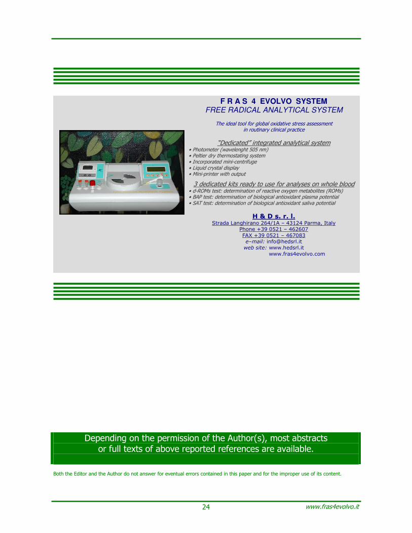
F R A S 4 EVOLVO SYSTEM FREE RADICAL ANALYTICAL SYSTEM
The ideal tool for global oxidative stress assessment in routinary clinical practice
“Dedicated” integrated analytical system • Photometer (wavelenght 505 nm) • Peltier dry thermostating system • Incorporated mini-centrifuge • Liquid crystal display • Mini-printer with output
3 dedicated kits ready to use for analyses on whole blood • d-ROMs test: determination of reactive oxygen metabolites (ROMs) • BAP test: determination of biological antioxidant plasma potential • SAT test: determination of biological antioxidant saliva potential
H & D s. r. l.
Strada Langhirano 264/1A – 43124 Parma, Italy Phone +39 0521 – 462607
FAX +39 0521 – 467083 e–mail: [email protected] web site: www.hedsrl.it
www.fras4evolvo.com