the appearance of ossification centres in …. humanitie… · the appearance of ossification...
TRANSCRIPT
THE APPEARANCE OF OSSIFICATION CENTRES IN STERNUM–A DIMORPHIC STUDY
IN NORTH INDIANS
GURDEEP S KALYAN 1 & GAURAV AGNIHOTRI 2 1Professor & Head, Department of Anatomy, Government Medical College, Patiala, Punjab, India
2Associate Professor, Department of Anatomy, Government Medical College, Amritsar, Punjab, India
ABSTRACT
The state and pattern of ossification is a reliable guide for the determination of age. The timing of appearance and
characteristics of ossification centres of sternum exhibit considerable variation on the basis of individuality, sex and race.
The study aims to lay down a standard in North Indians on which we can rely to evaluate the ossification against the
backdrop of known chronological age of the fetus. The study was conducted on 100 aborted North Indian fetuses of both
sexes (male female ratio 1:1) aged between 3 months to full term. The appearance of centres of ossification was studied
and the sterna were typed. There exists a female prevalence in appearance of centres of ossification. The ossification centre
for manubrium appears with the centre for the 1st sternebra.
Double centres are frequent and there is a tendency of rise in frequency of double centres from above
downwards(for the sternebrae). The incidence of Type I sterna was maximum amongst the fetuses (50%) followed by
Type II (30%) and Type III sterna(20%). The morphological variation is a fundamental feature for proper maturation and
ossification of sternum. The timing of appearance of ossification centres has medicolegal and clinical significance and
explains the chronology of development of an individual.
KEYWORDS: Ossification, Variation, Sternum, Typing
INTRODUCTION
The timing of appearance of ossification centres for a bone is of great medicolegal and clinical importance.
The timing serves as a reliable guide for the determination of age of an individual especially when the age is not known.
However considerable variation in the chronology of skeletal development does occur on the basis of individuality,
sex and race[1].
The events of ossification also exhibit a regional variation and vary according to the geography of the population.
It has been mentioned that bones ossify in Indians much earlier than in Western population both in prenatal as well as
postnatal life. Racial and clinical variation in the time of appearance of ossification and fusion of epiphysis and metaphysis
in the bones have been recorded [2]
Although some work has already been done on appearance of centres of ossification in the various segments of
the sternum in Western countries [3], [4], [5] not much information is available on the Indian sterna. The only work done in
India has been in Chandigarh North West Indian subjects [6] regarding the centres of ossification in sternum.
It is a documented fact[7]that adequate amount of proteins, fats, carbohydrates and vitamins are required for
normal development and ossification of bones. In India, studies on ossification and skeletal maturation have been few[8]
and in most cases differently oriented.
BEST: International Journal of Humanities, Arts, Medicine and Sciences (BEST: IJHAMS) ISSN(E): 2348-0521 Vol. 2, Issue 4, Apr 2014, 57-66 © BEST Journals
58 Gurdeep S Kalyan & Gaurav Agnihotri
In large areas of the world today including India, nutritional deficiency affecting young children is one of the
principal public health problem [9] and is responsible for high mortality in pre-school age children and also for long term
sequence of diminished learning capacity and permanently retarded physical and personality development.
As such the studies on ossification like the present one assume paramount significance.
The present study aims to find out the times of appearance of centres of ossification in the various segments of the
sterna in North Indian subjects as compared to other populations. The study aims to observe the sex differences and also
type the sterna on the basis of ossification.
It is believed that the results so deduced will have a significant value in laying down a standard on which we can
rely to evaluate the state and pattern of ossification against the backdrop of known chronological age of the foetus.
OBJECTIVES OF THE STUDY
The study aims to lay down a standard in North Indians on which we can rely to evaluate the ossification against
the backdrop of known chronological age of the fetus.
METHODS
The study was done on 100 aborted fetuses of both sexes (male female ratio 1:1) aged between 3 months to full
term (70 mm to 245 mm crown rump length). The fetuses were collected from dept. of Obstetrics and Gynaecology at
Government Medical Colleges of Patiala and Amritsar, Punjab, India.
A prior permission of the ethical committee of medical colleges was taken and the study was conducted over a
period of five years (2007-2012). The material was obtained either from the abortions or emcredil done. The fetuses were
collected and stored in jars containing 10% formalin. Tags were tied to the legs of fetuses so as to maintain the record of
identity of the fetus of a particular age. 50 male and female fetuses above 3 months age with crown rump length
(equal to and above )70 mm were selected for the study and rest were discarded.
The criteria adopted for determining the age of the abortus were:
• History of patient for finding out date of last menstrual period.
• Crown –Rump (C.R.) length of abortus.
After noting down C.R. length the sternum alongwith small segments of costal cartilages was dissected with help
of surgical scissors and toothed forceps. The sternum was then kept in 2% KOH for 1-7 days. The remnants of attached
tissues were removed daily by scraping and also by using toothed forceps. After keeping the sterna in 2% KOH the
periosteum was removed from the sternum alongwith the costal cartilages. A small sharp scissor was used alongwith a
plain pointed forceps to hold the periosteum. The periosteum was cleared gently as cartilage becomes brittle after removing
the periosteum.
Then the sterna were again kept in formalin and were X rayed in the antero-posterior view to find out the
appearance of various ossification centres and these radiographs were photographed. In cases where the radiographs
obtained were not satisfactory direct photography was done by keeping the specimens on a view box. The appearance of
centres of ossification was studied from the photographs and the data was collected, compiled, analysed and discussed.

The Appearance of Ossification Centres in Sternum-A Dimorphic Study in North Indians 59
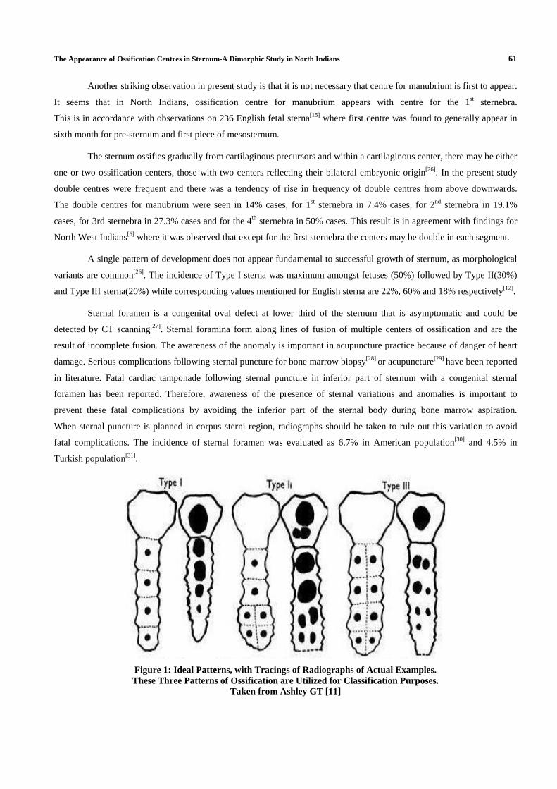
For typing of sterna, only those sterna were included in which centres of manubrium sterni , 1st, 2nd and
3rd sternebrae were present. The method adopted for typing sterna was as given by Ashley GT in his series of studies on
sterna [10], [11], [12]. The author has put forward evidence that development of three basic types of sterna depended upon the
number and arrangement of ossification centres in its segments. The ideal pattern and variations described by author for
typing the ossification pattern are provided in figure 1.
In type I, the body is ossified from four single midline centres. In type II, the centres for the first two segments are
single and midline, those for the lower two being double and bilaterally or obliquely placed. In type III the centres for the
first three segments are double and in the fourth segment a single or double centre may be present.
These variations in the ossification pattern are associated with varying degrees of cohesion of the fetal sterna bars
and are reflected in the final shape of the adult bone.
RESULTS
Sample Distribution According to Crown Rump Range
Table 1 shows the distribution of male and female fetuses according to crown rump length. From table 1,
it is evident that there seems to exist a female prevalence in appearance of centres of ossification.
Manubrium Sterni and 1st Sternebra
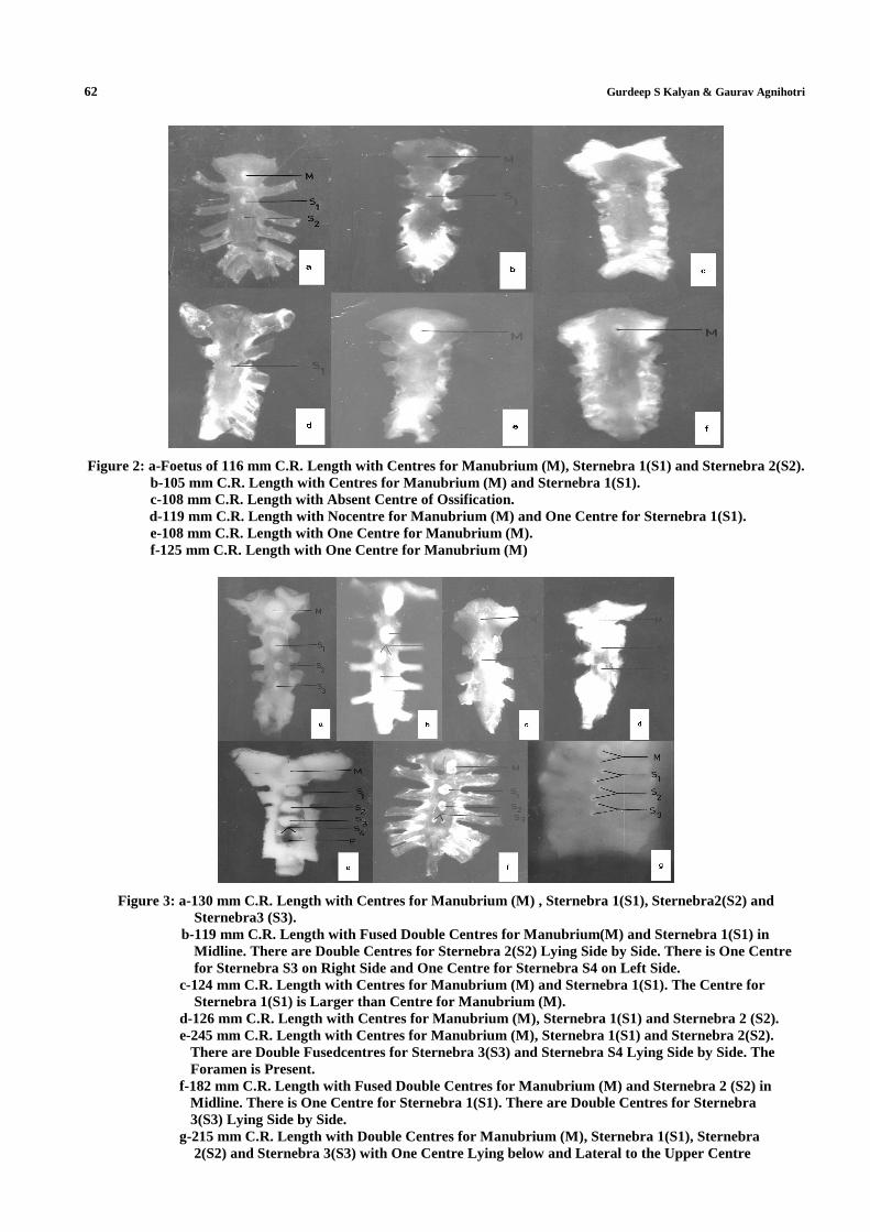
In males and females the earliest centre of ossification in the manubrium sterni and 1st sternebra was seen in
foetus of 116mm C.R. length (Figure 2a) and 105 mm C.R. length (Figure 2b) respectively. Thereafter, the centre was seen
in all older fetuses in males for manubrium sterni and 1st sternebra(except 2 fetuses). Similarly the centre was seen in all
older fetuses in females for manubrium sterni (except 4 fetuses; figures 2c and 2d) and1st sternebra
(except 6 fetuses; figures 2c, 2e and 2f). In 3 foetuses (Figure 2d) the centre for S1 was seen while the centre for
manubrium sterni was not seen. This indicates that it is not necessary that the centre for manubrium is the first to appear.
It seems that in North Indians the ossification centre for manubrium appears with the centre for the 1st sternebra.
2nd, 3rd and 4th Sternebrae
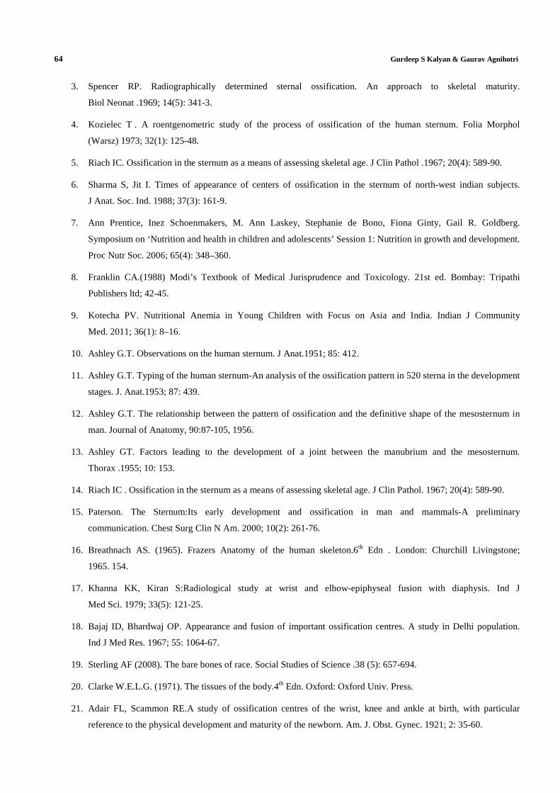
In males, earliest centre of ossification in 2nd, 3rd and 4th sternebrae was seen in foetus of C.R. lengths 116mm
(Figure 2a), 130 mm (Figure 3a) and 243 mm (Figure 3b) respectively. Thereafter centre for S2 sternebra was seen in all
older specimens except 4 fetuses (Figure 3c). The same was true for S3 sternebra except 8 fetuses. The centre for
S3 sternebra was seen to be present in all fetuses equal to or older than 180 mm C.R. length.
In females, earliest centre of ossification in the 2nd, 3rd and 4th Sternebrae was seen in fetus of C.R. lengths 126mm
(Figure 3d), 127 mm (Figure 3c) and 245 mm (Figure 3e) respectively. Thereafter centre for S2 sternebra was seen in all
older specimens except 4 fetuses. The same was true for S3 sternebra except 5 fetuses. The centre for S3 sternebra was
seen to be present in all (except 3) fetuses equal to or older than 160 mm C.R. length.
Xiphoid Process
No centre of ossification was seen in any specimen either in males or in females.
Sternal Foramen
Two female specimens having C.R. lengths 243mm and 245 mms respectively displayed a foramen in the fourth
segment of mesosternum.
60 Gurdeep S Kalyan & Gaurav Agnihotri
Double Centres
Double centres were frequent (Figures 3b, 3f and 3g). The number of fetuses with double centres in males and
females is depicted in table 2.
The percentage incidence of double centres was determined by dividing the total number of double centres
detected with total no of centres detected for each component of the sternum. Data extraction was done and table 3 was
prepared.
For each segment of the bone the numerator in above table was divided with the denominator to determine the
percentage frequency of double centres for that segment.
It was found that there is a tendency of rise in frequency of double centres from above downwards.
The double centres for manubrium were seen in 14% cases, for 1st sternebra in 7.4% cases, for 2nd sternebra in 18 % cases,
for 3rd sternebra in 25% cases and for the 4th sternebra in 50% cases.
In one specimen of 243mm C.R. length in males (Figure 2b) the centre for 3rd sternebra was single and was
present on one side ie. right side. This indicates that there is a likelihood of appearance of 2nd centre on the other side.
Pattern of Ossification and Typing of Sternum
To study the pattern of ossification only those specimens were selected where the centres of ossification
for 1st, 2nd and 3rd sternebrae were present. In the present study such specimens were 20 in number. Rest of the sterna were
discarded due to indeterminate pattern.
In 18 indeterminate specimens, pattern was such that they may ultimately become either type 1 or type 2 but never
type 3.
DISCUSSIONS
The shape, form and morphology of sternum are dependent upon the manner of ossification. This bone, though
present in vast majority of vertebrates and always more or less constant in position, varies greatly both in form and in
function. It is broad and flat and devoid of joints, as in tortoise or long and narrow and composed of many segments,
as in cat[13].
The knowledge of ossification pattern and appearance of ossification centres is helpful in assessing skeletal
maturity for pathological or forensic purposes[14]. The results indicate that corresponding centres of ossification seem to
appear earlier for Indian population as compared to Western populations[15],[16]. This is in agreement with earlier studies on
ossification which state that ossification seems to occur earlier in Indian subcontinent than in other regions[17],[18].
This observation reiterates and reinforces the concept that rate of bone growth, ossification and maturation are not only
dependent on age, sex and race[19] but also other variables like economic status (and hence nutrition ), total body weight
and the functional stress on the bone[20].
The results of the present study indicate that there exists a female prevalence in appearance of centres of
ossification. There is sufficient evidence in available literature that female bones ossify earlier[21],[22]. A similar study[ 6] on
15 male and 12 female fetuses belonging to North West India found that ossification of the various segments of the
sternum starts a little earlier in the female fetus than in the males, though some researchers feel that there are no significant
sexual differences in ossification patterns observed for embryos, fetuses and new born children[23] ,[24], [25].
The Appearance of Ossification Centres in Sternum-A Dimorphic Study in North Indians 61
Another striking observation in present study is that it is not necessary that centre for manubrium is first to appear.
It seems that in North Indians, ossification centre for manubrium appears with centre for the 1st sternebra.
This is in accordance with observations on 236 English fetal sterna[15] where first centre was found to generally appear in
sixth month for pre-sternum and first piece of mesosternum.
The sternum ossifies gradually from cartilaginous precursors and within a cartilaginous center, there may be either
one or two ossification centers, those with two centers reflecting their bilateral embryonic origin[26]. In the present study
double centres were frequent and there was a tendency of rise in frequency of double centres from above downwards.
The double centres for manubrium were seen in 14% cases, for 1st sternebra in 7.4% cases, for 2nd sternebra in 19.1%
cases, for 3rd sternebra in 27.3% cases and for the 4th sternebra in 50% cases. This result is in agreement with findings for
North West Indians[6] where it was observed that except for the first sternebra the centers may be double in each segment.
A single pattern of development does not appear fundamental to successful growth of sternum, as morphological
variants are common[26]. The incidence of Type I sterna was maximum amongst fetuses (50%) followed by Type II(30%)
and Type III sterna(20%) while corresponding values mentioned for English sterna are 22%, 60% and 18% respectively[12].
Sternal foramen is a congenital oval defect at lower third of the sternum that is asymptomatic and could be
detected by CT scanning[27]. Sternal foramina form along lines of fusion of multiple centers of ossification and are the
result of incomplete fusion. The awareness of the anomaly is important in acupuncture practice because of danger of heart
damage. Serious complications following sternal puncture for bone marrow biopsy[28] or acupuncture[29] have been reported
in literature. Fatal cardiac tamponade following sternal puncture in inferior part of sternum with a congenital sternal
foramen has been reported. Therefore, awareness of the presence of sternal variations and anomalies is important to
prevent these fatal complications by avoiding the inferior part of the sternal body during bone marrow aspiration.
When sternal puncture is planned in corpus sterni region, radiographs should be taken to rule out this variation to avoid
fatal complications. The incidence of sternal foramen was evaluated as 6.7% in American population[30] and 4.5% in
Turkish population[31].
Figure 1: Ideal Patterns, with Tracings of Radiographs of Actual Examples. These Three Patterns of Ossification are Utilized for Classification Purposes.
Taken from Ashley GT [11]
62 Gurdeep S Kalyan & Gaurav Agnihotri
Figure 2: a-Foetus of 116 mm C.R. Length with Centres for Manubrium (M), Sternebra 1(S1) and Sternebra 2(S2). b-105 mm C.R. Length with Centres for Manubrium (M) and Sternebra 1(S1). c-108 mm C.R. Length with Absent Centre of Ossification.
d-119 mm C.R. Length with Nocentre for Manubrium (M) and One Centre for Sternebra 1(S1). e-108 mm C.R. Length with One Centre for Manubrium (M). f-125 mm C.R. Length with One Centre for Manubrium (M)
Figure 3: a-130 mm C.R. Length with Centres for Manubrium (M) , Sternebra 1(S1), Sternebra2(S2) and Sternebra3 (S3).
b-119 mm C.R. Length with Fused Double Centres for Manubrium(M) and Sternebra 1(S1) in Midline. There are Double Centres for Sternebra 2(S2) Lying Side by Side. There is One Centre for Sternebra S3 on Right Side and One Centre for Sternebra S4 on Left Side. c-124 mm C.R. Length with Centres for Manubrium (M) and Sternebra 1(S1). The Centre for Sternebra 1(S1) is Larger than Centre for Manubrium (M). d-126 mm C.R. Length with Centres for Manubrium (M), Sternebra 1(S1) and Sternebra 2 (S2). e-245 mm C.R. Length with Centres for Manubrium (M), Sternebra 1(S1) and Sternebra 2(S2). There are Double Fusedcentres for Sternebra 3(S3) and Sternebra S4 Lying Side by Side. The Foramen is Present. f-182 mm C.R. Length with Fused Double Centres for Manubrium (M) and Sternebra 2 (S2) in Midline. There is One Centre for Sternebra 1(S1). There are Double Centres for Sternebra 3(S3) Lying Side by Side. g-215 mm C.R. Length with Double Centres for Manubrium (M), Sternebra 1(S1), Sternebra 2(S2) and Sternebra 3(S3) with One Centre Lying below and Lateral to the Upper Centre

The Appearance of Ossification Centres in Sternum-A Dimorphic Study in North Indians 63
Table 1: Number of Centres of Ossification According to Crown Rump Length Range
Crown Rump Range
Sex M S1 S2 S3 S4 F
70-110 Male - - - - - - Female 4 7 - - - -
111-150 Male 12 11 11 3 - - Female 14 12 13 4 - -
151-190 Male 8 8 6 4 - - Female 10 8 6 5 - -
191-230 Male 2 2 2 2 - - Female 2 2 2 2 - -
>231 Male 2 2 2 2 2 - Female 2 2 2 2 2 2
M-Manubrium;S1-1st Sternebra, S2-2nd Sternebra;S3-3rd Sternebra;S4-4th Sternebra; F-Foramen Sternale
Table 2: Number of Specimens with Double Centres
Sex M S1 S2 S3 S4 Male 6 4 6 4 0
Female 2 0 2 2 2 M-Manubrium;S1-1st Sternebra,S2-2nd Sternebra;S3-3rd
Sternebra;S4-4th Sternebra
Table 3: Showing Data to Determine the Percentage Frequency of Double Centres
Centres Detected M S1 S2 S3 S4 Number of double centres detected 8 4 8 6 2 Total number of centres detected 56 54 44 24 4
M-Manubrium;S1-1st Sternebra, S2-2nd Sternebra;S3-3rd Sternebra;S4-4th Sternebra
Table 4: Showing Classification of Ossification Pattern
Types Number Percentage (%) Type I 10 50 Type II 6 30 Type III 4 20
CONCLUSIONS
The study describes pattern and variations of ossification in sexes. It provides a module which can be relied upon
to evaluate ossification against backdrop of known chronological age of the fetus. The timing of appearance of ossification
centres has medicolegal/clinical significance and enriches the contemporary concept regarding sequence of events in
development of North Indian fetal sterna.
ACKNOWLEDGEMENTS
The authors wish to convey their thanks to dept. of Obstetrics and Gynaecology, Govt. Medical Colleges of
Punjab for providing material for the study.
REFERENCES
1. Işcan, M. Y. The aging process in the rib: An analysis of sex- and race-related morphological variation. Am. J.
Hum. Biol.1991; 3: 617–623.
2. N. Olarte L, A. Rubiano F, A. Mejía F. Comparison of valuation techniques for bone age assessment.
World Academy of Science, Engineering and Technology. 2012; 68: 1645-49.
64 Gurdeep S Kalyan & Gaurav Agnihotri
3. Spencer RP. Radiographically determined sternal ossification. An approach to skeletal maturity.
Biol Neonat .1969; 14(5): 341-3.
4. Kozielec T . A roentgenometric study of the process of ossification of the human sternum. Folia Morphol
(Warsz) 1973; 32(1): 125-48.
5. Riach IC. Ossification in the sternum as a means of assessing skeletal age. J Clin Pathol .1967; 20(4): 589-90.
6. Sharma S, Jit I. Times of appearance of centers of ossification in the sternum of north-west indian subjects.
J Anat. Soc. Ind. 1988; 37(3): 161-9.
7. Ann Prentice, Inez Schoenmakers, M. Ann Laskey, Stephanie de Bono, Fiona Ginty, Gail R. Goldberg.
Symposium on ‘Nutrition and health in children and adolescents’ Session 1: Nutrition in growth and development.
Proc Nutr Soc. 2006; 65(4): 348–360.
8. Franklin CA.(1988) Modi’s Textbook of Medical Jurisprudence and Toxicology. 21st ed. Bombay: Tripathi
Publishers ltd; 42-45.
9. Kotecha PV. Nutritional Anemia in Young Children with Focus on Asia and India. Indian J Community
Med. 2011; 36(1): 8–16.
10. Ashley G.T. Observations on the human sternum. J Anat.1951; 85: 412.
11. Ashley G.T. Typing of the human sternum-An analysis of the ossification pattern in 520 sterna in the development
stages. J. Anat.1953; 87: 439.
12. Ashley G.T. The relationship between the pattern of ossification and the definitive shape of the mesosternum in
man. Journal of Anatomy, 90:87-105, 1956.
13. Ashley GT. Factors leading to the development of a joint between the manubrium and the mesosternum.
Thorax .1955; 10: 153.
14. Riach IC . Ossification in the sternum as a means of assessing skeletal age. J Clin Pathol. 1967; 20(4): 589-90.
15. Paterson. The Sternum:Its early development and ossification in man and mammals-A preliminary
communication. Chest Surg Clin N Am. 2000; 10(2): 261-76.
16. Breathnach AS. (1965). Frazers Anatomy of the human skeleton.6th Edn . London: Churchill Livingstone;
1965. 154.
17. Khanna KK, Kiran S:Radiological study at wrist and elbow-epiphyseal fusion with diaphysis. Ind J
Med Sci. 1979; 33(5): 121-25.
18. Bajaj ID, Bhardwaj OP. Appearance and fusion of important ossification centres. A study in Delhi population.
Ind J Med Res. 1967; 55: 1064-67.
19. Sterling AF (2008). The bare bones of race. Social Studies of Science .38 (5): 657-694.
20. Clarke W.E.L.G. (1971). The tissues of the body.4th Edn. Oxford: Oxford Univ. Press.
21. Adair FL, Scammon RE.A study of ossification centres of the wrist, knee and ankle at birth, with particular
reference to the physical development and maturity of the newborn. Am. J. Obst. Gynec. 1921; 2: 35-60.
The Appearance of Ossification Centres in Sternum-A Dimorphic Study in North Indians 65
22. Jimenez-Castellanos J, Carmona A, Catalina-Herrera CJ, Vinuales M. Skeletal maturation of wrist and hand
ossification centers in normal Spanish boys and girls: a study using the Greulich-Pyle method. Acta Anat
(Basel). 1996; 155(3): 206-11.
23. Jit I: Observations on prenatal ossification with special reference to the bones of the hand and foot. J. Anat. Soc.
Ind.1957; 6: 12-23.
24. Jit, Verma U, Gandhi OP. Ossification of the bones of the hand, foot and knee in the new born children. J. Anat.
Soc. Ind. 1968; 17: 8.
25. Jit I, Singh S. Sexing of adult clavicle. Ind J Med Res.1966;54:551-76.
26. O"Neal ML, Dwornik JJ, Ganey TM, Ogden JA. Postnatal development of the human sternum. J Pediatr Orthop.
1998;18(3):398-405.
27. Fokin AA. Cleft sternum and sternal foramen. Chest Surg Clin N Am. 2000; 10(2):261-76.
28. Bhootra BL. Fatality following a sternal bone marrow aspiration procedure: a case report. Med Sci.
Law. 2004; 44: 170–72.
29. Halvorsen TB, Anda SS, Naess AB, Lewang OW. Fatal cardiac tamponade after acupuncture through congenital
sternal foramen. Lancet. 1995; 345: 1175.
30. Cooper PD, Stewart JH, McCormick WF. Development and morphology of the sternal foramen. Am J Forensic
Med Pathol. 1988; 9: 342–47.
31. Yekeler E, Tunaci M, Tunaci A, Dursun M, Acunas G. Frequency of sternal variations and anomalies evaluated
by MDCT. AJR Am J Roentgenol. 2006; 186: 956–60.