the antioxidant potentials of the new zealand surf clams
TRANSCRIPT

The Antioxidant Potentials of the New Zealand Surf
Clams
ODELEYE TINUOLUWA
A thesis submitted to
the Faculty of Health and Environmental Sciences
Auckland University of Technology
In fulfilment of the requirements for the Degree of
Master of Philosophy (MPhil)
March 2015.
Auckland, New Zealand.

The Antioxidant Potentials of the New Zealand Surf Clams
Approved by:
Supervisors:
Assoc. Prof. Jun Lu
Dr. Yan Li
9 March 2015
Date approved

Table of Contents Table of Contents .......................................................................................................... I
List of Figures ............................................................................................................... IV
List of Tables ................................................................................................................ VI
Attestation of Authorship ............................................................................................. VII
Acknowledgement ...................................................................................................... VIII
Abstract ........................................................................................................................ IX
1. INTRODUCTION .......................................................................................................... 1
1.1 BIOACTIVES DERIVED FROM SEAFOOD ...................................................................... 1
1.2 SURF CLAMS ........................................................................................................... 5
1.2.1 Biology of Surf Clam .................................................................................................... 5
1.2.2 Surf Clams in New Zealand ......................................................................................... 9
1.3 OXIDATIVE STRESS ................................................................................................ 10
1.4 OXIDATION OF BIOLOGICAL MOLECULES ............................................................... 11
1.4.1 Free Radicals ............................................................................................................. 13
1.4.2 Reactive Oxygen Species (ROS) ................................................................................. 14
1.4.3 The Two Sides of ROS ................................................................................................ 18
1.4.4 Oxidative Stress and Diseases in Man ....................................................................... 20
1.5 ANTIOXIDANTS ..................................................................................................... 25
1.5.1 Benefits of Antioxidants ............................................................................................ 25
1.5.2 Synthetic and Natural Antioxidants .......................................................................... 28
1.6 POTENTIAL THERAPEUTIC EFFECTS OF SURF CLAM EXTRACTS................................. 30
1.6.1 Anticancer Activity .................................................................................................... 31
1.6.2 Anticoagulant Activity ............................................................................................... 31
1.6.3 Antiviral Activity ........................................................................................................ 32
1.6.4 Antibacterial Activity ................................................................................................. 33
1.6.5 Antioxidant Activity ................................................................................................... 33
1.7 ANTIOXIDANTS DERIVED FROM SURF CLAM (DIETARY ANTIOXIDANTS) ................... 34
i

1.7.1 Carotenoids ............................................................................................................... 35
1.7.2 Free Amino Acids ...................................................................................................... 36
1.7.3 Polyunsaturated Fatty Acids ..................................................................................... 37
1.7.4 Nucleoside ................................................................................................................. 38
1.7.5 Other Antioxidant Compounds ................................................................................. 39
1.8 MECHANISM OF ACTION OF ANTIOXIDANTS ........................................................... 39
1.8.1 Primary Antioxidants................................................................................................. 39
1.8.2 Secondary Antioxidants ............................................................................................ 42
1.9 ANTIOXIDANT CAPACITY ASSAYS ........................................................................... 43
1.9.1 HAT Based Assays...................................................................................................... 43
1.9.2 ET Based Assays ........................................................................................................ 44
1.9.3 DPPH Radical Scavenging Assay ................................................................................ 44
1.9.4 Cupric Ion Reducing Antioxidant Capacity (CUPRAC) Assay ..................................... 45
1.9.5 Pros and Cons of DPPH and CUPRAC Assays ............................................................. 46
1.10 OBJECTIVES .......................................................................................................... 48
2. MATERIALS AND METHODS ...................................................................................... 49
2.1 MATERIALS ............................................................................................................. 49
2.1.1 Chemicals and reagents ............................................................................................ 49
2.1.2 Surf clam Processing ................................................................................................. 49
2.2 METHODS ............................................................................................................... 50
2.2.1 Sample Preparation .................................................................................................. 50
2.2.2 DPPH Radical Scavenging Activity ............................................................................. 52
2.2.3 CUPRAC Assay ........................................................................................................... 53
2.2.4 Statistical Analysis ..................................................................................................... 54
3. RESULTS ................................................................................................................... 55
3.1 ANTIOXIDANT ACTIVITY .......................................................................................... 55
3.2 DPPH Assay ............................................................................................................ 55
3.3 CUPRAC Assay ........................................................................................................ 59
3.4 Statistical Significance............................................................................................. 62
4. DISCUSSION ............................................................................................................. 69
4.1 Major Active Compounds in the NZ Surf Clam Extracts ............................................. 70
ii

4.1.1 Crude Extracts (cd) .................................................................................................... 70
4.1.2 Petroleum Ether Fraction (pe) .................................................................................. 71
4.1.3 Ethyl Acetate Fraction (ea) ........................................................................................ 72
4.1.4 n- Butanol Fraction (nb) ............................................................................................ 73
4.1.5 Aqueous Fraction (w) ................................................................................................ 74
4.2 Comparison of antioxidant activity among species .................................................. 75
4.3 Comparison of antioxidant activity in the NZ surf clam and other marine species and
plant products.............................................................................................................. 76
4.4 Correlations ............................................................................................................ 77
4.5 Differences in DPPH scavenging activity and CUPRAC reducing activity .................... 78
5. CONCLUSION ............................................................................................................ 80
5.1 Caveats ......................................................................................................................... 80
5.2 Future Research Directions .......................................................................................... 81
5.3 Overall Conclusion ....................................................................................................... 83
REFERENCES ................................................................................................................. 84
iii
List of Figures Figure 1: Classification of Surf clam……………………………………………………………………..……..…. 5
Figure 2: Surf clam sturcture………..……………………………………………………………………..…………. 8
Figure 3: The electron reduction products of oxygen………...……………………………….….……. 15
Figure 4: DPPH (2,2-diphenyl-1- picrylhydrazyl free radical) Inhibition………..………….…….45
Figure 5a: Flesh thawing prior to drying………………..…..…………………………………………...…….50
Figure 5b: Dried flesh………………………….…………………………………..………….………………….…….50
Figure 5c: Extraction process using the Rota evaporator..…………………………………….……….51
Figure 6: DPPH radical scavenging activity of Tua tua.……………………………………………….….56
Figure 7: DPPH radical scavenging activity of Diamond shell……………………………..………….57
Figure 8: DPPH radical scavenging activity of Storm shell..………………………..………………….58
Figure 9: Total antioxidant capacity of Tua tua with the CUPRAC reagent………………...….59
Figure 10: Total antioxidant capacity of Diamond shell with the CUPRAC reagent…….….60
Figure 11: Total antioxidant capacity of Storm shell with the CUPRAC reagent………….….61
Figure 12a: DPPH radical scavenging activity of all crude extracts …………..………………..….63
Figure 12b: Copper ion reducing activity of all crude extracts……………………………..………..63
Figure 13a: DPPH radical scavenging activity of all petroleum ether fractions….………..….64
Figure 13b: Copper ion reducing activity of all petroleum ether fractions .………….…….….64
Figure 14a: DPPH radical scavenging activity of all ethyl acetate fractions…………………….65
Figure 14b: Copper ion reducing activity of all ethyl acetate fractions…………………….…….65
Figure 15a: DPPH radical scavenging activity of all n-butanol fractions……………………...….66
Figure 15b: Copper ion reducing activity of all n-butanol fractions……………….……………….66
Figure 16a: DPPH radical scavenging activity of all aqueous fractions…….…...……………….67
Figure 16b: Copper ion reducing activity of all aqueous fractions…………..………………….….67
iv

Figure 17: Diamond shell CUPRAC assay: Sample stock solution 10mg/mL; Standard
stock solution 10mg/mL…….……………………………………………………………………………….……….68
Figure 18: Diamond shell CUPRAC assay: Sample stock solution 10mg/1mL; Standard
stock solution 1mg/mL………….………………………………………………………….……….……….……….68
v

List of Tables
Table 1: Reported marine seafood that possess antioxidant peptides and their mode of
action…………………………………………………………………………………………………………………………..……...4
Table 2: DPPH radical scavenging activities by the New Zealand surf clam extracts.…………………………………………………………………………………………………..……….…………....…58
Table 3: R2 values of surf clam fractions from CUPRAC assay …...…………………………………..……62
vi

Attestation of Authorship I hereby declare that this submission is my own work and that, to the best of my knowledge and
belief, it contains no material previously published or written by another person (except where
explicitly defined in the acknowledgements), nor material which to a substantial extent has been
submitted for the award of any other degree or diploma of a university or other institution of
higher learning.
Signed
Name ODELEYE TINUOLUWA
Date 9th March, 2015.
vii
Acknowledgement
I have taken efforts in this project. Nonetheless, it would not have been possible without the
kind support and help of many individuals. I would like to express my deepest appreciation to all
those who provided me the support, from start to finish.
I am highly indebted to my primary supervisor, Assoc. Prof. Jun Lu. I could never have done this
without you. Thank you for pushing me and making me think things through by myself (even
when I knew that you knew the answers ). For the sacrifices you made, your endless support
in ordering materials, and in the laboratory, thank you. I could never have wished for a better
supervisor. I consider myself honoured to be one of the people who have brought your ideas to
life.
I would like to thank my secondary supervisor, Dr. Yan Li for his kind words of encouragement.
Thank you for your input in the editing of this thesis.
I was blessed with the best set of staff and laboratories (Chemistry and Biology), and research
technicians in the Applied Sciences; Saeedah, Louise, Jinan, Dr. Chris Pook, Yan, Dr. Sonya,
Rachael and Brid Lorigan. There is no way I could have finished my studies without you. Thank
you for your patience.
Special thanks to all my friends and colleagues. Kelvin, for your help in the lab and with getting
stuff right; Loveena, for your diligence and determination that encouraged me; Harriet, for your
kind words of encouragement; Kevin, for your cookies; Joe, Roy, Reza, Riya, Jennifer,
Bhawana. Thank you for making the late nights and early starts enjoyable!
Especially to April, Karl, Shun, Theo, Kola, Muhammad, Izilda, and Ehsan- words cannot
express how much you did for me. Thank you! A big thanks to Jenny, Greg, Agatha, Kelvin,
Mai, Jacque and Grant for your prayers, and ALWAYS asking, ‘how’s it going?’
My love and gratitude goes to my beloved family, for your unconditional love, unflinching
support, understanding and unending prayers.
viii

Abstract The antioxidant activities of the aqueous and ethanolic extracts of three New Zealand surf clam
species (Tua Tua (Paphies donacina), Diamond Shell (Spisula aequilatera) and Storm shell
(Mactra murchisoni)) were evaluated and presented as relative activities by comparing with a
standard synthetic antioxidant (ascorbic acid). The ethanolic extracts were further fractioned
into four parts (petroleum ether (pe), ethyl acetate (ea), n-butanol (nb) and the final aqueous
residue (w)).
Two types of antioxidant assays were employed to test for antioxidant activity. The DPPH (2,2-
diphenyl-1-picrylhydrazyl) assay, to test the scavenging ability of the clam fractions, and the
Cupric Ion Reducing Antioxidant Capacity (CUPRAC) assay to evaluate their copper ion
reducing ability.
Results showed that the in vitro antioxidant activities of all fractions of the three NZ surf clams
tested exhibited dose dependency, and increased with increasing concentration of the extract.
The pe and ea fractions of all clam species showed higher cupric ion reducing capacity than
ascorbic acid and the other fractions tested. All fractions had very strong radical scavenging
ability, with the aqueous fraction of the Diamond shell having the least DPPH radical
scavenging ability of 57.08% at 50µg/mL.
This study forms the first comprehensive report on the nutraceutical properties of the New
Zealand surf clams as natural sources of antioxidants.
ix

CHAPTER ONE
1. INTRODUCTION
1.1 BIOACTIVES DERIVED FROM SEAFOOD The wide diversity of marine organisms contained in the world’s oceans offers a rich source of
natural products (Wijesekara, Pangestuti, & Kim, 2011, Aneiros, & Garateix, 2004) and, as
such, is regarded as the largest remaining reservoir of natural molecules to be evaluated for
drug activity (Gerwick, 1987). Marine organisms are rich in functional materials including
polyunsaturated fatty acids (PUFA), polysaccharides, essential minerals and vitamins,
antioxidants, enzymes and bioactive peptides (Kim et al., 2008, Kim & Wijesekara, 2010; Ngo,
Wijesekara, Vo, Van Ta, & Kim, 2010) and provide an ample scope for the extraction of drugs
and chemicals for therapeutic purposes (Chakraborty& Ghosh, 2010) and medical research
(Ruggieri, 1975).
Seafood contributes at least 15% of average animal protein consumption to 2.9 billion people
and as much as 50% for some small islands and West African states. As well as being a great
source of Omega-3 fatty acids essential for brain development, seafood also provides important
micronutrients for the poor. As a source of livelihood, capture fisheries and aquaculture
employed 43.5 million people in 2006 and 520 million people relied on income from seafood
production. Seafood is also the most highly traded food commodity internationally (Smith et al.,
2010).
Due to the increasing demand by consumers for high-quality healthy food, there is a focus on
trying to reduce the impact of some risk factors of current agricultural production methods on
human health. To date nutritional theories have focused on the health implications of the fatty
acid profile of the diet; in particular the relationship between saturated fatty acids (SFA),
monounsaturated fatty acids (MUFA), long-chain polyunsaturated fatty acids (PUFA) of both the
n-3 and n-6 categories. An important feature of polyunsaturated fatty acids, in particular n-3
fatty acids, is their role in the prevention and modulation of certain diseases that are common in
industrialized countries. Reports have shown that lipids of marine fish or seafood species are 1

characterised by low levels of linoleic acid (18:2 n-6) and linolenic acid (18:3 n-3), as well as
high levels of long chain n-3 polyunsaturated fatty acids. Eicosapentaenoic acid (20:5 n-3) and
docosahexaenoic acid (22:6 n-3) are the predominant n-3 fatty acids (Valfré, Caprino, &
Turchini, 2003).
While some marine animals especially bivalves (oysters, clams, scallops, etc.) contain
carotenoids which show structural diversity (Maoka, 2009) and are antioxidants (Table 1), there
is an increasing focus on another source of potential antioxidant activity, namely the marine-
derived peptides.
Marine-derived bioactive peptides have been shown to possess many physiological functions
including antihypertensive action or angiotensin-I-converting enzyme (ACE) inhibition (Je et al.,
2005). Several studies have indicated that peptides derived from marine fish proteins have
antioxidant properties (Jun, Park, Jung, & Kim, 2004) and a number of such peptides have
shown higher antioxidative potential than α-tocopherol (Ngo, Wijesekara, Vo, Ta, & Kim, 2011).
The peptide (Glu-Ser-Thr-Val-Pro-Glu-Arg-Thr-His-Pro-Ala-Cys-Pro-Asp-Phe-Asn) for example,
which was isolated from the peptic hydrolysate of hoki (Johnius belengerii) frame protein, has
inhibited lipid peroxidation higher than that of α-tocopherol and efficiently quenched different
sources of free radicals (Kim et al., 2007). An Antioxidant Reptide from Bullfrog Skin Protein
(APBSP) also inhibited lipid peroxidation higher than that of α-tocopherol (Qian, Jung, & Kim,
2008). Fish and shellfish protein hydrolysates have shown a greater lipid peroxidation inhibition
and are considered as useful candidates in search of effective non-toxic substances with
differing antioxidant mechanisms (Mendis, Rajapakse, Byun, & Kim, 2005).
Bioactive peptides can be obtained from an organism’s protein by enzymatic hydrolysis (Je et
al., 2005; Lee, Hong, Jeon, Kim, & Byun, 2009) and have been reported effective and safe in
recent years (Nazeer, Prabha, Kumar, & Ganesh, 2013).
The antioxidant activity of marine derived bioactive peptides has been determined by various in
vitro methods such as DPPH (2,2-diphenyl-1-picrylhydrazyl), peroxide, hydroxyl and superoxide
anion radical scavenging activities which have been detected by electron spin resonance (ESR)
2
spectroscopy method as well as intra-cellular free radical scavenging assays such as DNA
oxidation, reactive oxygen species scavenging, membrane protein oxidation and membrane
lipid oxidation (Ngo, Wijesekara, Vo, Ta, & Kim, 2011).
3

Table 1: Reported marine seafood that possess antioxidant peptides and their mode of
action.
Marine animal Mode of Peptide Action References
Meterix casta (Chemnitz i) Free radical scavenging
ii) Lipid peroxidation inhibition
Nazeer, Prabha, Kumar, & Ganesh,
2013
Giant squid (Dosidicus
gigas) i) Free radical scavenging Rajapakse, Mendis, Byun, & Kim,
2005
Fermented marine blue
mussel i) Superoxide radical
scavenging
ii) Hydroxyl radical scavenging
iii) Carbon-centered radical
scavenging
iv) Lipid peroxidation inhibition
v) DPPH radical scavenging
Rajapakse, Mendis, Jung, Je, & Kim,
2005
Jumbo squid (Dosidicus
gigas) i) Hydroxyl radical scavenging
ii) Carbon centered radical
scavenging
iii) Metal ion chelation
Mendis, Rajapakse, Byun, & Kim,
2005
Oyster (Crassostrea gigas) ii) Hydroxyl radical scavenging
iii) Superoxide radical
scavenging
Qian, Jung, Byun, & Kim, 2008
Side-gill Sea slug
(Pleurobranchus forskalii)
Wesson & Hamann, 1996
Jellyfish (Rhopilema) i) Superoxide anion
scavenging
ii) Hydroxyl radical scavenging
iii) Copper- chelating
Zhuang, Sun, Zhao, Wang, Hou, & Li,
2009
4

1.2 SURF CLAMS Extensive research has shown the usefulness of the surf clam, both nutritionally and
medically. Clams are important recreational and commercial resource in many countries
(Ludien, Brey, & Arntz, 2003; McLachlan et al., 1996). The flesh of Mactra veneriformis is
consumed in China as a delicious food and it is also used in traditional Chinese medicine
(Luan, Wang, Wu, Jin, & Ji, 2011).
Beentjes and Baird (2004) identified surf clams as one of the most important aquatic
organisms. Surf clams are also very important in the surf zone food webs (McLachlan et al.,
1996; Menn, 2002) as they feed on phytoplankton and detritus, and are consumed by birds,
fish and crabs (Laudien, Brey, & Arntz, 2003). As well as playing an important ecological
role, surf clams can be exploited for bait and harvested for human consumption (Ludien,
Brey, & Arntz, 2003). A large quantity of surf clam is traded internationally (Manalo &
Campos, 2010; Agasen et al. 1998).
1.2.1 Biology of Surf Clam
Following Linnaeus’ system of organism classification, Fay, Neves, & Pardue, (1983)
classified surf clams as shown in Figure 1 below.
Figure 1: Classification of surf clam (Redrawn from Fay, Neves, & Pardue, 1983).
Order: Veneroida
Class: Bivalvia
Phylum: Mollusca
Kingdom: Animalia
Veneridae Mactidae Family:
5

In 1980, Barnes reported the phylum Mollusca as one of the largest phyla in the animal
kingdom with more than 100,000 extant species living in marine, fresh water and terrestrial
habitats. Just two decades later, it was reported to have an estimated diversity of up to
200,000 species (Pechenik, 2000). Mollusca are subdivided into seven classes: Gastropoda,
Monoplacophora, Polyplacophora, Aplacophora, Bivalvia, Scaphopoda and Cephalopoda. Of
these, classes Gastropoda (over 75,000 extant species) and Bivalvia (over 20,000 living
species) constitute 95% of the representatives of molluscs (Kantha, 1989; Chapman, 2009).
The three prominent classes of molluscs are gastropods, bivalves and cephalopods (Kantha,
1989). They are prominent majorly because they are the commonly seen of the seven known
classes (Bunje, 2003). Other animals in this phylum are snails, squids, oysters, mussels,
scallops etc. Surf clams fall into the class Bivalvia because of their paired shells
(“Massachusetts Marine Fisheries Shellfish Purification Plant”, 2013).
A number of environmental variables such as water temperature (Ansell, 1968; Mann, 1979;
Robert & Debra, 1992; Orban et al., 2004; Karakoltsidis, Zotos, & Constantinides, 1995),
water depth (Jones, 1980; Ambrose et al., 1980) and population density (Cargnelli et al.,
1999) can influence the growth of surf clams (Douglas et al., 1983). It is reported that sea
water temperature variations also influence the immune functions in molluscs such as
Mactra veneriformis (Yu et al., 2009; Renault et al., 2006). In Chamelea gallina, for example,
the phagocytic activity of haemocytes was significantly decreased at 30°C (Marin et al.,
2007). The lysosomal and cell membranes of haemocytes from the mussel, Mytilus edulis,
appeared destabilised at 0 °C with respect to those of haemocytes from mussels acclimated
at 10 °C. Significant reductions in lysosomal stability were also recorded in haemocytes from
the oyster, Ostrea edulis, maintained at 15 °C (Camus, Grøsvikb, Børseth, Jones, &
Depledge, 2000; Hauton, Hawkins, & Hutchinson, 2001).
Other factors that may also influence the growth of surf clams are geographical location,
food availability (Robert & Debra, 1992; Karakoltsidis, Zotos, & Constantinides, 1995;
Beninger & Lucas, 1984), salinity (Orban et al., 2002;Gardner and Thompson, 2001; Islay,
2004; Orban et al., 2006), turbidity or quantity of sediments present in the water (Bricelj,
6

Malouf, & Quillfeldt, 1984; Aldridge, Payne, & Miller, 1987) and dissolved oxygen in ocean
bottom waters (Fay, Neves, & Pardue, 1983).
Sometimes, man’s activities such as dredging and navigational operations in water bodies,
where clams can be found, also cause some of these deleterious impacts on aquatic
organisms (Aldridge et al., 1987). Surf clams can live for up to 35 years. On average, those
living in open water live longer than those living inshore (Fishwatch U.S. Seafood Facts,
2014).
Surf clams have two shells which are mirror images of each other, joined at one end by a
flexible hinge (Klappenbach, 2014) and can close tightly together for protection with their
whole body inside (University of Michigan, 2002; Klappenbach, 2014). It has been reported
by the National Oceanic and Atmospheric Administration (2013) that Bivalves make their
own shells whereby the mantle secretes calcium carbonate so that as the inner invertebrate
grows, the outer shell provides a roomier home. The shells do not close fully and gape
slightly (Fishwatch U.S. Seafood Facts, 2014).
As explained by Gosling (2003), clams occupy the broadest range of habitats among the four
bivalve groups, and are found from open coasts to sheltered, saline and estuarine locations.
Like fish, bivalve molluscs breathe through their gills. As filter feeders, bivalves gather food
also through their gills (NOAA, 2013). Surf clams have a funnel-like siphon into which water
and food can flow, and use this siphon in reproduction and locomotion (Cargnelli, Griesbach,
Packer, & Weissberger, 1999).
All marine bivalves, except scallops, possess gonads that produce the gametes necessary
for their reproduction (Uddin et al., 2012). As male and female surf clams are identical
externally, the sexes are differentiated through the histological examination of gonads
(Gaspar, Ferreira, & Monteiro, 1999; Joaquim et al., 2008; Aljadani, 2013). Surf clams spawn
from late spring through early fall, shedding their eggs and sperm directly into the water
column. Larvae spend about three weeks in the water column as plankton before settling to
the bottom to live. Sometimes reproduction in surf clams is possible by the age of one but
most of them spawn by the end of their second year (Fishwatch U.S. Seafood Facts, 2014).
7
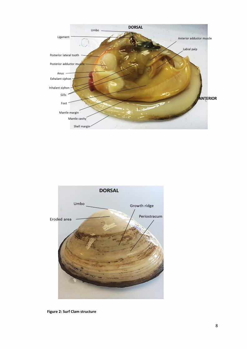
Figure 2: Surf Clam structure
8

1.2.2 Surf Clams in New Zealand Surf clams are found in the surf zone of exposed sandy beaches throughout New Zealand
(Cranfield, & Michael, 2001). According to the Ministry for Primary Industries (2012), three
families of sub-tidal surf clams occur in New Zealand: Veneridae, Mactridae, and
Mesodesmatidae. There are seven main species of surf clams in New Zealand; Paphies
donacina (PDO), Spisula aequilatera (SAE), Mactra discors (MDI), Mactra murchisoni (MMI),
Dosinia anus (DAN), Dosinia subrosea (DSU) and Bassina yatei (BYA), four out of which are
given more attention; PDO, SAE, MMI and DAN (Ministry for Primary Industries, 2012). The
reason for this could probably be as a result of their different depth ranges. The optimal
depth range for PDO, SAE, MMI and DAN in the North Island is 2-8m, while other species
like DSU and BYA range from 6-10m (Aljadani, 2013). Thus, the total catch across PDO,
SAE, MMI and DAN will probably be higher than the catch in other species.
Cloudy Bay Clams (2014) has described these four species; Paphies donacina (PDO),
Spisula aequilatera (SAE), Mactra discors (MDI), Mactra murchisoni (MMI), and Dosinia
anus (DAN). Tua Tua (Paphies donacina) which is harvested from the low tide zone (2 to 4
metres) has a smooth clean shell and a full meat at nearly 33% meat to shell. The smooth
low profile shell is cream to light moss in colour, weighing approximately 12-30 pieces per
kg. Moon Shell (Dosinia anus) is found in the deeper parts of the surf zone (at 5-10 metres)
with its beautiful mustard coloured circular shell. It has a depth of flavour and firm texture like
no other clam and has a 20-25% meat to shell ratio, approximately 12-25 pieces per kg.
Diamond Shell (Spisula aequilatera) is harvested from the 3 to 5 metre surf zone, naturally
full of plump meat, and yielding 28- 35% meat to shell. The Diamond Shell clam has a beige
coloured shell with a rich, deep coloured meat and white tongue, approximately 12-50 pieces
per kg. The Storm shell clam (Mactra murchisoni) is harvested in the deeper regions of the
surf zone (4 to 8 metres) and has a distinctive angular shell which is white with pale straw
coloured bands. The smoother deep cupped shell holds a clam which is almost two separate
parts- a long pearl white tongue and a deep ochre coloured body. With a total meat to shell
ratio of more than 30%, the Storm Clam is unique in the world of shellfish, approximately 5-
15 pieces per kg. 9
New Zealand surf clams grow rapidly in their first 3 years but considerably slower thereafter.
Individuals grow quickly in summer but hardly at all in winter. South Island clams grow faster
and to a larger size as compared to the same species from the North Island. It is also
reported that at a growth size of 7cm, the deeper water species, such as the moon shell
(Dosinia anus), and the diamond shell (Spisula aequilatera), a species most abundant in
shallower waters, had a life expectancy of up to 25 years and about 5 years respectively
(NIWA, 2008).
1.3 OXIDATIVE STRESS Individuals are exposed to oxidants, both endogenous and exogenous, since the moment of
conception (Fraga et aI., 1991). Reactive oxygen and nitrogen species are generated in vivo
and cause damage to DNA, lipids, proteins and other bio-molecules (Halliwell, 1996). DNA
damage can occur, for example, when hydroxyl radical (a highly ROS) reacts with DNA
bases by adding to double bonds of DNA bases and by abstracting an H atom from the
methyl group of thymine and each of the C-H bonds of 2´ -deoxyribose (Cooke, Evans,
Dizdaroglu, & Lunec, 2003). Antioxidants are, therefore, needed to prevent the formation and
to oppose the actions of oxidants.
Antioxidants can be consumed in the diet and are synthesised in vivo in humans. Synthetic
antioxidants have been invented, tested for acute toxicity and proposed as an addition to
naturally occurring antioxidants. They are chemically pure and produce consistent
antioxidant activity and are commercially available (Pokorný, 2007). Their advantages
include, in some cases, lower production cost and higher antioxidant capabilities than
naturally produced antioxidants. Synthetic antioxidants have also found to be less polar than
their natural equivalents and are therefore more soluble in lipids (Kulawik, Özogul, Glew, &
Özogul, 2013). Unfortunately, recent reports have revealed that these synthetic antioxidants
may be associated with toxic and carcinogenic effects (Zhang et al., 2010).
10

As a part of society’s demand for a better lifestyle and increased longevity, consumers have
developed an increasing interest towards consuming ‘nutraceuticals’ and functional foods
rich in natural bioactive compounds (Fung, 2012). In an attempt to address this interest,
there has been a dramatic increase in the number of investigations aimed at identifying
dietary compounds from natural sources which may be effective in preventing diseases
caused by oxidative damage (Tierney, Croft, & Hayes, 2010; Yangthong, Hutadilok-
Towatana, & Phromkunthong, 2009; Dudonné, Vitrac, Coutiére, Woillez, & Mérillon, 2009).
Antioxidants sourced from our diets generally contain radical chain reaction inhibitors, metal
chelators, oxidative enzyme inhibitors and antioxidant enzyme cofactors that can prevent
damage caused by oxidative stress (Huang, Ou, & Prior, 2005). However, it is important to
be able to measure the antioxidant potency of food material containing antioxidants in order
to assess the potential of the food to produce antioxidant effects and to assist in the
diagnosis and treatment of diseases (Özyürek, Güçlü, & Apak, 2011).
Marine organisms are known to be a rich source of many different kinds of bioactive
substances. The richness is believed to be due to the fact that they are living in a very
exigent, competitive and aggressive surroundings, very different in many aspects from the
terrestrial environment, a situation that demands the production of quite specific and potent
active molecules (Kim & Wijesekara, 2010). One such marine organism of great importance
to man as a dietary component and which also has antioxidant potential is the surf clam.
1.4 OXIDATION OF BIOLOGICAL MOLECULES
Oxygen is the most prevalent element in the earth’s crust (Halliwell & Gutteridge, 1999) and
is vital for most organisms for respiration. As well as being essential for most life,
paradoxically, oxidative respiration also leads to the generation of molecules that cause
sometimes life threatening damage to key biological sites (Serafini, 2006). The basis of
energy production in animals, oxidation, is at the same time a major cause of irreversible
deterioration and ultimate death (Scott, 1993).
11
Except for a small number of anaerobic bacteria, all living organisms use oxygen for energy
production through the coupling of oxidation to energy transfer via the phosphorylation of
Adenosine diphosphate (ADP) (Winston & Di Giulio, 1991) and it is thus essential for life as
we know it (Magder, 2006). In the absence of oxygen, the electron transport chain is
inhibited and glucose metabolism is shunted down the glycolytic pathways. The resultant
depression of cellular metabolism is incompatible with life in higher organisms (Maltepe &
Saugstad, 2009).
The reduction of molecular oxygen to H2O via mitochondrial respiration complexes provides
Adenosine triphosphate (ATP) (Bhattacharyya, Chattopadhyay, Mitra, & Crowe, 2014).
A consequence of this reaction is the formation of toxic ROS that can damage various
classes of biological molecules and contribute to cell death (Bhattacharyya, Chattopadhyay,
Mitra, & Crowe, 2014).
Oxygen is a diradical as it contains two electrons that are not spin-paired, with each electron
residing in an orbital of its own. This unconventional distribution of electrons in the oxygen
molecule makes it impossible for oxygen to accept a spin-matched pair of electrons until one
of its unpaired electrons undergoes a spontaneous spin reversal to make pairing possible.
Occasionally, oxygen does manage to steal away electrons from other molecules by non-
enzymatic auto-oxidations. Since it cannot accommodate a spin-matched pair, it must settle
for stealing electrons one at a time. This breaking-up of electron pairs results in the formation
of free radicals (McCord, 2000). The univalent reduction of oxygen is superoxide radical, the
divalent and trivalent reductions are hydrogen peroxide and hydroxyl radical (Cadenas &
Davies, 2000) respectively as shown in Figure 3. One can agree with Aruoma (1994) and
Halliwell (2007) that oxygen is a toxic gas.
Oxygen is the ultimate electron acceptor in the electron flow system that produces energy in
the form of ATP (Pérez & Aguilar, 2013). Oxidation, the transfer of electrons from one atom
to another, occurs when oxygen comes in contact with other substances, thereby causing
them to lose one or more electrons (Stevens, 2011). Oxidation occurs in over one-quarter of
12

the known chemical reactions catalysed by enzymes in living cells (Khan, Tania, Zhang, &
Chen, 2010).
Oxidants occur naturally as part of the normal body process; however harmful oxidants or
free radicals, which are forms of oxygen, can cause damage to body cells. Biological
systems are continuously exposed to oxidants, either generated endogenously by metabolic
reactions (e.g. from mitochondrial electron transport during respiration or during activation of
phagocytes) or exogenously like the air pollutants or cigarette smoke (Rahman & Adcock,
2006).
1.4.1 Free Radicals
Atoms contain a nucleus and electrons move around the nucleus, usually in pairs (Halliwell,
1994). A free radical is any atom or molecule that has a single unpaired electron in an outer
shell (Erbas & Sekerci, 2011). The unpaired electrons alter the chemical reactivity of an atom
or molecule, usually making it more reactive than the corresponding non-radical (Halliwell,
1994). Free radicals could be positively charged, negatively charged or electrically neutral
(Battino, Bullon, Wilson, & Newman, 1999), and are generated during normal cellular
function and are part of the natural physiological processes of all living beings (Dasgupta &
Klein, 2014). Free radicals can be formed in situ in our tissues and cells by;
(i) Impact of radiation, which may be ionizing, ultra-violet, visible or thermal.
(ii) Redox reactions catalysed by transition metals such as iron or copper which can undergo
unit changes in valence state (e.g. Fe3+ + e- ⟶ Fe2+).
(iii) enzymic-catalysis that often involves flavoproteins or hemoproteins (Slater, 1988;
Freeman & Crapo, 1982).
During normal conditions, free radicals are generated at a low rate and subsequently taken
care of by the well-developed scavenger and antioxidant system. However, a greatly
increased rate of free radicals may exceed the capacity of the cellular defence system
(Sjödin, Westing, & Apple, 1990). Free radicals, if not neutralised, can work against the
immune system and cause the development of degenerative diseases (Veg-it.com, 2001)
such as cancer, cardiovascular disease (CVD) and other disorders (Gutteridge, 1995) such 13

as inflammatory diseases (Butterfield et al., 2002) and even affect the aging process
(Harmon, 1956). Excess free radicals can also cause destructive effects on foods (Wang,
Zhao, Zhao, & Jiang, 2007).
1.4.2 Reactive Oxygen Species (ROS) Reactive oxygen species (ROS) are various forms of activated oxygen (Kumar et al., 2005)
which are unstable molecules with unpaired electrons. They are capable of initiating the
process of oxidation (Kirkham & Rahman, 2006). ROS include free radicals (such as
hydroxyl (OH•), superoxide (O2•−), nitric oxide (NO•) and peroxyl (ROO•) (Aruoma, 1994),
hydroperoxyl (HO2• ), alkoxyl (RO•), carbon dioxide radical (CO2
•−), (Halliwell, 2006) and non-
free-radical species (such as hydrogen peroxide (H2O2), ozone (O3), singlet oxygen ( 1O2)
and hypochlorous acid (HOCl) (Aruoma, 1994)).
Different ROS can be generated in different ways. Some sources of ROS are produced in
the human body (endogenous) while others are not (exogenous) (Serafini, 2006). As
explained by Temple and Machner in 2001, ROS can be generated in the body through
leakage of electrons from the biological membranes, reactions with polyunsaturated fat,
reduction of tissue oxygen by transitional metals such as copper and iron and from the
activated phagocytes such as neutrophils, macrophages and eosinophils.
When phagocytes are exposed to bacteria or other appropriate stimuli, they undergo a
radical metabolic change in which their oxygen uptake increases abruptly and they begin to
produce large quantities of superoxide (O2•−) (Babior & Curnutte, 1987) as part of their killing
mechanism (Halliwell, 1996). This oxidative burst is mediated by the Nicotinamide Adenine
Dinucleotide Phosphate (NADPH) oxidase system and this results in a marked increase in
oxygen consumption and the production of superoxide (O2•−). NADPH is composed of several
subunits that assemble at the plasma membrane and fuse with intracellular phagocytic
vesicles or the outer membrane. This allows the concentrated release of oxidants formed
subsequently.
NADPH oxidase: 2O2 + NADPH ⟶ 2O2•− + NADPH+ + H+
Superoxide
14

Superoxide is converted to hydrogen peroxide (H2O2) either spontaneously or more rapidly
when catalyzed by superoxide dismutase, an enzyme that occurs in two isoforms, one of
which is inducible by inflammatory cytokines such as tumour necrosis factor-α (TNF-α)
(Hitchon & El-Gabalawy, 2004).
Spontaneous conversion: 2O2•− + 2H+ ⟶ [2HO2
• ] ⟶ O2 + H2O2
Hydroperoxyl Radical
Superoxide dismutase: 2O2•− + 2H+ ⟶ O2 + H2O2
ROS is also known as Reactive Oxygen Intermediates (ROI) (Bhattacharyya,
Chattopadhyay, Mitra, & Crowe, 2014), and they are formed as a natural by-product of the
normal metabolism (Valko et al., 2007) of oxygen (Temple, 2000; Frei, 1994; Matés, 2000).
In other words, the incomplete reduction of molecular oxygen produces ROS (Baud &
Ardaillou, 1986). Thus, energy transfer or sequential univalent reduction of ground state
triplet oxygen leads to the generation of ROS (Powers & Hamilton, 1999), as shown below.
Dioxygen Superoxide Peroxide ion Oxene ion Oxide ion radical ion
3O2 e- O2•− e- O2
2− e- O23− O− e- O2−
H+ 2H+ 2H+ H+
1O2 HO2• H2O2 H2O OH• H2O
Singlet Perhydroxyl Hydrogen Water Hydroxyl Water Oxygen radical peroxide radical
Figure 3: The electron reduction products of oxygen Although molecular O2 contains an even number of electrons, it has two unpaired electrons
in its molecular orbitals, and is said to be in a triplet ground state (Cadenas, 1989). Ground
state triplet molecular oxygen is a bio-radical with its two outermost valence electrons
occupying separate orbitals with parallel spins. To oxidize a nonradical atom or molecule,
triplet oxygen would need to react with a partner that provides a pair of electrons with parallel
spins that fit into its free electron orbitals. However, pairs of electrons typically have opposite
15

spins and thus fortunately impose a restriction on the reaction of triplet molecular oxygen
with most organic molecules (Powers & Hamilton, 1999).
Hydrogen peroxide,H2O2, for example, can be produced in this way by a 2-electron reduction
of molecular oxygen as shown in (i) below.
O2 + 2e− + 2H+ ⟶ H2O2 (i)
It is also produced by dismutation of the superoxide anion radical (non-enzymatic) (Özyürek,
Bektaşoğlu, Güçlü, Güngör, & Apak, 2010) as shown in (ii) below;
O2•− + e− + 2H+ ⟶ H2O2 (ii)
As mentioned earlier, tissue oxygen can be reduced by transition metals to produce ROS.
Activation of oxygen to ROS is energy-dependent and requires an electron donation. In
biological systems, transition metal ions (Fe2+, Cu+) can act as electron donors. OH• radicals
are generated from the less-damaging ROS, superoxide radical anion and hydrogen
peroxide in a Fenton or Haber−Weiss reaction catalysed by ferrous or cuprous ions
(Hevroni, Sayer, Blum, & Fischer, 2014) as shown in the reactions below:
Fe3+ + O2
•− ⟶ Fe2+ + O2 (iii)
Fe2+ + H2O2 ⟶ Fe3+ + OH− + OH• (iv) (Kehrer, 2000)
The ability of these metal ions to occupy multiple valence states and to undertake facile
redox cycling and, thereby activating molecular oxygen has been utilised by a variety of
enzymes. However, unregulated redox-active metals will inappropriately react with oxygen to
generate ROS. Hence, the same properties that cells harness for beneficial means become
destructive when the regulatory processes breakdown (Barnham, Masters, & Bush, 2004).
Additionally, the neutrophil-associated enzyme myeloperoxidase can oxidize halides such as
chloride (Cl−) [(v) below)] and also converts hydrogen peroxide (H2O2) into hypochlorous
acid (HOCl), which then can interact with amino acids to form chloramines.
Myeloperoxidase: Cl− + H2O2 ⟶ OCl− + H2O (v)
16

Similar reactions can occur with other halides such as bromide and iodide. Further reaction
of hydrogen peroxide with hypochlorous acid produces singlet oxygen, another highly
reactive and damaging radical. Reactions of hypochlorous acid with amino acids lead to
aldehyde production (shown in (vi) below). Superoxide can also react with nitric oxide (NO)
{synthesized from the deimination of L-arginine by nitric oxide synthase (NOS)} and produce
the highly reactive peroxynitrite radical (ONOO−) (equation (vii) (Hitchon & El-Gabalawy,
2004).
R − CHNH2 − COOH + HOCl ⟶ R − CHNHCl − COOH + H2O ⟶ R − CHO + CO2 + NH4+ + Cl− (vi)
Amino acids Chloroamines Aldehydes
NO• + O2•− ⟶ ONOO− (vii)
Other endogenous sources of ROS in vivo are oxidative bursts in enzyme systems and the
metabolic pathways (Yan, 2014) such as xanthine oxidase (Serafini, 2006), the
mitochondria, peroxisomes, lipoxygenases, (Manea, 2010), cytochrome P450 oxidase
(Finkel & Holbrook, 2000), glucose oxidase, monoamine oxidase (Cadenas & Davies, 2000;
Yan, 2014) and cyclo-oxygenase (Bhattacharyya, Chattopadhyay, Mitra, & Crowe, 2014).
Exposure to electromagnetic radiation (both natural and man-made) can lead to the
production of ROS in the body. A typical example is hydroxyl radical (OH•). Hydroxyl radical
can be generated when low wavelength electromagnetic radiation (e.g. gamma rays) splits
water in the body (Halliwell, 1994). Ultraviolet light is insufficiently energetic to split H2O but it
can cleave the O-O covalent bond in H2O2 to give 2OH•. The viciously reactive OH•, once
generated, attacks whatever is next to it, that is, it reacts at its site of formation (Halliwell,
1996). UVA and UVB radiation can be absorbed by many cellular components and cause
oxidative damage by photo-dynamically generating reactive oxygen intermediates such as
hydroperoxyl radical HO2• , singlet oxygen 1O2, hydroxyl radical HO•, etc. (Dulap &
Yamamoto, 1995). Ultrasound and microwave radiation can also generate ROS (Sies, 1993).
Physical exercise is yet another source of ROS. The production of ROS is an outcome of the
obligatory increased oxidative metabolism associated with exercise (Ha & Zemel, 2003).
Physical activity increases the generation of free radicals in several ways which can result in
17
damage to cells (Clarkson & Thompson, 2000). As oxidative phosphorylation increases in
response to exercise, there will be a concomitant increase in free radicals. During exercise,
two of the potentially harmful free radical generating sources are semi-quinone in the
mitochondria, and xanthine oxidase in the capillary endothelial cells. During high intensity
exercise, the flow of oxygen through the skeletal muscle cells is greatly increased at the
same time as the rate of ATP utilisation exceeds the rate of ATP generation. The metabolic
stress in the cells causes several biochemical changes to occur, resulting in a markedly
enhanced rate of production of oxygen free radicals from semi-quinone and xanthine oxidase
(Sjödin, Westing, & Apple, 1990). Besides this, catecholamines that are released during
exercise can lead to free radical production (Urso & Clarkson, 2003).
Furthermore, pollutants, foods and nutrition, chemotherapy, drugs and xenobiotics can also
lead to the generation of ROS. A number of drugs (such as quinone, and hydroquinone) and
chemicals activate oxygen to oxygen radicals. Oxygen is enzymatically reduced by a one-
electron reduction step. The intermediates formed then transfer the extra electron to the
molecular oxygen (Kappus, 1987). A common mechanism with quinones and related species
is redox cycling, in which the compound is reduced by a flavoenzyme such as cytochrome
P450 reductase to a radical that then reacts with oxygen to generate superoxide. Examples
are the herbicide paraquat (as in equation (viii) below), the anticancer drug doxorubicin and
the diabetogenic compound, alloxan (Winterbourn, 2008).
Paraquat 𝑟𝑟𝑟𝑟𝑟𝑟𝑟𝑟𝑟𝑟𝑟𝑟𝑟𝑟𝑟𝑟𝑟𝑟�⎯⎯⎯⎯⎯⎯� paraquat radical + O2 ⇆ paraquat + O2
•− (viii)
1.4.3 The Two Sides of ROS Beneficial actions of ROS occur at low concentrations and they involve physiological roles in
cellular responses against infectious agents (Alexieva, Markova, Nikolova, Aragane, &
Higashino, 2010). ROS have important roles in cell signalling (Nemoto, Takeda, Yu, Ferrans,
& Finkel, 2000) and homeostasis (Harrison, Griendling, Landmesser, Hornig, & Drexler,
2003; Stocker & Keaney, 2004; Cooper, Patel, Brookes, & Darley-Usmar, 2002; Finkel &
Holbrock, 2000).
18
Hydrogen peroxide, for instance, is considered to be the most important signalling
messenger considering the specificity of its production, reaction and removal (Forman,
Maiorino, & Ursini, 2010). The generation of ROS by phagocytic cells also constitutes an
essential host defence mechanism necessary to combat infection. Likewise, cytosolic ROS
produced in response to stimulation by growth factors are involved in regulating the
proliferative response (Finkel, 1998). Studies have also shown that ROS actively participate
in a diverse array of biological processes including protein phosphorylation, transcription
factor activation (Rajendran et al., 2014), normal cell growth, induction and maintenance of
the transformed state, programmed cell death, immune function (Serafini, 2006), regulation
of vascular tone, monitoring of oxygen tension in the control of ventilation and erythropoietin
production (Alexieva et al., 2010), cellular senescence (Finkel, 2003), adaptation to stress
and the regulation of lifespan (Van Raamsdonk & Hekimi, 2010; D'Autreaux & Toledano,
2007).
During times of environmental stress {e.g., exposure to UV, heat, cigarette smoke, air
pollution (Temple, 2000)} and physical exercise (Serafini, 2006; Leeuwenburgh & Heinecke,
2001), however, the ROS levels can increase dramatically which is a situation known as
‘hyperoxia’ (Maltepe & Saugstad, 2009). This results in significant damage to cell structures
(Devasagayam et al., 2004). This is known as oxidative stress which leads to oxidative
damage (Sies, 1991; Joyner-Matos, Downs & Julian, 2006).
The production of ROS can also be increased due to certain conditions such as
hypercholesterolemia, diabetes, hypertension (Landmesser et al., 2003; Kushiro et al.,
2005), smoking and Chronic Obstructive Pulmonary Disease (COPD) (Chapple, 1997; Rytilä
et al., 2006; Boots, Haenen, & Bast, 2003), aging, nitrate intolerance (Harrison, et al., 2003),
Hepatitis C (Korenaga et al., 1995), hyperthermia, chemotherapeutic agents (Finkel &
Holbrook, 2000) and obesity (Furukawa et al., 2004).
ROS can cause direct tissue injury (Kirkham & Rahman, 2006) and can exert deleterious
effects by oxidizing biologically essential molecules such as lipids (Livingstone, 2001),
nucleic acids (Parthasarathy, Steinberg, & Witztum, 1992), proteins (Stadtman, 1992),
carbohydrates, and DNA (Niki, 2012; Devasagayam et al., 2004). Lipid peroxidation causes
aging in organisms and cancer promotion as well as food deterioration (Sakata, 1997). All
19
these happen either as a result of overproduction of ROS or inadequate antioxidant defence/
decrease in antioxidant defence system against ROS. (Farber 1994; Akpinar, Toker,
Ozdemir, Bostanci, & Aydin, 2013).
Low levels of certain free radicals and ROS can stimulate the growth of fibroblasts and
epithelial cells in culture whereas higher levels may result in tissue injury (Battino, Bullon,
Wilson, & Newman, 1999). Thus, ROS act as a double edged sword by exerting both
harmful and beneficial effects (Niki, 2012).
Reports have stated that ROS, formed in vivo and imported from outside, induce oxidative
damage of cellular membranes, tissues, and enzymes (Niki, 2012). This may eventually lead
to disorders and diseases such as atherosclerosis, neurological diseases (Temple, 2000),
cancer (Matanjun, Mohamed, Mustapha, Muhammad, & Ming, 2008) and even Alzheimer’s
disease (Chauhan and Chauhan, 2006). Retinal damage (Nath, Gupta, Prasad, Pandav, &
Thakur, 1999), schizophrenia (Reddy & Yao, 1999), skin-aging, nephritis, reperfusion injury,
asthma, diabetes mellitus (Harman, 1998; Nazeer, Saranya & Naqash, 2012) are all also
associated with ROS. It is reported that ROS are involved in apoptotic processes by
activating/deactivating many enzymes (Calió et al., 2014). In addition to established
concerns regarding ROS and chronic disease risk, some investigators believe that ROS
accumulation delays muscle recovery and may also impair performance (Ha, & Zemel,
2003).
Unfortunately, the higher the metabolic rate of an organism, the greater the production ROS
and hence the shorter is the life span (Finkel & Holbrook, 2000).
This only proves one thing that the metabolic rate of a species ultimately determines its life
expectancy (Sugamura & Keaney, 2011).
1.4.4 Oxidative Stress and Diseases in Man The oxygen consumption inherent in cell growth leads to the generation of a series of free
radicals of oxygen which are the most abundant and characteristic species in the
phenomenon known as ‘’oxidative stress’’ (Yen, Duh, & Tsai, 2002). Oxidative stress is a
serious imbalance between the generation of reactive oxygen species and the in vivo
antioxidant protection in favour of the former, causing excessive oxidative damage (Halliwell,
2010; Redón et al., 2003; MacNee, 2000; Palmer & Kitchin, 2010). The balance between 20

ROS production and antioxidant defences determines the degree of oxidative stress (Finkel
& Holbrook, 2000).
Alternatively, oxidative stress can also occur when there is a decrease in the antioxidant
capacity of a cell. Non-enzymatic antioxidant levels (vitamin E, vitamin C, glutathione, etc.)
and enzymatic antioxidant levels (superoxide dismutase, glutathione peroxidase, and
catalase) in the cell can be decreased through modification in gene expression, decrease in
their uptake in the diet or the levels can be overloaded in ROS production which creates a
net increase in the amount of oxygen free radicals present in the cell (Barber & Harris,
1994). The formation of oxidative stress may result in damage to critical cellular
macromolecules including DNA, membrane lipids, and proteins which can cause cell death
(Klaunig et al., 1998; Mahajan & Tandon, 2004).
Oxidative stress, which involves chronically elevated ROS levels (Paravicini & Touyz, 2006),
plays critical roles in the pathogenesis of various diseases (Brownlee, 2001; Furukawa et al.,
2004) such as cardiovascular disease (CVD) (Andallu, Shankaran, Ullagaddi, & Iyer, 2014),
Huntington’s (Chandra, Samali, & Orrenius, 2000), diabetes (Furukawa et al., 2004), septic
shock (Salvemini & Cuzzocrea, 2002), rheumatoid arthritis (Baynes & Thorpe, 1999), AIDS
and atherosclerosis (Ohara, Peterson, & Harrison, 1993). A case in which it fits and is
particularly well understood is the role of oxidative stress in CVD. Here, the oxidation of low
density lipoprotein (LDL) seems to trigger the process of ‘’atherogenesis’’ which leads to
atherosclerosis and then ultimately to CVD (Pérez & Aguilar, 2013).
In the diseases that have a high impact on the health sector, Diabetes Mellitus is one of the
most known. Diabetes is a risk factor for atherosclerosis. Like atherosclerosis, diabetes is
progressive and is associated with enhanced oxidative stress (Madamanchi, Vendrov, &
Runge, 2005). In the past few decades, type 2 diabetes mellitus (T2DM) has rapidly
increased in the world. It has been estimated that the number of diabetic patients will more
than double within 15 years. Moreover, although T2DM was previously considered a slow-
onset disease of middle-aged and older subjects, an emerging issue is the recent increase in
diagnosis of T2DM and pre-diabetic conditions in children (Ceriello & Motz, 2004). The World
Health Organization (WHO) estimates that there are just over 180 million diabetics worldwide
and the likelihood to double this number in 2030 is quite high. Countries like China, India,
21

United States of America and Mexico are at the top of this pathology. In Mexico, this
condition is a major cause of mortality and morbidity with approximately 10 million individuals
suffering from diabetes, out of which 22.7% did not know they are sick while 55% do not
have good control of their condition (Pérez & Aguilar, 2013).
Cancer is the second leading cause of death after myocardial infarction. Despite the great
advances in modern medical science in the last century, most cancers are not yet curable.
This is partly due to the complexity of the pathogenesis of cancer and the difficulties in
developing efficient treatments. Among the numerous factors, oxidative stress plays an
important role in cancer initiation, promotion and progression by inducing the DNA damage
and by interfering with the intracellular signal transduction pathways. Since antioxidant
enzymes play crucial roles in protecting cells from oxidative stress, dysregulation or defects
in the activity of antioxidant enzymes [such as superoxide dismutase (SOD), catalase (CAT)
and glutathione-peroxidase (GPX)] are associated with cancer (Khan, Tania, Zhang, & Chen,
2010).
The decreased activities of antioxidant enzymes or the down-regulation of their expression
were found to be associated with several types of cancers, including prostate cancer,
bladder cancer, breast cancer, hepatic cancer, multiple myeloma (Khan et al., 2013).
More than 50 million Americans experience systolic hypertension. Hypertension is a risk
factor for many other vascular diseases including atherosclerosis and stroke (Madamanchi,
Vendrov, & Runge, 2005). Oxidative stress is thought to play a critical role in the
pathogenesis of hypertension. The precise mechanisms remain to be elucidated. However,
there is general consensus that ROS play a role by mediating oxidative damage to target
organs, decreasing nitric oxide bioavailability and giving rise to endothelial dysfunction (Ward
et al., 2004). Observations have strongly suggested that oxidative stress is a modulator of
hypertension and a risk factor for atherosclerosis (Madamanchi, Vendrov, & Runge, 2005).
The importance of oxidative stress in chronic heart failure can be gauged by the fact that
antioxidants prevent the progression of several pathological processes, such as cardiac
hypertrophy, cardiac myocyte apoptosis, ischemia-reperfusion and myocardial stunning-
which lead to heart failure in animal models. Emerging evidence demonstrates that oxidative
stress, in general, and NAD(P)H oxidase–derived ROS, in particular, are important in human
22
cardiac failure. In the failing myocardium of patients with ischemic or dilated cardiomyopathy,
NAD(P)H oxidase–derived ROS were up regulated. Plasma TNF- levels and platelet-derived
NAD(P)H oxidase activity were also elevated in patients with heart failure. In addition,
NAD(P)H oxidase activation and increased translocation of regulatory p47phox from the
cytosol to the sarcolemmal membrane were recently observed in failing human myocardium.
These combined results suggest that oxidative stress has a role in the pathophysiologic
cardiac dysfunction in heart failure (Madamanchi, Vendrov, & Runge, 2005).
Rheumatoid arthritis is a chronic systemic disease that affects the joints, connective tissues,
muscle, tendons and fibrous tissue. It tends to strike during the most productive years of
adulthood, between the ages of 20 and 40, and is a chronic disabling condition often causing
pain and deformity (WHO, 2014). Rheumatoid arthritis has been ranked as the 42nd highest
contributor to global disability, according to analysis of data from the Global Burden of
Disease 2010 study and it was ranked just below malaria and one place above iodine
deficiency (Arthritis Research UK, 2014). Based on a 2010 - 2012 data from the National
Health Interview Survey (NHIS), an estimated 52.5 million (22.7%) of adults have self-
reported and doctor-diagnosed arthritis and 22.7 million (9.8% of all adults) have arthritis and
arthritis-attributable activity limitation. Unfortunately, based on another NHIS data (2003), a
projected 67 million (25%) adults aged 18 years or older will have doctor-diagnosed arthritis
by the year 2030 and an estimated 37% (25 million adults) of those with arthritis will report
arthritis-attributable activity limitations by the year 2030 (Centers for Disease Control and
Prevention (CDC), 2014). Arthritis is the single greatest cause of disability in New Zealand.
More than half a million people will be affected by arthritis during their lifetime (Ministry of
Health, 2014).
Chronic obstructive pulmonary disease (COPD) is a major worldwide health problem that
has an increasing prevalence and mortality (Repine et al., 1997).
According to WHO (2014), 65 million people have moderate to severe COPD. In 2002,
COPD was the fifth leading cause of death. Total deaths from COPD are projected to
increase by more than 30% in the next 10 years unless urgent action is taken to reduce the
underlying risk factors, especially the tobacco usage. Estimates show that COPD becomes,
in 2030, the third leading cause of death worldwide.
23

COPD has a substantial impact on the health of New Zealanders. Although often
undiagnosed, it affects an estimated 15 percent of the adult population over the age of 45
years (at least 200,000 New Zealanders). In New Zealand, COPD is the 4th leading cause of
death after cancer, heart disease and stroke. It is ranked 2nd in men and 5th in women with
regards to its health impact (Asthma Foundation, 2012).
Exacerbations of obstructive airway diseases (whether asthma or COPD) are among the
most common emergency admissions to hospitals and they place a large burden on health
resources. There has been considerable interest in the hypothesis that an
oxidant/antioxidant imbalance may be important in the pathogenesis of COPD (Rahman,
Morrison, Donaldson, & Macnee, 1996).
A current hypothesis in the pathogenesis of COPD is that the increased oxidant burden, both
directly as a result of smoking and/or indirectly by the release of increased amounts of
reactive oxygen species from airspace leucocytes may not be adequately counterbalanced
by the lung antioxidant systems and thence resulting in oxidative stress. An excess of
oxidants may then lead to enhanced pro-inflammatory gene expression and protein release,
inactivation of anti-proteases and oxidative tissue injury leading to COPD (Drost et al., 1996).
Oxidative stress also accounts for the dysfunction or death of hepatocytes and other liver
cells and subsequently contributes to the pathogenesis of acute and chronic liver diseases
(Medina & Moreno-Otero, 2005). The deleterious effects of increased free radicals causing
potential biological damage (oxidative stress) are termed as “oxidative damage” (Chapple,
1997; Valko et al., 2007). Whiteman and Halliwell (2004) defined oxidative damage as the
bio-molecular damage caused by attack of reactive species upon the constituents of living
organisms.
Ultimately, under the normal conditions, increased radical concentrations would lead to
damage of most biomolecules and, among them, of the antioxidant enzymes. Increased rate
of radical production frequently elicits as response, an increase in the levels of antioxidant
enzymes. However, in the initial rates of radicals input and/or under high rates of radicals
input, the enzyme inactivation prevails and the enzymatic activities are reduced leading to
autocatalysis of the oxidative damage process (Escobar, Rubio, & Lissi, 1996).
24

The basic tenet of oxidative stress hypothesis is that senescence-related loss of function is
due to the progressive and irreversible accrual of molecular oxidative damage (Sohal, &
Weindruch, 1996).
1.5 ANTIOXIDANTS An anti-oxidant is any substance that when present at low concentrations, compared to
those of an oxidise-able substrate, significantly delays or prevents oxidation of that substrate
(Halliwell, 1997; Battino, Bullon, Wilson, & Newman, 1999). It does so by inhibiting the
initiation or propagation of oxidizing chain reactions (Velioglu, Mazza, Gao & Oomah, 1998).
Antioxidants are man-made or natural substances that may prevent or delay some types of
cell damage (MedLine Plus, 2014) caused by free radicals. In its broadest sense, an
antioxidant denotes an agent which is capable of inhibiting various types of oxidation
reactions (Kahl & Hildebrandt, 1986). Antioxidants interfere with the oxidation process by
reacting with free radicals, chelating catalytic metals, acting as oxygen scavengers (Shahidi,
Janitha & Wanasundara, 1992; Buyukokuroglu et al., 2001), breaking chain reactions and by
repairing the molecules damaged by free radicals (Temple and Machner, 2001), thereby
preventing oxidative damage.
1.5.1 Benefits of Antioxidants
A number of diseases are associated with oxidative stress, being the basis of an antioxidant
therapy (Sies, 1991). Experimental evidence has shown that plasma total antioxidant
capacity (TAC) of patients affected by different chronic diseases such as diabetes, AIDS,
ulcerative colitis, Crohn’s disease, meningitis, CVD, colorectal, lung and breast cancer is
lower compared to healthy controls, suggesting the deep impairment of the antioxidant
network during the development of these pathologies (Serafini, 2006). The body has several
defence systems to synthesize and accumulate molecules that would avidly react with and
annihilate active oxygen species before they could inflict oxidative damage to vital
components (McCord, 2000). To counter oxidative stress, cells constitutively express
25

enzymes that detoxify the reactive oxygen species and repair the damage caused by them
(Storz & Imlay, 1999).
Aerobic organisms survive the presence of oxygen only because they contain antioxidant
defences (Halliwell, 2007). These comprise of the endogenous enzymes including catalase,
glutathione reductase [for the elimination of hydrogen peroxide and organic peroxides] and
superoxide dismutase [for the elimination of superoxide radical]. Endogenous factors
(including glutathione, urate and coenzyme Q), nutritional factors (principally the antioxidant
nutrients, especially β-carotene and other carotenoids), vitamin C [ascorbic acid], vitamin E
[α-tocopherol], bioflavonoids and selenium (Temple, 2000; Tierney, Croft, & Hayes, 2010)
and metabolic antioxidants like ceruloplasmin, albumin, bilirubin, ferritin, transferrin, uric acid
and lactoferrin are also a part of these defences (Mahajan & Tandon, 2004).
Catalase is a porphyrin-containing enzyme which catalyses two electron-dismutation of
hydrogen peroxide into oxygen and water (Chaudièare & Ferrari-Iliou, 1999). It is located in
the cytoplasm of red blood cells but compartmentalised in the peroxisomes of other cells.
2H2O2 + O2Catalase �⎯⎯⎯⎯⎯� O2 + 2H2O
(Parker, 1995).
Glutathione peroxidase (𝐺𝐺𝐺𝐺𝑥𝑥), which contains active selenium, is involved not only in
hydrogen peroxide removal but also in converting lipid hydroperoxides (LOOH) to their
corresponding alcohols (LOH) and in oxidising GSH to glutathione disulphide (GSSG)
(Serafini, 2006).
ROOH + GSH Glutathione peroxidase �⎯⎯⎯⎯⎯⎯⎯⎯⎯⎯⎯⎯⎯⎯⎯⎯� GSSG + ROH
Reduced Thiol Glutathione (Parker, 1995).
Since iron and copper catalyse radical generation, they need to be sequestered in order to
prevent toxicity. Transferrin and lactoferrin-bound iron, ceruloplasmin-bound copper and
albumin can scavenge radicals and can bind copper ions (Serafini, 2006).
β -Carotene, a water-soluble provitamin A, is a free-radical scavenger that controls the
propagation of reactive species and influences lipoxygenase activity. Vitamin C (ascorbic
acid), one of the first lines of defence from oxidative stress, can prevent lipid peroxidation by 26

trapping water-soluble peroxyl radicals before their diffusion into lipid membranes. It reacts
with superoxide, peroxy and hydroxyl radicals and is important in recycling other antioxidants
such as vitamin E. Vitamin E has lipid-soluble properties that allow it to act as a chain-
breaking reagent in lipid peroxidation (Hitchon & El-Gabalawy, 2004).
Reports have shown that cells are protected by secondary and tertiary layers of damage
removal and repair systems (e.g. proteinases, lipases, DNA repair enzymes). These layers
help to make life in an oxygen- rich environment possible (Forman, Davies, & Ursini, 2014).
The rate of an antioxidant-oxidant reaction, which would be a second-order reaction, is
determined by the equation:
𝐑𝐑𝐑𝐑𝐑𝐑𝐑𝐑𝐑𝐑𝐑𝐑𝐑𝐑𝐑𝐑 𝐫𝐫𝐑𝐑𝐑𝐑𝐑𝐑 = 𝒌𝒌[𝐀𝐀][𝐁𝐁]
where 𝑘𝑘 is a second rate constant, [A] is the concentration of the antioxidant and [B] is the
concentration of the reactive species.
Hydroxyl radical, for example, is produced by radiolysis of water or decomposition of
hydrogen peroxide. This extremely reactive radical reacts with practically all molecules
present in a cell with a rate constant approaching the rate of diffusion limitation.
HO• + AH ⟶ A• + H2O > 109M−1s−1
The only efficient protection mechanism is to prevent its formation instead of trying to
scavenge it after it is formed. Such prevention in vivo is by reduction of H2O2 to water and
this antioxidant reaction is enzyme catalysed. Catalase, an endogenous antioxidant enzyme,
can dismutate H2O2 to H2O and O2 (Forman, Davies, & Ursini, 2014).
To sum it up, the major classes of endogenous protective mechanisms (i.e., enzymatic and
non-enzymatic antioxidants) work as a unit to reduce the harmful effects of ROS in cells
(Powers, & Hamilton, 1999).
These defences are inadequate, however, in cases where the rates of intracellular O2•−
andH2O2 formation are accelerated (Storz & Imlay, 1999) or if the balance between ROS and
27
antioxidants is disturbed (Chapple, 1997) or if the balance between the generation and
elimination of ROS is broken (Qian, Jung, & Kim, 2008).
Abiotic stresses such as dehydration, low and high temperatures and excess irradiation can
disturb this balance such that increased ROS initiates some signalling responses that
include enzyme activation, gene expression, programmed cell death (PCD) and cellular
damage (Neill, Desikan, & Hancock, 2002). It is also worth knowing that these enzymatic
antioxidants are not 100% effective in eliminating the formation of all free radicals. The very
reactive hydroxyl free radical, HO•, for example, is not eliminated by these mechanisms
(Rose & Bode, 1993). Therefore, identification and development of safer, cheaper and
effective antioxidant from natural sources is essential (Rehman, 2003).
Antioxidants have, therefore, become a topic of increasing interest recently. A literature
search revealed that the number of publications on antioxidants and oxidative stress has
nearly quadrupled in the past decade (Huang, Ou, & Prior, 2005).
Antioxidants are micronutrients that have gained importance in recent years due to their
ability to neutralise free radicals (Shukla, Mehta, Mehta, & Bajpai, 2012). Antioxidants have
become very important in almost all industries (Huang, Ou, & Prior, 2005; Shen, Ji, & Zhang,
2007) such as the pharmaceutical industry, in the production of cosmeceuticals (Kim,
Ravichandran, Khan, & Kim, 2008), nutraceuticals (Ye, Jia, Tang, &Chen, 2014) and
cosmetics and in the food and nutrition industry (McCarthy, Kerry, Kerry, Lynch, & Buckley,
2001).
1.5.2 Synthetic and Natural Antioxidants There are two basic categories of antioxidants, namely, synthetic and natural (Velioglu,
Mazza, Gao, & Oomah, 1998). In general, synthetic antioxidants are compounds with
phenolic structures of various degrees of alkyl substitution, whereas, natural antioxidants can
be phenolic compounds (tocopherols, flavonoids, and phenolic acids), nitrogen compounds
(alkaloids, chlorophyll derivatives, amino acids, and amines), carotenoids as well as ascorbic
acid (Hall and Cuppett, 1997).
28

The interest in natural antioxidants has increased considerably (Löliger, 1991), this might be
due to their reduced or non-cytotoxicity or their low side effects compared with chemically
synthesised drugs (Fung, 2012). Reports have shown that there has been a growing interest
in replacing commercial antioxidants with natural ingredients due to the possible adverse
effects of synthetic antioxidants such as butylated hydroxy anisole (BHA) and butylated
hydroxy toluene (BHT) which now have restricted uses in foods as they are suspected to be
carcinogenic (Madhavi et al., 1995).
As a result of their safety and toxicity problems, synthetic antioxidants such as BHA, BHT,
propyl gallate (PG), and tert-butylhydroquinone (TBHQ) have been reported to cause the
following symptoms; enlarged liver, increased liver microsomal enzyme activity and
conversion of some ingested materials into toxic and carcinogenic substances, especially if
they are present in excessive amounts (Amarowicz, Naczk, & Shahidi, 2000; Jayathilakan,
Sharma, Radhakrishna, & Bawa, 2007; Ito et al., 1986). BHA has been implicated in
stomach cancer and urinary bladder cancer. BHT has been linked to urinary bladder cancer
and thyroid cancer. In a study conducted in rats, moreover, BHT was shown to cause
haemorrhage and death (Kulawik, Özogul, Glew, & Özogul, 2013). In fact, most chemically
synthesised drugs are highly cytotoxic, not only to tumour cells but also to normal cells. Thus
these agents cannot be used for cancer prevention (Ravindran, Prasad, & Aggarwal, 2009).
Also, the lack of biocompatibility of many of the synthetic metal chelators triggered the
search for naturally occurring metal chelators (Hevroni, Sayer, Blum, & Fischer, 2014). Some
Cu/Fe-chelators, for example phytic acid, proved to be promising antioxidants. However,
most others which include tea catechins, hydroxytyrosol, and chlorophyll derivatives are
found showing low antioxidant activity. Some of them even exhibited pro-oxidant properties
(Yoshioka, Semba, Saito, & Hayakawa, 2001; Hevroni, Sayer, Blum, & Fischer, 2014).
Such problems are not seen when natural antioxidants are used (Aparadh, Naik, & Karadge,
2012). Naturally-occurring antioxidants can be formulated to give nutraceuticals that can help
to prevent oxidative damage from occurring in the body (Wong, Leong, & Koh, 2006).
Because of this, there is a motivation to search for safe and natural antioxidants from various
bio-resources (Byun,Lee, Park, Jeon, & Kim, 2009) which are considered to be safe,
environment-friendly, and play a very important role in the physician’s therapeutic
armamentarium (Andallu, Shankaran, Ullagaddi, & Iyer, 2014).
29

When ROS are generated in a living system, a wide variety of antioxidants comes into play.
The relative importance of these as protective agents depends on which ROS is generated,
how it is generated, where it is generated, and what target of damage is measured (Halliwell
& Gutteridge, 1995). The enormous benefits of antioxidants to man cannot be over-
emphasized. One of these is its ability to shield the lungs from free proteolytic and oxidative
damage (Rahman & MacNee, 2000; Hector, Griese, & Hart, 2014).
1.6 POTENTIAL THERAPEUTIC EFFECTS OF SURF CLAM EXTRACTS
It has been reported that the extracts of certain molluscs like abalone, oyster and clams have
shown both antibacterial and antiviral activities (Li et al., 1972; Lin-rui, 2012). Research on
surf clams have been carried out in different parts of the world, as it is known to exhibit a
variety of biological activities. It is common practice in China that the molluscs of Mactra
veneriformis are not only utilised as a delicious food but also as a traditional Chinese
medicine with the functions as antihyperglycemic, anticancer, anticoagulant, protecting
vascular system and so on (Liu et al., 2012; Ji et al., 2013). Certain substances present in
the common clam (Mercenaria mercenaria) possess significant antibacterial and antiviral
activity in vitro and in vivo (Li et al., 1965). An extract from the edible clam Mercenaria
mercenaria prevented the development of transplanted sarcoma 180 and Krebs-2-ascites
tumors in Swiss mice (Schmeer & Huala, 1965). It has also been reported to show
detoxification, cyst elimination, protecting the body cells from mutation and decrepitude
(Leng, Liu, & Chen, 2005), as well as decreasing blood sugar and blood fat levels (Levy,
1985).
Clam antioxidant properties have been supported by several studies in different species
including Cyclina sinensis (Jiang, Wang, Liu, Gan, & Zeng, 2011), Venerupis (Ruditapes)
philippinarum (Lassudrie et al., 2014; Zhang, Pan, & Tao, 2014), Corbicula fluminea (Ren,
Luo, Ma, Wang, & Ma, 2013; dos Santos & Martinez, 2014), Mactra veneriformis (Wang, Wu,
Chang, & Zhang, 2011), Ruditapes decussatus (Geret, Serafim, & Bebianno, 2003; Sellami
30
et al., 2014), Meretrix casts (Chemnitz) (Nazeer, Prabha, Sampath, Kumar & Ganesh, 2013)
and Merecenaria merecenaria (Rubin, Tanguy, Perrigault, Espinosa, & Allam, 2014).
Some health benefits of clam extracts are briefly described below.
1.6.1 Anticancer Activity Clinical interest in the pharmacology of medicinals from marine organisms has heightened
due to anticancer effects indicated for Mercenaria marine clam components (Schmeer,
1979). Evaluations into the apoptotic effects of the hard clam extract have shown it to
possess inhibitory effects on human cancer cell proliferation (Pan, Huang, Chan, Ho, & Pan,
2007). The liver extract of this clam is a general inhibitor of cancer growth, as it has been
reported to have a therapeutic effect upon Leukaemia 1210 in mice and a prophylactic effect
against adenovirus 12-induced-tumor formation in hamsters (Ruggieri, 1975). The hot water
extract of Corbicula fluminea and the aqueous, ethanol, and ethyl acetate extracts of
Meretrix lusoria have been reported to have significant antitumor activities and significant
inhibitory effects on growth of human gastric cancer cells (SGC7901) and human ovarian
carcinoma cells (SKOV3 and A2780) respectively (Liao, Chen, & Xingqian, 2013; Kong,
Chiang, Fang, Shinohara, & Pan, 1997; Pan, Huang, Chang, Ho, & Pan, 2006; Pan, Huang,
Chan, Ho, & Pan, 2008). Furthermore, extracts derived from Cyclina sinensis have been
found to induce apoptosis in human gastric cancer cells (Jiang, Wang, Liu, Gan, & Zeng,
2011).
1.6.2 Anticoagulant Activity
A substance with potent blood anticoagulant properties was first isolated by McLean in 1916
from bovine liver and heart; it was called heparin (McLean, 1959).
Subsequently, similar substances have been isolated from a variety of mammalian tissues
including lung and liver of whale and tumours of animal and human. Potent anti-coagulants
with comparable structure have also been isolated from such disparate sources as fish
scales and other organs of fish and from the common surf and ocean clam (Lasker, 1984).
31

Heparin-like polysaccharides with high anticoagulant activity have been isolated from 2
species of clams, Mactra spissula and Artica islandica. These purified polysaccharides,
termed as mactin-A and mactin-B, are related to but not identical with mammalian heparin.
They possess a greater in vivo activity and a favourable therapeutic index when compared
with heparin preparations of equal in vitro activity (Frommhagen, Fahrenbach, Brockman Jr.,
& Stokstad, 1953). They also possess structural features and anticoagulant activity identical
to the mammalian polysaccharides (Hovingh & Linker, 1993). The presence of this heparin-
like substance in the common clam, Spissula solidissima was demonstrated by Thomas in
1951 (De Meio, Lin, & Narasimhulu, 1967). According to Thomas, the 1954 discovery of the
Spissula anticoagulant stemmed from previous studies by Heilbrunn and his students which
were concerning the biological significance of heparin and related substances (in 1949).
There is an increasing amount of evidence that such substances may be of rather general
importance (Thomas, 1954). After this, further research went into the anticoagulant
properties of clams. One of such was by Burson et al., (1956) and they showed that the
alkali and ammonium sulphate extract of Cyprina (Arctica) islandica had anti-coagulating
effects.
1.6.3 Antiviral Activity Studies have been carried out on the effects of a number of substances from marine animals
as potential antiviral agents. The results have indicated that certain substances present in
the abalone (Holiotis rufescens), oyster (Crassosfrea virginica), common clam (Mercenaria
rnercenaria), queen conch (Strombus gigas), squid (Loligo pealii), sea snail (Tegulu gallina),
and Mya arenaria (Dang, Benkendorff, & Speck, 2011) possess significant antiviral activity in
vitro and in vivo (Li et al., 1965).
Recent investigations conducted by Chatterji et al. in 2002 showed that extracts prepared
from economically important Indian marine bivalves including the estuarine clam
(Meretrixcasta), black clam (Villorita cyprinoides) and mud clam (Polymesoda erosa) were
found to possess high antiviral activity when tested with influenza virus strains type-A
(A/Missisipi 1/85/H3N2) and type-B (B/Harbin 7/94).
Li and Traxler (1972) reported that the aqueous extract of Mye arenaria had antiviral activity
in vivo. Moreover, a new type of D-galactan sulfate was isolated from Meretrix petechialis. Its 32

structure was characterised and its antiviral activity was examined (Amornrut et al., 1999). It
has been reported that these polysulfates exert their anti-HIV activity by either interfering
with CD4 binding to gp 120 thus inhibiting the syncytica formation (Uzair, Mahmood, &
Tabassum, 2011) or by binding to the V3 loop, stopping infectivity (Hayashi, Hayashi, &
Kojima, 1996). Most of the sulphated polysaccharides, that have been found to inhibit HIV
replication, appear to inhibit syncytia formation (Amornrut et al., 1999).
1.6.4 Antibacterial Activity
More than 100 species of marine organisms have been shown to exhibit antimicrobial activity
(Ramasamy & Murugan, 2005). Two clam species; Meretrix casta and Tridacna maxima
were screened for antibacterial activity using whole body tissues extracts obtained from
different solvents. The ethanol and methanol extracts were able to inhibit all the pathogens,
exhibiting broad spectral antibiotic activity. Ethanol extracts of Meretrix casta showed highest
activity against Escherichia coli, Staphylococcus aureus while aqueous extracts showed
highest activity against Escherichia coli, Salmonella typhi and Staphylococcus aureus. And
the other extracts showed lowest activity against Klebsiella pneumoniae and Lactobacillus
vulgaris. Similarly, the ethanol extract of Tridacna maxima exhibited highest activity against
Klebsiella oxytoca, Proteus mirabilis and Staphylococcus aureus (Mariappan &
Balasubramanian, 2012).
1.6.5 Antioxidant Activity The antioxidant activity of surf clams has been evaluated over the years. The role of P.
malabarica extracts in inhibition of free radical mediated oxidation of lipids as well as its
reducing potential and radical scavenging properties have been demonstrated, showing that
the seafood clam, P. malabarica, is a natural source of antioxidants (Pawar, Nagvenkar, &
Jagtap, 2013).
Chang, Li, Sun, Yang, & Sun (2012) reported that polysaccharides from the Chinese surf
clam (Mactra chinensis) have scavenging activity against superoxide anion and hydroxyl free
radicals in a concentration-dependent way within a certain range of concentrations. Also, the
33

aqueous and alcoholic extracts from Mactra veneriformis showed antioxidant potentials
using various in vitro assays (Luan, Wang, Wu, Jin, & Ji, 2011). The protein hydrolysates,
prepared from the body, foot and viscera of the marine clam, Meretrix casta by enzymatic
hydrolysis, have been reported to have potent antioxidant properties (Nazeer, Prabha,
Kumar, & Ganesh, 2013).
Clam therapeutic properties are not limited to the above only. Surf clam extracts have also
shown to have hepatoprotective (Hsu, Hsu, & Yen, 2010; Chen, Lin, Hiao, & Pan, 2011; Lin,
2013), antihypertensive (Tsai, Lin, Chen, & Pan, 2006), antineoplastic (Schmeer, 1964; Li,
Prescott, Eddy, Chu & Martino, 1968), antimicrobial (Defer, Bourgougnon, & Fleury, 2009;
Ramasamy & Balasubramanian, 2012), hypocholesterolemic (Lin, Tsai, Hungi, & Pan, 2010),
and antiulcer (Ajithkumar, 2012) effects.
Clam extracts, besides the aforementioned, have been associated with ACE inhibition (Tsai,
Chen, & Pan, 2008), leukaemia therapy (Li, Prescott, Liu, & Martino, 1968), Low-Density
Lipoprotein (LDL) oxidation inhibition (Chen, Lin, Hiao, & Pan, 2008), chronic hepatitis
treatment (Peng et al., 2008), enhanced IgM secretion (Kong, Chiang, Fang, Shinohara, &
Pan, 1997), anti-HIV activity (Amornrut et al., 1999), skin wound healing (Badiu, Luque,
Dumitrescu, Craciun, & Dinca, 2010), decreasing Haemorrhagic Shock–Induced Liver (HSL)
injury (Lee, Subeq, Lee, Hsu, & Peng, 2011), anti-hyperglycaemia (Wang, Wu, Chang, &
Zhang, 2011) and reducing cholesterol level and hepatic lipids (Chijimatsu, Tatsuguchi, Oda,
& Mochizuki, 2009).
Surf clams are known not only for their abundance in nutrition. Consumption of these foods
by humans may also significantly influence their health status (Peng, 2008).
1.7 ANTIOXIDANTS DERIVED FROM SURF CLAM (DIETARY ANTIOXIDANTS)
Many antioxidants have been found in shellfish which are of marine origin such as
carotenoids, taurine and n-3 polyunsaturated fatty acids (Luan et al., 2011). Surf clams
contain antioxidant substances that may either be hydrophilic or lipophilic. Antioxidants
34
released during digestion of seafood, including peptides with antioxidative capacity (AOC),
have recently attracted considerable attention.
Early reports concluded that the AOC of seafood is mainly due to the aqueous fraction,
referred to as press juice (PJ) (Jensen, Abrahamsen, Maehre, & Elvevoll, 2009).
This section will cover some active antioxidant compounds in clams.
1.7.1 Carotenoids Carotenoids are lipophilic colorants present in numerous fruits and vegetables (Mangels,
Holden, Beecher, Forman, & Lanza, 1993). There have been several reports on carotenoids
in marine shellfish such as Mytilus edulis, Pectene maximus, Patinopectene yessoensis,
Mytilus coruscus, Mactra chinensis, Crassostrea gigas and Fushinus perplexus. Carotenoids
have also been extracted from clams; Corbicula japonica, Corbicula sandai, Corbicula sp.
(Chinese freshwater corbicula clam) (Maoka, Fujiwara, Hashimoto, & Akimoto, 2005),
Rudiapes philippinarum and Meretrix petechialis (Maoka, Akimoto, Murakoshi, Sugiyama, &
Nishino, 2010). Carotenoids interact synergistically with other antioxidants (Stahl & Sies,
2003). Carotenoids are reported as effective radical scavengers, responsible for scavenging
radicals including singular molecular oxygen and peroxyl radicals, and forming a part of the
antioxidant defence system in the human organism (Yan, Chuda, Suzuki, & Nagata, 1999;
Stahl & Sies, 2003).
Fucoxanthin, a marine carotenoid possessing allenic, conjugated carbonyl, epoxide, and
acetyl groups in its molecule, and fucoxanthinol esters have been isolated from the Chinese
surf clam, Mactra chinensis (Maoka, Fujiwara, Hashimoto, & Akimoto, 2007). Carotenoids,
including fucoxanthin, are also well known as antioxidants due to their singlet oxygen-
quenching activity. The physical quenching reaction involves the transfer of excitation energy
from 3O2 to the carotenoid, resulting in the formation of the carotenoid triplet, as shown in
reaction (i). Then in a subsequent reaction (ii), the excitation energy is harmlessly dissipated
through rotational and vibrational interactions between the carotenoid triplet and the solvent,
regenerating the original carotenoid molecule (Krinsky, 1989).
35

1O2 + CAROTENOID 3O2 + 3CAROTENOID (i)
3CAROTENOID CAROTENOID + HEAT (ii)
Besides scavenging reactive oxygen species, carotenoids also influence cellular signalling.
Carotenoids may cause triggering of redox sensitive regulatory pathways, stimulation of gap
junctional communications and production of an impact on the regulation of cell growth and
induction of detoxifying enzymes, such as cytochrome P450-dependent monooxygenases
(Stahl, Ale-Agha, & Polidori, 2002). Carotenoids have attracted attention because a number
of epidemiological studies have revealed that an increased consumption of a diet rich in
carotenoids is correlated with a diminished risk for several degenerative disorders, and in the
prevention of various types of cancers, cardiovascular or ophthalmological diseases (Mayne,
1996).
1.7.2 Free Amino Acids Free amino acids are substrates for energy metabolism, protein synthesis and
osmoregulation (Hummel et al., 1996). Extracts of fish meat contains free amino acids
(FAAs) and peptides which are not incorporated in proteins. The FAAs have been implicated
to be responsible for the characteristic taste of seafood, playing an important role in free
radical scavenging activity (Wun, Chen, & Shiau, 2003). Also, a good correlation between
the content of FAAs and antioxidant activity has been proved (Wu, Chen & Shiau, 2003).
FFAs (e.g. alanine, β-alanine, arginine, aspartic acid, glutamine, Glycine, isoleucine, lysine,
etc.) have been isolated and investigated in the soft tissues of clams, including Macoma
balthica (Sokolowski, Wolowicz, & Hummel, 2003) and Mercenaria mercenaria (Jeffries,
1972).The contents of FAAs investigated in Mactra veneriformis have been positively
correlated with its metal chelating activities (Luan et al., 2011).
Taurine (2-aminoethanesulfonic acid) is a free acid amino that is found in high
concentrations in most types of animal tissues (Militante & Lombardini, 2004). Several
different roles have been ascribed to taurine. These include conjugation of bile acids,
calcium flux modulator, a neuronal activity modulator, a cellular osmo-regulator (Rose,
1996). The benefits of taurine supplementation have been reviewed with respect to diabetes, 36

cardiomyopathy, and hypertension (Militante & Lombardini, 2004). Claims have also been
made for membrane stabilisation via antioxidant properties, a role essentially carried out by
hypotaurine (the metabolic precursor of taurine (Rose, 1996) and cysteamine (Aruoma,
Halliwell, Hoey, & Butler, 1988).
Taurine is formed by the oxidation of hypotaurine, which is a scavenger of hypochlorous acid
(HOCl) and hydroxyl radical (OH●) (Green, Fellman, Eicher, & Pratt, 1991).
Taurine maintains the viability of human lymphocyte-derived cultured lymphoblastoid cells
after long periods of aerobic incubation in the presence of the oxidants like ferrous sulfate
and ascorbate (Redmond, Stapleton, Neary, & Bouchier-Hayes, 1998).The observation of a
protective role for taurine against oxidant induced damage in the lymphocyte would suggest
that it acts as an antioxidant in these cells (Pasantes-Morales, Wright, Gaull, 1985). Also, in
a cell culture system where taurine concentrations can be controlled, taurine enhances the
proliferation of human lymphoblastoid cells in a concentration-dependent manner. This
action of taurine could result from its role of counteracting the antiproliferative effects of
oxidative and peroxidative products, which may otherwise cause extensive damage and
subsequent cellular death (Wright, Tallan, & Lin, 1986). Furthermore, previous studies have
reported that taurine can act as a direct antioxidant by scavenging reactive oxygen species
(ROS) and/or as an indirect antioxidant by preventing changes in membrane permeability
due to oxidant injury (Gürer, Özgünes, Saygin, & Ercal, 2001). Other studies have revealed
that the administration of taurine partially protected cells from oxidative damage and showed
morphological protection for surviving cells (Rodríguez-Martínez, Rugerio-Vargas,
Rodríguez, Borgonio-Pérez, & Rivas-Arancibia, 2004).
1.7.3 Polyunsaturated Fatty Acids Free fatty acids can function as an energy source, as a constituent of the cell membrane and
as a physiologically active substance. In nature the three types of fatty acids are; saturated
fatty acids, monounsaturated fatty acids and polyunsaturated fatty acids (PUFAs) (Nishikiori,
Iizuka, Ichiba, Sadamoto, & Fukushima, 2014). Mammals do not possess enzymes able to
synthesise essential nutrients with double bonds at the n-3 and n-6 positions of the carbon 37

chain of a fatty acid, therefore, humans must obtain the essential fatty acids linoleic acid
(C18:2n-6) and alpha linolenic acid (ALA, C18:3n-3) from dietary sources (Demott, & Müller-
Navarra, 1997; Kromhout, Yasuda, Geleijnse, & Shimokawa, 2012). In the last 40 years, a
growing body of evidence has accumulated on the beneficial effects of n-3 poly-unsaturated
fatty acids (n-3 PUFAs) on cardiovascular diseases (CVD) in reducing triglyceride levels,
controlling inflammation processes, improving endothelial function, regulating platelet
aggregation and decreasing the incidence of arrhythmias. n-3 PUFAs are among the drugs
recommended for managing dyslipidemia and preventing CVD. In addition, higher circulating
n-3 PUFA levels have very recently been reported to be associated with lower total mortality,
especially deaths due to coronary heart disease (CHD) in older adults (Bilato, 2013).
Consumption of n-3 fatty acids like eicosapentaenoic acid (EPA) and docosahexaenoic acid
(DHA) can mitigate the progression of diseases in which oxidative stress represents a
common underlying biochemical process (Gao et al., 2007). It has been concluded that
these unsaturated fatty acids extracted from surf clams contribute to its antioxidant activity
(Luan et al., 2011).
1.7.4 Nucleoside
Nucleoside analogues display a wide range of biological activities as antitumor, antiviral and
chemotherapeutic agents. In the last decades, nucleosides with a six-membered
carbohydrate moiety have been evaluated for their potential antiviral and antibiotic properties
and as building blocks in nucleic acid synthesis. One series of uncommon six-membered
nucleoside analogues, the unsaturated ketonucleosides, is well established for its
antineoplastic activity and immunosuppressive effects. It appears that these nucleosides not
only exhibit growth inhibitory activity against a variety of tumor cells in vitro and L1210
leukemia in vivo but they may also constitute important synthetic intermediates in the
nucleoside field owing to their chemical reactivity in various media (Spanou, Manta,
Komiotis, Dervishi, & Kouretas, 2007). It was also proved that the introduction of a fluorine
atom in the sugar moiety of the unsaturated nucleosides increases the activity, raises the
lipophilicity and makes the penetration of the drug through the cell membrane easier (Lipnick
& Fissekis, 1980). A study reports the antioxidant activity of a series of modified nucleoside
analogues of N4-benzoyl cytosine and N6-benzoyl adenine for the first time. It is noteworthy
38

that most of the tested compounds showed significant activity to protect DNA from the strand
breaking activity of ROO• radicals. What is also important is that the molecules with a α,β-
unsaturated keto system were the most potent against activity of the ROO• radicals which
can be explained by a radical stabilisation resonance effect (Spanou, Manta, Komiotis,
Dervishi, & Kouretas, 2007).
1.7.5 Other Antioxidant Compounds Various proteins and amino acids present in seafood have a capacity to scavenge free
radicals by virtue of their ability to chelate metals, thereby, serving as synergists with other
antioxidants (Jensen, Abrahamsen, Maehre, & Elvevoll, 2009). Other antioxidants present in
seafood include flavonoids or polyphenols. Polyphenols act as metal chelators and
synergists which enhance the activity of other antioxidants such as α-tocopherol. Many
seafood species also contain significant amounts of chlorophyll a, purpurin, and
bromophenols, which have antioxidant activity (Kulawik, Özogul, Glew, & Özogul, 2013).
1.8 MECHANISM OF ACTION OF ANTIOXIDANTS
Different responses of radical chain reactions are anticipated with regard to the type of
antioxidants involved (Fung, 2012). For convenience, antioxidants have been traditionally
divided into two classes; primary or chain-breaking antioxidants and secondary or
preventative antioxidants, depending on their mode of operation (Vulic, Vitarelli, & Zenner,
2002).
1.8.1 Primary Antioxidants Primary antioxidants are radical scavengers or hydrogen donors or chain reaction breakers.
The major molecules of primary antioxidants include hindered phenols and secondary aryl
amines (Wang, 2000).
39
Chain-breaking mechanisms:
L• + AH ⟶ LH + A•
LO• + AH ⟶ LOH + A•
LOO• + AH ⟶ LOOH + A•
(Where L• is the lipid radical, LO • peroxyl radical, LOO
• alkoxyl radical, and AH; antioxidant).
As illustrated above, radical initiation (by reacting with a lipid radical) or propagation (by
reacting with peroxyl or alkoxyl radicals) steps are inhibited (Apak et al., 2013).
Four different reactions have been proposed for the initial step of the chain-stopping action.
And each is supported by some experimental evidence. Each of the reactions provides for
interruption of the free radical chain mechanism by interaction of a propagating radical with
the antioxidant. The four mechanisms that have been proposed are:
(a) Hydrogen donation to RO2• by the antioxidant,
(b) Electron donation to RO2• by the antioxidant,
(c) Addition of RO2• to the aromatic ring of the antioxidant and
(d) Formation of a 𝜋𝜋-complex between RO2• and the aromatic ring of the antioxidant.
The first reaction [(a)] listed above in which the antioxidant donates a labile hydrogen to a
peroxy radical to form a hydroperoxide has generally been considered to be the most
probable reaction in the case of phenols and amines (other than tertiary) (Shelton, 1959).
This is illustrated below;
RH ⟶ R• Initiation (i)
R• + O2 ⟶ RO2• Addition of O2 (ii)
RO2• + RH ⟶ ROOH + R• H- Atom exchange (iii)
Once a free radical R• has been generated, then reactions (ii) and (iii) form a chain reaction.
As the chain cycles through (ii) and (iii), many lipid molecules (R-H) are converted into lipid
hydroperoxide (ROOH) and thus resulting in oxidation and rancidity of fats. Reaction (ii) is
40

very fast, ca. 109 M-1 s-1, whereas (iii) is much slower, typically 101 M-1 s-1 (Wright, Johnson,
& DiLabio, 2001; Lucarini et al., 1999).
The second reaction [(b)] listed above by which an antioxidant can deactivate a free radical
is electron transfer, in which the radical cation is first formed and then followed by rapid and
reversible deprotonation in solution, according to the equations below;
RO2 • + ArOH ⟶ RO2
− + ArOH+ Electron transfer (iv)
ArOH+ + H2O ⇌ ArO• + H3O+ Deprotonation equilibrium (v)
RO2− + H3O+ ⇌ ROOH + H2O Hydroperoxide formation (vi)
However, if the radical cation, ArOH+ has sufficient lifetime it can attack suitable substrates
(Wright, Johnson, & DiLabio, 2001).
Polyphenols (strong chain- breaking antioxidants) are the most abundant antioxidants in the
human diet. Current evidence strongly supports a contribution of polyphenols in the
prevention of cardiovascular diseases, cancers and osteoporosis and suggests a role in the
prevention of neurodegenerative diseases and diabetes mellitus (Scalbert, Johnson, &
Saltmarsh, 2005). It employs the first mechanism [(a) above], inhibiting lipid peroxidation by
rapidly donating a hydrogen atom to the peroxyl radical (ROO•) to form alkyl hydroperoxide
(ROOH).
ROO• + PPH ⟶ ROOH + PP•
The polyphenol phenoxyl radical (PP•) produced is then stabilised by further donation of a
hydrogen atom and formation of quinines or by reacting with another radical, including
another phenoxyl radical, thereby, interrupting the initiation of a new chain reaction (Prior,
Wu, & Schaich, 2005; Fung, 2012).
It has thus been reported that non-tertiary aromatic amines and phenols are highly effective
radical-trapping antioxidants. This retards the rates of oxidative degradation of organic
materials of commercial and biological importance (MacFaul, Ingold, & Lusztyk, 1996).
41

1.8.2 Secondary Antioxidants Secondary antioxidants, on the other hand, retard the rate of oxidation e.g., transition-metal
ion chelators may inhibit Fenton-type reactions that produce hydroxyl radicals;
Fe2+ + H2O2 ⟶ Fe3+ + OH • + OH− (Apak et al., 2013).
Deactivation of metal catalysts of oxidation is very important since it is well known that many
metal ions, such as iron and copper, when present will accelerate the oxidation process and
bring about a rapid deterioration of the material. Organic compounds capable of forming
coordination complexes in which the metal is held in an inactive form will thus retard the
oxidation process. These compounds usually have more than one coordination centre, so
that cyclic structures are formed. This effectively encloses the metal ion in a cage. For this
reason they are frequently referred to as chelating agents and as sequestering agents
(Shelton, 1959).
Secondary antioxidants are also peroxide decomposers (Wang, 2000). Peroxide
decomposers include those materials which stoichiometrically reduce hydroperoxides to the
corresponding alcohol;
ROOH 2H �⎯⎯� ROH + H2O (Burton & Ingold, 1984).
Where 2H = preventive antioxidant
ROH Preventiveantioxidant�⎯⎯⎯⎯⎯⎯⎯� ROOH Chain ⋯⋯⋯⋯⋯
Initiativeprocesses�⎯⎯⎯⎯⎯� ROO•; RO•; HO• (Burton &
Ingold, 1983).
Or catalytically decompose it to non- radical products;
R3COOH �H+��⎯� R2C = O + ROH
In living organisms a variety of these enzymes (such as catalase, glutathione, and
peroxidase) act as preventive antioxidants by destroying hydroperoxides without generating
free radicals (Burton & Ingold, 1984).
This type of antioxidant thus inhibits oxidation by reducing the rate of peroxide initiation of
the free radical oxidation mechanism (Shelton, 1959).
42

1.9 ANTIOXIDANT CAPACITY ASSAYS Antioxidant capacity is related with compounds which are capable of protecting a biological
system against the potentially harmful effect of processes or reactions involving reactive
oxygen species (ROS). These protective effects of antioxidants have resulted in the
requirement of simple, convenient, and reliable antioxidant capacity determination methods.
Many methods which differ from each other in terms of reaction mechanisms, oxidant and
target/probe species, reaction conditions and expression of results have been developed
and tested in the literature (Karadag, Ozcelik, & Saner, 2009). The most commonly used
antioxidant capacity assays are divided into two main categories: the hydrogen atom transfer
(HAT) reaction-based assays and the electron transfer (ET) reaction-based assays (Tierney,
Croft, & Hayes, 2010).
1.9.1 HAT Based Assays HAT based assays measure the ability of an antioxidant to quench a free radical by
hydrogen donation to form stable compounds (Prior et al. 2005). The majority of HAT-based
assays involve a reaction scheme in which antioxidant and substrate compete for thermally
generated peroxyl radicals through the decomposition of azo compounds. These assays
include inhibition of induced low-density lipoprotein autoxidation, oxygen radical absorbance
capacity (ORAC), total radical trapping antioxidant parameter (TRAP) and crocin bleaching
assays (Huang, Ou, &. Prior, 2005). Most of these assays are kinetic-based, meaning that
they are more concerned with the rate rather than thermodynamic conversion efficiency of
the radical reaction with the antioxidant (Apak, Güçlü, Özyürek, Çelik, 2008).
AH + X• ⟶ XH + A•
HAT reactions are solvent and pH independent and are usually quite rapid, typically
completed in seconds to minutes. However, the presence of reducing agents, including
metals, is a complication in HAT assays and this can lead to erroneously high apparent
reactivity (Prior, Wu, & Schaich, 2005).
43

1.9.2 ET Based Assays In ET-based antioxidant assay methods, the ability of a potential antioxidant to transfer an
electron to reduce any compound (including metals, carbonyls and radicals) is detected
(Tierney, Croft, & Hayes, 2010). ET-based methods involve two components in the reaction
mixture, antioxidants and oxidant (probe) (Karadag, Ozcelik, & Saner, 2009).
Spectrophotometric ET-based assays measure the capacity of an antioxidant in the
reduction of an oxidant, which changes colour when reduced (Apak, Güçlü, Özyürek, Çelik,
2008). The degree of colour change (either an increase or decrease of absorbance of the
probe at a given wavelength) is correlated to the concentration of antioxidants in the sample
(Apak et al., 2013).
M(III) + AH ⟶ AH• + M(II)
ET-based assays include ABTS/TEAC, DPPH [though ABTS/TEAC and DPPH are
considered as mixed HAT/ET-based assays by some researchers (Özyürek, Güçlü, & Apak,
2011)], Folin–Ciocalteu reagent (FCR), FRAP, ferricyanide and CUPRAC using different
chromogenic redox reagents with different standard potentials (Apak et al., 2013).
ET and HAT mechanisms cannot be differentiated with distinct boundaries (Huang, Ou,
Prior, 2005; Özyürek, Güçlü, & Apak, 2011), as both almost always occur together in all
samples (Karadag, Ozcelik, & Saner, 2009).
1.9.3 DPPH Radical Scavenging Assay This method was developed by Blois in 1958 with the viewpoint to determine the antioxidant
activity in a similar manner by using a stable free radical α, α-diphenyl-β-picrylhydrazyl
(DPPH; C18H12N5O6, M=394.33) (Kedare & Singh, 2011).
DPPH (2,2-diphenyl-1-picrylhydrazyl or 1,1-diphenyl-2-picrylhydrazyl) radical is one of the
few stable organic nitrogen centred free radical, which is effectively scavenged by
antioxidants (Fung, 2012). DPPH is considered as a stable radical because of the
paramagnetism conferred by its odd electron (delocalisation of the spare electron over the
molecule as a whole) (Jansuk, 2007). The odd electron in the DPPH free radical gives a
44

strong absorption maximum at 517 nm and is purple in colour. The colour turns from purple
to yellow as the molar absorptivity of the DPPH radical at 517nm reduces from 9660 to 1640
when the odd electron of DPPH radical becomes paired with hydrogen from a free radical
scavenging antioxidant to form the reduced DPPH-H. The resulting decolourization is
stoichiometric with respect to number of electrons captured (Prakash, Rigelhof, & Mille,
2001).
Representing the DPPH radical by R• and the donor molecule by AH, the primary reaction is;
R• + AH = RH + A• (Where RH is the reduced form and A• is free radical produced in this first step).
DPPH- Free radical R• - Donor molecule DPPH- Reduced form Figure 4: DPPH Inhibition. Retrieved from http://commons.wikimedia.org/wiki/File:DPPHInhibition.png
1.9.4 Cupric Ion Reducing Antioxidant Capacity (CUPRAC) Assay The CUPRAC method of antioxidant capacity measurement, which is introduced by the
analytical chemistry laboratory of Istanbul University to world literature, is based on the
absorbance measurement of the CUPRAC chromophore, Cu(I)-neocuproine (Nc) chelate
which were formed as a result of the redox reaction of antioxidants with the CUPRAC
reagent, bis(neocuproine)copper(II) cation [Cu(II)-Nc] where absorbance is recorded at the
maximal light absorption wavelength of 450 nm (Apak et al., 2013). CUPRAC method is
hinged on the reduction of light-blue colored cupric neocuproine complex (Cu(II)–Nc) by
antioxidants to the yellow-orange coloured cuprous chelate (Cu(I)–Nc) (Özyürek et al., 2014;
Çelik, Özyürek, Güçlü, Çapanoğlu, & Apak, 2014) in the following manner;
45

𝑛𝑛𝑛𝑛𝑛𝑛(𝑁𝑁𝑁𝑁)₂2+ + 𝑛𝑛- 𝑒𝑒𝑒𝑒𝑒𝑒𝑁𝑁𝑒𝑒𝑒𝑒𝑒𝑒𝑛𝑛 𝑒𝑒𝑒𝑒𝑟𝑟𝑛𝑛𝑁𝑁𝑒𝑒𝑟𝑟𝑛𝑛𝑒𝑒 → 𝑛𝑛𝑛𝑛𝑛𝑛(𝑁𝑁𝑁𝑁)₂+ + 𝑛𝑛- 𝑒𝑒𝑒𝑒𝑒𝑒𝑁𝑁𝑒𝑒𝑒𝑒𝑒𝑒𝑛𝑛 𝑒𝑒𝑜𝑜𝑜𝑜𝑟𝑟𝑜𝑜𝑜𝑜𝑒𝑒𝑟𝑟 𝑝𝑝𝑒𝑒𝑒𝑒𝑟𝑟𝑛𝑛𝑁𝑁𝑒𝑒 + 𝑛𝑛𝑛𝑛⁺
(Fung, 2012).
The use of iron in this assay rather than copper ions is referred to as the ferric reducing
antioxidant power (FRAP) assay. However, copper ions have a lower redox potential than
iron and hence giving the CUPRAC assay less interference from sugars and other common
interfering substances. The copper reaction also responds faster than iron which
consequently gives the CUPRAC assay a shorter completion time than the FRAP assay
(Prior, Wu, & Schaich, 2005; Mak, 2012). Also, the CUPRAC reagent has been shown to be
much less dependent on phenolic lipophilicity, steric effects, solvent (mobile phase)
composition, pH, dissolved oxygen, and daylight (Çelik, Özyürek, Güçlü, & Apak, 2010).
1.9.5 Pros and Cons of DPPH and CUPRAC Assays The DPPH radical scavenging assay is a technically simple procedure (Huang, Ou, & Prior,
2005) that is rapid, inexpensive (Mensor at al., 2001) and widely used (Sagar, Kedare, &
Singh, 2011). Also, antioxidant efficiency is measured at ambient temperature, which
eliminates the risk of the thermal degradation of molecules tested (Bondet, Brand-Williams,
& Berset, 1997). The drawbacks of this technique, however, include the loss of DPPH colour
through mechanisms like radical reaction, reduction and steric accessibility. Antioxidants that
react quickly with peroxyl radicals involved in lipid peroxidation may not react to DPPH due
to steric inaccessibility (Prior, Wu, & Schaich, 2005; Fung, 2012). One very important
drawback is that the reactional mechanism between the antioxidant and DPPH• depends on
the structural conformation of the antioxidant. Some compounds react very quickly with the
DPPH radical, reducing a number of DPPH• molecules equal to their number of available
hydroxyl groups. However, for the majority of the compounds tested, the reactions are
slower and the mechanisms seem to be more complex (Bondet, Brand-Williams, & Berset,
1997). Also, various research groups have used widely different protocols which differed in
the concentration of DPPH (22.5 - 250µM), incubation time (5 minutes to 1 hour), reaction
solvent (methanol, buffered methanol, ethanol) and pH of the reaction mixture. As a result of
these differences in reaction conditions, the 1C50 values for even the standard antioxidants
46

like ascorbic acid and BHT have been reported to vary a lot (Sharma & Bhat, 2009). These
complications can lead to imprecise estimations of antioxidant capacity (AOC) (Prior, Wu, &
Schaich, 2005).
The CUPRAC method, on the other hand, is advantageous over other ET- Based methods
as it measures the antioxidant capacity at nearly physiological pH (i.e. pH 7) so it better
simulates the physiological action of these antioxidants. The chromogenic oxidising reagent
of the CUPRAC assay, bis (2,9-dimethyl-1,10-phenanthroline) copper (II) (abbreviated as
Cu(II)-Nc), is simple, stable and easily available at low cost (Özyürek et al., 2014).The
CUPRAC reagent is fast enough to oxidise thiol-type antioxidants, whereas the FRAP
method does not measure certain thiol-type antioxidants like glutathione. The reagent is
more stable and accessible than other chromogenic reagents (e.g., ABTS, DPPH). The
redox reaction which gives rise to a coloured chelate of Cu(I)-Nc is relatively insensitive to a
number of parameters adversely affecting certain reagents such as DPPH, i.e., air, sunlight,
humidity and pH to a certain extent (Apak, Güçlü, Özyürek, & Çelik, 2008). Also, the
univalent- charged CUPRAC chromophore (Cu(Nc)2 +) is soluble in both aqueous and
organic solvents, enabling the simultaneous determination of hydrophilic and lipophilic
antioxidants (Özyürek, Güçlü, & Apak, 2011).
47
1.10 OBJECTIVES
The aims of this study were as follows:
(1) to explore the pharmacological activity and the potential of three New Zealand surf
clam’s species as a natural resource of bioactive compounds;
(2) to investigate the antioxidant activities of extracts, fractions and sub-fractions of New
Zealand surf clam’s species;
(3) to compare and identify which fraction of surf clam has the highest antioxidant
activity;
(4) to examine the differences in the DPPH scavenging activity and the reducing activity
(using CUPRAC assay) between fractions of three New Zealand surf clam species.
48

CHAPTER TWO
2. MATERIALS AND METHODS
2.1 MATERIALS
2.1.1 Chemicals and reagents Methanol, n-Butanol, Petroleum spirit and Ethyl acetate were purchased from Global
Science (Auckland, New Zealand) while ethanol was purchased from Thermofisher
(Auckland, New Zealand) and were all of HPLC grade. 1,1-Diphenyl-2-picrylhydrazyl radical
(DPPH) and Neocuproine, ≥98% were purchased from Sigma- Aldrich (St. Louis, MO, USA).
Stock cultures of DPPH were prepared in methanol. All other reagents, solvents and
chemicals used were obtained from the Auckland University of Technology (AUT) Applied
Sciences laboratory and were of analytical grade.
2.1.2 Surf clam Processing All clams used were cleaned and blanched. Clams were obtained from Cloudy Bay Clams
and they were ready to eat. As such, no further processing was required. But according to
Cloudy Bay Clams (2014), the surf clams are harvested using their environmentally friendly
“winnowing clam rake” and sorted by species on board by the harvester. The shellfish are
then held submersed in sea water in bulk bins on the deck of the vessel with frequent
exchanges of aerated water for allowing them to continue natural respiration. At the
conclusion of harvesting, the water is drained and the fish remain in the bulk bins in this “dry”
state until they arrive at the wet store facility. The clams are placed in a single layer on
perforated trays and stacked in specially designed tanks in a manner that allows uniform flow
of water across them. They remain in this oxygen enhanced seawater that is drawn in from
the open sea through a vast infiltration gallery, used once and then returned to sea. Both
flesh and water are tested regularly for any contamination from either bacterial and bio toxin
49

causes. This process allows them to purge themselves of any sand or grit they may have in
them. The fish remain in this environment with regular cleaning and care until dispatched live
into both the local and international market or to the local processor. All water used in this
process is USFDA approved and regularly monitored to maintain the highest possible
quality. Prior to export, the water is cooled to below 10 degrees Celsius as required by
NZFSA. The clams are packed into Polystyrene boxes (to regulate temperature) and
freighted dry to the end user (AUT University).
2.2 METHODS
2.2.1 Sample Preparation Drying
Frozen clams were prepared. The flesh was taken out by shucking the shells with a knife.
The clam juice was drained and the frozen flesh was thawed at room tepmperature. The
flesh was laid onto plastic trays as shown in Figure 5 (below) and then it was dried in an hot
air oven at 60° to constant weight for 4 days. Flesh was turned two to three times daily to
prevent it from sticking.
Dried sample was then pulverised at low speed in a laboratory blender (model: 800watts
Sunbeam Multiblender-Pro blender). Milled clam flesh was transferred into glasswares,
weighed and recorded. Sample was stored in the dark at room temperature until when it was
needed.
Figure 5a: Flesh thawing prior to drying Figure 5b: Dried flesh
50
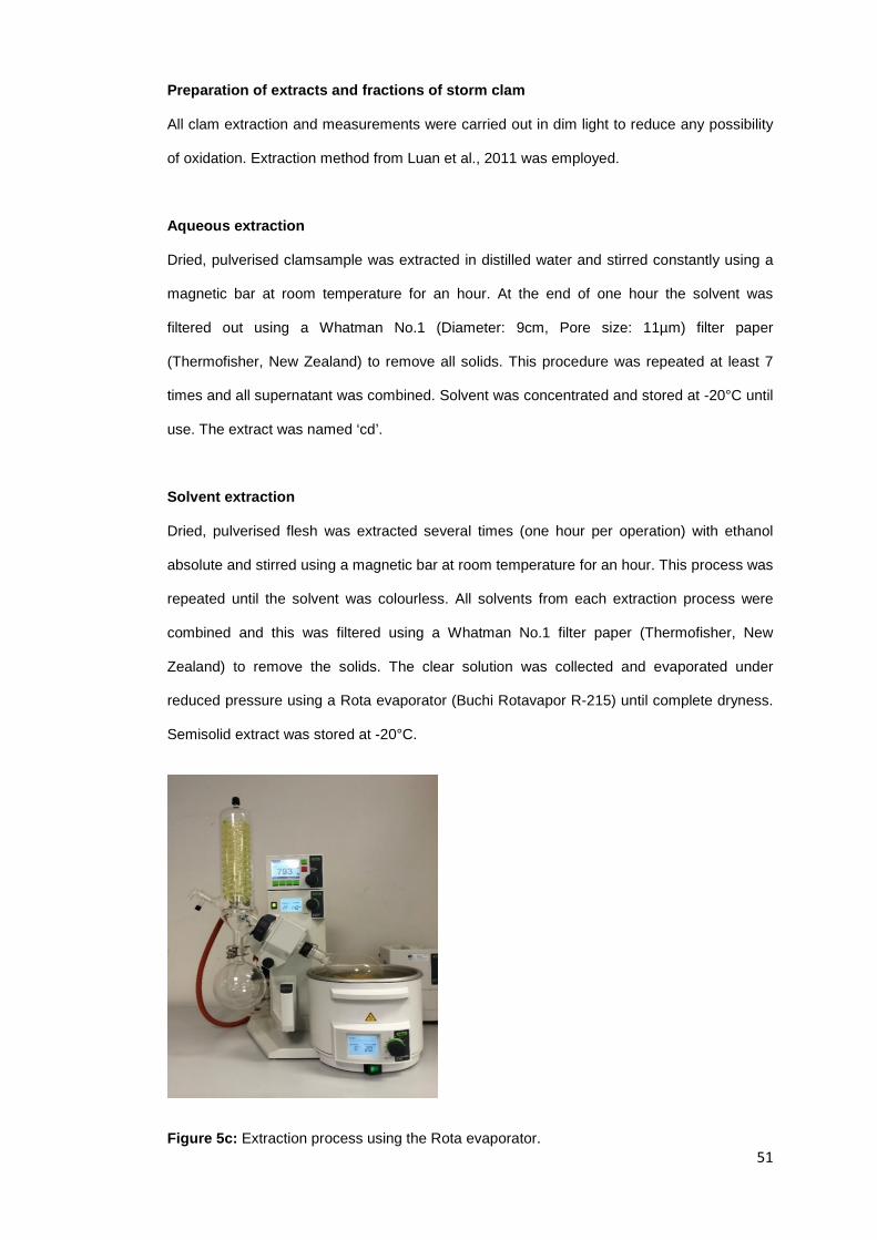
Preparation of extracts and fractions of storm clam
All clam extraction and measurements were carried out in dim light to reduce any possibility
of oxidation. Extraction method from Luan et al., 2011 was employed.
Aqueous extraction
Dried, pulverised clamsample was extracted in distilled water and stirred constantly using a
magnetic bar at room temperature for an hour. At the end of one hour the solvent was
filtered out using a Whatman No.1 (Diameter: 9cm, Pore size: 11µm) filter paper
(Thermofisher, New Zealand) to remove all solids. This procedure was repeated at least 7
times and all supernatant was combined. Solvent was concentrated and stored at -20°C until
use. The extract was named ‘cd’.
Solvent extraction
Dried, pulverised flesh was extracted several times (one hour per operation) with ethanol
absolute and stirred using a magnetic bar at room temperature for an hour. This process was
repeated until the solvent was colourless. All solvents from each extraction process were
combined and this was filtered using a Whatman No.1 filter paper (Thermofisher, New
Zealand) to remove the solids. The clear solution was collected and evaporated under
reduced pressure using a Rota evaporator (Buchi Rotavapor R-215) until complete dryness.
Semisolid extract was stored at -20°C.
Figure 5c: Extraction process using the Rota evaporator. 51

Fractionation
The crude extracts of the ethanol extraction were further fractionated by liquid-liquid
extraction. The ethanol extract was fractioned into four sub-fractions according to the polarity
of the extractive medium and using petroleum ether, ethyl acetate, n-butanol, and water. The
extracts were dissolved in 100ml distilled water and fractionated with petroleum ether.
Extracts (‘pe’) were collected and concentrated under reduced pressure. Further step-by-
step fractionation was done using ethyl acetate and lastly with n-butanol, which resulted in
the fractions of ‘ea’ and ‘nb’ respectively. Each fractionation process was repeated until the
solvent was colourless. The final aqueous fraction was named ‘w’. All four fractions (pe, ea,
nb, and w) were evaporated to dryness and stored in the fridge at -20⁰C until they were
ready to be used for the antioxidant analyses (DPPH and CUPRAC).
2.2.2 DPPH Radical Scavenging Activity Preparation of stock solution
The working solutions of test extracts were prepared by dissolving the required quantity of
test extracts in distilled water and then diluting in methanol absolute to give the
concentrations of 0.5µg/mL, 1µg/mL, 10µg/mL, and 20µg/mL.
Preparation of standard solution
Ascorbic acid was used as the reference standard. The standard solution was prepared by a
series of dilution in methanol absolute.
DPPH Analysis
The free radical scavenging capacity of the aqueous and ethanol extracts was determined
using DPPH (Duan, Zhang, & Wang, 2006). 0.16mM DPPH solution (6.31mg/100mL) was
prepared in absolute methanol. 2mL of freshly prepared DPPH solution was mixed with 2mL
of the sample in a cuvette so that the final volume was 4mL. The reaction tubes in triplicates
were shaken properly and after 30 min of incubation at room temperature, the absorbance
was read at 517nm using a spectrophotometer (ULTROSPEC 7000). Control sample was
prepared containing the same volume without any extracts. Absolute methanol was used to
52

zero the spectrophotometer as the blank. Percentage scavenging of the DPPH free radical
was measured using the following equation:
Scavenging (%) =�1 – �𝐴𝐴𝐴𝐴𝑟𝑟𝑠𝑠𝑠𝑠𝑠𝑠𝑠𝑠𝑠𝑠𝑠𝑠 − 𝐴𝐴𝐴𝐴𝑟𝑟𝑠𝑠𝑠𝑠𝑠𝑠𝑠𝑠𝑠𝑠𝑠𝑠𝑠𝑠𝑠𝑠𝑠𝑠𝑠𝑠𝑠𝑠
𝐴𝐴𝐴𝐴𝑟𝑟𝑐𝑐𝑐𝑐𝑠𝑠𝑐𝑐𝑐𝑐𝑐𝑐𝑠𝑠�� 𝑋𝑋 100
Where 𝐴𝐴𝐴𝐴𝐴𝐴𝑟𝑟𝑟𝑟𝑠𝑠𝑠𝑠𝑠𝑠𝑟𝑟 = the absorbance of the sample with treatment
𝐴𝐴𝐴𝐴𝐴𝐴𝑟𝑟𝑟𝑟𝑠𝑠𝑠𝑠𝑠𝑠𝑟𝑟𝐴𝐴𝑠𝑠𝑟𝑟𝑠𝑠𝑠𝑠 = the absorbance of sample with 2.0ml of methanol
𝐴𝐴𝐴𝐴𝐴𝐴𝑟𝑟𝑐𝑐𝑠𝑠𝑟𝑟𝑟𝑟𝑐𝑐𝑠𝑠 = the absorbance of 2.0ml of methanol with the treatment.
2.2.3 CUPRAC Assay Preparation of stock solution
The working solutions of test extracts were prepared by dissolving the required quantity of
test extracts in distilled water and then diluting in ethanol absolute to give the concentrations
of 0.1µg/mL, 0.5µg/mL, 1.5µg/mL, 2.5µg/mL, 3.5µg/mL, 5µg/mL, 7.5µg/mL, 10µg/mL,
15µg/mL, and 20µg/mL.
Preparation of other solutions
Ascorbic acid was used as the reference standard. The standard solutions (0.1µg/mL,
0.5µg/mL, 1.5µg/mL, 2.5µg/mL, 3.5µg/mL, 5µg/mL, 7.5µg/mL, 10µg/mL, 15µg/mL, and
20µg/mL) were prepared by a series of dilution in ethanol absolute.
The copper (II) chloride (CuCl2) solution (10mM) and ammonium acetate (NH2Ac) buffer
solution (1M, pH7) were prepared in pure distilled water and Neocuproine solution (7.5mM)
was prepared in absolute ethanol (Çelik, Özyürek, Güçlü, & Apak, 2010).
CUPRAC Analysis
Cupric reducing antioxidant power (CUPRAC) was determined as described by Apak, Güçlü,
Özyürek, & Karademir (2004). 1mL each of Cu (II), Nc, and NH2Ac buffer solutions were
53

added to a cuvette. Antioxidant standard solutions (1.0mL) and distilled water (0.1mL) were
added to the initial mixture so as to make the final volume of 4.1mL. The cuvettes were
manually shaken and after 30 min standing at room temperature, the absorbance at 450nm
(A450) was recorded against a reagent blank. The reading on the spectrophotometer
(ULTROSPEC 7000) was zeroed using distilled water as the blank. The absorbance of the
samples was recorded. The standard calibration curves of each antioxidant compound were
constructed in this manner as Absorbance vs. Concentration. The CUPRAC molar
absorptivity of each antioxidant was found from the slope of the calibration line concerned.
The scheme for normal measurement of antioxidants is summarized as:
Cu (II) (1mL) + Nc (1mL) + NH2Ac buffer (1mL) + sample (1mL) + H2O (0.1mL);
Total volume = 4.1mL (measured at 450nm against a reagent blank after 30 min of reagent
addition).
2.2.4 Statistical Analysis Analysis of data was done with GraphPad Prism (GraphPad Software, San Diego, CA).
Analyses of variance were performed using one-way ANOVA, with post hoc Barlett’s test in
GraphPad Prism, where significant differences occurred. Assays for both the DPPH and
CUPRAC analyses were repeated three times. Where shown, results were expressed as
mean ± standard deviation. A nominal two sided p < 0.05 was used to assess significance.
Correlation analysis was done using Microsoft Excel (2013).
54

CHAPTER THREE
3. RESULTS
3.1 ANTIOXIDANT ACTIVITY Crude extract and four fractions of three New Zealand surf clam species were tested for the
antioxidant activity using the DPPH and CUPRAC assays. The experimental results for both
assays are presented in the figures below, where crude extracts and all four fractions were
established to possess antioxidant activity. The antioxidant properties of all fractions,
determined using DPPH and CUPRAC assays, were compared with that of ascorbic acid.
3.2 DPPH Assay Proton-radical scavenging action is an important mechanism of an antioxidant, which is
measured by 2,2-diphenyl-1-picrylhydrazyl (DPPH) radical scavenging assay.
DPPH is a stable free radical that shows maximum absorbance at 517 nm in ethanol. When
DPPH encounters a proton-donating substance such as an antioxidant, the radical would be
scavenged and the absorbance is reduced. Based on this principle, the antioxidant activity of
the substance can be expressed as its ability in scavenging the DPPH (Wu, Chen, & Shiau,
2003).
The radical scavenging activity was observed in all tested samples in a concentration-
dependent manner (Figures 6-10 below). The present results reveal that the clam extracts
probably contained peptides, which were electron donors and could react with free radicals
to convert them to more stable products and terminate the radical chain reaction.
The percentages of inhibition of all clam extracts and fractions at concentrations ranging
from 0.5 - 20µg/mL were however lower than ascorbic acid. The ea fraction in all samples
showed very high scavenging ability. At a concentration of 20µg/mL, the percentage
inhibitions for Diamond shell, Tua tua and Storm shell were 73.29%, 76.14% and 73.69%
respectively. It can be observed that the pe, cd, and w fractions had very similar scavenging
55

abilities, as they closely followed the ea fraction. As shown in Table 2, the DPPH radical
scavenging activity was significantly higher (p < 0.05) in Diamond shell (p < 0.0001) and
Storm shell (p < 0.0001), followed by Tua tua (p = 0.017). It was observed that DPPH had
radical scavenging in the range of 0.5µg/mL - 20µg/mL.
Figure 6: DPPH radical scavenging activity of Tua tua (crude extract- cd, petroleum ether-pe, ethyl acetate- ea, n-butanol-nb, and aqueous-w fractions), and Ascorbic acid. Values are means from three independent tests.
0
10
20
30
40
50
60
70
80
90
100
0 2 4 6 8 10 12 14 16 18 20
% In
hibi
tion
Concentration (µg/mL)
Tua tua
Ascorbic acidTTcdTTpeTTeaTTnbTTw
56

Figure 7: DPPH radical scavenging activity of Diamond shell (crude extract-cd, petroleum ether-pe, ethyl acetate- ea, n-butanol-nb, and aqueous-w fractions), and Ascorbic acid. Values are means from three independent tests.
0
10
20
30
40
50
60
70
80
90
100
0 5 10 15 20
% In
hibi
tion
Concentration (µg/mL)
Diamond shell
Ascorbic acid
DScd
DSpe
DSea
DSnb
DSw
57

Figure 8: DPPH radical scavenging activity of Storm shell (crude extract-cd, petroleum ether-pe, ethyl acetate- ea, n-butanol-nb, and aqueous-w fractions), and Ascorbic acid. Values are means from three independent tests.
Table 2: DPPH radical scavenging activities by the New Zealand surf clam extracts.
Fraction Diamond shell (%) Tua tua (%) Storm shell (%)
cd 73.01 ± 1.53 64.02 ± 8.27 72.83 ± 4.89
pe 71.78 ± 1.34 77.25 ± 6.46 68.52 ± 1.83
ea 73.81 ± 2.40 75.83 ± 2.15 73.59 ± 0.76
nb 73.24 ± 1.58 68.93 ± 2.09 72.18 ± 2.41
w 74.73 ± 1.41 68.90 ± 9.52 74.48 ± 2.26
All the values are mean ± standard error of three samples.
0
10
20
30
40
50
60
70
80
90
100
0 2 4 6 8 10 12 14 16 18 20
% In
hibi
tion
Concentration (µg/mL)
Storm shell
Ascorbic acid
SScd
SSpe
SSea
SSnb
SSw
58
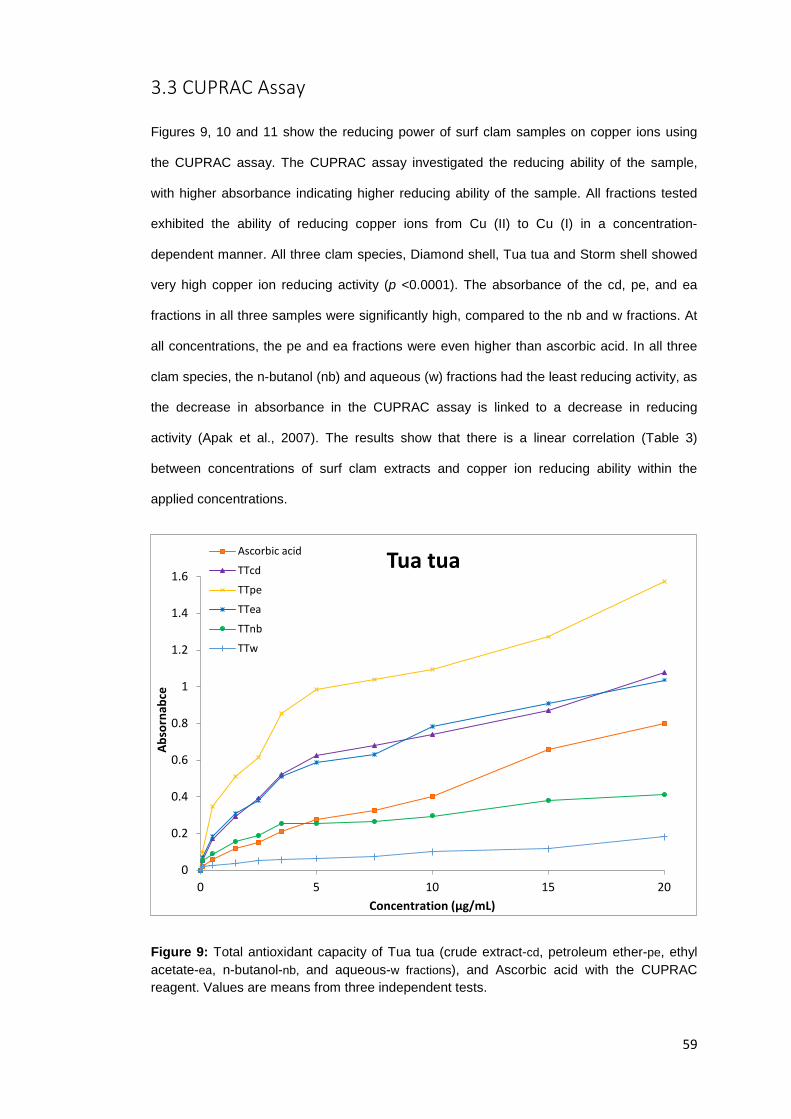
3.3 CUPRAC Assay Figures 9, 10 and 11 show the reducing power of surf clam samples on copper ions using
the CUPRAC assay. The CUPRAC assay investigated the reducing ability of the sample,
with higher absorbance indicating higher reducing ability of the sample. All fractions tested
exhibited the ability of reducing copper ions from Cu (II) to Cu (I) in a concentration-
dependent manner. All three clam species, Diamond shell, Tua tua and Storm shell showed
very high copper ion reducing activity (p <0.0001). The absorbance of the cd, pe, and ea
fractions in all three samples were significantly high, compared to the nb and w fractions. At
all concentrations, the pe and ea fractions were even higher than ascorbic acid. In all three
clam species, the n-butanol (nb) and aqueous (w) fractions had the least reducing activity, as
the decrease in absorbance in the CUPRAC assay is linked to a decrease in reducing
activity (Apak et al., 2007). The results show that there is a linear correlation (Table 3)
between concentrations of surf clam extracts and copper ion reducing ability within the
applied concentrations.
Figure 9: Total antioxidant capacity of Tua tua (crude extract-cd, petroleum ether-pe, ethyl acetate-ea, n-butanol-nb, and aqueous-w fractions), and Ascorbic acid with the CUPRAC reagent. Values are means from three independent tests.
0
0.2
0.4
0.6
0.8
1
1.2
1.4
1.6
0 5 10 15 20
Abso
rnab
ce
Concentration (µg/mL)
Tua tuaAscorbic acid
TTcd
TTpe
TTea
TTnb
TTw
59

Figure 10: Total antioxidant capacity of Diamond shell (crude extract-cd, petroleum ether-pe, ethyl acetate-ea, n-butanol-nb, and aqueous-w fractions), and Ascorbic acid with the CUPRAC reagent. Values are means from three independent tests.
0
0.2
0.4
0.6
0.8
1
1.2
1.4
1.6
0 5 10 15 20
Abso
rban
ce
Concentration (µg/mL)
Diamond shellAscorbic acid
DScd
DSpe
DSea
DSnb
DSw
60

Figure 11: Total antioxidant capacity of Storm shell (crude extract- cd, petroleum ether-pe, ethyl acetate-ea, n-butanol-nb, and aqueous-w fractions), and Ascorbic acid with the CUPRAC reagent. Values are means from three independent tests.
0
0.2
0.4
0.6
0.8
1
1.2
1.4
1.6
0 5 10 15 20
Abso
rban
ce
Concentration (µg/mL)
Storm shellAscorbic acidSScdSSpeSSeaSSnbSSw
61
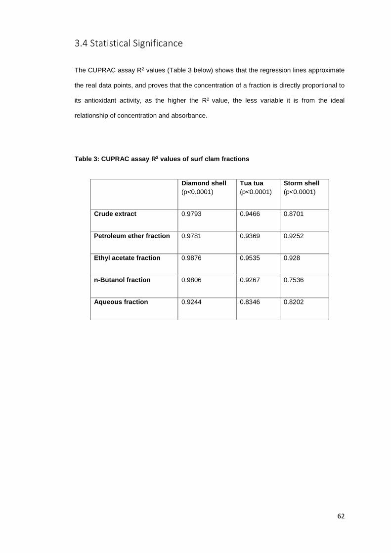
3.4 Statistical Significance
The CUPRAC assay R2 values (Table 3 below) shows that the regression lines approximate
the real data points, and proves that the concentration of a fraction is directly proportional to
its antioxidant activity, as the higher the R2 value, the less variable it is from the ideal
relationship of concentration and absorbance.
Table 3: CUPRAC assay R2 values of surf clam fractions
Diamond shell
(p<0.0001) Tua tua (p<0.0001)
Storm shell (p<0.0001)
Crude extract 0.9793 0.9466 0.8701
Petroleum ether fraction 0.9781 0.9369 0.9252
Ethyl acetate fraction 0.9876 0.9535 0.928
n-Butanol fraction 0.9806 0.9267 0.7536
Aqueous fraction 0.9244 0.8346 0.8202
62

The crude extracts and solvent fractions of all samples were further analysed at a higher concentration of 50µg/mL and then compared amongst each other. The
results represented in Figures 12a- 16b below show that the antioxidant activity of fractions tested increased with an increase in concentration.
Figure 12a: DPPH radical scavenging activity of all crude extracts Figure 12b: Copper ion reducing activity of all crude extracts
0
10
20
30
40
50
60
70
80
90
100
0 10 20 30 40 50
% In
hibi
tion
Concentration (µg/mL)
Crude extracts
Ascorbic acid
DScd
TTcd
SScd0
0.5
1
1.5
2
2.5
0 10 20 30 40 50
Abso
rban
ceConcentration (µg/mL)
Crude extractsAscorbic acid
DScd
TTcd
SScd
63

Figure 13a: DPPH radical scavenging activity of all pe fractions Figure 13b: Copper ion reducing activity of all pe fractions
0
10
20
30
40
50
60
70
80
90
100
0 10 20 30 40 50
% In
hibi
tion
Concentration (µg/mL)
Petroleum ether fractions
Ascorbic acid
DSpe
TTpe
SSpe0
0.5
1
1.5
2
2.5
3
3.5
0 10 20 30 40 50
Abso
rban
ce
Concentration (µg/mL)
Petroleum ether fractionsAscorbic acid
DSpe
TTpe
SSpe
64

Figure 14a: DPPH radical scavenging activity of all ea fractions Figure 14b: Copper ion reducing activity of all ea fractions
0
10
20
30
40
50
60
70
80
90
100
0 10 20 30 40 50
% In
hibi
tion
Concentration (µg/mL)
Ethyl acetate fractions
Ascorbic acid
DSea
TTea
SSea0
0.5
1
1.5
2
2.5
3
3.5
0 10 20 30 40 50
Abso
rban
ce
Concentration (µg/mL)
Ethyl acetate fractionsAscorbic acid
DSea
TTea
SSea
65

Figure 15a: DPPH radical scavenging activity of all nb fractions Figure 15b: Copper ion reducing activity of all nb fractions
0
10
20
30
40
50
60
70
80
90
100
0 10 20 30 40 50
% In
hibi
tion
Concentration (µg/mL)
n-Butanol fractions
Ascorbic acid
DSnb
TTnb
SSnb0
0.5
1
1.5
2
2.5
0 10 20 30 40 50
Abso
rban
ce
Concentration (µg/mL)
n-Butanol fractionsAscorbic acid
DSnb
TTnb
SSnb
66
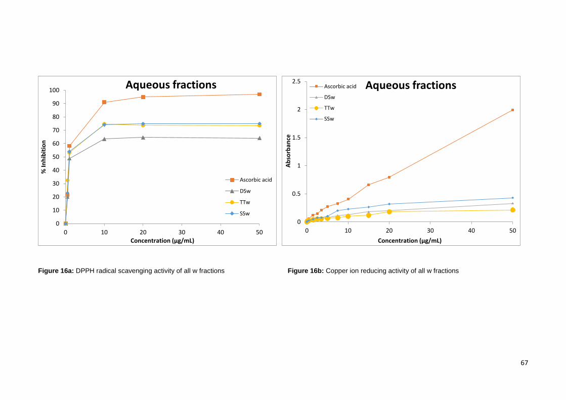
Figure 16a: DPPH radical scavenging activity of all w fractions Figure 16b: Copper ion reducing activity of all w fractions
0
10
20
30
40
50
60
70
80
90
100
0 10 20 30 40 50
% In
hibi
tion
Concentration (µg/mL)
Aqueous fractions
Ascorbic acid
DSw
TTw
SSw0
0.5
1
1.5
2
2.5
0 10 20 30 40 50
Abso
rban
ce
Concentration (µg/mL)
Aqueous fractionsAscorbic acid
DSw
TTw
SSw
67

Figure 17: Diamond shell CUPRAC assay: Sample stock solution 10mg/mL; Standard stock solution 10mg/mL.
Figure 18: Diamond shell CUPRAC assay: Sample stock solution 10mg/mL; Standard stock solution 1mg/mL.
0
0.5
1
1.5
2
2.5
3
3.5
4
4.5
5
0 0.5 1 1.5 2 2.5 3 3.5 4 4.5 5
Abso
rban
ce
Concentration (µg/mL)
Diamond shellAscorbic acid
DScd
DSpe
DSea
0
0.1
0.2
0.3
0.4
0.5
0.6
0.7
0 0.5 1 1.5 2 2.5 3 3.5 4 4.5 5
Abso
rban
ce
Concentration (µg/mL)
Diamond shellAscorbic acidDScdDSpeDSea
68

CHAPTER FOUR
4. DISCUSSION Free radical and active oxygen species oxidise biological molecules such as lipids, proteins, DNA,
and sugars to induce their modification (Wang, Sun, Cao, & Wang, 2010) which could lead to
oxidative stress. Shellfish of marine origin have been found to be good sources of antioxidants (Luan
et al., 2011). Three New Zealand surf clam species were evaluated for their antioxidant potentials.
The extraction of all clam species yielded five clam fractions; cd (crude extract), pe (petroleum ether
fraction), ea (ethyl acetate fraction), nb (n-butanol fraction) and w (aqueous fraction). Due to the
presence of different components in the crude extracts of biological tissue samples, it was relatively
difficult to measure each antioxidant component separately. Therefore, two assays (DPPH and
CUPRAC) were employed to evaluate the total antioxidant activity of the three NZ surf clam extracts
in this study. The CUPRAC assay measures the ability of reducing antioxidants to reduce copper ions
from Cu(II) to Cu(I) utilising bis(neocuproine) (Fung, 2012). Hence, CUPRAC measured the reducing
activity of the surf clam extract. In this study, the experimental results showed that all fractions
exhibited the ability to reduce copper ions from Cu (II) to Cu (I).
It is well accepted that the antioxidants can scavenge the DPPH radical through hydrogen-donating
behaviour (Chen & Ho, 1995). DPPH has been used as a free radical to determinate reducing
substances (Cotelle et al., 1996; Luan et al., 2011). All fractions tested exhibited a dose dependent
behaviour on DPPH radical scavenging activity within the test range of 0.5- 50μg/mL. This might be
due to the fact that the surf clam extracts possibly contained substances which were electron donors
and could react with free radicals to convert them to more stable products and terminate the radical
chain reaction. This data clearly demonstrates that the NZ surf clam extracts have significant
antioxidant properties.
69

4.1 Major Active Compounds in the NZ Surf Clam Extracts
The correlations between the major active compounds in the NZ surf clam extracts with their
antioxidant activities have been briefly reported below.
4.1.1 Crude Extracts (cd)
The inhibition of ascorbic acid towards DPPH radicals was higher than that obtained by the crude
extracts. (Figure 12a). Conversely, with the CUPRAC assay (Figure 12b), it can be seen that at
concentrations between 0.1µg/mL and 10µg/mL, all three crude extracts from the clam species
showed higher inhibitions than ascorbic acid, indicating that they possess stronger ability to reduce
copper ions.
The component contents evaluation in the surf clam, Mactra veneriformis, revealed that the crude
extract possessed free amino acids (140.49 ± 5.93µmolg-1) and oligosaccharides (360.52 ± 11.64
µmolg-1) (Luan et al., 2011).
Previous reports have shown that superoxide dismutase (SOD), catalase (CAT) and glutathione
peroxidase (GSH-Px) (endogenous antioxidant enzymes) activities increased significantly in plasma
from rats fed with Feruloyl oligosaccharides- FOs (the ferulic acid ester of oligosaccharides from
wheat bran). The increase in activities of SOD, CAT and GSH-Px in plasma from rats fed coincides
with the decrease of the levels of oxidative stress biomarkers (Wang, Sun, Cao, & Wang, 2010).
Also, Ou et al. (2007) reported that feeding FOs increased the level of antioxidant capacity and
significantly increased SOD and GSH-Px activity in the serum of diabetic rats compared to feeding the
sodium ferulate and vitamin C. Furthermore, FOs can enhance the ability of human lymphocytes to
resist H2O2 induced oxidative DNA damage and inhibit the non-enzymatic glycation of protein in a
glucose/BSA system under in vitro conditions. Therefore the favourable influence of phenolic
compounds on antioxidant enzyme activity merits attention. And this would be very helpful in
understanding the mechanism of prevention of chronic diseases by intake of diets rich in phenolic
compounds (Wang, Sun, Cao, & Wang, 2010).
The amino acid composition of the Meretrix casta peptides showed the presence of both essential
and non-essential amino acids including Proline, Glycine, Alanine, Phenylalanine, Histidine, Glutamic
70

acid, etc. The HPLC analysis for amino acid composition and clam peptides revealed that the
quantity of essential amino acids (milligrams of amino acid/gram of protein) in all samples was higher
than the recommended values for human adults. These antioxidant active peptides contained
glutamic acid, histidine, threonine, arginine in good percentage than remaining amino acids. The
presence of amino acids like histidine and methionine might be the reason for good antioxidant
activity (Nazeer, Prabha, Kumar, & Ganesh, 2013). Some amino acids such as Tau, Tyr, Met, Cys,
and Arg have shown obvious antioxidant activities and constituted 14.59% of the total amino acid
contents of Mactra veneriformis. Tau could scavenge reactive oxygen species and prevent changes in
membrane permeability following oxidant injury (Luan et al., 2011).
Several amino acids such as Tyr, Met, His, Lys, and Trp are generally accepted as antioxidants and
the presence of all these amino acids makes the clam peptide a potent antioxidant (Chen, Muramoto,
Yamauchi, & Nokihara, 1996; Nazeer, Prabha, Kumar, & Ganesh, 2013).
4.1.2 Petroleum Ether Fraction (pe) The results showed that the petroleum ether fraction was the strongest DPPH radical scavenger with
a 77.25% inhibition (Table 2) and a very strong copper ion reducer, hence, there must be some
compounds possessing very strong antioxidant properties occurring in the fraction. The pe results
obtained in this study corresponds with Psoralea corylifolia L, (Jiangning, Xinchu, Hou, Qinghua, &
Kaishun, 2005). Upon analysis, it was observed that the petroleum ether fraction contained a phenolic
hydroxyl group and that there is a double bond directly connecting the phenol in the para-position of
the phenolic hydroxyl group. This double bond extends the conjugation system of the phenolic oxygen
free radical of the extract after donating a hydrogen atom to active free radicals. Hence, this group
stabilises the phenolic oxygen free radical a lot and greatly strengthens the antioxidant activities of the
compound. In other words, more resonance structures can be formed to stabilize the free radical
because of the alkenic group (Jiangning, Xinchu, Hou, Qinghua, & Kaishun, 2005).
According to Luan et al. (2011), the highest content of unsaturated fatty acid was detected in the
petroleum ether fraction. The n-3 series unsaturated fatty acid could act as indirect antioxidant in
vascular endothelial cells, hence diminishing inflammation, and in turn the risk of atherosclerosis and
cardiovascular disease.
71

The 4, 7, 10, 13, 16, 19-docosahexaenoic acid (DHA) and 5, 8, 11, 14, 17-eicosapentaenoic acid
(EPA) lower serum lipid concentration, reduce incidence of CVD and prevent platelet aggregation and
they have been used in clinical practice (Luan et al., 2011). It has also been established that these
polyunsaturated fatty acids decrease the extent of cell proliferation in cultures of smooth muscle or
medial cells (Gavino, Miller, Ikharebha, Milo, & Cornwell, 1981). In vivo studies (Gao et al., 2007;
Frenoux, Prost, Belleville, &. Prost, 2001) have shown that the consumption of n-3 unsaturated fatty
acid, especially EPA, lowered blood pressure, improved the total antioxidant activity status and
decreased oxidative stress.
Furthermore, cellular evidence for the antioxidant actions of polyunsaturated acids has been provided.
Fatty acid micelles scavenged superoxide in an unsaturation dependent manner, up to EPA which
was the most effective O2•− scavenger. In particular, EPA and DHA decreased ROS production by
16.9% and 15.8%, respectively, as compared to stearic acid-supplemented controls. LC-PUFAs,
including EPA, are also effective inhibitors of superoxide production by HAEC at more physiological
concentrations. The inhibition of NAD(P)H oxidase by DHA and (presumably) other PUFAs might
greatly explain the observed effects on ROS production. One other potential mechanism of action of
LC-PUFAs would be that they act as a “sink” to trap free radicals, hence, becoming oxidised
themselves (Richard, Kefi, Barbe, Bausero, & Visioli, 2008). These and more may be the reasons why
the pe fractions of all samples showed very high antioxidant activities, especially with the CUPRAC
assay.
4.1.3 Ethyl Acetate Fraction (ea) At all concentrations evaluated (0.1µg/mL - 50µg/mL), the copper ion reducing abilities of the ea
fraction of all three surf clam species were higher than ascorbic acid (Figure 14b). The highest
correlation between a clam extract and the CUPRAC assay was found in the ea fraction (R2 = 0.9876,
p <0.0001). Compared with other fractions, the ea fraction (of Tua tua) also had the second highest
DPPH radical scavenging ability (Table 2) with a 75.83% inhibition. This result was identical with
those of Hypericum hyssopifolium (Cakir et al., 2003) where the ea fraction contained the highest
amount of total phenolic compounds, which could be accepted as indication of antioxidant potential
(Yıldırım, Mavi, & Kara, 2001), and Polysiphonia urceolata (Duan, Zhang, Li,& Wang, 2006) in which
72

the ethyl acetate-soluble fraction showed the most potent activity and indicated that compounds with
strongest radical scavenging activity in P. urceolata are of medium polarity. Furthermore, the
distribution of phenolic compounds in P. urceolata demonstrated that the ea-soluble fraction
contained the highest amount and a high correlation was found between the total phenolic content
and DPPH radical scavenging in P. Urceolata (Duan, Zhang, Li, & Wang, 2006).
The phenolic compounds of the ea fraction showed very high antioxidant activity. This could be
responsible for its antioxidant activity as the free –OH groups in phenolic compounds have been
linked with antioxidant activity (Weng & Wang, 2000; Cakir et al., 2003). It has been reported that if
there is an electron donating group, especially a hydroxyl group located on phenolic compounds, their
antioxidant activities are increased greatly (Weng, 1993).
The gas chromatography-mass spectrometric analysis of the ea fraction of the freshwater clam
showed that it was composed mainly of fatty acids and steroids (Hua et al., 2012). Steroids have been
found in plant and animal extracts (Akinmoladun, Ibukun, Afor, Obuotor, & Farombi, 2007) and may
be responsible for their medicinal value. Steroids have been reported to be an active compound in
Solanum aculeastrum where they are showing high antioxidant properties (Koduru, Jimoh, Grierson,
& Afolayan, 2007).
Fatty acids, on the other hand, have the ability to augment free radical generation (Das, 1991).
4.1.4 n- Butanol Fraction (nb) The nb fraction revealed the highest nucleoside contents. And the identification of hypoxanthine,
xanthine, inosine and guanosine was determined by high-performance liquid chromatography (HPLC)
analysis (Luan et al., 2011). Xanthine is known to have a quenching effect on the production of
hydroxyl radicals, as well as on oxidative DNA breakage by hydroxyl radicals. Xanthine bind to copper
ions, leading to the reduction of Cu(II) to Cu(I). This leads to a prooxidant action capable of inducing
DNA breakage. However, such an activity requires a relatively high concentration of the compounds
and copper ions (Azam, Hadi, Khan, & Hadi, 2003). Nucleoside analogues with the α,β-unsaturated
keto system, compounds 9, 10 and 11 were potent against the activity of ROO• (Spanou, Manta,
Komiotis, Dervishi, & Kouretas, 2007). In their study, Gudkov et al. (2006) stated that purine
nucleotides (especially guanosine and inosine) are natural antioxidants and protect DNA in vitro from
73
damage by ROS generated by ionizing radiation and heat. These nucleotides decrease heat-induced
generation in aqueous solutions of such ROS as hydrogen peroxide and hydroxyl radical. It was found
that guanosine and inosine, when injected intraperitoneally, function as radio-protectors that increase
the survival rate of mice exposed to lethal doses of γ-radiation and that they are most effective when
injected shortly after irradiation. A considerable radioprotective effect was also observed in mice that
consumed nucleosides after irradiation with drinking water. Today, inosine (under the commercial
name riboxin) is used in clinical practice for treating for the ischemic heart disease, arrhythmias,
myocardial infarction and other pathologies. The conversion of hypoxanthine to uric acid is particularly
principal as uric acid is regarded to be an important antioxidant in human adult plasma. It can directly
react with free radicals and may interfere with Fenton's chemistry by chelating catalytic metal ions
(Žitňanová et al., 2004). However, consistent with previous observations, the nb fraction showed the
least antioxidant activity (especially with the CUPRAC assay) (Figure 15b). This might be due to the
fact that little correlation was found between the nucleoside contents and the antioxidant activity of the
extract, due to their stearic inaccessibility or the fact that nucleosides have higher antioxidant activity
in vivo than in vitro (Luan et al., 2011; Spanou, Manta, Komiotis, Dervishi, & Kouretas, 2007; Gudkov,
Shtarkman, Smirnova, Chernikov, & Bruskov, 2006).
4.1.5 Aqueous Fraction (w) The w fraction of all three clam species showed antioxidant activity. Although this fraction was the
least copper ion reducing fraction (CUPRAC assay), it showed relatively high DPPH radical
scavenging activity. A spectrum of oligosaccharides was analysed from the aqueous fraction of
Mactra veneriformis. This mainly consisted of monosaccharide, disaccharide and trisaccharide which
positively correlated with its metal ion chelating activities (Luan et al., 2011). In a dose-response
relation, agar oligosaccharides exhibited lipid peroxidation inhibition. They also showed antioxidative
activities in scavenging superoxide anion and hydroxyl free radical (Wang, Jiang, Mou, & Guan,
2004). In vitro antioxidant studies showed that agaro-oligosaccharides expressed different antioxidant
abilities. And it is quite significant that the in vivo animal experiment for agaro-oligosaccharides is
quite consistent with the in vitro assays. Besides successful protection of liver damage by efficiently
inhibiting MDA formation and decreasing AST and ALT, agaro-oligosaccharides enhance the activities
74

of antioxidant enzyme system of the host including SOD, GSH-Px etc (Chen, Yan, Zhu, & Lin, 2006).
The w fractions of two species (Diamond shell and Storm shell) showed the highest DPPH radical
scavenging ability, with 74.73% and 74.48% respectively (Table 2). Interestingly, this agrees with
Yuan et al. (2005) and Chen & Yan, (2005) that oligosaccharides showed excellent scavenging
activity on DPPH radicals, which may be attributable to its strong hydrogen-donating ability compared
to other samples.
4.2 Comparison of antioxidant activity among species
All species tested showed very high antioxidant activity. Tua tua had the highest DPPH radical
scavenging activity of 76.14% at 20µg/mL, and the fraction with the least scavenging activity was
TTcd with a 67.53% inhibition at 20µg/mL (Figure 6). Storm shell was the next on the DPPH radical
scavenging scale, with SSw as its fraction with the highest inhibition, and SSpe as its fraction with the
lowest inhibition. SSw had a 74.88% inhibition at 20µg/mL, while SSpe had an inhibition of 68.79% at
20µg/mL (Figure 8). The Diamond shell was the surf clam species with the least DPPH radical
scavenging activity. DSw had the highest inhibition, while DSnb had the least with 74.6% and 65.12%
inhibitions respectively (Figure 7). Comparing among species, the fractions with the highest inhibition
rate would be in this order; TTea > SSw > DSw, and fractions with the lowest inhibition rate as
follows; SSpe > TTcd > DSnb. It is interesting to observe that among all three species tested in this
study, Diamond shell was the species with the least DDPH radical scavenging ability.
The Diamond shell (which was the species with the least DPPH radical scavenging ability) had the
highest copper reducing activity (Figure 14b), with DSea having an absorbance of 1.596 at 20µg/mL.
DSw was the fraction that showed the least copper reducing ability, with an absorbance of 0.201 at
20µg/mL (Figure 10). The Diamond shell was closely followed by Tua tua, with TTpe (highest Tua tua
copper reducing fraction) having a 1.575 absorbance and TTw (lowest Tua tua copper reducing
fraction) a 0.182 absorbance at 20µg/mL respectively (Figure 9). Storm shell was the species with the
least copper reducing ability. At 20µg/mL, its highest absorbance was 1.327 (SSea), and its lowest
(SSw) 0.321. The order of copper ion reducing activities among surf clam species is DSea > TTpe >
75

SSea. As a matter of interest, the ea fraction seemed to have very high copper reducing abilities than
other fractions tested. The DSea and SSea were the highest copper reducers in the Diamond shell
and Storm shell respectively. In Tua tua however, the ea fraction was the second highest copper
reducer (after the pe fraction), with an absorbance of 1.036 at 20µg/mL.
4.3 Comparison of antioxidant activity in the NZ surf clam and other marine species and plant products
Compared with other bivalves, the free radical scavenging ability of the NZ surf clam can be said to
have very strong antioxidant activity. In contrast with concentrations used in this study, the free radical
scavenging ability of most bivalves in many researches was analysed at rather high concentration
ranges, e.g. Oyster (Crassostrea talienwhannensis) - 0.63 to 20 mg/mL (Dong et al., 2010), Oyster
(Saccostrea cucullata) - 0.25 to 1mg/mL (Umayaparvathi et al., 2014), Black mussel (Mytilus
galloprovincialis) - 2mg/mL (Monchevaa et al., 2004), and Mytilus edulis - 10mg/mL (Wang et al.,
2013). The percentage of free radical scavenging activity of the smooth hound (Mustelus mustelus)
hydrolysate (76.7% at 3mg/mL) (Bougatef et al. 2009) was significantly lower than the TTea fraction
which was 76.45% at 50µg/mL (Figure 14a). Hence, one can say that the NZ surf clam is a potential
source for antioxidant activity.
In comparison with seaweed (1000µg), Sargassum marginatum (11.00%), Padina tetrastomatica
(14.78%), Turbinaria conoides (17.23%) (Chandini, Ganesan, & Bhaskar, 2008) had relatively lower
percentage inhibitions than DSw which had an inhibition of 64.09% at 50µg/mL, and was the fraction
with the lowest free radical inhibition (Figure 16a). At 2mg/mL, Undaria pinnatifida (neutrase extract)
had a percentage inhibition of 57.08% (Je et al., 2009), while Gelidiella acerosa had an inhibition of
72.5% at 100μg/mL (Devi, Suganthy, Kesika, Pandian, 2008). These results were attained by most
clam fractions at concentrations less than 50µg/mL. TTpe, TTea, TTw, SScd, SSea, SSnb, SSw and
all DS fractions had over 70% inhibition at a concentration as low as 20μg/mL, whereas the F1, F2
and F3 fractions of the New Zealand Undaria pinnatifida (Harvey) Suringar required a concentration of
4000μg/mL to reach similar percentage (Mak, 2012).
76

At concentrations of 10, 20 and 30μg/mL, the kiwifruit had DPPH radical scavenging percentages of
4.9, 14.8 and 16.7% respectively (Bursal & Gülçin, 2011), which was nowhere close to any of the
fractions tested in this study at 0.5μg/mL. An earlier study on Apples reported that the maximal
percentage inhibition of 69% was reached at a concentration of 1mg/mL (Chaudhary et al., 2006),
which was attained by most fractions, including all ea and pe (with the exception of SSpe which had
an inhibition of 68.79%) fractions at 20 μg/mL (Figures 13a & 14a). In another study (Leontowicz et
al., 2003), the apple pulp extract at a concentration of 5 mg/mL was reported to quench only 54.4% of
DPPH radicals, and Pear pulp extract at concentrations of 2 and 5 mg/mL quenched only 6.5 and
10.8% of DPPH radicals, respectively. It is evident that the NZ clam extracts showed very strong
proton donating ability and could serve as free radical inhibitors or scavengers, and could possibly act
as primary antioxidants.
Unfortunately, the CUPRAC assay values could only be compared with a couple of studies due to
more common application of other assays in other studies. One of such studies (Fung, Hamid, & Lu,
2013) reported that in July (the month with the highest copper reducing activity), processed New
Zealand U. pinnatifida had an absorbance of 0.93, while fresh U. pinnatifida had an absorbance of
1.41. These absorbances were obtained at 1mg/g, which was several times higher than
concentrations used in this study. Boğa, Hacıbekiroğlu, & Kolak, (2011) also reported the copper ion
reducing ability of some edible plants. At 100μg/mL, the dichloromethane extracts of Celery, Spinach,
Red cabbage and Cinnamon had absorbance values of 0.369, 0.171, 0.729, and 1.073 respectively.
These values were very low compared to those obtained in this study, as they were exceeded by all
cd, pe and ea fractions at 50μg/mL. Thus the NZ surf clam, compared with edible plants and marine
algae, possess higher copper reducing abilities
4.4 Correlations The results revealed that most fractions tested had very high copper reducing activity. Comparing
between clam species, the Diamond shell had the highest correlation, as all DS fractions had higher
R2 values than the same fraction in other clam species. The DSea fraction had the highest correlation
of all surf clam fractions, with an R2 value of 0.9876.
77

Interestingly, the aqueous fractions of both the Diamond shell and Tua tua species were the fractions
with the least correlation, with R2 values of 0.9244 and 0.8346 respectively. This is indicated by the
antioxidant activity of the fractions, as they (DSw and TTw) were the fraction samples with the least
antioxidant activity in the two species (Figures 9 and 10).
Among all fractions tested, the least linear correlation based on concentration versus absorbance was
found in the SSnb fraction with an R2 value of 0.7536, followed by the SSw and TTw fractions with R2
values of 0.8202 and 0.8346 respectively. It should be known, however, that the R2 value does not
always indicate the adequacy of a model (Frost, 2013). This is true with the SSnb fraction which had
the least R2 value of 0.7536, yet had higher absorbance at concentrations between 0.1 and 20µg/mL
than SSw (Figure 11), which had an R2 value of 0.8202. The same is also true for the pe and ea
fractions of the Tua tua species. TTpe showed higher copper reducing activity than TTea (Figure 9),
but had lower linear correlation (R2 value = 0.9369) than TTea (R2 value = 0.9535).
In this study, a nominal two sided p < 0.05 was used to assess significance. A decrease in the p value
of an experiment indicates an increase in its significance. The results show that all fractions tested
with the CUPRAC assay were statistically significant, with p < 0.0001 in all three clam species. With
the DPPH assay, results showed that surf clam species also had significantly high radical scavenging
ability- Diamond shell (p < 0.0001) Storm shell (p < 0.0001), and Tua tua (p = 0.017). Thus the
observed copper reducing and DPPH radical scavenging abilities actually reflect characteristics of the
New Zealand surf clam rather than just sampling errors.
4.5 Differences in DPPH scavenging activity and CUPRAC reducing activity
Comparing the DPPH radical scavenging capacity of the New Zealand surf clam to their copper ion
reducing ability, it can be seen that all samples possessed higher copper ions reducing abilities than
radical scavenging, with most fractions showing higher reducing values than ascorbic acid. This might
be due to the difference in stock solution concentrations used, not necessarily because the extracts
are better copper ion reducers than DPPH radical scavengers (See 5.1 - Caveats). Another reason for
78

the differences in the radical scavenging ability and copper reducing ability of the surf clam could be
due to the selectivity of antioxidants that react differently in each assay. The CUPRAC assay,
moreover, can simultaneously measure hydrophilic and lipophilic antioxidants, whereas the DPPH
radical scavenging assay is solvent dependent (Prior et al., 2005; Fung, 2012).
The differences in antioxidant activities of each fraction (or active compounds in each fraction),
moreover, may be due to their solubility effect. Phenolic compounds of the ethyl acetate fraction have
been reported to be soluble in oil phase, hence the very high antioxidant activity (Figures 14a & b).
Lower antioxidant activities of other fractions (which naturally choose to move water phase in an oil-
in-water emulsion) may be due to this solubility effect (Cakir et al., 2003). Compared to all other
fractions tested, a very lower copper ion reducing rate was observed in the w fraction. This could be
due to the fact that enzymatic extraction is aqueous based and may not be as effective in extracting
the active principles like in the case of solvent extraction (Ganesan, Kumar, & Bhaskar, 2008).
79

CHAPTER FIVE
5. CONCLUSION
5.1 Caveats Sample stock solutions for the DPPH assay were prepared at a very low concentration of 1mg/10mL
to investigate the strength of the surf clam antioxidant capacity.
Low working sample concentrations were also used (0.1µg/mL- 20µg/mL) as opposed to those of
previous studies (Ganesan, Kumar, & Bhaskar, 2008; Cakir et al., 2003; Srivastava, Harish, &
Shivanandappa, 2006; Mak, 2012). Interestingly, the results have shown that the three New Zealand
surf clams tested have very high DPPH radical scavenging activity at such low concentrations. This
(low working sample concentration) may be one of the reasons why none of the fractions tested had
higher antioxidant abilities than ascorbic acid in the DPPH test. A higher concentration of the stock
solution (and perhaps higher working sample solution concentrations) would probably have shown
higher scavenging activity. Furthermore, reports (Zhou et al., 2012) have shown that an increase in
incubation time is directly proportional to an increase in antioxidant activity. In this study, incubation
time was 30 minutes. An increase in incubation time might have yielded higher percentage inhibition
values.
DPPH assay is based on kinetic derived process, that is, it may take one minute (e.g. ascorbic acid)
to more than 180 min (e.g. curcumin) and therefore the assay should be performed in kinetic mode
and estimation of antioxidant capacity of unknown antioxidant compounds must be counter-measured
by using other antioxidant assays e.g. ABTS, FRAP, ORAC, etc (Mishra, Ojha, & Chaudhury, 2012).
The different relative radical scavenging capacity of individual clam fractions against different DPPH
radicals may be explained by the different mechanisms involved in the radical–antioxidant reactions.
Other factors, such as stereoselectivity of the radicals or the solubility of the clam extracts in different
testing systems, may also affect its capacity to react and quench different radicals. The mechanisms
involved in the beneficial actions of antioxidants in biological systems included directly quenching of
free radicals to terminate the radical chain reaction, chelating transition metals to suppress the
80

initiation of radical formation, acting as reducing agents, or stimulating the antioxidative defence
enzyme activities (Monchevaa et al., 2004).
On the other hand, with the CUPRAC assay, an initial standard solution of 10mg/mL against a
10mg/mL sample stock solution was used. But the results showed very little to no effect vis-à-vis
copper ion reduction (Figure 17). The standard stock solution was reduced to 1mg/mL. Again, results
showed a minuscule reducing effect (Figure 18). To obtain results within the standard curve range,
the concentration of sample stock solution was increased. A sample stock solution of 50mg/mL was
finally used and results showed very high copper ion reducing effect.
Processed food have been found to contain fewer antioxidant vitamins than fresh and uncooked
foods, as preparation exposes food to heat and oxygen (Henry & Heppell, 2002). The New Zealand
surf clam, like most foods, might have had some vitamins destroyed, or some enzymes deactivated
due to heat processing. There might have been a significant increase in the antioxidant activities of
the New Zealand surf clam if they were not oven dried.
In this study, whole soft dried parts of surf clams were used. This implies that different tissues with
different antioxidant capacities were mixed together, without distinguishing what is happening in a
particular tissue, for instance, in the digestive gland, gills (Monchevaa et al., 2004), foot, mantle,
mouth or viscera.
It should be known that many natural antioxidants are less potent than synthetic antioxidants but they
can be used at higher concentrations than the synthetic ones, due to the very restrictive toxicological
parameters of the latter (Kim, Je, & Kim, 2007). Thus, consuming foods possessing natural
antioxidants like the surf clam could deliver desirable nutritional and functional properties.
5.2 Future Research Directions
This in vitro study has shown that the New Zealand surf clam have antioxidant effects, with some
fractions more potent than the ascorbic acid standard. The significantly potent antioxidant effects was
81

found in DPPH free radical scavenging, and in reducing copper ion. This indicates that the clams may
possess some novel compounds with antioxidant activities. It would be of great research interest to
further elucidate the exact molecular structures responsible for its antioxidant activities.
The New Zealand surf clam extracts showed higher antioxidant activities than many seaweed,
bivalves and marine products. This could be due to the fact that the NZ surf clam contained peptides
with higher antioxidant activities than other most samples. A post-column HPLC and the application of
nuclear magnetic resonance (NMR) spectroscopy could be used to analyse a wide range these
endogenous metabolites from surf clam tissues, detect and obtain more structural information on any
polysaccharides present that may be responsible for the clams’ high antioxidant ability. In order to
identify new compounds, moreover, gel permeation chromatography- based separation could be
employed.
Surf clam extracts have been reported to have therapeutic effect on Leukaemia 1210 in mice, and
proven to inhibit the growth of other human cancer cells (Pan, Huang, Chan, Ho, & Pan, 2008;
Ruggieri, 1975). It would be useful to test the anticancer/ antitumor activity of the New Zealand surf
clam on human cancer cell lines, especially breast, lung, melanoma and colorectal cancer cell lines,
as these have been reported to have significantly increased in New Zealand (Ministry of Health,
2008).
It has been reported that for a number of compounds tested for DPPH free radical scavenging ability,
the reactions are slow with very complex mechanisms (Bondet, Brand-Williams, & Berset, 1997).
Further research to help understand the mechanism of action of the surf clam vis-à-vis its DPPH
scavenging activity could also be carried out.
Among all the oxidative radicals, hydroxyl radicals are reported to be the most reactive free radicals
formed in biological systems. They can almost react with all the substances in the living cells and
induce severe damage to the cells (Luan et al., 2011). Hydroxyl radical scavenging is therefore said to
be the most effective method to estimate the behaviour of antioxidants. It would be highly
advantageous if the hydroxyl radical scavenging activity of the surf clam is also tested.
82

5.3 Overall Conclusion In conclusion, the surf clams from Cloudy Bay Clams, New Zealand, were found to have great
antioxidant potential for pharmaceutical developments. The Diamond shell, Tua tua and Storm shell
showed high DPPH radical scavenging and copper reducing properties.
The broad range of antioxidant activity of the extracts indicates the potential of the clam as a source
of natural antioxidants or nutraceuticals with potential application to reduce oxidative stress with
consequent health benefits.
83

REFERENCES
Agasen, E. V., Del Mundo, C. M., & Matias, G. O. (1998). Assessment of Paphia undulata in Negros Occidental/ Guimaras Strait waters. Journal of Shellfish Research, 17(5), 1613-1617.
Ajithkumar, P. (2012). Evaluation of Anti-ulcer Activity of Villorita cyprinoides extract (Black water clams) Against Immobilization Stress Induced Ulcer in Albino Rats. Journal of Pharmaceutical Research & Opinion, 2(6).
Akinmoladun, A. C., Ibukun, E. O., Afor, E., Obuotor, E. M., & Farombi, E. O. (2007). Phytochemical constituent and antioxidant activity of extract from the leaves of Ocimum gratissimum. Scientific Research and Essay, 2(5), 163-166.
Akpinar, A., Toker, H., Ozdemir, H., Bostanci, V., Aydin, H. (2013). The effects of non-surgical periodontal therapy on oxidant and anti-oxidant status in smokers with chronic periodontitis. Archives of Oral Biology, 58, 717–723.
Aldridge, D. W., Payne, B. S., & Miller, A. C. (1987). The effects of Intermittent Exposure to Suspended Solids and Turbulence on Three Species of Freshwater Mussels. Environmental Pollution. 45, 17-25.
Alexieva, B., Markova, T., Nikolova, E., Aragane, Y., & Higashino, H. (2010). Free radicals, antioxidants and cancer chemotherapy. Acta Medica Kinki University, 35(2), 57- 65.
Aljadani, N. (2013). The reproductive biology of the surf clams Triangle shell (Spisula auquilatera), Ringed dosinia (Dosinia anus) and Deep water Tuatua (Paphies donacina) from the North- East of South Island, New Zealand (Unpublished master’s thesis). Auckland University of Technology, Auckland, New Zealand.
Amarowicz, R., Naczk, M., & Shahidi, F. (2000). Antioxidant Activity of Various Fractions of Non-Tannin Phenolics of Canola Hulls. Journal of Agricultural and Food Chemistry, 48, 2755-2759.
Ambrose, W. G., Jones, D. S., & Thompson, I. (1980). Distance from shore and growth rate of the suspension feeding bivalve, Spisula solidissima. Proceedings of the National Shellfisheries Association, 70, 207-215.
Amornrut, C., Toida, T., Imanari, T., Woo, E-R., Park, H., Linhardt, R., Wu, S, J., & Kim, Y.S. (1999). A new sulfated b-galactan from clams with anti-HIV activity. Carbohydrate Research, 321, 121-127.
Andallu, B., Shankaran, M., Ullagaddi, R., & Iyer, S. (2014). In vitro free radical scavenging and in vivo antioxidant potential of mulberry (Morus indica L.) leaves. Journal of Herbal Medicine, 4, 10-17.
Aneiros, A., & Garateix, A. (2004). Bioactive peptides from marine sources: pharmacological properties and isolation procedures. Journal of Chromatography B, 803, 41–53.
Ansell, A. D. (1968). The Rate of Growth of the Hard Clam Mercenaria mercenaria (L) throughout the Geographical Range. Journal du Conseil / Conseil Permanent International pour l'Exploration de la Mer, 31(3), 364-409.
Anthony, K. P., & Saleh, M. A. (2013). Free Radical Scavenging and Antioxidant Activities of Silymarin Components. Antioxidants, 2, 398-407. doi: 10.3390/antiox2040398
Apak, R., Gorinstein, S., Böhm, V., Schaich, K. M., Özyürek, M., & Güçlü, K. (2013). Methods of measurement and evaluation of natural antioxidant capacity/activity (IUPAC Technical Report). Pure and Applied Chemistry, 85(5), 957–998.
Apak, R., Güçlü, K., Özyürek, M., & Çelik, S. E. (2008). Mechanism of antioxidant capacity assays and the CUPRAC (cupric ion reducing antioxidant capacity) assay. Microchimica Acta, 160, 413-419.
Apak, R., Güçlü, K., Demirata, B., Özyürek, M., & Çelik, S. E., Bektaşoğlu, B., Berker, K. I., & Özyurt, D. (2007). Comparative Evaluation of Various Total Antioxidant Capacity Assays Applied to Phenolic Compounds with the CUPRAC Assay. Molecules, 12, 1496-1547.
Apak, R., Güçlü, K., Özyürek, M., & Karademir, S. E. (2004). Novel total antioxidant capacity index for dietary polyphenols and vitamins C and E, using their cupric ion reducing capability in the
84

presence of neocuproine: CUPRAC method. Journal of Agricultural and Food Chemistry, 52(26), 7970–7981.
Aparadh, V. T., Naik, V. V., & Karadge, B. A. (2012). Antioxidative Properties (Tpc, Dpph, Frap, Metal Chelating Ability, Reducing Power and Tac) within Some Cleome Species. Annali di Botanica, 2, 49–56.
Apel, K. & Hirt, H. (2004). Reactive Oxygen Species: Metabolism, Oxidative Stress, and Signal Transduction. Annual Review of Plant Biology, 55, 373-99.
Arthritis Research UK. (2014). Global study highlights ongoing global burden of rheumatoid arthritis. Retrieved October 1, 2014, from http://eastmidslibdems.org.uk/en/article/2014/0778096/global-study-highlights-ongoing-global-burden-of-rheumatoid-arthritis
Aruoma, O. I. (1994). Nutrition and Health Aspects of Free Radicals and Antioxidants. Food and Chemical Toxicology, 32(7), 671-683.
Aruoma, O. I., Halliwell, B., Hoey, B. M. & Butler, J. (1988).The antioxidant action of taurine, hypotaurine and their metabolic Precursors. Biochemical Journal, 256, 251-255.
Asthma Foundation. (2012). COPD in NZ. Retrieved November 6, 2014, from http://asthmafoundation.org.nz/your-health/living-with-copd/copd-in-nz/
Azam, S., Hadi, N., Khan, N. U., & Hadi, S. M. (2003). Antioxidant and prooxidant properties of caffeine, theobromine and xanthine. Medical Science Monitor, 9(9), BR325-330.
Babior, B. M., & Curnutte, J. T. (1987). Chronic Granulomatous Disease-Pieces of a Cellular and Molecular Puzzle. Blood Reviews, 1, 215-218.
Badiu, D. L., Luque, R., Dumitrescu, E., Craciun, A., & Dinca, D. (2010). Amino acids from Mytilus galloprovincialis (L.) and Rapana venosa molluscs accelerate skin wounds healing via enhancement of dermal and epidermal neoformation. The Protein Journal, 29, 81-92.
Barber, D. A, & Harris, S.R. (1994). Oxygen free radicals and antioxidants: a review. Journal of the American Pharmacists Association, 34, 26-35.
Barnes, R. D. (1980). Invertebrate Zoology, Philadelphia, USA: Saunders College. Barnham, K. J., Masters, C. L., & Bush, A. I. (2004). Neurodegenerative Diseases and Oxidative
Stress. Nature Reviews, 3, 205-214. Battino, M., Bullon, P., Wilson, M., & Newman, H. (1999). Oxidative injury and inflammatory
periodontal diseases: the challenge of anti-oxidants to free radicals and reactive oxygen species. Critical Reviews in Oral Biology and Medicine, 10(4), 458-76.
Baud, L., & Ardaillou, R. (1986). Reactive oxygen species: production and role in the kidney. American Journal of Physiology, 251, F765-F776.
Baynes, J. W., & Thorpe, S. R. (1999). Role of oxidative stress in diabetic complications: a new perspective on an old paradigm. Diabetes, 48(1), 1-9. doi: 10.2337/diabetes.48.1.1
Bhattacharyya, A., Chattopadhyay, R., Mitra, S., & Crowe, S. E. (2014). Oxidative Stress: An Essential Factor in the Pathogenesis of Gastrointestinal Mucosal Diseases. Physiological Reviews, 94, 329-354.
Bilato, C. (2013). n-3 Fatty acids and cardiovascular disease: the story is not over yet. Aging Clinical and Experimental Research, 25, 357-363.
Blois, M. S. (1958). Antioxidant determinations by the use of a stable free radical. Nature, 181, 1199-1200.
Boğa, M., Hacıbekiroğlu, I., & Kolak, U. (2011). Antioxidant and anticholinesterase activities of eleven edible Plants. Pharmaceutical Biology, 49(3), 290–295. doi: 10.3109/13880209.2010.517539
Bondet, V., Brand-Williams, W., & Berset, C. (1997). Kinetics and Mechanisms of Antioxidant Activity using the DPPH• Free Radical Method. Lebensmittel-Wissenschaft & Technologie, 30, 609–615.
Boots, A. W., Haenen, G. R. M. M., Bast, A. (2003). Oxidant metabolism in chronic obstructive pulmonary disease. European Respiratory Journal, 22(46), 14s-27s.
85
Bougatef, A., Hajji, M., Balti, R., Lassoued, I., Triki-Ellouz, Y., & Nasri, M. (2009). Antioxidant and free radical-scavenging activities of smooth hound (Mustelus mustelus) muscle protein hydrolysates obtained by gastrointestinal proteases. Food Chemistry, 114, 1198-1205.
Brownlee, M. (2001). Biochemistry and molecular cell biology of diabetic complications. Nature, 414, 813-820.
Bunje, P. (2003). The Mollusca. Retrieved May 25, 2015, from http://www.ucmp.berkeley.edu/taxa/inverts/mollusca/mollusca.php
Bursal, E., & Gülçin, I. (2011). Polyphenol contents and in vitro antioxidant activities of lyophilised aqueous extract of kiwifruit (Actinidia deliciosa). Food Research International, 44, 1482-1489.
Burson Jr., S. L., Fahrenbach, M. J., Frommhagen, L. H., Riccardi, B. A., Brown, R. A., Brockman, J. A., Lewry, H. V., & Stokstad, E. L. R. (1956). Isolation and Purification of Mactins, Heparin-like Anticoagulants from Mollusca. Journal of the American Chemical Society, 78(22), 5874-5878.
Burton, G. W & Ingold, K. U., McBrien, D. C. H., & Slater, T. F. (1983). Protective Agents in Cancer. London, England: Pitman Books Ltd.
Burton, G. W. & Ingold K. U. (1984). β-Carotene: An Unusual Type of Lipid Antioxidant. Science, 224(4649), 569-573, doi: 10.1126/science.6710156
Butterfield, D. A., Castenga, A., Pocernich, C. B., Drake, J., Scapagnini, G., & Calabrese, V. (2002). Nutritional approaches to combat oxidative stress in Alzheimer’s disease. Journal of Nutritional Biochemistry, 13, 444-461.
Buyukokuroglu, M. E., Gulcin, I., Oktay, M., & Kufrevioglu, O. I. (2001). In vitro antioxidant properties of dantrolene sodium. Pharmacological Research, 44, 491-494.
Byun, H., Lee, J. K., Park, H. G., Jeon, J. & Kim, S. (2009). Antioxidant peptides isolated from the marine rotifer, Brachionus rotundiformis. Process Biochemistry, 44, 842-846.
Cadenas E. (1989). Biochemistry of oxygen toxicity. Annual Review of Biochemistry, 58, 79-110. Cadenas, E. & Davies, K. J. (2000). Mitochondrial free radical generation, oxidative stress, and aging.
Free Radical Biology and Medicine, 29, 222-230. Cakir, A., Mavi, A., Yıldırım, A., Duru, M. E., Harmandar, M., & Kazaz, C. (2003). Isolation and
characterization of antioxidant phenolic compounds from the aerial parts of Hypericum hyssopifolium L. by activity-guided fractionation. Journal of Ethnopharmacology, 87(1), 73-83. doi:10.1016/S0378-8741(03)00112-0
Calió, M. L., Marinho, D. S., Ko, G. M., Ribeiro, R. R., Carbonel, A. F., Oyama, L. M., Ormanji, M., Guirao, T. P., Calió, P. L., Reis, L. A., Simões, M. J., Lisbôa-Nascimento, T., Ferreira, A. T., & Bertoncini, C. R. A. (2014). Transplantation of bone marrow mesenchymal stem cells decreases oxidative stress, apoptosis, and hippocampal damage in brain of a spontaneous stroke model. Free Radical Biology and Medicine, 70, 141-154.
Camus, L., Grøsvikb, B. E., Børseth, J. F., Jones, M. B., & Depledge, M. H. (2000). Stability of lysosomal and cell membranes in haemocytes of the common mussel (Mytilus edulis): effect of low temperatures. Marine Environmental Research, 50, 325–329.
Cardoso, R. S. & Veloso, V. G. (2003). Population dynamics and secondary production of the wedge clam Donax hanleyanus (Bivalvia: Donacidae) on a high-energy, subtropical beach of Brazil. Marine Biology, 142, 153-162.
Cargnelli, L. M., Griesbach, S. J., Packer, D. B., & Weissberger, E. (1999). Essential Fish Habitat Source Document: Atlantic Surf clam, Spisula solidissima, Life History and Habitat Characteristics. NOAA Technical Memorandum NMFS-NE, 142.
Cargnelli, L., Griesbach, S., Packer, D., & Weissberger, E. (1999). Essential Fish Habitat Source Document: Ocean Quahog, Arctica islandica, Life History and Habitat Characteristics. NOAA Technical Memorandum NMFS-NE, 148, 1-12.
Çelik, S. E., Özyürek, M., Güçlü, K., Çapanoğlu, E & Apak, R. (2014). Identification and Anti-oxidant Capacity Determination of Phenolics and their Glycosides in Elderflower by On-line HPLC–CUPRAC Method. Phytochemical Analysis, 25, 147–154.
86

Çelik, S. E., Özyürek, M., Güçlü, K., & Apak, R. (2010). Determination of antioxidants by a novel on-line HPLC-cupric reducing antioxidant capacity (CUPRAC) assay with post-column detection. Analytica Chimica Acta, 674, 79-88.
Centers for Disease Control and Prevention, CDC. (2014). Arthritis- Data and Statistics. Retrieved October 1, 2014, from http://www.cdc.gov/arthritis/data_statistics.htm
Ceriello, A., & Motz, E. (2004). Is Oxidative Stress the Pathogenic Mechanism Underlying Insulin Resistance, Diabetes, and Cardiovascular Disease? The Common Soil Hypothesis Revisited. Arteriosclerosis, Thrombosis, and Vascular Biology, 24, 816-823.
Chakraborty, S., & Ghosh, U. (2010). Oceans: A Store house Of Drugs - A Review. Journal of Pharmacy Research, 3(6), 1293-1296.
Chandini, S. K., Ganesan, P., & Bhaskar, N. (2008). In vitro antioxidant activities of three selected brown seaweeds of India. Food Chemistry, 107, 707-713.
Chandra, J., Samali, A., & Orrenius, S. (2000). Triggering and Modulation of Apoptosis by Oxidative Stress. Free Radical Biology & Medicine, 29, (3-4), 323-333.
Chang, L., Li, Q., Sun, Z., Yang, Y., & Sun, L. (2012). Antioxidant and Antibacterial Activities of Polysaccharides from Chinese Surf Clam (Mactra chinensis). Food science.
Chapman, A. D. (2009). Numbers of living species in Australia and the world (2nd ed.). Canberra, Australia: Australian Biological Resources Study.
Chapple, I. L. (1997). Reactive oxygen species and antioxidants in inflammatory diseases. Journal of Clinical Periodontology, 24(5), 287–96.
Chatterji, A., Ansari, Z. A., Ingole, B. S., Bichurina, M. S., Sovetova, M., & Boikov, Y. A. (2002). Indian marine bivalves: Potential source of antiviral drugs. Current Science, 82(10), 1279-1282.
Chaudhary, P., Shukla, S. K., Kumar, I. P., Namita, I., Afrin, F., & Sharma, R. K. (2006). Radioprotective properties of apple polyphenols: An in vitro study. Molecular and Cellular Biochemistry, 288, 37-46. doi: 10.1007/s11010-005-9116-0
Chaudièare, J., & Ferrari-Iliou, R. (1999). Intracellular Antioxidants: from Chemical to Biochemical Mechanisms. Food and Chemical Toxicology, 37, 949- 962.
Chauhan, V., & Chauhan, A. (2006). Oxidative stress in Alzheimer’s disease. Pathophysiology, 13, 195–208.
Chen, C. W., & Ho, C. T. (1995). Antioxidant properties of polyphenols extracted from green and black tea. Jounal of Food Lipids, 2, 35–46.
Chen, D., Su, J., Liu, X., Yan, D., Lin, Y., Jiang, W., & Chen, X. (2012). Amino Acid Profiles of Bivalve Mollusks from Beibu Gulf, China. Journal of Aquatic Food Product Technology, 21, 369–379.
Chen, H., Yan, X., Zhu, P., & Lin, J. (2006). Antioxidant activity and hepatoprotective potential of agaro-oligosaccharides in vitro and in vivo. Nutrition Journal, 5(31). doi:10.1186/1475-2891-5-31
Chen, H-M., & Yan, X-J. (2005). Antioxidant activities of agaro-oligosaccharides with different degrees of polymerization in cell-based system. Biochimica et Biophysica Acta, 1722, 103-111.
Chen, H-M., Muramoto, K., Yamauchi, F., & Nokihara, K. (1996). Antioxidant Activity of Designed Peptides Based on the Antioxidative Peptide Isolated from Digests of a Soybean Protein. Journal of Agricultural and Food Chemistry, 44, 2619-2623.
Chen, T-Y., Lin, B-C., Hiao, M-S. S, & Pan, B. S. (2008). Lipid-Lowering and LDL- Oxidation Inhibitory Effects of Aqueous Extract of Freshwater Clam (Corbicula fluminea) - Using Tilapia as an Animal Model. Journal of Food Science, 73(7), H148–H154.
Chijimatsu, T., Tatsuguchi, I., Oda, H., & Mochizuki, S. (2009). A Freshwater Clam (Corbicula fluminea) Extract Reduces Cholesterol Level and Hepatic Lipids in Normal Rats and Xenobiotics-Induced Hypercholesterolemic Rats. Journal of Agricultural and Food Chemistry, 57, 3108–3112.
Clarkson, P. M., & Thompson, H. S. (2000). Antioxidants: what role do they play in physical activity and health? The American Journal of Clinical Nutrition, 72(2), 637S-646S.
Cloudy Bay Clam. (2014). Clam Species. Retrieved June 25, 2014, from http://www.cloudybayclams.com/species.asp
87

Cooke, M. S., Evans, M. D., Dizdaroglu, M., & Lunec, J. (2003). Oxidative DNA Damage: Mechanisms, Mutation, and Disease. The Faseb Journal, 17, 1195-1214.
Cooper, C. E., Patel, R. P., Brookes, P. S., & Darley-Usmar, V. M. (2002). Nanotransducers in cellular redox signaling: modification of thiols by reactive oxygen and nitrogen species. Trends in Biochemical Sciences, 27, 489-492.
Cotelle, N., Bernier, J. L., Catteau, J. P., Pommery, J., Wallet, J. C., & Gaydou, E. M. (1996). Antioxidant properties of hydroxy-flavones. Free Radical Biology and Medicine, 20, 35-43.
Cranfield, H. J., & Michael, K. P. (2001). The surf clam fishery in New Zealand: description of the fishery, its management, and the biology of surf clams. New Zealand Fisheries Assessment Report 2001/62, 24.
Dang, V. T., Benkendorff, K., & Speck, P. (2011). In vitro antiviral activity against herpes simplex virus in the abalone Haliotis laevigata. Journal of General Virology, 92, 627–637. doi:10.1099/vir.0.025247-0
Das, U. N. (1991). Tumoricidal action of &-unsaturated fatty acids and their relationship to free radicals and lipid peroxidation. Cancer Letters, 56, 235-243.
Dasgupta, A. & Klein, K. (2014). Antioxidants in Food, Vitamins and Supplements. doi: doi:10.1016/B978-0-12-405872-9.00019-7
D'Autreaux, B., & Toledano, M. B. (2007). ROS as signalling molecules: mechanisms that generate specificity in ROS homeostasis. Nature Reviews Molecular Cell Biology, 8, 813-824.
De Meio, R. H., Lin, Y. C., & Narasimhulu, S. (1967). Some Aspects of the Biosynthesis of Mactin. Comparative Biochemistry and Physiology, 20, 581- 591.
Defeo, O., Ortiz E., & Castilla J. (1992). Growth, mortality and recruitment of the yellow clam Mesodesma mactroides on Uruguayan beaches. 114(3), 429-437.
Defer, D., Bourgougnon, N., & Fleury, Y. (2009). Screening for antibacterial and antiviral activities in three bivalve and two gastropod marine molluscs. Aquaculture, 293, 1–7.
Demott, W. R., & Müller- Navarra, D. C. (1997). The importance of highly unsaturated fatty acids in zooplankton nutrition: evidence from experiments with Daphinia, a cyanobacterium and lipid emulsions. Freshwater Biology, 38, 649- 664.
Devasagayam, T. P. A., Tilak, J. C., Boloor, K. K., Sane Ketaki, S., Ghaskadbi Saroj, S., Lele, R. D. (2004). Free Radicals and Antioxidants in Human Health: Current Status and Future Prospects. Journal of Association of Physicians of India (JAPI), 52, 796.
Devi, K. P., Suganthy, N., Kesika, P., Pandian, S. K. (2008). Bioprotective properties of seaweeds: In vitro evaluation of antioxidant activity and antimicrobial activity against food borne bacteria in relation to polyphenolic content. BMC Complementary and Alternative Medicine, 8(38). doi:10.1186/1472-6882-8-38
Dong, X- P., Zhu, B-W., Zhao, H-X., Zhou, D-Y., Wu, H-T., Yang, J-F., Li, D-M., & Murata, Y., (2010). Preparation and in vitro antioxidant activity of enzymatic hydrolysates from oyster (Crassostrea talienwhannensis) meat. International Journal of Food Science and Technology, 45, 978–984.
dos Santos, K. C., & Martinez, C. B. R. (2014). Genotoxic and biochemical effects of atrazine and Roundup®, alone and in combination, on the Asian clam Corbicula fluminea. Ecotoxicology and Environmental Safety, 100, 7–14.
Douglas, S. J., Douglas, F. W., & Michael, A. A. (1983). Growth History and Ecology of the Atlantic Surf Clam, Spisula Solidissima (Dillwyn), As Revealed By Stable Isotopes and Annual Shell Increments. Journal of Experimental Marine Biology and Ecology, 13, 225-242.
DPPH Inhibition [Photograph]. (2013). Retrieved July 30, 2014, from http://commons.wikimedia.org/wiki/File:DPPHInhibition.png
Drost, E. M., Skwarski, K. M., Sauleda, J., Soler, N., Roca, J., Agusti, A., & MacNee, W. (2005). Oxidative stress and airway inflammation in severe exacerbations of COPD. Thorax, 60, 293–300. doi: 10.1136/thx.2004.027946
Duan, X. J., W. W., Zhang, X. M. L., & Wang, B. G. (2006). Evaluation of antioxidant property of extract and fractions obtained from a red alga, Polysiphonia urceolata. Food Chemistry, 95, 37-43.
88
Dudonné, S., Vitrac, X., Coutiére, P., Woillez, M., & Mérillon, J. M. (2009). Comparative Study of Antioxidant Properties and Total Phenolic Content of 30 Plant Extracts of Industrial Interest Using DPPH, ABTS, FRAP, SOD, and ORAC Assays. Journal of Agricultural and Food Chemistry, 57, 1768–1774.
Dulap, W. C., & Yamamoto, Y. (1995). Small-molecule antioxidants in marine organisms: antioxidant activity of mycosporine- glycine. Comparative Biochemistry and Physiology, 112B(1), 105-114.
Erbas, M., & Sekerci, H. (2011). Importance of Free Radicals and Occurring During Food Processing. Review, 36(6), 349-356.
Escobar, J. A., Rubio, M. A., & Lissi, E. A. (1996). SOD and Catalase Inactivation by Singlet Oxygen and Peroxyl Radicals. Free Radical Biology and Medicine, 20(3), 285-290.
Evseev, G.A., & Lutaenko, K.A. (1998). Bivalves of the subfamily Anadarinae (Arcidae) from Vietnam. Malacological Review, 7, 1-37.
Extract Against CCl4-Induced Hepatic Damage in Rats. The American Journal of Chinese Medicine, 38 (5), 881-894.
Fan, S. P., Wu, H. S., Lei, S. L., Zhong, P. R., Luo, C. H., & Xie, Y. (2003). Isolation and extraction of glycosaminoglycan from Ruditapes philippinarum and the research on its anti-tumor activity. Science and Technology of Food Industry, 24, 73-76.
FAO Fisheries and Aquaculture Department Mercenaria mercenaria (Linnaeus, 1758). (2014). Retrieved March 27, 2014, from http://www.fao.org/fishery/species/3547/en
FAO Fisheries and Aquaculture Department Species Fact Sheets Ruditapes decussatus. (2014). Retrieved March 27, 2014, from http://www.fao.org/fishery/species/3542/en
Farber, J. L. (1994). Mechanisms of cell injury by activated oxygen. Environmental Health Perspectives, 102, 17–24.
Fay, C.W., R.J. Neves, and G.R. Pardue. 1983. Species profiles: life histories and environmental requirements of coastal fishes and invertebrates (Mid- Atlaintic) -- surf clam. U.S. Fish and Wildlife Service, Division of Biological Services, FWS/OBS-82/11.13. U.S. Army Corps of Engineers, TR EL-82-4. 23.
Finkel, T. (2003). Oxidant signals and oxidative stress. Current Opinion in Cell Biology, 15, 247-254. Finkel, T. & Holbrook, N. J. (2000). Oxidants, oxidative stress and the biology of ageing. Nature, 408,
239–247. Finkel, T., & Holbrook, N. J. (2000). Oxidants, oxidative stress and the biology of ageing. Nature,
408(9), 239-247. doi:10.1038/35041687 Finkel, T. (1998). Oxygen radicals and signalling. Current Opinion in Cell Biology, 10, 248-253. Fishwatch U.S. Seafood Facts. (2014). Atlantic Surfclam. Retrieved April 2, 2014, from
http://www.fishwatch.gov/seafood_profiles/species/clams/species_pages/atlantic_surfclam.htm
Forman, H. J., Davies, K. J. A., & Ursini, F. (2014). How do nutritional antioxidants really work: Nucleophilic tone and para-hormesis versus free radical scavenging in vivo. Free Radical Biology and Medicine, 66, 24–35.
Forman, H. J., Maiorino, M. & Ursini, F. (2010). Signalling functions of reactive oxygen species. Biochemistry, 49, 835- 842.
Fraga, C. G., Motchnik, P. A., Shigenaga, M. K., Helbock, H. J., Jacob, R. A. & Ames, B. N. (1991). Ascorbic acid protects against endogenous oxidative damage in human sperm. Proceedings of the National Academy of Sciences, 88, 11003-11006.
Freeman, B. A & Crapo, J. D. (1982). Biology of disease: free radicals and tissue injury. Laboratory Investigation; a Journal of Technical Methods and Pathology, 47(5), 412-426.
Frei, B. (1994). Reactive Oxygen Species and Antioxidant Vitamins: Mechanisms of Action. The American Journal of Medicine, 97(3A).
Frenoux, J. R., Prost, E. D., Belleville, J. L., &. Prost, J. L. (2001). A Polyunsaturated Fatty Acid Diet Lowers Blood Pressure and Improves Antioxidant Status in Spontaneously Hypertensive Rats. Journal of Nutrition, 131, 39-45.
89

Frommhagen, L. H., Fahrenbach, M. J., Brockman Jr., J. A., & Stokstad, E. L.R. (1953). Heparin-like anticoagulants from mollusca. Experimental Biology and Medicine, 82(2), 280-283.
Frost, J. (2013). Regression Analysis: How Do I Interpret R-squared and Assess the Goodness-of-Fit? Retrieved February 27, 2015 from http://blog.minitab.com/blog/adventures-in-statistics/regression-analysis-how-do-i-interpret-r-squared-and-assess-the-goodness-of-fit
Fung, A. (2012). The Fucoxanthin Content and Antioxidant Properties of Undaria Pinnatifida from Marlborough Sound, New Zealand. Auckland University of Technology University, Auckland.
Furukawa, S., Fujita, T., Shimabukuro, M., Iwaki, M., Yamada, Y., Nakajima, Y., Nakayama, O., Makishima, M., Matsuda, M., & Shimomura, I. (2004). Increased oxidative stress in obesity and its impact on metabolic syndrome. Journal of Clinical Investigation, 114, 1752–1761. doi:10.1172/JCI200421625
Ganesan, P., Kumar, C. S., & Bhaskar, N. (2008). Antioxidant properties of methanol extract and its solvent fractions obtained from selected Indian red seaweeds. Bioresource Technology, 99, 2717–2723.
Gao, L., Wang, J., Sekhar, K. R., Yin, H., Yared, N. F., Schneider, S. N… Chan, J. Y. (2007). Novel n-3 fatty acid oxidation products activate Nrf2 by destabilizing the association between Keap1 and Cullin3. Journal of Biological Chemistry, 282, 2529–2537.
Gardner, P. A., & Thompson, R. J. (2001). The effects of coastal and estuarine conditions on the physiology and survivorship of the mussels Mytilus edulis, M. trossulus and their hybrids. Journal of Experimental Marine Biology and Ecology, 265,119–140.
Gaspar, M. B., Ferreira, R., & Monteiro, C. C. (1999). Growth and reproductive cycle of Donax trunculus L., (Mollusca: Bivalvia) off Faro, southern Portugal. Fisheries Research, 41, 309-316.
Gasper, M. B., Chicharo, L. M., Vasconcelos, P., Garcia, A., Santos, A. R., & Monteiro, C. C. (2002). Depth segregation phenomenon in Donaz trunculus (Bivalve: Donacidae) populations of Algarve coast (southern Portugal). Scentia Marina, 66(2), 111-121.
Gavino, V. C., Miller, J. S., Ikharebha, S. O., Milo, G. E., & Cornwell, D. G. (1981). Effect of polyunsaturated fatty acids and antioxidants on lipid Peroxidation in tissue cultures. Journal of Lipid Research, 22, 763-769.
Geret, F., Serafim, A., & Bebianno, M. J. (2003). Antioxidant Enzyme Activities, Metallothioneins and Lipid Peroxidation as Biomarkers in Ruditapes decussatus. Ectotoxicology, 12, 417-426.
Gerwick, W. H. (1987). Drugs from the sea: The search continues. Journal of Pharmacy Technology, 3,136-141.
Gosling, E. M. (2003). Bivalve Molluscs: Biology, Ecology and Culture. Oxford, England: Blackwell Publishing.
Green, T. R., Fellman, J. H., Eicher, A. L., & Pratt, K. L. (1991). Antioxidant role and subcellular location of hypotaurine and taurine in human neutrophil. Biochemica et Biophysical Acta, 1073, 91-97.
Gudkov, S. V., Shtarkman, I. N., Smirnova, V. S., Chernikov, A. V., & Bruskov, V. I. (2006). Guanosine and Inosine as Natural Antioxidants and Radioprotectors for Mice Exposed to Lethal Doses of γ-Radiation. Doklady Biochemistry and Biophysics, 407, 47–50.
Guo, L., Zhu, W., Xu, F., Liu, M., Xie, Y., & Zhang, J. (2014). Optimized ultrasonic-assisted extraction of polysaccharides from Cyclina sinensis and evaluation of antioxidant activities in vitro. Journal of Food, 12(1), 32-39. doi:10.1080/19476337.2013.785982
Gürer, H., Özgünes, H., Saygin, E., & Ercal, N. (2001). Antioxidant Effect of Taurine against Lead-Induced Oxidative Stress. Archives of Environmental Contamination and Toxicology, 41, 397-402. doi: 10.1007/s002440010265
Gutteridge, J. M. (1995). Lipid peroxidation and antioxidants as biomarkers of tissue damage. Clinical Chemistry, 41, 1819–28.
Ha, E., & Zemel, M. B. (2003). Functional properties of whey, whey components, and essential amino acids: mechanisms underlying health benefits for active people (Review). Journal of Nutritional Biochemistry, 14, 251-258.
90

Hall, C. A. & Cuppett, S. L. (1997). Structure-activities of natural antioxidants. In Antioxidant Methodology In Vivo and In Vitro Concepts; Aruoma, O. I., Cuppett, S. L. Champaign, IL, AOCS Press.
Halliwell, B. (2010). Free radicals and antioxidants- quo vadis? Pharmacological Sciences, 32(3). Halliwell, B. (2006). Oxidative stress and neurodegeneration: where are we now? Journal of
Neurochemistry, 97, 1634–1658. Halliwell, B. & Whiteman, M. (2004). Measuring reactive species and oxidative damage in vivo and in
cell culture: how should you do it and what do the results mean? British Journal of Pharmacology, 142, 231-255.
Halliwell, B., & Gutteridge, J. M. C. (1999). Free Radicals in Biology and Medicine (3rd ed.). Oxford, England: Oxford University Press
Halliwell, B. (1997). Antioxidants: the basics-what they are and how to evaluate them. In: Antioxidants in disease mechanisms and therapy. San Diego, U.S.A: Academic Press.
Halliwell, B. (1996). Antioxidants in human health and disease. Annual Review of Nutrition, 16, 33-50. Halliwell, B., & Gutteridge, J. M. C. (1995). The Definition and Measurement of Antioxidants in
Biological Systems. Free Radical Biology and Medicine, 18(1), 125-126. Halliwell, B. (1994). Free radicals, antioxidants, and human disease: curiosity, cause, or
consequence? The Lancet, 344, 8924. Harman, D. (1956). Aging: a theory based on free radical and radiation chemistry. Journal of
Gerontology, 11 (3): 298–300. doi:10.1093/geronj/11.3.298 Harrison, D., Griendling, K. K., Landmesser, U., Hornig B., & Drexler, H. (2003). Role of oxidative
stress in atherosclerosis. American Journal of Cardiology, 91, 3A, 7A–11A. Hauton, C., Hawkins, L. E., & Hutchinson, S. (2001). Response of haemocyte lysosomes to bacterial
inoculation in the oysters Ostrea edulis L. and Crassostrea gigas (Thunberg) and the scallop Pecten maximus (L.). Fish and Shellfish Immunology, 11, 143–153.
Hayashi, K., Hayashi, T., & Kojima, I. (1996). A Natural Sulfated Polysaccharide, Calcium Spirulan, Isolated from Spirulina platensis: In Vitro and ex Vivo Evaluation of Anti-Herpes Simplex Virus and Anti-Human Immunodeficiency Virus Activities. AIDS Research and Human Retroviruses, 12, 1463-1471.
Hector, H., Griese, M., & Hart, D. (2014). Oxidative stress in cystic fibrosis lung disease: an early event, but worth targeting? European Respiratory Journal, 44, 17-19. doi: 10.1183/09031936.00038114
Heilbrunn, L. V., & Wilson, W. L. (1949). The effect of heparin on cell division. Proceedings of the Society for Experimental Biology and Medicine, 70, 179-182.
Henry, C., & Heppell, N. (2002). Nutritional losses and gains during processing: future problems and issues. Proceedings of the Nutrition Society, 61(1), 145-148. doi:10.1079/PNS2001142.
Herrmann, M. (2008). Population dynamics of the Argentinean surf clams Donax hanleyanus and Mesodesma mactroides from open-Atlantic beaches off Argentina (Unpublished Doctoral thesis). University of Bremen, Germany.
Hevroni, B. L., Sayer, A. H., Blum, E., & Fischer, B. (2014). Nucleoside2'3'/3'5' -Bis(thio)phosphate Analogues are Promising Antioxidants Acting Mainly via Cu+/Fe2+ Ion Chelation. Inorganic Chemistry, 53, 1594−1605.
High Food Liner. (2014). Atlantic Surf Clams: Did you know? Retrieved from http://www.highlinerfs.com/resources/species-information/atlantic-surf-clams/
Hitchon, C. A., & El-Gabalawy, H. S. (2004). Oxidation in rheumatoid arthritis. Arthritis Research & Therapy, 6(6), 265-278.
Hmida, L., Fassatoui, C., Ayed, D., Ayache, N., & Romdhane, M. (2012). Genetic characterization of the razor clam Solen marginatus (Mollusca: Bivalvia: Solenidae) in Tunisian coasts based on isozyme markers. Biochemical Systematics and Ecology, 40, 146-155.
Hovingh, P., & Linker, A. (1993). Glycosaminoglycans in Anodonta californiensis, a Freshwater Mussel. The Biological Bulletin, 185, 263-216.
Hsu, C. L., Hsu, C.C., & Yen, G. C. (2010). Hepatoprotection by Freshwater Clam. The American Journal of Chinese Medicine, 38, 881. doi: 10.1142/S0192415X10008329
91

Hua, K. F., Chen, G. M., Ho, C. L., Chen, M. C., Chen, Y. L., Chen, W. J., Huang, J. F., Perng, Y. S., Lin, C. C. (2012). Freshwater clam extract inhibits inflammatory responses in LPS-activated macrophages by reducing the activation of mitogen-activated protein kinases and NF-kappaB National Product Communications. 7(11), 1435-40.
Huang, D., Ou, B., &. Prior, R. L. (2005). The chemistry behind antioxidant capacity assays. Journal of Agricultural and Food Chemistry, 53, 1841–1856.
Hummel, H., Amiard-Triquet, C., Bachelet, G., Desprezd, M., Marchand, B., Sylvand, J. C… de Wolf., L. (1996). Free amino acids as a biochemical indicator of stress in the estuarine bivalve Macoma balthica. Science of the Total Environment, 188, 233-241.
Islay, D. M. (2004). Effects of reduced salinity and seston availability on growth of the New Zealand little-neck clam. Marine Ecology Progress Series, 266, 157-171.
Ito, N., Hirose, M., Fukushima, G., Tauda, H., Shira, T., Tatematsu, M. (1986). Studies on antioxidant. Their carcinogenic and modifying effects on chemical carcinogenesis. Food and Chemical Toxicology, 24, 1071-1081.
Jackson, A. L. & Loeb, L. A. (2001). The contribution of endogenous sources of DNA damage to the multiple mutations in cancer. Mutation Research, 477, 7–21.
Jacobson, L., & Weinberg, J. (2006). Status of Fishery Resources off the Northeastern US NEFSC - Resource Evaluation and Assessment Division.
Jansuk, P. (2007). Pomegranate rind extract preparation and preliminary formulation study (Master’s thesis, Prince of Songkla University, Songkla, Thailand). Retrieved from http://translate.googleusercontent.com/translate_c?depth=1&hl=en&prev=/search%3Fq%3Dkb.psu.ac.th%26rls%3Dcom.microsoft:en-NZ%26biw%3D1768%26bih%3D982&rurl=translate.google.co.nz&sl=th&u=http://kb.psu.ac.th/psukb/browse%3Ftype%3Dauthor%26order%3DASC%26rpp%3D20%26value%3DPaweena%2BJansuk&usg=ALkJrhiqfIjvxi6BNZYVWaHERv2M3XFl8Q
Jayathilakan, K., Sharma, G. K., Radhakrishna, K., & Bawa, A. S. (2007). Antioxidant potential of synthetic and natural antioxidants and its effect on warmed-over-flavour in different species of meat. Food Chemistry, 105, 908–916.
Je, J. Y., Park, P. J., Byun, H. K., Jung, W. K., & Kim, S. K. (2005). Angiotensin I converting enzyme (ACE) inhibitory peptide derived from the sauce of fermented blue mussel, Mytilus edulis. Bioresource Technology, 96, 1624–1629.
Je, J. Y., Park, P. J., Kim, E. K., Park, J. S., Yoon, H. D., Kim, K. R., & Ahn, C. B. (2009). Antioxidant activity of enzymatic extracts from the brown seaweed Undaria pinnatifida by electron spin resonance spectroscopy. Food Science and Technology, 42, 874–878.
Jeffries, P. H. (1972). A stress syndrome in the hard clam Marcenaria marcenaria. Journal of Invertebrate Pathology, 20, 242–251.
Jensen, I-D., Abrahamsen, H., Maehre, H. K., & Elvevoll, E. O. (2009). Changes in Antioxidative Capacity of Saithe (Pollachius virens) and Shrimp (Pandalus borealis) during in Vitro Digestion. Journal of Agricultural and Food Chemistry, 57(22), 10928–10932. doi:10.1021/jf9023849
Ji, J., Hu, J., Chen, S., Liu, R., Wang, L., Cheng, J., &Wu, H. (2013). Development and application of a method for determination of nucleosides and nucleobases in Mactra veneriformis. Pharmacognosy Magazine. 9(34), 96-102. doi:10.4103/0973-1296.111241
Jiang, C., Wang, M., Liu, J., Gan, D., & Zeng, X. (2011). Extraction, preliminary characterization, antioxidant and anticancer activities in vitro of polysaccharides from Cyclina sinensis. Carbohydrate Polymers, 84, 851–857.
Jiangning, G., Xinchu, W., Hou, W., Qinghua, L., & Kaishun, B. (2005). Antioxidants from a Chinese medicinal herb – Psoralea corylifolia L. Food Chemistry, 91, 287-292.
Jun, S. Y., Park, P. J., Jung, W. K., & Kim, S. K. (2004). Purification and characterization of an antioxidative peptide from enzymatic hydrolysate of yellowfin sole (Limanda aspera) frame protein. European Food Research Technology, 219, 20-26.
92

Joaquim, S., Matias, D., Lopes, B., Arnold, W., & Gaspar, M. (2008). The reproductive cycle of white clam Spisula solida (L.) (Mollusca: Bivalvia): Implications for aquaculture and wild stock management. Aquaculture, 281, 43-48.
Jones, D. S., Thompson, I., & Ambrose, W. (1978). Age and growth rate determinations for the Atlantic surf clam Spisula solidissima (Bivalvia: Mactracea), based on internal growth lines in shell cross-sections. Marine Biology, 47(1), 63-70. doi:10.1007/BF00397019
Joyner-Matos, J., Downs, C.A., & Julian, D. (2006). Increased expression of stress proteins in the surf clam Donax variabilis following hydrogen sulfide exposure. Comparative Biochemistry and Physiology, Part A, 145, 245–257.
Kahl, R. & Hildebrandt, A. G. (1986). Methodology for studying antioxidant activity and mechanisms of action of antioxidants. Food and Chemical Toxicology, 24(10/11), 1007-1014.
Kantha, S. S. (1989). Carotenoids of Edible Molluscs; A Review. Journal of Food Biochemistry, 13(6), 429–442.
Kappus, H. (1987). Oxidative stress in chemical toxicity. Archives of Toxicology, 60(1-3), 144-149. Karadag, A., Ozcelik, B., & Saner, S. (2009). Review of Methods to Determine Antioxidant Capacities.
Food Anal. Methods, 2, 41–60. doi:10.1007/s12161-008-9067-7 Karakoltsidis, P. A., Zotos, A., & Constantinides, S. M. (1995). Composition of the commercially
important Mediterranean finfish, crustaceans and molluscs. Journal of Food Composition and Analysis, 8, 258–273.
Kedare, S. B., & Singh, R. P. (2011). Genesis and development of DPPH method of antioxidant assay. Journal of Food Science and Technology, 48(4), 412–422.
Kehrer, J. P. (2000). The Haber–Weiss reaction and mechanisms of toxicity. Toxicology, 149, 43–50. Khan, A., Chen, H-C., Wan, X-X., Tania, M., Xu, A-H., Chen, F-Z., & Zhang, D-Z. (2013). Regulatory
Effects of Resveratrol on Antioxidant Enzymes: a Mechanism of Growth Inhibition and Apoptosis Induction in Cancer Cells. Molecules and Cells, 35, 219-225. doi:10.1007/s10059-013-2259-z
Khan, A., Tania, M., Zhang, D., & Chen, H. (2010). Antioxidant Enzymes and Cancer. Chinese Journal of Cancer Research, 22(2), 87-92.
Kim, S. K., & Wijesekaraa, I. (2010). Development and biological activities of marine-derived bioactive peptides: A review. Journal of Functional Foods, 2, 1-9.
Kim, S. K., Ravichandran, Y. D. Khan, S. B. & Kim, Y. T. (2008). Prospective of the Cosmeceuticals Derived from Marine Organisms. Biotechnology and Bioprocess Engineering, 13, 511-523.
Kirkham, P., & Rahman, I. (2006). Oxidative stress in asthma and COPD: Antioxidants as a therapeutic strategy. Pharmacology & Therapeutics, 111, 476-494.
Klappenbach L. (2014). Bivalves - Bivalvia - The Animal Encyclopedia. Retrieved April 7, 2014, from http://animals.about.com/od/molluscs/p/bivalves.htm
Klaunig, J. E., Xu,Y., lsenberg, J. S., Bachowski, S., Kolaja, K. L., Jiang, J., Stevenson, D. E., & Walborg, E. F. (1998). The Role of Oxidative Stress in Chemical Carcinogenesis. Environmental Health Perspectives, 106(1), 289-295.
Koduru, S., Jimoh, F. O., Grierson, D. S., & Afolayan, A. J. (2007). Antioxidant Activity of Two Steroid Alkaloids Extracted from Solanum aculeastrum. Journal of Pharmacology and Toxicology, 2(2), 160-167.
Kong, Z. L., Chiang, L. C., Fang, F., Shinohara, K., & Pan, P. (1997). Immune Bioactivity in Shellfish toward Serum-free Cultured Human Cell Lines. Bioscience, Biotechnology and Biochemistry, 61(I). 24-28.
Korenaga, M., Wang, T., Li, Y., Showalter, L. A., Chan, T., Sun, J., & Weinman, S. A. (1995). Hepatitis C Virus Core Protein Inhibits Mitochondrial Electron Transport and Increases Reactive Oxygen Species (ROS) Production. The Journal of Biological Chemistry, 280(45), 37481–37488.
Krinsky, N. I. (1989). Antioxidant Functions of Carotenoids. Free Radical Biology and Medicine, 7, 617- 635.
Kromhout, D., Yasuda, S., Geleijnse, J. M., & Shimokawa, H. (2012). Fish oil and omega-3 fatty acids in cardiovascular disease: do they really work? European Heart Journal, 33, 436-443.
93

Kulawik, P., Özogul, F., Glew, R., & Özogul, Y. (2013). Significance of Antioxidants for Seafood Safety and Human Health. Journal of Agricultural and Food Chemistry, 61, 475−491. doi:10.1021/jf304266s
Kumar, R. S., Sivakumar, T., Sunderam, R. S., Gupta, M., Mazumdar, U. K., Gomathi, P., Rajeshwar, Y., Saravanan, S., Kumar, M. S., Murugesh, K., & Kumar, K.A. (2005). Antioxidant and antimicrobial activities of Bauhinia racemosa L. stem bark. Brazilian Journal of Medical and Biological Research, 38(7).
Kushiro, T., Fujita, H., Hisaki, R., Asai, T., Ichiyama, I., Kitahara, Y., Koike, M., Sugiura, H., Saito, F., Otsuka, Y., & Kanmatsuse, K. (2005). Oxidative Stress in the Dahl Salt-Sensitive Hypertensive Rat. Clinical and Experimental Hypertension, 1, 9-15.
Landmesser, U., Dikalov, S., Price, S. R., McCann, L., Fukai, T., Holland, S. M., Mitch, W. E., & Harrison D. G. (2003). Oxidation of tetrahydrobiopterin leads to uncoupling of endothelial cell nitric oxide synthase in hypertension. The Journal of Clinical Investigation 111, 1201–1209. doi:10.1172/JCI200314172.
Lasker, S. E. (1985). Antithrombotic agents. United States Patent, No 4,533,549 Lassudrie, M., Soudant, P., Richard, G., Henry, N., Medhioub, W., Mirella da Silva, P., Donval, A.,
Bunel, M., Le Goïc N., Lambert, C., de Montaudouin, X., Fabioux, C., & Hégaret, H. (2014). Physiological responses of Manila clams Venerupis (Ruditapes) philippinarum with varying parasite Perkinsus olseni burden to toxic algal Alexandrium ostenfeldii exposure. Aquatic Toxicology, 154, 27–38.
Laudien, J., T. Brey, et al. (2003). Population structure, growth and production of the surf clam Donax serra (Bivalvia, Donacidae) on two Namibian sandy beaches. Estuarine, Coastal and Shelf Science, 58, 105-115.
Lee, J. K., Hong, S., Jeon, J., Kim, S., & Byun, H. (2009). Purification and characterization of angiotensin I converting enzyme inhibitory peptides from the rotifer, Brachionus rotundiformis. Bioresource Technology, 100, 5255–5259.
Lee, R. P., Subeq, Y. M., Lee, C. J., Hsu, B. G., & Peng, T. C. (2011). Freshwater Clam Extract Decreased Hemorrhagic Shock–Induced Liver Injury by Attenuating TNF-α Production. Biological Research for Nursing, 000(00), 1-8. doi:10.1177/1099800411408881
Leeuwenburgh, C. & Heinecke, J. W. (2001). Oxidative Stress and Antioxidants in Exercise. Current Medicinal Chemistry, 8, 829-838.
Leng, B., Liu, X., & Chen, Q. (2005). Inhibitory effects of anticancer peptide from Mercenaria on the BGC-823 cells and several enzymes. FEBS Letters, 579, 1187-1190.
Leontowicz, M., Gorinstein, S., Leontowicz, H., Krzeminski, R., Lojek, A., Katrich, E., Číž, M., Martin-Belloso, O., Soliva-Fortuny, R., Haruenkit, R. & Trakhtenberg, S. (2003). Apple and Pear Peel and Pulp and Their Influence on Plasma Lipids and Antioxidant Potentials in Rats Fed Cholesterol-Containing Diets. Journal of Agricultural and Food Chemistry, 51, 5780 5785.
Levy, R. I. (1985). Primary prevention of coronary heart disease by lowering lipids: results and implications. American Heart Journal, 110(5), 1116.
Li, C. P., Prescott, B., Eddy, B., Caldes, G., Green, W. R., Martino, E. C., & Young, A. M. (1965). Antiviral Activity of Paolins from Clams. Annals of the New York Academy of Sciences, 130, 374–382. doi:10.1111/j.1749-6632.1965.tb12571.x
Li, C. P., Prescott, B., Eddy, B. E., Chu, E. W., Martino, E. C. (1968). Studies on Inhibition of Viral Oncogenesis. I. Reduced Tumor Incidence in Hamsters Inoculated With Adenovirus 12 and Treated With Clam Extracts. Journal of the National Cancer Institute, 41, 1249-1253.
Li, C. P., Prescott, B., Liu, O. C., & Martino, E. C. (1968). Antineoplastic activity of clam liver extract. Nature (London), 219, 1163-1164.
Li, C. P., Tauraso, N. M., Prescott, B., Eddy, B. E., Hoye, R. C., Martino, E. C., Caldes, G., & Gorschboth, C. (1972). Intratumor therapy in rodents with aqueous clam extracts. Cancer Research, 32, 1201-1205.
Li, M. F., & Traxler, G. S. (1972). Antiviral activity of aqueous clam (Mya arenaria) extract on amphibian virus (LT-1). Canadian Journal of Microbiology, 18(4), 397-402.
94
Li., Y., Jiang, B., Zhang, T., Mu, W., & Liu, J. (2008). Antioxidant and free radical-scavenging activities of chickpea protein hydrolysate (CPH). Food Chemistry, 106, 444-450.
Liao, N., Chen, S., Ye, X., Zhong, J., Wu, N., Dong, S., Yang, B., & Liu, D. (2013). Antioxidant and anti-tumor activity of a polysaccharide from freshwater clam, Corbicula fluminea. Food Function, 4, 539-548. doi:10.1039/c2FO70178D
Lin, Y. H., Tsai, J. S., Hung, L. B., & Pan, B. S. (2010). Hypocholesterolemic effect of compounded freshwater clam protein hydrolysate and Gracilaria. Food Chemistry, 123, 395-399.
Lin-rui, C. (2012).Antioxidant and Antibacterial Activities of Polysaccharides from Chinese Sur2012f Clam (Mactra chinensis). Food Science, 33(7), 101-104.
Liochev, S. I., (1999). The mechanism of ‘Fenton-like’ reactions and their importance for biological systems. A biologist’s view. Metal Ions in Biological Systems, 36, 1-39.
Lipnick, R. L. & Fissekis, J. D. (1980). A Comparative Conformational Study of Certain 2΄-Deoxy-2΄- Fluoro-Arabinofuranosylcytosine Nucleosides. Biochimica et Biophysica Acta, 608, 96-102.
Liu, R., Ji, J., Wang, L., Chen, S., Guo, S., & Wu, H. (2012). Characterisation of nucleosides and nucleobases in Mactra veneriformis by high performance liquid chromatography coupled with diode array detector-mass spectrometry (HPLC–DAD–MS). Food Chemistry, 135, 548–554.
Livingstone, D. R., (2001). Contaminant-stimulated reactive oxygen species production and oxidative damage in aquatic organisms. Marine Pollution Bulletin, 42, 656–666.
Löliger, J. (1991). The use of antioxidants in foods. In Free Radicals and Food Additives; Arouma, O. I., Halliwell, B., Eds.; Taylor and Francis: London, 121-150.
Luan, H., Wang, L., Wu, H., Jin, Y., & Ji, J. (2011). Antioxidant activities and antioxidative components in the surf clam, Mactra veneriformis. Natural Product Research: Formerly Natural Product Letters, 25(19), 1838-1848.
Lucarini, M., Pedrielli, P., Pedulli, G. F., Valgimigli, L., Gigmes, D., & Tordo, P. (1999). Bond Dissociation Energies of the N-H Bond and Rate Constants for the Reaction with Alkyl, Alkoxyl, and Peroxyl Radicals of Phenothiazines and Related Compounds. Journal of the American Chemical Society, 121, 11546-11553.
MacFaul, P. A., Ingold, K. U., & Lusztyk, J. (1996). Kinetic Solvent Effects on Hydrogen Atom Abstraction from Phenol, Aniline, and Diphenylamine. The Importance of Hydrogen Bonding on Their Radical-Trapping (Antioxidant) Activities. Journal of Organic Chemistry, 61, 1316-1321.
MacNee, W. (2000). Oxidants/Antioxidants and COPD. Chest, 117 (5), 303S-317S. Madamanchi, N. R., Vendrov, A., & Runge, M. S. (2005). Oxidative Stress and Vascular Disease.
Arteriosclerosis, Thrombosis, and Vascular Biology, 25, 29-38. Madhavi, D. L., Deshpande, S. S., Salunkhe, D. K., editors. (1995). Toxicological aspects of food
antioxidants. In: Food antioxidants. New York: Marcel Dekker Inc., 267–90. Mahajan, A., & Tandon, V. R. (2004). Antioxidants and Rheumatoid Arthritis. J Indian Rheumatol
Assoc., 12, 139 - 142 Mak, W. W. F. (2012). Extraction, Characterization and Antioxidant Activity of Fucoidan from New
Zealand Undaria pinnatifida (Harvey) Suringar. Auckland University of Technology University, Auckland.
Maltepe, E. & Saugstad, O. D. (2009). Oxygen in Health and Disease: Regulation of Oxygen Homeostasis–Clinical Implications. Paediatric Research, 65(3), 261- 268.
Manalo, L. M., & Campos, A. N. (2010). Filtration and respiration rates of the short-necked clam Paphia undulata (Born, 1778) (Mollusca, Pelecypoda: Veneridae) under laboratory conditions. Science Diliman, 22(2), 21-29.
Manea, A. (2010). NADPH oxidase-derived reactive oxygen species: involvement in vascular physiology and pathology. Cell and Tissue Research, 342, 325-339.
Mangels, A. R., Holden, J. M., Beecher, G. R., Forman, M. R. & Lanza, E. (1993). Carotenoid content of fruits and vegetables: an evaluation of analytical data. Journal of the American Dietetic Association, 93, 284-296.
95

Mann, R. (1979). Some Biochemical and Physiological Aspects of Growth and Gametogenesis in Crassostrea Gigas and Ostrea Edulis Grown at Sustained Elevated Temperatures. Journal of the Marine Biological Association of the United Kingdom, 59, 95-110.
Maoka, T. (2009). Recent progress in structural studies of carotenoids in animals and plants. Archives of Biochemistry and Biophysics, 483, 191-195.
Maoka, T., Akimoto, N., Murakoshi, M., Sugiyama, K. & Nishino, H. (2010). Carotenoids in Clams, Ruditapes philippinarum and Meretrix petechialis. Journal of Agricultural and Food Chemistry, 58, 5784–5788.
Maoka, T., Fujiwara, Y., Hashimoto, K., & Akimoto, N. (2005). Carotenoids in Three Species of Corbicula Clams, Corbicula japonica, Corbicula sandai, and Corbicula sp. (Chinese Freshwater Corbicula Clam). Journal of Agricultural and Food Chemistry, 53, 8357-8364.
Maoka, T., Fujiwara, Y., Hashimoto, K., & Akimoto, N. (2007). Characterization of Fucoxanthin and Fucoxanthinol Esters in the Chinese Surf Clam, Mactra chinensis. Journal of Agricultural and Food Chemistry, 55, 1563-1567.
Mariappan, R. M., & Balasubramanian, U. (2012). Antibacterial Activity of Bivalves Meretrix Casta and Tridacna Maxima from South East Coast of India. International Journal of Pharmaceutical Sciences Review and Research, 13 (2).
Marin, M. G., Monari, M., Matozzo, V., Foschi, J., Cattani, O., Serrazanetti, G. P. (2007). Effects of high temperature on functional responses of haemocytes in the clam Chamelea gallina. Fish Shellfish Immunology, 22, 1–17.
Marko, H. (2008). Population dynamics of the Argentinean surf clams Donax hanleyanus and Mesodesma mactroides from open-Atlantic beaches off Argentina. Retrieved from http://epic.awi.de/19559/1/Her2008j.pdf
MarlboroughNet Online Media © 2014. Cloudy Bay Clams. (2014). Retrieved March 31, 2014, from http://www.cloudybayclams.com/processing.asp
Massachusetts Division of Marine Fisheries Shellfish Purification Plant. (2013). What’s a clam? Facts and fun with soft shell clams. Newburyport, Massachusetts. Retrieved from www.mass.gov/eea/agencies/dfg/dmf/education/whats-a-clam.pdf
Matanjun, P., Mohamed, S., Mustapha, N. M., Muhammad, K., & Ming, C. H. (2008). Antioxidant activities and phenolics content of eight species of seaweeds from north Borneo. Journal of Applied Phycology, 20(4), 367-373.
Matés, J. M. (2000). Effects of antioxidant enzymes in the molecular control of reactive oxygen species toxicology. Toxicology, 153, 83–104
Matozzo, V., Chinellato, A., Munari, M., Bressan, M., & Marin, M. G. (2013). Can the combination of decreased pH and increased temperature values induce oxidative stress in the clam Chamelea gallina and the mussel Mytilus galloprovincialis? Marine Pollution Bulletin, 72, 34-40.
Matsukuma, A., & Okutani, T. (2000). Family Arcidae, order Arcoida. In: Okutani, T. (Ed.), Marine Mollusks in Japan. Tokai University Press, Tokyo, 844–855.
Mayne, S. T. (1996). Beta-carotene, carotenoids, and disease prevention in humans. The FASEB Journal, 10, 690–701.
McCarthy, T. L., Kerry, J. P., Kerry, J. F., Lynch, P. B., & Buckley, D. J. (2001). Evaluation of the antioxidant potential of natural food/plant extracts as compared with synthetic antioxidants and vitamin E in raw and cooked pork patties. Meat Science, 57, 45- 52.
McCord, J. M. (2000). The Evolution of Free Radicals and Oxidative Stress. The American Journal of Medicine, 108, 652- 659.
McLachlan, A., Dugan, J. E., Defeo, O., Ansell, A. D., Hubbard, D. M., Jaramillo, E., & Penchaszadeh, P. (1996). Beach clam fisheries. In A. D. Ansell, R. N. Gibson, & M. Barnes (Eds.). Oceanography and marine biology: An annual review, London: UCL Press.
McLean, J. (1959). The discovery of heparin. Circulation, 19(1), 75-78. Medina, J., & Moreno-Otero, R. (2005). Pathophysiological Basis for Antioxidant Therapy in Chronic
Liver Disease. Drugs, 65(17), 2445-2461.
96

Mendis, E., Rajapakse, N., Byun, H., & Kim, S. (2005). Investigation of jumbo squid (Dosidicus gigas) skin gelatin peptides for their in vitro antioxidant effects. Life Sciences, 77, 2166-2178.
Menn, I. (2002). Beach morphology and food web structure: comparison of an eroding and an accreting sandy shore in the North Sea. Helgoland Marine Research, 56, 177–189.
Mensor, L. L., Menezes, F. S., Leitão, G. G., Reis, A. S., dos Santos, T. C., Coube, C. S., & Leitão, S. G. (2001). Screening of Brazilian Plant Extracts for Antioxidant Activity by the Use of DPPH Free Radical Method. Phytotherapy Research, 15, 127-130.
Militante, J. D. & Lombardini, J. B. (2004). Dietary taurine supplementation: hypolipidemic and antiatherogenic effects. Nutrition Research, 24, 787–801.
Ministry of Fisheries. (2014). Surf clam catch limits increased. Retrieved August 5, 2014, from http://www.google.co.nz/url?sa=t&rct=j&q=&esrc=s&source=web&cd=3&cad=rja&uact=8&ved=0CCoQFjAC&url=http%3A%2F%2Fwww.fish.govt.nz%2FNR%2Frdonlyres%2F3EFE0DB8-9C73-47CD-9DF5-DA5797F82E05%2F0%2FSurf_clam_catch_limit_increases.pdf&ei=snzgU6bxB5Lk8AXrgoCADg&usg=AFQjCNFwOOPScjjCqywqx8HjWR5Wsjad7A&bvm=bv.72197243,d.dGc
Ministry of Health. (2008). Cancer: New Registrations and Deaths. Retrieved February 26, 2015, form http://www.health.govt.nz/publication/cancer-new-registrations-and-deaths-2008
Ministry of Health. (2014). Arthritis. Retrieved from http://www.health.govt.nz/your-health/conditions-and-treatments/diseases-and-illnesses/arthritis
Mishra, K., Ojha, H., & Chaudhury, N. K. (2012). Estimation of antiradical properties of antioxidants using DPPH_ assay: A critical review and results. Food Chemistry, 130, 1036-1043.
Monchevaa, S. Trakhtenberg, S., Katrich, E., Zemser, M., Goshev, I., Toledo, F., Arancibia-Avila, P., Doncheva, V., & Gorinstein, S. (2004). Total antioxidant capacity in the black mussel (Mytilus galloprovincialis) from Black Sea coasts. Estuarine, Coastal and Shelf Science, 59, 475- 484.
Morton, B. (1986). Corbicula in Asia - an updated synthesis. American Malacological Bulletin, Special Edition, 2, 113-124.
Nath, R., Gupta, A., Prasad, R., Pandav, S. S., Thakur, R. (1999). Reactive oxygen species and age-related macular degeneration. In: Basu, T. K., Temple, N. J., Garg, M. L., eds. Antioxidants in Human Health and Disease. Wallingford, Oxon, UK: CAB International, 285-92.
National Oceanic and Atmospheric Administration [NOAA]. (2013). Retrieved from http://oceanservice.noaa.gov/facts/bivalve.html
Nazeer, R. A., Prabha, K. R. D, Kumar, N. S. S, & Ganesh, R. J. (2013). Isolation of antioxidant peptides from clam, Meretrix casta (Chemnitz). Journal of Food Science and Technology, 50(4), 777-783.
Nazeer, R. A., Saranya, M. A. V., & Naqash, S. Y. (2012). Radical scavenging and amino acid profiling of wedge clam, Donax cuneatus (Linnaeus) protein hydrolysates. Journal of Food Science and Technology. doi:10.1007/s13197-012-0894-6
Neill, S., Desikan, R., & Hancock, J. (2002). Hydrogen peroxide signalling. Current Opinion in Plant Biology, 5, 388-395.
Nemoto, S., Takeda, K., Yu, Z. X., Ferrans, V. J. & Finkel, T. (2000). A role for mitochondrial oxidants as regulators of cellular metabolism. Molecular and Cell Biology, 20, 7311-7318.
New York Invasive Species Information. Asian clam (corbicula fluminea). (2014). Retrieved March 27, 2014, from http://www.nyis.info/?action=invasive_detail&id=52
Ngo, D., Wijesekara, I., Vo, T., Ta, Q. V., Kim, S. (2011).Marine food-derived functional ingredients as potential antioxidants in the food industry: An overview. Food Research International, 44, 523–529.
Niki, E. (2012). Do antioxidants impair signalling by reactive oxygen species and lipid oxidation products? FEBS Letters, 586, 3767–3770.
Nishida, K., Ishimura, T., Suzuki, A., & Sasaki, T. (2012). Seasonal changes in the shell microstructure of the bloody clam, Scapharca broughtonii (Mollusca: Bivalvia: Arcidae). Palaeogeography, Palaeoclimatology, Palaeoecology, 363, 99–108.
97

Nishikiori, M., Iizuka, H., Ichiba, H., Sadamoto, K., & Fukushima, T. (2014). Determination of Free Fatty Acids in Human Serum by HPLC with Fluorescence Detection. Journal of Chromatographic Science, 1–5. doi:10.1093/chromsci/bmu081
NIWA. (2008). Surf clam research: coming soon to a beach near you! Retrieved June 25, 2014, from https://www.niwa.co.nz/publications/wa/vol16-no1-march-2008/surf-clam-research-coming-soon-to-a-beach-near-you
Ohara, Y., Peterson, T.E., & Harrison, D.G. (1993). Hypercholesterolemia increases endothelial superoxide anion production. Journal of Clinical Investigation, 91, 2546–2551.
Orban, E., Di Lena, G. Nevigato, T., Casini, I. Caproni, R., Santaroni, G., & Giulini, G. (2006). Nutritional and commercial quality of the striped venus clam, Chamelea gallina, from the Adriatic sea. Food Chemistry, 101, 1063–1070.
Orban, E., Di Lena, G., Masci, M., Nevigato, T., Casini, I., & Caproni, R., (2004). Growth, nutritional quality and safety of oysters (Crassostrea gigas) cultured in the lagoon of Venice (Italy). Journal of the Science of Food and Agriculture, 84, 1929-1938.
Orban, E., Di Lena, G., Nevigato, T., Casini, I., Marzetti, A., & Caproni, R. (2002). Seasonal changes in meat content, condition index and chemical composition of mussels (Mytilus galloprovincialis) cultured in two different Italian sites. Food Chemistry, 77, 57–65.
Osawa, T., & Namiki, M. (1985). Natural antioxidants isolated from eucalyptus leaf waxes. Journal of Agricultural and Food Chemistry, 33, 777–780.
Özyürek, M., Akpınar, D., Bener, M., Türkkan, B., Güçlü, K., & Apak, R. (2014). Novel oxime based flavanone, naringin-oxime: Synthesis, characterization and screening for antioxidant activity. Chemico-Biological Interactions, 212, 40–46.
Özyürek, M., Bektaşoğlu, B., Güçlü, K, Güngör, N., & Apak, R. (2010). A novel hydrogen peroxide scavenging assay of phenolics and flavonoids using cupric reducing antioxidant capacity (CUPRAC) methodology. Journal of Food Composition and Analysis, 23, 689–698.
Özyürek, M., Güçlü, K, Apak, R. (2011). The main and modified CUPRAC methods of antioxidant measurement. Trends in Analytical Chemistry, 30(4).
Palmer, D. M. & Kitchin, J. S. (2010). Oxidative damage, Skin Aging, Antioxidants and a Novel Antioxidant Rating System. Journal of Drugs in Dermatology, 9(1).
Pan, M. H., Huang, Y. T., Chang, C. L., Ho, C. T., & Pan, B. S. (2007). Apoptotic-inducing epidioxysterols identified in hard clam (Meretrix lusoria). Food Chemistry, 102, 788–795.
Paravicini, T. M., & Touyz, R. M. (2006). Redox signaling in hypertension. Cardiovascular Research, 71, 247-258.
Parker, L. (1995). Oxidative stress and aging (1st ed.). Basel, Switzerland: Springer. Parthasarathy, S., Steinberg, D., & Witztum, J. L. (1992). The role of oxidized low-density lipoproteins
in the pathogenesis of atherosclerosis. Annual Review of Medicine, 43, 219-225. Pasantes-Morales, H., Wright, C. E., Gaull, G. E. (1985). Taurine protection of lymphoblastoid cells
from iron-ascorbate induced damage. Biochemical Pharmacology, 34(12), 2205-2207. Pawar, R. T., Nagvenkar, S. S., & Jagtap, T. G. (2013). Protective role of edible clam Paphia
malabarica (Chemnitz) against lipid peroxidation and free radicals. Turkish Journal of Biochemistry, 38(2), 138-144.
Pechenik, J. A. (2000). Biology of the Invertebrates (4th ed.). New York, U.S.A: McGraw Hill. Peng, T. C., Subeq, Y. M., Lee, C. J., Lee, C. C., Tsai, C. J., Chang, F. M., & Lee, R. P. (2008).
Freshwater Clam Extract Ameliorates Acute Liver Injury Induced by Hemorrhage in Rats. The American Journal of Chinese Medicine, 36(6), 1121-1133.
Penta-Ramos, E. A., & Xiong, Y. L. (2001). Antioxidative activity of whey protein hydrolysates in a liposomal system. Journal of Dairy Science, 84, 2577−2583.
Pérez, J. A. M., & Aguilar, T. A. F. (2013). Chemistry of Natural Antioxidants and Studies Performed with Different Plants Collected in Mexico, Oxidative Stress and Chronic Degenerative Diseases - A Role for Antioxidants. doi:10.5772/52247.
Pokorny, J. (1991). Natural antioxidants for food use. Trends in Food Science & Technology, 2, 223–227.
98
Pokorný, J. (2007). Are natural antioxidants better-and safer-than synthetic antioxidants? European Journal of Lipid Science and Technology, 109, 629–642.
Poppe, G. T. & Goto, Y. (1991). European seashells: Volume 1: (Polyplacophora, Caudofoveata, Solenogastra, Gastropoda). Verlag Christa Hemmen
Powers, S. K, & Hamilton, K. (1999). Antioxidants and Exercise. Nutritional Aspects of Exercise, 18(3), 525-536.
Prakash, A., Rigelhof, F. & Mille, E. (2001). Antioxidant activity. Medallion Laboratories Analytical Progress, 19(2), 1-4.
Prior, R. L., Wu, X., & Schaich, K. (2005). Standardized methods for the determination of antioxidant capacity and phenolics in foods and dietary supplements. Journal of Agricultural and Food Chemistry, 53, 4290-4302.
Qian, Z. J., Jung, W. K., Byun, H. G., & Kim, S. K. (2008). Protective effect of an antioxidative peptide purified from gastrointestinal digests of oyster, Crassostrea gigas against free radical induced DNA damage. Bioresource Technology, 99, 3365−3371.
Qureshi, N. N., Kuchekar, B. S., Logade, N. A., & aHaleem, M. A. (2009). Antioxidant and hepatoprotective activity of Cordia macleodii leaves. Saudi Pharmaceutical Journal, 17, 299-302.
Rahman, I. & Adcock, I. M. (2006). Oxidative stress and redox regulation of lung inflammation in COPD. European Respiratory Journal, 28, 219–242.
Rahman, I., & MacNee, W. (2000). Oxidative stress and regulation of glutathione in lung inflammation. European Respiratory Journal, 16, 534-554.
Rahman, I., Morrison, D., Donaldson, K., & Macnee, W. (1996). Systemic Oxidative Stress in Asthma, COPD, and Smokers. American Journal of Respiratory and Critical Care Medicine, 154, 1055-60.
Rajapakse, N., Mendis, E., Byun, H., & Kim, S. (2005). Purification and in vitro antioxidative effects of giant squid muscle peptides on free radical-mediated oxidative systems. Journal of Nutritional Biochemistry, 16, 562-569.
Rajapakse, N., Mendis, E., Jung, W., Je, J., & Kim, S. (2005). Purification of a radical scavenging peptide from fermented mussel sauce and its antioxidant properties. Food Research International, 38, 175-182.
Rajendran, P., Nandakumar, N., Rengarajan, T., Palaniswami, R., Gnanadhas, E. N. Lakshminarasaiah, U., Gopas, J., & Nishigaki, I. (2014). Antioxidants and human diseases. Clinica Chimica Acta. doi: 10.1016/j.cca.2014.06.004
Ramasamy, M. & Balasubramanian, U. (2012). Identification of Bioactive Compounds and Antimicrobial Activity of Marine Clam Anadara Granosa (Linn.). International Journal of Security and Networks, 3(2), 263-266.
Ramasamy, M. S., & Murugan, A. (2005). Potential Antimicrobial Activity of Marine Molluscs from Tuticorin, Southeast Coast of India against 40 Biofilm Bacteria. Journal of Shellfish Research, 24(1), 243-251.
Ravindran, J., Prasad, S., & Aggarwal, B. B. (2009). Curcumin and cancer cells: How many ways can curry kill tumor cells selectively? American Association of Pharmaceutical Scientists, 11, 495- 510.
Reddy, R., Yao, J. K. (1999). Schizophrenia: role of oxidative stress and essential fatty acids. In Basu, T. K., Temple, N. J., Garg, M. L., eds. Antioxidants in Human Health and Disease. Wallingford, Oxon, United Kingdom: CAB International, 35l-366.
Redmond, H. P., Stapleton, P. P., Neary, P., & Bouchier-Hayes, D. (1998). Immunonutrition: The Role of Taurine. Nutrition, 14(7/8).
Redón, J., Oliva, M. R., Tormos, C., Giner, V., Chaves, J., Iradi, A., & Sáez, G. T. (2003). Antioxidant Activities and Oxidative Stress Byproducts in Human Hypertension. Dallas, Texas: American Heart Association. Hypertension, 41, 1096-1101. DOI: 10.1161/01.HYP.0000068370.21009.38
Rehman, Z. (2003). Evaluation of antioxidant activity of methanolic extract from peanut hulls in fried potato chips. Plant Foods for Human Nutrition, 58, 75-83.
99

Ren, J., Luo, J., Ma, H., Wang, X., & Ma, L. Q. (2013). Bioavailability and oxidative stress of cadmium to Corbicula fluminea. Environmental Science: Processes and Impacts, 15(4), 860- 869.
Ren, J., Zhao, M., Shi, J., Wang, J., Jiang, Y., Cui, C., Kakuda, Y., & Xue, S. J. (2008). Purification and identification of antioxidant peptides from grass carp muscle hydrolysates by consecutive chromatography and electrospray ionization-mass spectrometry. Food Chemistry, 108, 727-736.
Renault, T., Gagnaire, B., Frouin, H., Moreau, K., Thomas-Guyon, H. (2006). Effects of tempertature and salinity on haemocyte activities of the Pacific oyster, Crassostrea gigas (Thunberg). Fish and Shellfish Immunology, 20, 536–547.
Repine, J. E., Bast, A., Lankhorst, I., & The Oxidative Stress Study Group. (1997). Oxidative Stress in Chronic Obstructive Pulmonary Disease. American Journal of Respiratory and Critical Care Medicine, 156, 341-357.
Richard, D., Kefi, K., Barbe, U., Bausero, P., & Visioli, F. (2008).Polyunsaturated fatty acids as antioxidants. Pharmacological Research, 57, 451–455.
Robert, M. C. & Debra, L. K. (1992). Age structure, growth, and morphometric variations in the Atlantic surf clam, Spisula solidissima, from estuarine and inshore waters. Marine biology, 114(4), 581-593.
Rodríguez-Martínez, E., Rugerio-Vargas, C., Rodríguez, A.I., Borgonio-Pérez, G., & Rivas-Arancibia, S. (2004). Antioxidant effects of taurine, vitamin C, and vitamin E on oxidative damage in hippocampus caused by the administration of 3-nitropropionic acid in rats. International Journal of Neuroscience, 114, 1133-1145.
Ropes. J. W. (1980). Biological and fisheries data on the Atlantic surf clam, Spisula solidissima. Environmental and Technical Services Division, National Marine Fisheries Services, 24, 1–88.
Rose, R. C., & Bode, A. M. (1993). Biology of Free Radical Scavengers: and evaluation of Ascorbate. FASEB Journal, 7, 1135-1142.
Rose, S. J. (1996). Taurine in Neonatal Nutrition. Seminars in Neonatology, 1, 35-41. Rubin, E., Tanguy, A., Perrigault, M., Espinosa, E. P. & Allam, B. (2014). Characterization of the
transcriptome and temperature-induced differential gene expression in QPX, the thraustochytrid parasite of hard clams. Rubin et al. BMC Genomics, 15, 245.
Ruggieri, G. D. (1975). Aquatic Animals in Biomedical Research. Annals of the New York Academy of Sciences, 245(1), 39-54.
Rytilä, P., Rehn, T., Ilumets, H., Rouhos, A., Sovijärvi, A., Myllärniemi, M., & Kinnula, V. L. (2006). Increased oxidative stress in asymptomatic current chronic smokers and GOLD stage 0 COPD. Respiratory Research, 7, 69.
Sakata, K. (1997). Antioxidative Compounds from Marine Organisms. Food and Free Radicals, 85-99. Salvemini, D., & Cuzzocrea, S. (2002). Oxidative stress in septic shock and disseminated
intravascular coagulation. Free Radical Biology and Medicine, 33, 1173-1185. Scalbert, A., Johnson, I. T., & Saltmarsh, M. (2005). Polyphenols: antioxidants and beyond. American
Journal of Clinical Nutrition, 81, 215S– 217S. Schmeer, A. C. (1979). Chemical characterization and biological activity of an anticancer agent of
marine origin. Physiological chemistry and physics, 11(5), 415-424. Schmeer, M. R. & Huala, C. V. (1965). Mercene: in vivo effects of mollusk extracts on sarcoma 180.
Annals of the New York Academy of Sciences, 118, 605-610. Schmeer, M. R. (1964). Growth-inhibiting agents from mercenaria extracts: Chemical and biological
properties. Science, 144, 413-414. Scott, G. (1993). Atmospheric oxidation and antioxidants. Amsterdam, The Netherlands: Elsevier
Science Sellami, B., Louati, H., Dellali, M., Aissa, P., Mahmoudi, E., Coelho, A. V. & Sheehan, D. (2014).
Effects of permethrin exposure on antioxidant enzymes and protein status in Mediterranean clams Ruditapes decussatus. Environmental Science and Pollution Research, 21, 4461–4472.
100
Serafini, M. (2006). The role of antioxidants in disease prevention. Medicine, 34, 12. Serviddio, G., Bellanti, F., & Vendemiale, G. (2013). Free radical biology for medicine: learning from
non-alcoholic fatty liver disease. Free Radical Biology and Medicine, 65, 952–968. Shahidi, F., Janitha, P. K., & Wanasundara, P. D. (1992). Phenolic Antioxidants. Critical Reviews in
Food Science and Nutrition, 32(1), 67- 103. doi:10.1080/10408399209527581 Sharma, O. P., & Bhat, T. K. (2009). DPPH antioxidant assay revisited. Food chemistry, 113(4), 1202-
1205. Shelton, J. R. (1959). Mechanism of Antioxidant Action in the Stabilization of Hydrocarbon Systems.
Journal of Applied Polymer Science, 2(6), 345-350. Shen, L., Ji, H., & Zhang, H. (2007). How to understand the dichotomy of antioxidants. Biochemical
and Biophysical Research Communications, 362, 543–545. Shukla, S., Mehta, A., Mehta, P., & Bajpai, V. K. (2012). Antioxidant ability and total phenolic content
of aqueous leaf extract of Stevia rebaudiana Bert. Experimental and Toxicologic Pathology, 64, 807-811.
Sies, H. (1991). Oxidative Stress: From Basic Research to Clinical Application. The American Journal of Medicine, 91(3).
Sies, H. (1991). Oxidative Stress: introduction. In Oxidants and Antioxidants. London, England: Academic Press.
Sies, H. (1993). Strategies of antioxidant defence. European Journal of Biochemistry, 215, 213-219. Sjödin, B., Westing, Y. H., & Apple, F. S. (1990). Biochemical Mechanisms for Oxygen Free Radical
Formation during Exercise. Sports Medicine, 10(4), 236-254. Slater, T. F. (1988). Cell Function and Disease. doi: 10.1007/978-1-4613-0813-3_18 Smith, M. D., Roheim, C. A., Crowder, L. B., Halpern, B. S., Turnipseed, M., Anderson, J. L., …
Selkoe, K. A. (2007). Sustainability and Global Seafood. Science, 327 Sohal, R. S., & Weindruch, R. (1996). Oxidative stress, Caloric Restriction and Aging. Science, 273,
59-63. Sokolowski, A, Wolowicz, M., & Hummel, H. (2003). Free amino acids in the clam Macoma balthica L.
(Bivalvia, Mollusca) from brackish waters of the southern Baltic Sea. Comparative Biochemistry and Physiology, Part A, 134, 579–592.
Spanou, C., Manta, S., Komiotis, D., Dervishi, A. & Kouretas, D. (2007). Antioxidant Activity of a Series of Fluorinated Pyrano-nucleoside Analogues of N4-benzoyl Cytosine and N6-benzoyl Adenine. International Journal of Molecular Sciences, 8, 695-704.
Srivastava, A., Harish, S. R., & Shivanandappa, T. (2006). Antioxidant activity of the roots of Decalepis hamiltonii (Wight & Arn.). LWT, 39, 1059-1065.
Stadtman, E. R. (1992). Protein Oxidation and Aging. Science, 257(5074), 1220-1224. doi: 10.1126/science.1355616
Stahl, W., & Sies, H. (2003). Antioxidant activity of carotenoids. Molecular Aspects of Medicine, 24, 345-351.
Stahl, W., Ale-Agha, N., Polidori, M. C. (2002). Non-antioxidant properties of carotenoids. Journal of Biological Chemistry, 383, 553–558.
Stanley, J. & DeWitt. R. (1983). Species profiles: life histories and environmental requirements of coastal fishes and invertebrates (North Atlantic) Hard Clam. U.S. Fish and Wildlife Service Biological Report, 82(11.18), 19.
Stevens, M. M. (2011). Oxidation: Explaining Free-Radicals, Cell Damage, & Antioxidants. Retrieved from http://mariamaestevens.com/2011/04/29/oxidation-explaining-free-radicals-cell-damage-antioxidants/
Stocker, R. & Keaney Jr., J. F. (2004). Role of oxidative modifications in atherosclerosis. Physiological Reviews, 84, 1381-1478.
Storz, G., & Imlay, J. A. (1999). Oxidative stress. Current Opinion in Microbiology, 2, 188-l94. Sugamura, K. & Keaney, Jr J. F. (2011). Reactive oxygen species in cardiovascular disease. Free
Radical Biology & Medicine, 51, 978-992. Surf Clams (QMA 2 AND QMA 3) – Final Advice Paper (n.d.). Retrieved from
http://www.fish.govt.nz/en-
101

nz/Consultations/Archive/2010/Review+of+Sustainability+Measures+for+Surf+Clam+Stocks+in+QMA+2+and+QMA+3+for+1+April+2010/default.htm
Takamatsu, S., Hodges, T. W., Rajbhandari, I., Gerwick, W. H., Hamann, M. T., Nagle, D. G. (2003). Marine natural products as novel antioxidant prototypes. Journal of Natural Products, 66, 605-608.
Tebble, N. (1966). British bivalve seashells. A handbook for identification, 2nd ed. Edinburgh HMSO: British Museum (Natural History).
Temple, N. J. & Machner, A. (2001). Antioxidants in Health and Disease. Nutrition and Health, 89-100.
Temple, N. J. (2000). Antioxidants and disease: more questions than answers. Nutrition Research, 20, 449-459.
Thomas, L. J. (1954). The Localization of Heparin-Like Blood Anticoagulant Substances in the Tissues of Spisula Solidissima. The Biological Bulletin, 106(1), 129-138.
Thomas, L. J. Jr. (1951). A blood anticoagulant from surf-clams. Biological Bulletin (Woods Hole), 101, 230-231.
Tierney, M. S., Croft, A. K., & Hayes, M. (2010). A review of antihypertensive and antioxidant activities in macroalgae. Botanica Marina, 53(5), 387-408.
Tiwari, & Ashok, K. (2001). Imbalance in antioxidant defense and human diseases: Multiple approach of natural antioxidants therapy. Current Science, 81, 1179-1187.
Tsai, J. S., Lin, T. C., Chen, J. L., & Pan, B. S. (2006). The inhibitory effects of freshwater clam (Corbicula fluminea, Muller) muscle protein hydrolysates on angiotensin I converting enzyme. Process Biochemistry, 41, 2276-2281.
Tsai, J., Chen, J., & Pan, B. S. (2008). ACE-inhibitory peptides identified from the muscle protein hydrolysate of hard clam (Meretrix lusoria). Process Biochemistry, 43, 743-747.
Ueta, K., Takenaka, S., Yabuta, Y., & Watanabe, F. (2011). Broth from canned clams is suitable for use as an excellent source of free vitamin B12. Journal of Agricultural & Food Chemistry, 59, 12054-8.
Umayaparvathi, S., Arumugam, M., Meenakshi, S., Dräger, G., Kirschning, A., & Balasubramanian, T. (2014). Purification and Characterization of Antioxidant Peptides from Oyster (Saccostrea cucullata) Hydrolysate and the Anticancer Activity of Hydrolysate on Human Colon Cancer Cell Lines. International Journal of Peptide Research and Therapeutics, 20, 231-243. doi:10.1007/s10989-013-9385-5
University of Michigan. (2002). BioKids: Bivalvia. Retrieved April 7, 2014, from http://www.biokids.umich.edu/accounts/Bivalvia/
Urso, M. L., & Clarkson, P. M. (2003). Oxidative stress, exercise, and antioxidant supplementation. Toxicology, 189, 41-54.
USGS (2001): Nonindigenous species information bulletin: Asian clam, Corbicula fluminea (Müller, 1774) (Mollusca: Corbiculidae). Retrieved from http://www.google.co.nz/url?sa=t&rct=j&q=&esrc=s&source=web&cd=2&ved=0CCIQFjAB&url=http%3A%2F%2Ffl.biology.usgs.gov%2Fcorbicula4.pdf&ei=tyDkVJjSJoOm8AW8hIHgDA&usg=AFQjCNFry34By3v4Wi8aINHaBCzpGupl4Q&bvm=bv.85970519,d.dGc
Uzair, B., Mahmood, Z., & Tabassum, S. (2011). Antiviral Activity of Natural Products Extracted from Marine Organisms. BioImpacts, 1(4), 203-211. doi: 10.5681/bi.2011.029
Valfré, F., Caprino, F., &. Turchini, G. M. (2003). The Health Benefit of Seafood. Veterinary Research Communications, 27, 1, 507–512.
Valko, M., Leibfritz, D., Moncol, J., Cronin, M. T. D., Mazur, M., & Telser, J. (2007). Free radicals and antioxidants in normal physiological functions and human disease. The International Journal of Biochemistry & Cell Biology, 39, 44–84.
Van Raamsdonk, J. M., & Hekimi, S. (2010). Reactive oxygen species and aging in Caenorhabditis elegans: causal or casual relationship? Antioxidants and Redox Signaling, 13, 1911-1953.
Veg-it.com. (2001). Retrieved from http://www.vegit.com/nutrition/oxidants_antioxidants.htm Vulic, I., Vitarelli, G., & Zenner, J. M. (2002). Structure–property relationships: phenolic antioxidants
with high efficiency and low colour contribution. Polymer Degradation and Stability, 78, 27–34.
102

Wang, B., Li, L., Chi, C-F., Ma, J-H., Luo, H-Y., & Xu, Y-F. (2013). Purification and characterisation of a novel antioxidant peptide derived from blue mussel (Mytilus edulis) protein hydrolysate. Food Chemistry, 138, 1713–1719.
Wang, F.C.-Y. (2000). Polymer additive analysis by pyrolysis–gas chromatography IV. Antioxidants Journal of Chromatography A, 891, 325–336.
Wang, J. S., Zhao, M. M., Zhao, Q. Z., & Jiang, Y. M. (2007). Antioxidant properties of papain hydrolysates of wheat gluten in different oxidation systems. Food Chemistry, 4, 1658-1663.
Wang, J., Jiang, X., Mou, H., & Guan, H. (2004). Anti-oxidation of agar oligosaccharides produced by agarase from a marine bacterium. Journal of Applied Phycology, 16, 333–340.
Wang, J., Sun, B., Cao, Y., & Wang, C. (2010). Wheat bran feruloyl oligosaccharides enhance the antioxidant activity of rat plasma. Food Chemistry, 123, 472-476.
Wang, L., Wu, H., Chang, N., & Zhang, K. (2011). Anti-hyperglycemic effect of the polysaccharide fraction isolated from Mactra veneriformis. Frontier of Chemical Science and Engineering, 5, 238-244.
Ward, N. C., Hodgson, J. M., Puddey, I. B., Mori, T. A., Beilin, L. J., & Croft, K. D. (2004). Oxidative Stress in Human Hypertension: Association with Antihypertensive Treatment, Gender, Nutrition, and Lifestyle. Free Radical Biology & Medicine, 36(2), 226-232.
Weinberg, J. (1995). Ocean quahog. In Conservation and Utilization Division, Northeast Fisheries Science Center eds. Status of the fishery resources off the northeastern United States for 1994. p. 121-122. NOAA Technical Memorandum NMFS-NE-108.
Weng, X. C. (1993). Antioxidants and their antioxidant mechanism. Journal of Zhengzhou Grain College, 3, 20-29.
Weng, X. C., & Wang, W., (2000). Antioxidant activity of compounds isolated from Salvia plebeia. Food Chemistry, 71, 489-493.
Wesson, K. J., & Hamann, M. T. (1996). Keenamide A, a bioactive cyclic peptide from the marine mollusk Pleurobranchus forskalii. Journal of Natural Products (Lloydia), 59, 629.
WHO. (2014). Chronic diseases and health promotion. Retrieved on October 1, 2014, from http://www.who.int/chp/topics/rheumatic/en/
WHO. (2014). Chronic respiratory diseases- Burden of COPD. Retrieved November 6, 2014, from http://www.who.int/respiratory/copd/burden/en/
Wijesekara, I, Pangestuti, R., & Kim, S. K. (2011). Biological activities and potential health benefits of sulfated polysaccharides derived from marine algae. Carbohydrate Polymers, 84, 14-21.
Winston, G. W., & Di Giulio, R.T. (1991). Prooxidant and antioxidant mechanisms in aquatic organisms. Aquatic Toxicology, 19, 137-161.
Winterbourn, C. C. (2008).Reconciling the chemistry and biology of reactive oxygen species. Nature Chemical Biology, 4(5).
Wong, S. P., Leong, L. P., & Koh, J. H. W. (2006). Antioxidant activities of aqueous extracts of selected plants. Food Chemistry, 99, 775–783.
Wright, C. E., Tallan, H. H. & Lin, Y. Y. (1986). Taurine: Biological Update. Annual Review of Biochemistry, 55, 427-53.
Wright, J. S., Johnson, E. R., & DiLabio, G. A. (2001). Predicting the activity of phenolic antioxidants: Theoretical method, analysis of substituent effects, and application to major families of antioxidants. Journal of the American Chemical Society, 123, 1173-1183.
Wu, H. C., Chen, H. M. & Shiau, C. Y. (2003). Free amino acids and peptides as related to antioxidant properties in protein hydrolysates of mackerel (Scomber austriasicus). Food Research International, 36, 949-957.
Yan, L. (2014). Positive oxidative stress in aging and aging-related disease tolerance. Redox Biology, 2, 165-169.
Yan, X., Chuda, Y., Suzuki, M., & Nagata, T. (1999). Fucoxanthin as the major antioxidant in Hijikia fusiformis, a common edible sea weed. Bioscience, Biotechnology, and Biochemistry, 63 (3), 605-607.
103

Yangthong, M., Hutadilok-Towatana, N., & Phromkunthong, W. (2009). Antioxidant activities of four edible seaweeds from the southern coast of Thailand. Plant Foods for Human Nutrition, 64, 218-223.
Ye, Y. Jia, R. Tang, L. & Chen, F. (2014). In Vivo Antioxidant and Anti-Skin-Aging Activities of Ethyl Acetate Extraction from Idesia polycarpa Defatted Fruit Residue in Aging Mice Induced by D-Galactose. Evidence-Based Complementary and Alternative Medicine. doi: 10.1155/2014/185716
Yen, G. Y., & Chen, H. Y. (1995). Antioxidant activity of various tea extracts in relation to their antimutagenicity. Journal of Agricultural and Food Chemistry, 43, 27–32.
Yen, G., Duh, P., & Tsai, H. (2002). Antioxidant and pro-oxidant properties of ascorbic acid and gallic acid. Food Chemistry, 79, 307-313.
Yıldırım, A., Mavi, A., & Kara, A. A. (2001). Determination of Antioxidant and Antimicrobial Activities of Rumex crispus L. Extracts. Journal of Agricultural and Food Chemistry, 49, 4083-4089.
Yoshioka, H., Semba, Y., Saito, K., & Hayakawa, T. (2001). Spin- Trapping Study on The Hydroxyl Radical Formed from a Tea Catechin- CU(II) System. Bioscience, Biotechnology, and Biochemistry, 65(8), 1679-1706.
You, L., Zhao, M., Regenstein, J. M., & Ren, J. (2010). Purification and identification of antioxidative peptides from loach (Misgurnus anguillicaudatus) protein hydrolysate by consecutive chromatography and electrospray ionization-mass spectrometry. Food Research International, 43, 1167-1173.
Yu, J. H., Song, J. H., Choi, M. C., & Park, S. W. (2009). Effects of water temperature change on immune function in surf clams, Mactra veneriformis (Bivalvia: Mactridae). Journal of Invertebrate Pathology, 102, 30-35.
Yuan, H., Zhang, W., Lu, X., Li, N., Gao, X., & Song, J. (2005). Preparation and in vitro antioxidant activity of κ-carrageenan oligosaccharides and their oversulfated, acetylated, and phosphorylated derivatives. Carbohydrate Research, 340(4), 685-692.
Zhang, H., Pan, L., & Tao Y. (2014). Toxicity assessment of environmental pollutant phenanthrene in clam Venerupis philippinarum using oxidative stress biomarkers. Environmental Toxicology and Pharmacology, 37, 697-704.
Zhang, Y., Yang, L., Zu, Y., Chen, X., Wang, F., & Liu, F. (2010). Oxidative stability of sunflower oil supplemented with carnosic acid compared with synthetic antioxidants during accelerated storage. Food Chemistry, 118, 656-662.
Zhou, D-Y., Tang, Y., Zhu, B-W., Qin, L., Li, D-M., Yang, J-F., Lei, K., & Murata, Y. (2012). Antioxidant activity of hydrolysates obtained from scallop (Patinopecten yessoensis) and abalone (Haliotis discus hannai Ino) muscle. Food Chemistry, 132, 815-822.
Zhuang, Y., Sun, L., Zhao, X., Wang, J., Hou, H., & Li, B. (2009). Antioxidant and melanogenesis-inhibitory activities of collagen peptide from jellyfish (Rhopilema esculent um). Journal of the Science of Food and Agriculture, 89, 1722-1727.
Žitňanová, I., Korytár, P., Aruoma, O. I., Šustrová, M., Garaiová, I., Muchová, J., Kalnovičová, T., Pueschel, S., & Ďuračková, Z. (2004). Uric acid and allantoin levels in Down syndrome: antioxidant and oxidative stress mechanisms? Clinica Chimica Acta, 341, 139-146.
104

105