the antidepressant-like effects of pioglitazone in a chronic mild
TRANSCRIPT
RESEARCH Open Access
The antidepressant-like effects ofpioglitazone in a chronic mild stress mousemodel are associated with PPARγ-mediatedalteration of microglial activationphenotypesQiuying Zhao1, Xiaohui Wu1, Shuo Yan1, Xiaofang Xie2, Yonghua Fan1, Jinqiang Zhang1, Cheng Peng2*
and Zili You1*
Abstract
Background: Discoveries that microglia-mediated neuroinflammation is involved in the pathological process ofdepression provided a new strategy for novel antidepressant therapy. Peroxisome proliferator-activated receptor γ(PPARγ) is a nuclear receptor regulating inflammation and microglial polarization and, therefore, a potential targetfor resolving depressive disorders. Our hypothesis was that antidepressant effects could be achieved through anti-inflammatory and neuroprotective activities by PPARγ-dependent microglia-modulating agents.
Methods: Chronic mild stress (CMS) treatment was performed on C57BL/6 mice for 6 weeks. After 3 weeks with theCMS procedure, depressive-like behaviors were evaluated by sucrose preference (SP), tail suspension test (TST), forcedswimming test (FST), and locomotor activity. Pioglitazone was administered intragastrically once per day for 3 weeks atdifferent doses. Neuroinflammatory cytokines were determined by real time-PCR (RT-PCR), enzyme-linked immunosorbentassay (ELISA), and western blot. The activated microglial state was confirmed by immunohistochemistry. N9 microglialcells were subjected to lipopolysaccharide, pioglitazone, and GW9662 to discuss the phenotype of activated microglia byRT-PCR, ELISA, and western blot.
Results: It was demonstrated that the PPARγ agonist pioglitazone (2.5 mg/kg) ameliorated depression-like behaviors inCMS-treated mice, as indicated by body weight (BW), the SP test, the FST, and the TST. The amelioration of the depressionwas blocked by the PPARγ antagonist GW9662. The expression of M1 markers (IL-1β, IL-6, TNFα, iNOS, and CCL2) increased,and the gene expression of M2 markers (Ym1, Arg1, IL-4, IL-10, and TGFβ) decreased in the hippocampus of the stress-treated mice. Pioglitazone significantly inhibited the increased numbers and morphological alterations ofmicroglia in the hippocampus, reduced the elevated expression of microglial M1 markers, and increased thedowngraded expression of microglial M2 markers in C57BL/6 mice exposed to CMS. In an in vitro experiment,pioglitazone reversed the imbalance of M1 and M2 inflammatory cytokines, which is correlated with theinhibition of nuclear factor kB activation and is expressed in LPS-stimulated N9 microglial cells.(Continued on next page)
* Correspondence: [email protected]; [email protected] Key Laboratory Breeding Base of Systematic Research, Developmentand Utilization of Chinese Medicine Resources, Pharmacy College, ChengduUniversity of Traditional Chinese Medicine, Chengdu 6111376, China1School of Life Science and Technology, Center for Informational Biology,University of Electronic Science and Technology of China, Chengdu 610054,China
© 2016 The Author(s). Open Access This article is distributed under the terms of the Creative Commons Attribution 4.0International License (http://creativecommons.org/licenses/by/4.0/), which permits unrestricted use, distribution, andreproduction in any medium, provided you give appropriate credit to the original author(s) and the source, provide a link tothe Creative Commons license, and indicate if changes were made. The Creative Commons Public Domain Dedication waiver(http://creativecommons.org/publicdomain/zero/1.0/) applies to the data made available in this article, unless otherwise stated.
Zhao et al. Journal of Neuroinflammation (2016) 13:259 DOI 10.1186/s12974-016-0728-y

(Continued from previous page)
Conclusions: We showed that pioglitazone administration induce the neuroprotective phenotype of microgliaand ameliorate depression-like behaviors in CMS-treated C57BL/6 mice. These data suggested that the microglia-modulating agent pioglitazone present a beneficial choice for depression.
Keywords: Pioglitazone, PPARγ, Antidepressant, CMS, Microglia, Alternative activation, Cytokine
BackgroundMajor depressive disorder (MDD) is one of the mostfrequently occurring mental disorders and has a consid-erable rate of mortality [1]. The medications currentlyavailable to treat depression, including monoamineoxidase inhibitors (MAOIs), selective serotonin reuptakeinhibitors (SSRIs), and tricyclic antidepressants, arepredicated on the monoamine hypothesis. Clinical evi-dence shows that these medications produce adequateremission of depressive symptoms in only two thirds ofpatients [2]. Moreover, monoamine antidepressants suchas SSRIs or MAOIs often have a slow onset of actionand serious side effects [3]. Increasing evidence indicatesthat depression is accompanied by inflammatory re-sponses [4, 5]. A recent study reported that the imbal-ance of pro- and anti-inflammatory cytokines plays acritical role in chronic mild stress (CMS)-induceddepression [6]. Research into the role of neuroinflamma-tory responses has brought new insights into the poten-tial etiologies of MDD and provided new strategies fornovel antidepressant therapies.Microglia are resident innate immune cells within the
central nervous system (CNS) and play a central role inthe neuroinflammatory response. Microglia populate theCNS during early fetal life and are present in a restingstate in the healthy brain but readily become activated inresponse to brain trauma, injury, and infection [7, 8]. Inclinical research, microglial activation has also been ob-served in patients with depression who have committedsuicide [9]. Microglial activation is often categorized intoclassical (M1) and alternative (M2). M1 microglia maycontribute to dysfunction of the neurotrophic system byexpressing pro-inflammatory cytokines, such as tumornecrosis factor-α (TNFα), IL-1β, IL-6, iNOS, and CCL2[10]. In the M1 phenotype of microglia, the activation ofnuclear factor kB (NF-kB) may play a critical role in theproduction of proinflammatory cytokines, leading toneurotoxic outcomes [11]. The M2 phenotype, some-times called the neuroprotective microglial phenotype,releases different mediators including Ym1, arignase1(Arg1), IL-4, IL-10, and TGFβ [10] to antagonizeinflammation-induced damage in the CNS [12, 13].Stress triggers neuroinflammatory processes in severalbrain regions including the frontal cortex, hypothalamus,and hippocampus [14, 15]. More specifically, the hippo-campus is an area particularly sensitive to chronic stress,
which was associated with evidence of inflammationcharacterized by microglial activation [16]. In this study,the hippocampus was chosen for investigation becauseof the effects of hippocampal microglial activation onstress-induced behaviors.Pioglitazone is a highly selective agonist for PPARγ,
which causes the transcription of several genes involvedin glucose and lipid metabolism as well as with the pro-duction of inflammatory mediators [17, 18]. In recentyears, researchers have found that PPARγ agonists mightbe useful in a number of central nervous system diseasesthat have a microglia-induced anti-inflammatory re-sponse [19, 20]. Pioglitazone is primarily used as an anti-diabetic drug [21] and recently has been used in clinicaltrials for psychiatric and neurodegenerative diseases. Ithas been reported that pioglitazone promotes cognitiveand functional improvements in schizophrenia and autism[22]. Pioglitazone is also being tested in Alzheimer’s dis-ease [23], Parkinson’s disease [24], multiple sclerosis [25],and stroke [26]. In clinical trials, pioglitazone worked asan adjuvant treatment for depression in insulin-resistantsubjects and was associated with improvement in glucosemetabolism [27]. Researchers have also reported thatpioglitazone had antidepressant-like effects in an acuteanimal model of depression, although the mechanismsunderlying their beneficial effect remain largely un-known [28, 29]. Accumulating evidence indicates thatinhibition of the microglia-mediated inflammatoryresponse is an important strategy for the treatment ofmood disease [30].Based on the effects of PPARγ on the anti-inflammatory
response, in this study we evaluated the antidepressant-like effects of the PPARγ agonist pioglitazone in CMS-induced depression using a mouse model and analyzedthe role of M2 microglia in the antidepressant activity ofpioglitazone.
MethodsAnimalsMale C57BL/6 mice weighing 18–22 g were purchasedfrom the Institute of Experimental Animals (Chengdu,China). The animals were housed individually undercontrolled conditions (23–25 °C, humidity 50–60 %, on a12-h light/dark cycle with lights on at 8:00 a.m. and waterand food available ad libitum) and acclimatized for at least1 week before they were used for the experiments. All the
Zhao et al. Journal of Neuroinflammation (2016) 13:259 Page 2 of 17

behavioral experiments were conducted in isolated behav-ioral testing rooms and performed by an experimenterwho was blind to the identity of the experimental groups.
CMS procedureThe CMS procedure was performed according to previ-ously described methods, but with small modifications[31, 32]. The stress method followed a random scheduleof commonly used mild stressors, such as fooddeprivation for 24 h, water deprivation for 24 h, cagetilting (45°) for 24 h, damp bedding for 24 h, strobo-scopic illumination for 24 h, overnight illumination, andrestraint stress for 2 h. The mice received one stressoronce per day with the same stressor never applied intwo consecutive days. The stress regimen lasted for sixconsecutive weeks. The control group was housed in aseparate room and had no contact with the stressed ani-mals. After the 3 weeks with the CMS procedure, thestress-treated mice were divided into a vehicle groupand drug-treatment groups. The body weight (BW) andsucrose preference (SP) data during the first 3 weekswhile the mice were being stressed showed no statisticaldifferences between the mice. The additional filesshowed this in more details (see Additional file 1 andAdditional file 2).
Drug treatmentDepressive-like behaviors appeared after the 3 weeks ofCMS induction. At the end of the 3-week inductionperiod, the drugs and vehicle were administered intra-gastrically once per day for an additional 3 weeks. Inorder to determine the optimal dosage, for experiment1, the mice were allocated into six experimental groupsaccording to treatment: Control (control group not sub-mitted to CMS, n = 26), CMS + Vehicle (CMS groupwithout pioglitazone, n = 26), CMS + 2.5 mg/kg (CMSanimals and administered with pioglitazone at 2.5 mg/kg, n = 26), CMS + 5 mg/kg (CMS animals and adminis-tered with pioglitazone at 5 mg/kg, n = 26), CMS +10 mg/kg (CMS animals and administered with pioglita-zone at 10 mg/kg, n = 26), CMS + 20 mg/kg (CMS animalsand administered with pioglitazone at 20 mg/kg, n = 26).In experiment 1, BW and SP test were evaluated in thesame group (n = 10). The tail suspension test (TST) andlocomotor activity were assessed in the same animals(n = 8). The forced swimming test (FST) was estimatedin the six groups (n = 8). In all the experiments, thevehicle was 10 % dimethyl sulfoxide diluted with 0.9 %saline.In order to investigate the possible involvement of a
PPARγ pathway, for experiment 2, the mice wereallocated into five experimental groups according totreatment: control (control group not submitted toCMS, n = 26), CMS + Vehicle (CMS group and 0.9 %
saline, n = 26), CMS + Piog (CMS animals and adminis-tered with pioglitazone at 2.5 mg/kg, n = 26), CMS +GW (CMS animals and administered with GW, the micewere pretreated for 1 h with 1 mg/kg of GW, a spe-cific PPARγ inhibitor, before pioglitazone administra-tion, n = 26), CMS + Piog + GW (CMS animals andadministered with pioglitazone at 2.5 mg/kg and GWat 1 mg/kg, n = 26). In experiment 2, BW and SP testwere evaluated in the same animals (n = 10). The TSTand locomotor activity were assessed in the samemice (n = 8). The FST was estimated in the five group ofanimals (n = 8). The immunofluorescence experiment wasperformed for microglial phenotype in the control (n = 5),CMS + Vehicle (n = 5), CMS + Piog (n = 5), CMS + GW(n = 5), and CMS + Piog + GW (n = 5) animals. Therelative expression of the M1 and M2 markers wasquantified by real time-PCR (RT-PCR) in all of thefive groups (n = 5 for each group). The pioglitazone(Sigma, USA) and GW9662 (Sigma, USA) were all di-luted with 10 % dimethyl sulfoxide (Sigma, USA) in0.9 % saline. Thirty minutes after the last drug ad-ministration, the mice were used in the behavioraltests as well as in cellular and molecular experiments.
Body weight measurement and sucrose preference testAs the primary assessment indicators for BW and anhe-donia in an animal model of depression, BW and SPwere measured at the onset of the CMS schedule andevery week until the end of the 6-week CMS test. BWwas measured once a week at 5:00 p.m. on Mondays tocalculate the BW gain during the CMS period. The BWtest and the SP test were administered to all the mice inall the groups.Before the SP test, individually housed mice were
habituated to consume 1 % sucrose solution. The testinvolved 20 h of food and water deprivation, followed by1 % sucrose and water intake for 2 h. The position ofthe two bottles (left/right sides of the cages) was variedrandomly in each trial. The intake of sucrose solutionand water was recorded every week at 5:00 p.m. onTuesdays for 6 weeks throughout the CMS experimentto evaluate the anhedonia of the mice. The SP wascalculated according to the ratio: SP = sucrose intake (g)/[sucrose intake (g) + water intake (g)].
Locomotor activity measurementTo measure the locomotor activities of the mice, weused a 36-point infrared ray passive sensor system(model No. ZZ-6, Taimeng Tech Ltd. Chengdu, China)in the sixth week of the experimental procedure, that is,at the end of the test. Each of the mice was placed in achamber of an autonomous movement device to accom-modate to the environment for 1 min before the test.The total locomotor activity (standing and movement)
Zhao et al. Journal of Neuroinflammation (2016) 13:259 Page 3 of 17
of the mice was automatically recorded by the autono-mous movement equipment for 10 min.
Tail suspension testThe TST is based on the fact that mice subjected to theshort-term, inescapable stress of being suspended bytheir tail will develop an immobile posture [33]. In thistest, which was also done once to all mice in the sixthweek of the experiment, adhesive tape (approximately1 cm from the tip of each mouse’s tail) was used to sus-pend the mice by the tail from a ledge 20 cm above atabletop. Each animal was isolated to avoid interferenceduring the experiment. The mice were recorded by avideo camera. Immobility was defined as the absence ofmovement for 6 min and was used as an evidence ofhopelessness.
Forced swimming testThe FST models depressant-like behavior as decreasedimmobility. We used the method described by Porsolt[34] with a slight modification. The mice were placed inan open glass cylindrical container (20 cm in height and14 cm in diameter) with 10-cm-deep water (25 ± 1 °C)for 6 min. Immobility time was recorded as the amountof time the mice floated passively without struggling inthe water. This test was also done once to all mice inthe sixth week of the experiment. The duration ofimmobility in the last 4 min of the total 6 min wasrecorded as evidence of despair.
ImmunofluorescenceThe animals were anesthetized with pentobarbitalsodium, perfused transcardially with pH 7.2 phosphate-buffered saline (PBS) and 4 % paraformaldehyde.Coronal sections were cut into 35-μm slices with asliding vibratome (CM1900; Leica Microsystems,Wetzlar, Germany). The immunofluorescence and statis-tical methods were based on our previously describedmethods [35]. The brain sections that contained thehippocampus and N9 microglial cells were permeabilizedwith 0.5 % Triton X-100 for 10 min. The samples wereplaced in 10 % donkey serum for 2 h; incubated with a pri-mary antibody, goat anti-ionized calcium-binding adaptorprotein-1 (Iba1) (1:600; Abcam), overnight at 4 °C; andthen incubated with a fluorescent-dye-conjugated second-ary antibody, DyLight 549-conjugate donkey anti-goat(1:500; Jackson ImmunoResearch). Positive cells weremanually counted under a ×40 objective microscope(Olympus BX51). The photomicrographs were saved asTIF files and quantitatively analyzed using the cell counterin the ImageJ software (version 1.45J; National Institutesof Health, Bethesda, MD, USA).
Cell cultureMurine N9 microglial cells (kindly provided by Dr. H.Han, Institute of Neuroscience, Fourth Military MedicalUniversity, China) were cultured in DMEM (Invitrogen,USA) with the addition of 10 % fetal bovine serum(Invitrogen, USA), 100 U/ml penicillin (Invitrogen,USA), and 0.1 mg/ml streptomycin (Invitrogen, USA) ina humidified atmosphere of 5 % CO2 and 95 % air at37 °C. The cells were passaged two to three times perweek and then distributed into 24-well plates at a dens-ity of 5 × 10⁵ cells per well. The cultivated N9 cells weretreated with or without lipopolysaccharide (LPS) (forsimulating an organism’s inflammatory environment invitro), pioglitazone, and GW9662. The concentrationswere as follows: LPS (Sigma, USA): 100 ng/ml; pioglita-zone (Sigma, USA): 10 μmol, and GW9662 (Sigma,USA): 1 μmol. After culturing for 24 h, the cells wereused in RT-PCR, ELISA, and western blot detection.
ELISAThe N9 cells were treated with RIPA Lysis Buffer (Beyo-time Institute of Biotechnology, China) and centrifugedat 12,000 rpm for 5 min. The supernatants were col-lected to detect the protein levels by ELISA and westernblot. The expression of IL-1β and TNFα was assayedusing ELISA kits (NeoBioscience Technology Co., Ltd.,China) according to the manufacturer’s protocol. Thedetection limits for IL-1β and TNFα were 1 pg/ml. Theabsorbance at 450 nm was recorded using a microplatereader.
Western blot analysisThe protein concentrations were determined with aBCA kit (Beyotime Institute of Biotechnology, China)following the manufacturer’s guidelines. Equal amountsof protein were separated by SDS-polyacrylamide gelelectrophoresis, transferred to polyvinylidene difluoridemembranes (Millipore, USA), and blocked with 5 % skimmilk incubated with primary antibody either overnightat 4 °C or for 1 h at room temperature. The membraneswere washed and incubated with anti-rabbit IgG horse-radish peroxidase-conjugated secondary antibody (1:500;Beyotime Institute of Biotechnology, China) using ECL-Plus kits (Millipore, USA) as a detection system. Densi-tometry was performed to quantify the signal intensityusing ImageJ software (Version 1.45 J; National Institutesof Health, Bethesda, MD, USA). The primary antibodieswere rabbit anti-Ym1 (1:800; Abcam, USA), rabbit anti-Arg1 (1:800; Abcam, USA), and rabbit anti-IkBα (1:1000;Cell Signaling Technology, USA).
Real-time PCRThe mice were sacrificed by decapitation, and the hippo-campus was quickly dissected out and placed in sterile
Zhao et al. Journal of Neuroinflammation (2016) 13:259 Page 4 of 17

tubes. The total RNA of the hippocampus and the N9cells were harvested using Trizol reagent (Invitrogen,USA) according to the manufacturer’s instructions. Thefirst strand complementary DNA (cDNA) was synthe-sized with a cDNA Synthesis Kit (TAKARA, Japan). ThePCR amplifications were conducted in a 10-μl reactionvolume. RT-PCR was carried out in a Bio-Rad CFX 96 at95 °C for 10 min followed by 38 cycles of 95 °C for 3 s,60 °C for 30 s, and 72 °C for 5 s. The relative gene ex-pression was calculated using the Ct method, as in ourprevious study [12]; the loading control was β-actin.Primer sequences were as follows: IL-1β, 5′-CCAGCAGGTTATCATCATCATCC-3′, 5′-CTCGCAGCAGCACATCAAC-3’, [GenBank: NM_008361.4]; IL-6, 5′-AGAGATACAAAGAAATGATGGA-3′, 5′-AGCTATGGTACTCCACAAGACCA-3′, [GenBank: NM_031168.2]; TNFα, 5′-CAGCCGATGGGTTGTACCTT-3′, 5′-TGTGGGTGAGGAGCACGTAGT-3′, [GenBank: NM_013693.3]; iNOS, 5′-GCAGAGATTGGAGGCCTTGTG-3′, 5′-GGGTTGTTGCTGAACTTCCAGTC-3′, [Gen-Bank: NM_010927.4]; CCL-2, 5′-CTGATGCAGGTCCCTATGGT-3′, 5′-GCAGGATTTTGAGGTCCAGA-3′, [GenBank: NM_009137.2]; Ym1, 5′-TCACTTACACACATGAGCAAGAC-3′, 5′-CGGTTCTGAGGAGTAGAGACCA-3′, [GenBank: NM_009892.3]; Arg1, 5′-GGAAGACAGCAGAGGAGGTG-3′, 5′-TATGGTTACCCTCCCGTTGA-3′, [GenBank: NM_007482.3]; IL-4, 5′-CAGCTAGTTGTCATCCTGCTCTTC-3′, 5′-GCCGATGATCTCTCTCAAGTGA-3′, [GenBank: NM_021283.2]; IL-10, 5′-GGCAGAGAACCATGGCCCAGAA-3′,5′-AATCGATGACAGCGCCTCAGCC-3′, [GenBank: NM_010548.2]; TGFβ, 5′-GACCGCAACAACGCCATCTA-3′, 5′-GGCGTATCAGTGGGGGTCAG-3′, [Gen-Bank: NM_011577.2]; β-actin, 5′-CCGTGAAAAGATGACCCAGATC-3′, 5′-CACAGCCTGGATGGCTACGT-3′, [GenBank: NM_007393.5].
Statistical analysisThe data were expressed as means ± SEM. The results ofthe BW and SP test were analyzed with a two-way ana-lysis of variance (ANOVA). The other statistical signifi-cances were assessed by one-way ANOVAs followed byBonferroni’s multiple comparison test using Windows®v.17 (SPSS Inc., Chicago, USA). A value of p < 0.05 wasconsidered statistically significant.
ResultsThe effects of different doses of pioglitazone on CMS-treated miceThe experimental design is presented in Fig. 1a. Therewere no differences in the BW or SP between the sixgroups at the baseline measurement (week 0: p > 0.05,respectively). The BW of the CMS mice slowly increasedcompared with that of the control group throughout the
3 weeks of the CMS procedure. After 3 weeks of con-tinuous treatment with pioglitazone for the CMS mice,the doses of 2.5 and 5.0 mg/kg pioglitazone had restoredthe CMS-induced BW reduction in the mice to a levelcomparable to that of the CMS + Vehicle group (p < 0.01and p < 0.05, respectively; Fig. 1b, c). A reduction in therelative sucrose intake (anhedonia) was observed in themice after the 3-week period of CMS. The anhedoniaimproved following the 2.5 and 5.0 mg/kg dosages to alevel that was comparable to that of the CMS + Vehiclemice (p < 0.01, both; Fig. 1d, e). Additional file 1 pro-vides the 6-week details of BW and SP in experiment 1.With 6 weeks of CMS induction, the immobility time
increased in the CMS + Vehicle mice. Three weeks oftreatment with pioglitazone at 2.5, 5.0, and 10 mg/kgsignificantly reduced the duration of immobility com-pared to the untreated CMS group (p < 0.01; Fig. 1f ).In the TST, after 6 weeks of CMS induction, the dur-
ation of the immobility increased in the CMS groupcompared with the control group. The immobility timeof the pioglitazone (2.5 and 5.0 mg/kg)-treated mice wasshorter than that of the CMS + Vehicle mice (p < 0.05;Fig. 1g).As shown in Fig. 1h, the CMS + Vehicle group showed
decreased spontaneous movement times compared withthe control animals (p < 0.05; Fig. 1h). The spontaneousactivity levels increased in the 2.5, 5.0, and 10 mg/kgpioglitazone groups compared with the stressed, butuntreated, mice. According to the depression indicatorsabove, the antidepressant effect was greatest at the2.5 mg/kg pioglitazone level.
The effects of pioglitazone on behaviors of CMS-treatedmice blocked by GW9662As shown in Fig. 2a, GW9662 was chosen to inhibit thePPARγ activity. The BW and SP baselines did not differbetween the five groups (p > 0.05, both; Fig. 2b, d), butthe CMS induction reduced the BW and SP in week 6compared with the control group. After 3 weeks of drugadministration, pioglitazone reversed the body loss andincreased the sucrose intake compared with the CMS +Vehicle mice. GW9662 aggravated the weight loss andanhedonia after the CMS procedure (p < 0.01, p < 0.05;Fig. 2c, e). Mice treated with pioglitazone and GW9662in the CMS groups also decreased in BW, but not SP,compared to the CMS + Vehicle group. Only the week 6data for the BW and SP tests are shown in Fig. 2.Additional file 2 shows more details for the BW and SP.The mice exposed to CMS induction showed reduced
locomotor movement during the 6 weeks of stress treat-ment compared with the control animals. Pioglitazoneincreased the durations of their spontaneous movementafter 3 weeks of administration, whereas GW9662 treat-ment decreased the spontaneous activity level compared
Zhao et al. Journal of Neuroinflammation (2016) 13:259 Page 5 of 17

Fig. 1 (See legend on next page.)
Zhao et al. Journal of Neuroinflammation (2016) 13:259 Page 6 of 17
with the controls and the CMS + Piog (p < 0.05). Pioglit-azone and GW9662 combined did not significantlychange the effect of the CMS (Fig. 2f ).In the FST, the duration of the immobility increased in
the CMS + Vehicle animals, whereas the time was re-stored to the control level with a 3-week pioglitazonetreatment. After administration with the PPARγ antag-onist GW9662, the immobility time increased, comparedto the control mice (p < 0.05; Fig. 2g). There were no sig-nificant differences in the FST between the treatment ofthe CMS-exposed mice with the two-drug (pioglitazoneand GW9662) combination and the control group.Figure 2h depicts the effect of PPARγ on depression
improvement in the TST experiment. The immobilitytime of the CMS + Vehicle as well as of the CMS +GW9662 mice was longer than that of the controlgroup. Mice that received pioglitazone either alone or incombination with GW9662 had a shorter immobilityduration than the CMS + Vehicle mice (p < 0.05; Fig. 2h).
The activity of PPARγ affects the microglial activatedstatusAfter 6 weeks of CMS induction, the morphology of theIba1+ microglia was amoeboid (Fig. 3a) and their num-bers increased in the hippocampus of the CMS + Vehiclemice compared with the control animals (Fig. 3b). Thenumber of Iba1+ microglia in the mice which receivedthe CMS induction was lower in the animals that re-ceived the PPARγ agonist, pioglitazone. The number ofIba1+ microglia increased and their amoeboid shaperemained following the administration of the PPARγ an-tagonist GW9662. Pioglitazone and GW9662 treatmenttogether did not change the number or morphology ofthe Iba1+ microglia compared with the CMS + Vehiclegroup (p < 0.01; Fig. 3b).We next studied the activated phenotype of the micro-
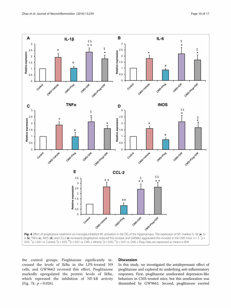
glia in the hippocampus. The expression of M1 markers,IL-1β, IL-6, TNFα, iNOS, and CCL2, increased in theCMS + Vehicle mice. After treatment with pioglitazone,the expression of M1 markers decreased. Administrationof pioglitazone and GW9662 did not change theactivation status of the microglia in the CMS animals(Fig. 4a: p = 0.018; Fig. 4b: p = 0.051; Fig. 4c: p = 0.045;Fig. 4d: p = 0.008; Fig. 4e: p = 0.032). In the M2 status,the messenger RNA (mRNA) expression (Ym1, Arg1,IL-4, IL-10, and TGFβ) was lower following the 6-week
CMS procedure. The decreases were attenuated byadministration with pioglitazone (Fig. 5a: p = 0.001;Fig. 5b: p = 0.004; Fig. 5c: p = 0.001; Fig. 5d: p = 0.037;Fig. 5e: p = 0.046).
The effects of pioglitazone on LPS-stimulated N9microglial phenotypes in vitroIn order to confirm the effect of pioglitazone on themicroglial phenotypes, we detected the microglial activa-tion status by using LPS simulate an organism’s inflam-matory environment in an N9 microglial line. As shownin Fig. 6, the expression of the M1 phenotype (IL-1β, IL-6, TNFα, iNOS, and CCL2) increased significantly afterthe LPS treatment compared with the control, Con +Piog, and Con + GW microglial cells. Pioglitazone mark-edly blocked the upregulation of these markers in theLPS groups. Co-treatment with pioglitazone, GW9662,and LPS showed no difference in the LPS microglia(Fig. 6a: p = 0.025; Fig. 6b: p = 0.023; Fig. 6c: p = 0.001;Fig. 6d: p = 0.001; Fig. 6e: p = 0.036). In contrast, the ex-pression of the M2 markers, Ym1, Arg1, IL-4, IL-10, andTGFβ, decreased after LPS stimulation, but theseexpressions improved after treatment with pioglitazone.The microglial activated phenotypes of the LPS + Piog+ GW group were similar to the LPS microglia(Fig. 7a: p = 0.010; Fig. 7b: p = 0.012; Fig. 7c: p = 0.025;Fig. 7d: p = 0.040; Fig. 7e: p = 0.032). Consistent withthese mRNA expressions, protein expression of theM1 pro-inflammatory cytokines (IL-1β and TNFα)was also upregulated by LPS, and these effects weresignificantly inhibited by pioglitazone. There were nodifferences between the LPS and the LPS + Piog + GWcells (Fig. 6f: p < 0.001; Fig. 6g: p < 0.001). The proteinlevels of the M2 anti-inflammatory mediators (Ym1and Arg1) were also reduced with LPS treatment, andthese decreases were attenuated by treatment with pi-oglitazone. There was no difference between the LPSand the LPS + Piog + GW microglia in their proteinexpression of either Ym1 or Arg1 (Fig. 7g: p = 0.041;Fig. 7i: p = 0.026). The results for the M1 and M2markers in the N9 microglia cells in vitro were con-sistent with the consequences in the mice in vivo.Next, we investigated whether pioglitazone exerted
its anti-inflammatory effects through regulating NF-kB activity. Following the LPS treatment, the level ofIkBα in the N9 microglia dropped compared with all
(See figure on previous page.)Fig. 1 Effects of pioglitazone at different doses on CMS mice. a Schematic diagram of the experimental design. BW was measured every week(b). The 2.5 mg/kg dose showed the best result in recovering the BW in the CMS mice (c, n = 10). Anhedonia was detected by SP over the courseof the 6-week-long experiment (d, e, n = 10). The duration of immobility in the FST (f) and TST (g) increased after CMS induction at the sixth week(n = 8 in both tests). Pioglitazone (2.5 mg/kg) reduced this increase. In the locomotor activity test, pioglitazone improved the spontaneous activitylevel after CMS induction (h, n = 8). *p < 0.05, * *p < 0.01 vs. Control; #p < 0.05, ##p < 0.01 vs. CMS + Vehicle. Data are expressed as means ± SEM
Zhao et al. Journal of Neuroinflammation (2016) 13:259 Page 7 of 17
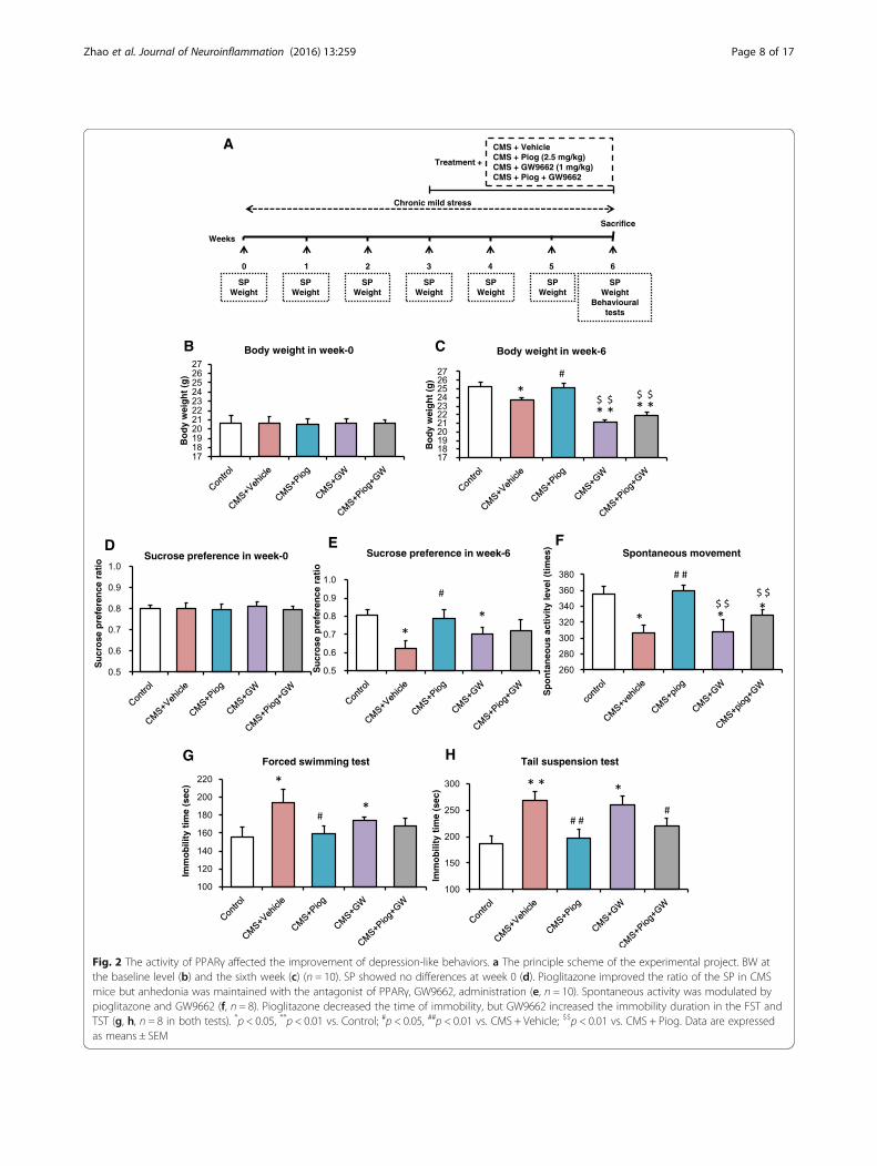
Fig. 2 The activity of PPARγ affected the improvement of depression-like behaviors. a The principle scheme of the experimental project. BW atthe baseline level (b) and the sixth week (c) (n = 10). SP showed no differences at week 0 (d). Pioglitazone improved the ratio of the SP in CMSmice but anhedonia was maintained with the antagonist of PPARγ, GW9662, administration (e, n = 10). Spontaneous activity was modulated bypioglitazone and GW9662 (f, n = 8). Pioglitazone decreased the time of immobility, but GW9662 increased the immobility duration in the FST andTST (g, h, n = 8 in both tests). *p < 0.05, **p < 0.01 vs. Control; #p < 0.05, ##p < 0.01 vs. CMS + Vehicle; $$p < 0.01 vs. CMS + Piog. Data are expressedas means ± SEM
Zhao et al. Journal of Neuroinflammation (2016) 13:259 Page 8 of 17

Fig. 3 Microglial activated status was influenced by PPARγ in the hippocampus. Pioglitazone decreased the number of Iba1+ microglia compared withthe CMS mice. The Iba1+ microglial morphology was amoeboid, and the number of microglia increased in the animals after GW9662 treatment.Administration of pioglitazone and GW9662 had no effect on the microglia (b, n = 5). Enlarged figures indicate typical microglia (a). Scale bars: 10 μm.**p < 0.01 vs. Control; ##p < 0.01 vs. CMS + Vehicle; $$p < 0.01 vs. CMS + Piog. Data are expressed as means ± SEM
Zhao et al. Journal of Neuroinflammation (2016) 13:259 Page 9 of 17
the control groups. Pioglitazone significantly in-creased the levels of IkBα in the LPS-treated N9cells, and GW9662 reversed this effect. Pioglitazonemarkedly upregulated the protein levels of IkBα,which repressed the inhibition of NF-kB activity(Fig. 7k: p = 0.026).
DiscussionIn this study, we investigated the antidepressant effect ofpioglitazone and explored its underlying anti-inflammatoryresponses. First, pioglitazone ameliorated depression-likebehaviors in CMS-treated mice, but this amelioration wasdiminished by GW9662. Second, pioglitazone exerted
Fig. 4 Effect of pioglitazone treatment on microglia-inhibited M1 activation in the DG of the hippocampus. The expression of M1 markers IL-1β (a), IL-6 (b), TNFα (c), iNOS (d), and CCL2 (e) increased; pioglitazone reduced this increase and GW9662 aggravated this increase in the CMS mice. n = 5. *p <0.05, **p < 0.01 vs. Control; #p < 0.05, ##p < 0.01 vs. CMS + Vehicle; $p < 0.05, $$p < 0.01 vs. CMS + Piog. Data are expressed as means ± SEM
Zhao et al. Journal of Neuroinflammation (2016) 13:259 Page 10 of 17

antidepressant effects through the anti-inflammatory activ-ity of the PPARγ pathway. Third, pioglitazone improvedCMS-induced depression-like behavior by regulating whichof the microglial phenotypes (M1 or M2) was activated.MDD is a common and sometimes fatal disorder that
has increasingly become a public health concern. Accu-mulating evidence suggests that chronic low-grade in-flammation plays an important role in the pathology of
depression. Over the past two decades, great effort hasbeen made to identify novel targets for antidepressanttherapies. It has been proposed that pioglitazone, work-ing as an anti-inflammatory agent, could produce anti-depressant responses in patients with concomitantmetabolic syndrome and diabetes [36, 37]. In our study,we assessed the antidepressant efficacy of pioglitazoneon a CMS model, which is commonly used to induce
Fig. 5 Effect of pioglitazone treatment on the microglial shift toward an M2 phenotype. The mRNA expression of M2: Ym1 (a), Arg1 (b), IL-4 (c),IL-10 (d), and TGFβ (e) reduced with a 6-week duration of CMS stress. The decreases were attenuated by pioglitazone administration. GW9662 stillreduced the expression of these markers. Pioglitazone and GW9662 treated together had no effect on the M2 level of CMS mice. n = 5. *p < 0.05,**p < 0.01 vs. Control; #p < 0.05, ##p < 0.01 vs. CMS + Vehicle; $p < 0.05, $$p < 0.01 vs. CMS + Piog. Data are expressed as means ± SEM
Zhao et al. Journal of Neuroinflammation (2016) 13:259 Page 11 of 17
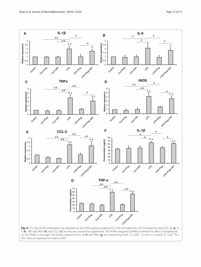
Fig. 6 LPS-induced M1 polarization was alleviated by the PPARγ agonist pioglitazone in N9 microglial cells. LPS increased the level of IL-1β (a), IL-6 (b), TNFα (c), iNOS (d), and CCL2 (e), but this was reversed by pioglitazone. The PPARγ antagonist GW9662 inhibited the effect of pioglitazoneon the PPARγ of microglia. The protein expression of IL-1β (f) and TNFα (g) was detected by ELISA. *p < 0.05, **p < 0.01 vs. Control; #p < 0.05, ##p <0.01. Data are expressed as means ± SEM
Zhao et al. Journal of Neuroinflammation (2016) 13:259 Page 12 of 17
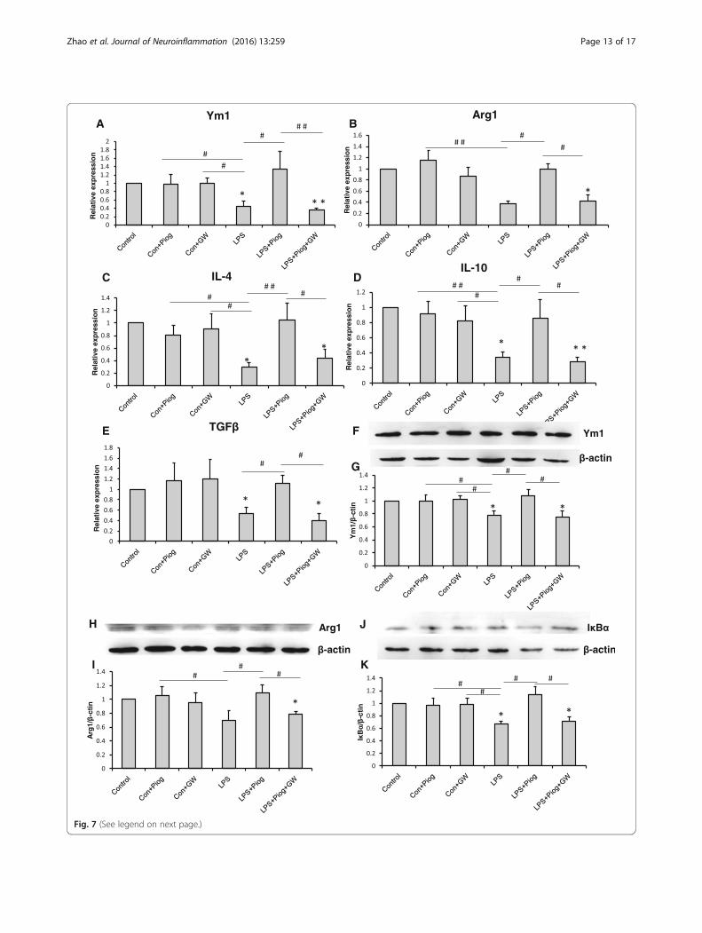
Fig. 7 (See legend on next page.)
Zhao et al. Journal of Neuroinflammation (2016) 13:259 Page 13 of 17
and measure depression (Fig. 1). Because pioglitazonecan cross the blood brain barrier, intragastric injectionwas selected for the CMS mice. Pioglitazone increasedthe SP in CMS-induced depressant model, which wasblocked by the PPAR-γ antagonist GW9662. Pioglitazoneas well decreased the time of immobility in FST and TSTin the CMS-treated mice, indicating that pioglitazone hasa therapeutic action in CMS-induced depression-like be-haviors. CMS induced a decrease in locomotor activity indi-cated a depressive state of animals because of a loss ofinterest for the movement, or for exploring the environ-ment. Pioglitazone increased spontaneous motor activity inthe CMS-exposed mice, without effect on locomotor activ-ity in normal mice [38]. Gavage administration of pioglita-zone was effective at the 2.5 and 5.0 mg/kg doses forameliorating the symptoms of depression, but the2.5 mg/kg dosage was more effective. Dosages of 10and 20 mg/kg were not efficacious in the CMS mice.As a result, the 2.5 mg/kg dose was chosen to treatthe CMS mice in the subsequent experiments.PPARγ is a ligand-dependent transcription factor be-
longing to the nuclear hormone receptor superfamilyimplicated in adipocyte differentiation, insulin sensitivity,and inflammatory processes [39, 40]. PPARγ is constitu-tively expressed in macrophages and CNS-resident micro-glia, acting as a key regulator of microglial activation [41].Activation of PPARγ signaling has a protective role byreducing neuroinflammation [42], a finding which maypresent a novel therapeutic approach for diseases, in-cluding Alzheimer’s disease [20, 43], Parkinson’s disease[24, 44], stroke [45], schizophrenia, and autism [22]. Inlight of the anti-inflammatory and neuroprotective ac-tivities of the PPARγ-dependent signaling pathway, weinvestigated the antidepressant effects of pioglitazone ina CMS mice model of depression. GW9662, a selective an-tagonist for PPARγ, was administered in this investigationin order to explore the possible role of PPARγ activationon the antidepressant activities of pioglitazone (Fig. 2).After treatment with GW9662, the BW loss was greaterthan that in the CMS-treated mice. GW9662 significantlyinhibited the pioglitazone-induced reduction in theduration of immobility in the FST and TST. GW9662also counteracted the effect of pioglitazone on PPARγactivation. These results suggest that pioglitazone ex-erts antidepressant-like effects at least in part due tothe activation of the PPARγ pathway.
Microglia play a crucial role in inflammation modula-tion in the CNS, and microglial dysfunction is believedto result in CNS immune disorders [46, 47]. The role ofmicroglia has long been considered in some neurode-generative diseases [48, 49]. Some interesting evidenceabout this role in depression has also began to emergein recent years, e.g., microglial activation and the expres-sion of inflammatory mediators have been reported inan olfactory bulbectomised (OB) rat model of depression[50]. According to in vitro research, microglia and theirpolarization status play an essential role in depressioninitiation [51]. These data indicated that the immunesystem was activated in the stressed depression models.This may have been due to the activation of M1 micro-glia [35]. Here, we showed that the microglial cells hadlarge somas, short thick processes, and the amoeboidmorphology typical of activated microglia after CMS ini-tiation (Fig. 3a). This morphology has been explained inour previous experiments [12, 35]. Microglial activatedphenotypes were determined by their polarized markers.We confirmed that the M1 markers (IL-1β, IL-6, TNFα,iNOS, and CCL2) were induced and that the M2 mole-cules (Ym1, Arg1, IL-4, IL-10, and TGFβ) were impairedin depression. Pioglitazone restored the balance of theM1 and M2 microglia in the hippocampus of the CMSmice (Figs. 4 and 5). The hippocampus has been impli-cated in the inhibition of stress responses [52] and in theregulation of affective states and emotional behavior[53]. Prior studies have shown that stress primed neu-roinflammatory processes characterized by microglialactivation [14, 15]. Microglial activation plays a vital rolein the pathogenesis of many neurodegenerative diseases.Experiments in animal models have provided moredirect evidence for a role of activated microglia in de-pression [50, 54]. In our experiment, we found thatpioglitazone can control microglia activation and neuro-inflammation in CMS-induced depression. These data in-dicated that microglia-modulating agents had therapeuticbenefits for MDD, thereby defining a new biologicalactivity for pioglitazone by showing that pioglitazone actsin modulating microglial phenotypes in a CMS-inducedrodent model of depression.Inflammatory cytokines play a significant role in de-
pression. In our previous study, an imbalance betweenthe pro- and anti-inflammatory cytokines may be one ofthe pathogeneses of depression [6]. In this current
(See figure on previous page.)Fig. 7 The M2 molecules: Ym1 (a), Arg1 (b), IL-4 (c), IL-10 (d), and TGFβ (e) decreased with LPS treatment. Pioglitazone increased the expressionof the M2 markers. The impact on the LPS+Piog+GW group was similar to that of the LPS microglial cells. The level of Ym1 (f & g) and Arg1 (h &i) was also confirmed by western blot. The protein expression of IκBα decreased in LPS-stimulated cells; pioglitazone significantly ameliorated theexpression. GW9662 suppressed the rise after pioglitazone-treated LPS in microglia (j & k).*p < 0.05, **p < 0.01 vs. Control; #p < 0.05, ##p < 0.01. Dataare expressed as means ± SEM
Zhao et al. Journal of Neuroinflammation (2016) 13:259 Page 14 of 17

research, a higher expression of IL-1β, IL-6, and TNFα,and a lower expression of IL-4, IL-10, and TGF-β wereobserved in the hippocampus of CMS-induced mice(Figs. 4 and 5). A relatively large body of evidence hassuggested that the PPARγ agonist pioglitazone canregulate the inflammatory response and oxidative stress[55, 56]. After pioglitazone treatment of the CMS mice,the expression of pro-inflammatory molecules wasreduced and the levels of anti-inflammatory cytokineswere increased. These results were consistent with re-cent findings that PPARγ agonists can transition M1 intoM2 phenotype as well as enhance the production ofanti-inflammatory cytokines such as IL-10 and TGFβ,both of which are pro-neurogenic [57, 58]. In order toensure the specific response of activated microglia instress-induced depression, experiments using LPS treat-ment of N9 microglial cells were performed. LPS cancause behavioral changes that indicate depression [59].LPS stimulation can break the balance of microglial acti-vation (M1 vs. M2), but the resulting imbalance in thelevels of mRNA and protein can be ameliorated by pioglit-azone administration (Fig. 6 and Fig. 7a–i). The results ofan in vitro study were consistent with the mice test. Thus,the M1-M2 imbalance should be a focus in studying ani-mal models of depression [50]. NF-kB pathway activationplays an important role in pro-inflammatory gene expres-sion after stimulation [60, 61]. NF-kB normally exists inthe cytoplasm, binding to its inhibitory proteins (IkB) andremaining inactive. Dissociation of IkB induces activationof NF-kB and facilitates the transcription of inflammatorygenes [62]. In our experiment, the protein expression ofIkB was reduced after an LPS stimulation of N9 microglialcells. Pioglitazone upregulated the IkBα expression in theLPS-treated N9 cells and GW9662 reversed the inhibitoryeffects of pioglitazone on NF-kB activity (Fig. 7k). Theseresults indicated that the role of pioglitazone in preventingthe NF-kB activation was partially linked to the upregula-tion of IkB expression. The present findings suggest thatthe anti-inflammatory effects of pioglitazone are associ-ated with a PPARγ-mediated suppression of the NF-kBsignaling pathway with consequential inflammatory cyto-kine expression in microglial cells.
ConclusionsIn summary, the present study demonstrated for the firsttime that the chronic administration of pioglitazoneinduced the neuroprotective phenotype of microglia inparallel with the amelioration of depression-like behaviorsin CMS-treated C57BL/6 mice. Pioglitazone, a microglia-modulating drug which regulates anti-inflammatory activ-ity, may partially account for the observed antidepressantresponse. This finding suggests that targeting microgliacould pave the way for new depression treatments.
Additional files
Additional file 1: BW and SP ratio in different weeks of experiment 1.(PDF 77 kb)
Additional file 2: BW and SP ratio in different weeks of experiment 2.(PDF 77 kb)
AbbreviationsANOVA: Analysis of variance; Arg1: Arignase1; BW: Body weight; CMS: Chronicmild stress; CNS: Central nervous system; FST: Forced swimming test;GW: GW9662; Iba1: Ionized calcium-binding adaptor protein-1; IkB: Inhibitor ofNF-kB; LPS: Lipopolysaccharide; M1: Classical activation; M2: Alternativeactivation; MAOIs: Monoamine oxidase inhibitors; MDD: Major depressivedisorder; NF-kB: Nuclear factor kB; OB: Olfactory bulbectomised;Piog: Pioglitazone; PBS: Phosphate-buffered saline; PPARγ: Peroxisomeproliferator-activated receptor γ; RT-PCR: Real time-PCR; SP: Sucrose preference;SSRIs: Selective serotonin reuptake inhibitors; TNFα: Tumor necrosis factor-α;TST: Tail suspension test
AcknowledgementsWe are grateful to Prof. Zujun Yang for his assistance and facilities inimmunohistochemistry and to Rhoda E and Edmund F. Perozzi, PhDs, fortheir extensive review and English language assistance on this paper.
FundingThis work was supported by the National Natural Science Foundation of China(No. 81571174, 81603503), 863 project (No. 2015AA020505), Key Technologies R& D Program of Sichuan Province (2015SZ0058-5), and the Open Research Fundof State Key Laboratory Breeding Base of Systematic Research, Developmentand Utilization of Chinese Medicine Resources (No. 2015003).
Availability of data and materialsThe datasets and materials supporting the conclusions of this article areincluded within the article and its additional files.
Authors’ contributionsQZ and ZY designed the study and drafted of the manuscript. QZ, XW, and SYperformed the experiments. XX and YF managed the literature searches andanalyses. QZ and JZ analyzed the data. QZ, CP, and ZY provided interpretationof the data and discussed the manuscript. All authors read and approved thefinal manuscript.
Competing interestsThe authors declare that they have no competing interests.
Consent for publicationNot applicable
Ethics approval and consent to participateAll animals care and experimental procedures were approved by theInstitutional Animal Care and Use Committee, University of ElectronicScience and Technology of China.
Received: 29 January 2016 Accepted: 21 September 2016
References1. Centers for Disease Control and Prevention (CDC). Current depression
among adults—United States, 2006 and 2008. MMWR Morb Mortal WklyRep. 2010;59:1229–1235.
2. Rush AJ, Trivedi MH, Wisniewski SR, Nierenberg AA, Stewart JW, Warden D,Niederehe G, Thase ME, Lavori PW, Lebowitz BD, et al. Acute and longer-term outcomes in depressed outpatients requiring one or several treatmentsteps: a STAR*D report. Am J Psychiatry. 2006;163:1905–17.
3. Anderson HD, Pace WD, Libby AM, West DR, Valuck RJ. Rates of 5 commonantidepressant side effects among new adult and adolescent cases ofdepression: a retrospective US claims study. Clin Ther. 2012;34:113–23.
4. Fonseka TM, McIntyre RS, Soczynska JK, Kennedy SH. Novel investigationaldrugs targeting IL-6 signaling for the treatment of depression. Expert OpinInvestig Drugs. 2015;24:459–75.
Zhao et al. Journal of Neuroinflammation (2016) 13:259 Page 15 of 17

5. Raison CL, Capuron L, Miller AH. Cytokines sing the blues: inflammation andthe pathogenesis of depression. Trends Immunol. 2006;27:24–31.
6. You Z, Luo C, Zhang W, Chen Y, He J, Zhao Q, Zuo R, Wu Y. Pro- and anti-inflammatory cytokines expression in rat’s brain and spleen exposed to chronicmild stress: involvement in depression. Behav Brain Res. 2011;225:135–41.
7. Hanisch UK, Kettenmann H. Microglia: active sensor and versatile effectorcells in the normal and pathologic brain. Nat Neurosci. 2007;10:1387–94.
8. Tambuyzer BR, Ponsaerts P, Nouwen EJ. Microglia: gatekeepers of centralnervous system immunology. J Leukoc Biol. 2009;85:352–70.
9. Steiner J, Bielau H, Brisch R, Danos P, Ullrich O, Mawrin C, Bernstein HG,Bogerts B. Immunological aspects in the neurobiology of suicide: elevatedmicroglial density in schizophrenia and depression is associated withsuicide. J Psychiatr Res. 2008;42:151–7.
10. Michelucci A, Heurtaux T, Grandbarbe L, Morga E, Heuschling P.Characterization of the microglial phenotype under specific pro-inflammatory and anti-inflammatory conditions: effects of oligomeric andfibrillar amyloid-beta. J Neuroimmunol. 2009;210:3–12.
11. Park J, Min JS, Kim B, Chae UB, Yun JW, Choi MS, Kong IK, Chang KT, Lee DS.Mitochondrial ROS govern the LPS-induced pro-inflammatory response inmicroglia cells by regulating MAPK and NF-kappaB pathways. Neurosci Lett.2015;584:191–6.
12. Zhao Q, Xie X, Fan Y, Zhang J, Jiang W, Wu X, Yan S, Chen Y, Peng C, You Z.Phenotypic dysregulation of microglial activation in young offspring rats withmaternal sleep deprivation-induced cognitive impairment. Sci Rep. 2015;5:9513.
13. Kobayashi K, Imagama S, Ohgomori T, Hirano K, Uchimura K, Sakamoto K,Hirakawa A, Takeuchi H, Suzumura A, Ishiguro N, Kadomatsu K. Minocyclineselectively inhibits M1 polarization of microglia. Cell Death Dis. 2013;7:54.
14. Frank MG, Hershman SA, Weber MD, Watkins LR, Maier SF. Chronic exposureto exogenous glucocorticoids primes microglia to pro-inflammatory stimuliand induces NLRP3 mRNA in the hippocampus. Psychoneuroendocrinology.2014;40:191–200.
15. Han A, Yeo H, Park MJ, Kim SH, Choi HJ, Hong CW, Kwon MS. IL-4/10prevents stress vulnerability following imipramine discontinuation.J Neuroinflammation. 2015;12:015–0416.
16. Calcia MA, Bonsall DR, Bloomfield PS, Selvaraj S, Barichello T, Howes OD.Stress and neuroinflammation: a systematic review of the effects of stresson microglia and the implications for mental illness. Psychopharmacology(Berl). 2016;233:1637–50.
17. Jafari M, Khodayari B, Felgner J, Bussel II, Rose MR, Mueller LD. Pioglitazone:an anti-diabetic compound with anti-aging properties. Biogerontology.2007;8:639–51.
18. Kemp DE, Ismail-Beigi F, Ganocy SJ, Conroy C, Gao K, Obral S, Fein E, FindlingRL, Calabrese JR. Use of insulin sensitizers for the treatment of majordepressive disorder: a pilot study of pioglitazone for major depressionaccompanied by abdominal obesity. J Affect Disord. 2012;136:1164–73.
19. Carta AR, Pisanu A. Modulating microglia activity with PPAR-gamma agonists:a promising therapy for Parkinson’s disease? Neurotox Res. 2013;23:112–23.
20. Mandrekar-Colucci S, Karlo JC, Landreth GE. Mechanisms underlying therapid peroxisome proliferator-activated receptor-gamma-mediated amyloidclearance and reversal of cognitive deficits in a murine model ofAlzheimer’s disease. J Neurosci. 2012;32:10117–28.
21. Spiegelman BM. PPAR-gamma: adipogenic regulator and thiazolidinedionereceptor. Diabetes. 1998;47:507–14.
22. Boris M, Kaiser CC, Goldblatt A, Elice MW, Edelson SM, Adams JB, FeinsteinDL. Effect of pioglitazone treatment on behavioral symptoms in autisticchildren. J Neuroinflammation. 2007;4:3.
23. Sato T, Hanyu H, Hirao K, Kanetaka H, Sakurai H, Iwamoto T. Efficacy ofPPAR-gamma agonist pioglitazone in mild Alzheimer disease. NeurobiolAging. 2011;32:1626–33.
24. NINDS Exploratory Trials in Parkinson Disease (NET-PD) FS-ZONEInvestigators. Pioglitazone in early Parkinson's disease: a phase 2,multicentre, double-blind, randomised trial. Lancet Neurol. 2015;14:795–803.
25. Kaiser CC, Shukla DK, Stebbins GT, Skias DD, Jeffery DR, Stefoski D, KatsamakisG, Feinstein DL. A pilot test of pioglitazone as an add-on in patients withrelapsing remitting multiple sclerosis. J Neuroimmunol. 2009;211:124–30.
26. Ji S, Kronenberg G, Balkaya M, Farber K, Gertz K, Kettenmann H, Endres M.Acute neuroprotection by pioglitazone after mild brain ischemia withouteffect on long-term outcome. Exp Neurol. 2009;216:321–8.
27. Lin KW, Wroolie TE, Robakis T, Rasgon NL. Adjuvant pioglitazone forunremitted depression: clinical correlates of treatment response. PsychiatryRes. 2015;230:846–52.
28. Salehi-Sadaghiani M, Javadi-Paydar M, Gharedaghi MH, Zandieh A,Heydarpour P, Yousefzadeh-Fard Y, Dehpour AR. NMDA receptorinvolvement in antidepressant-like effect of pioglitazone in the forcedswimming test in mice. Psychopharmacology (Berl). 2012;223:345–55.
29. Sadaghiani MS, Javadi-Paydar M, Gharedaghi MH, Fard YY, Dehpour AR.Antidepressant-like effect of pioglitazone in the forced swimming test inmice: the role of PPAR-gamma receptor and nitric oxide pathway. BehavBrain Res. 2011;224:336–43.
30. Kemp DE, Schinagle M, Gao K, Conroy C, Ganocy SJ, Ismail-Beigi F,Calabrese JR. PPAR-gamma agonism as a modulator of mood: proof-of-concept for pioglitazone in bipolar depression. CNS Drugs. 2014;28:571–81.
31. Gamaro GD, Manoli LP, Torres IL, Silveira R, Dalmaz C. Effects of chronicvariate stress on feeding behavior and on monoamine levels in different ratbrain structures. Neurochem Int. 2003;42:107–14.
32. Micale V, Kucerova J, Sulcova A. Leading compounds for the validation ofanimal models of psychopathology. Cell Tissue Res. 2013;354:309–30.
33. Cryan JF, Mombereau C, Vassout A. The tail suspension test as a model forassessing antidepressant activity: review of pharmacological and geneticstudies in mice. Neurosci Biobehav Rev. 2005;29:571–625.
34. Porsolt RD, Le Pichon M, Jalfre M. Depression: a new animal model sensitiveto antidepressant treatments. Nature. 1977;266:730–2.
35. Zhao Q, Peng C, Wu X, Chen Y, Wang C, You Z. Maternal sleep deprivationinhibits hippocampal neurogenesis associated with inflammatory responsein young offspring rats. Neurobiol Dis. 2014;68:57–65.
36. Sepanjnia K, Modabbernia A, Ashrafi M, Modabbernia MJ, Akhondzadeh S.Pioglitazone adjunctive therapy for moderate-to-severe major depressivedisorder: randomized double-blind placebo-controlled trial.Neuropsychopharmacology. 2012;37:2093–100.
37. Kashani L, Omidvar T, Farazmand B, Modabbernia A, Ramzanzadeh F,Tehraninejad ES, Ashrafi M, Tabrizi M, Akhondzadeh S. Does pioglitazoneimprove depression through insulin-sensitization? Results of a randomizeddouble-blind metformin-controlled trial in patients with polycystic ovariansyndrome and comorbid depression. Psychoneuroendocrinology. 2013;38:767–76.
38. Sanchis-Gomar F, Pareja-Galeano H, Martinez-Bello VE. PPARgamma agonistpioglitazone does not enhance performance in mice. Drug Test Anal. 2014;6:922–9.
39. Li ZY, Song J, Zheng SL, Fan MB, Guan YF, Qu Y, Xu J, Wang P, Miao CY.Adipocyte Metrnl antagonizes insulin resistance through PPARgammasignaling. Diabetes. 2015;64:4011–22.
40. McIntyre RS, Soczynska JK, Woldeyohannes HO, Lewis GF, Leiter LA,MacQueen GM, Miranda A, Fulgosi D, Konarski JZ, Kennedy SH.Thiazolidinediones: novel treatments for cognitive deficits in mooddisorders? Expert Opin Pharmacother. 2007;8:1615–28.
41. Carta AR, Pisanu A, Carboni E. Do PPAR-gamma agonists have a future inParkinson’s disease therapy? Parkinsons Dis. 2011;689181:29.
42. Pan J, Jin JL, Ge HM, Yin KL, Chen X, Han LJ, Chen Y, Qian L, Li XX, XuY. Malibatol A regulates microglia M1/M2 polarization in experimentalstroke in a PPARgamma-dependent manner. J Neuroinflammation. 2015;12:015–0270.
43. Skerrett R, Pellegrino MP, Casali BT, Taraboanta L, Landreth GE. Combinedliver X receptor/peroxisome proliferator-activated receptor gamma agonisttreatment reduces amyloid beta levels and improves behavior in amyloidprecursor protein/presenilin 1 mice. J Biol Chem. 2015;290:21591–602.
44. Pisanu A, Lecca D, Mulas G, Wardas J, Simbula G, Spiga S, Carta AR. Dynamicchanges in pro- and anti-inflammatory cytokines in microglia after PPAR-gamma agonist neuroprotective treatment in the MPTPp mouse model ofprogressive Parkinson’s disease. Neurobiol Dis. 2014;71:280–91.
45. Cuartero MI, Ballesteros I, Moraga A, Nombela F, Vivancos J, Hamilton JA,Corbi AL, Lizasoain I, Moro MA. N2 neutrophils, novel players in braininflammation after stroke: modulation by the PPARgamma agonistrosiglitazone. Stroke. 2013;44:3498–508.
46. Saijo K, Glass CK. Microglial cell origin and phenotypes in health anddisease. Nat Rev Immunol. 2011;11:775–87.
47. David S, Kroner A. Repertoire of microglial and macrophage responses afterspinal cord injury. Nat Rev Neurosci. 2011;12:388–99.
48. Jimenez S, Baglietto-Vargas D, Caballero C, Moreno-Gonzalez I, Torres M,Sanchez-Varo R, Ruano D, Vizuete M, Gutierrez A, Vitorica J. Inflammatoryresponse in the hippocampus of PS1M146L/APP751SL mouse model ofAlzheimer’s disease: age-dependent switch in the microglial phenotypefrom alternative to classic. J Neurosci. 2008;28:11650–61.
Zhao et al. Journal of Neuroinflammation (2016) 13:259 Page 16 of 17

49. Varnum MM, Ikezu T. The classification of microglial activation phenotypeson neurodegeneration and regeneration in Alzheimer’s disease brain. ArchImmunol Ther Exp. 2012;60:251–66.
50. Burke NN, Kerr DM, Moriarty O, Finn DP, Roche M. Minocycline modulatesneuropathic pain behaviour and cortical M1-M2 microglial gene expressionin a rat model of depression. Brain Behav Immun. 2014;42:147–56.
51. Pusic KM, Pusic AD, Kemme J, Kraig RP. Spreading depression requiresmicroglia and is decreased by their M2a polarization from environmentalenrichment. Glia. 2014;62:1176–94.
52. Lopez JF, Akil H, Watson SJ. Neural circuits mediating stress. Biol Psychiatry.1999;46:1461–71.
53. McEwen BS, Magarinos AM. Stress and hippocampal plasticity: implicationsfor the pathophysiology of affective disorders. Hum Psychopharmacol. 2001;16:S7–19.
54. Henry CJ, Huang Y, Wynne A, Hanke M, Himler J, Bailey MT, Sheridan JF,Godbout JP. Minocycline attenuates lipopolysaccharide (LPS)-inducedneuroinflammation, sickness behavior, and anhedonia. J Neuroinflammation.2008;5:1742–2094.
55. De Nuccio C, Bernardo A, Cruciani C, De Simone R, Visentin S, Minghetti L.Peroxisome proliferator activated receptor-gamma agonists protectoligodendrocyte progenitors against tumor necrosis factor-alpha-induceddamage: effects on mitochondrial functions and differentiation. Exp Neurol.2015;271:506–14.
56. El-Gowilly SM, Helmy MM, El-Gowelli HM. Pioglitazone amelioratesmethotrexate-induced renal endothelial dysfunction via amendingdetrimental changes in some antioxidant parameters, systemic cytokinesand Fas production. Vascul Pharmacol. 2015;74:139–50.
57. Yu Z, Sun D, Feng J, Tan W, Fang X, Zhao M, Zhao X, Pu Y, Huang A, XiangZ, et al. MSX3 switches microglia polarization and protects frominflammation-induced demyelination. J Neurosci. 2015;35:6350–65.
58. Papadopoulos P, Rosa-Neto P, Rochford J, Hamel E. Pioglitazone improvesreversal learning and exerts mixed cerebrovascular effects in a mousemodel of Alzheimer's disease with combined amyloid-beta andcerebrovascular pathology. PLoS One. 2013;8:e68612.
59. Adzic M, Djordjevic J, Mitic M, Brkic Z, Lukic I, Radojcic M. The contributionof hypothalamic neuroendocrine, neuroplastic and neuroinflammatoryprocesses to lipopolysaccharide-induced depressive-like behaviour in femaleand male rats: involvement of glucocorticoid receptor and C/EBP-beta.Behav Brain Res. 2015;291:130–9.
60. Qin ZH, Tao LY, Chen X. Dual roles of NF-kappaB in cell survival andimplications of NF-kappaB inhibitors in neuroprotective therapy. ActaPharmacol Sin. 2007;28:1859–72.
61. Lawrence T. The nuclear factor NF-kappaB pathway in inflammation. ColdSpring Harb Perspect Biol. 2009;1:7.
62. Baeuerle PA, Baltimore D. NF-kappa B: ten years after. Cell. 1996;87(1):13–20.
• We accept pre-submission inquiries
• Our selector tool helps you to find the most relevant journal
• We provide round the clock customer support
• Convenient online submission
• Thorough peer review
• Inclusion in PubMed and all major indexing services
• Maximum visibility for your research
Submit your manuscript atwww.biomedcentral.com/submit
Submit your next manuscript to BioMed Central and we will help you at every step:
Zhao et al. Journal of Neuroinflammation (2016) 13:259 Page 17 of 17