the american society of neuroimaging 2016 annual … annual meeting/2016abstracts.pdf · the...
TRANSCRIPT

1. TB Vasculitis and Strokes in a Immucompetent: Rare Diagnosis
becomes more Rarer if not Thought.
Zain Guduru1,2,3, Abhishek Purohit1,2,3, Sandeep Rana1,2,3
1Allegheny General Hospital Pittsburgh, PA, USA, 2Drexel School of Medicine
Philadelphia, PA, USA, 3Temple School of Medicine Philadelphia, PA, USA
BACKGROUND & PURPOSE: TB Meningitis with infarcts is fatal upto 3 times more
often than in those without infarcts. Location of strokes includes Deep Sylvain
region (presence of TB meningitis exudates at the level of basal cisterna). It is
assumed that infarcts are due to vasculitis/ subsequent intimal proliferation
+/- superadded thrombosis. Treatment includes combination of anti-TB drugs
and dexamethasone.
METHODS: 76 year Caucasian male with history of hypertension presented with
2 months history of altered mental status. No TB risk factors. Ammonia (17)
blood cultures, HIV, Hepatitis B, C and rheumatological panel were negative.
CSF showed: opening pressure (20), WBC(230), RBC(233), Lymphs (58%),
Neu(37%), Glu(30), Pro(195), IgG(24.5), ACE(4.7). VDRL, gram stain, strep B,
H.influenza, S.pneumo: negative. All viral studies: negative. ADA: 1. Culture:
negative. CSF cytology: mature lymphocytes, monocytes and few neutrophils.
No malignant cells. Immunophenotype: findings were NOT diagnostic of
lymphoproliferation. TB PCR was positive. Quantiferon gold test for TB was
indeterminate. Dexamethasone and anti-TB drugs were started which showed
improvement gradually.
RESULTS: MRI brain showed multifocal infarctions and leptomeningeal
enhancement. CT angiography head showed shows diffuse narrowing and
irregularity involving the circle of Willis and the proximal vasculature. Meningeal
biopsy AFB stain shows AFB positive structure in area of lymphohistocytic
infiltrate.
CONCLUSIONS: Even though diagnosis of CNS TB is rare in a person with no risk
factors, it is very much important to be considered in the differential diagnosis
The American Society of Neuroimaging 2016 Annual Meeting Abstracts

as failure of starting appropriate treatment causes higher mortality. TB
vasculitis should be considered if any neurological deterioration arising during
the course of TB.

2. Impact of phenomena " Distal Embolization " in current acute ischemic
stroke (AIS) treatment for emergent large vessel occlusion (ELVO).
Vishal Jani1, Mohammad-Rauf Afzal2, Muhammad Shah Miran2, Ahmed Riaz 2,
Jillian Schurr1, Anmar Razzak1, Syed Hussain1, Adnan I Qureshi2
1Michigan State University/ Department of Vascular and Endovascular
Neurology East Lansing, MI, USA, 2 University of Minnesota/Department of
Neurology and Neurosurgery Minneapolis, MN, USA
BACKGROUND & PURPOSE: Periprocedural acute distal embolization is a known
complication of acute ischemic stroke (AIS) treatment for emergent large vessel
occlusion (ELVO). To date there is limited data regarding its impact on
complications and clinical outcomes.
METHODS: A cohort of retrospectively maintained prospective ELVO
embolectomy registry patients (n=92) between the years of 2012 and 2014 was

reviewed for post treatment ADE, which was defined as appearance of an
occlusion on a downstream vessel. The cohort was dichotomized into two
groups (with and without distal embolization). Post-treatment 24-hour CT
brain scan, 30 day mortality, intracerebral hemorrhage (ICH), and 30-day
modified Rankin scale were collected and frequency analysis was performed to
assess the complications and compare clinical outcomes.
RESULTS: The 92-patient cohort was dichotomized in ELVO with distal
embolization (n=28) and ELVO without distal embolization (n=64). Overall rate
of complications in patients with distal embolization versus patients without
distal embolization was similar, e.g. symptomatic ICH (2 of 28 (7.1)% vs 9 of 64
(14.0)%, p = 0.5), 30-day mortality (2 of 28(7.1)% vs 10 of 64 (15.6)%, p=0.4),
composite endpoint of in-hospital mortality, and ICH(symptomatic and
Asymptomatic) 8 of 28 (28.5)% vs 20 of 64 (31.5)%, p = 0.9)
CONCLUSIONS: Our initial experience demonstrates that phenomena of distal
embolization during AIS treatment does not have any major clinical impact in
terms of complications and long-term clinical outcomes. Further studies are
needed to assess the impact of distal embolization phenomena to understand
clinical sequelae and to design the next generation of instrumentation.
3. Let's Tango: Approach to the Tandem Lesion
NIKIL SWAMY, SONAL MEHTA
PALMETTO HEALTH RICHLAND COLUMBIA, SC, USA
BACKGROUND & PURPOSE: Symptomatic acute basilar artery thrombosis with
associated bilateral vertebral occlusion is a unique entity, occurring in ~3% of
posterior circulation ischemic events common etiologies being atherosclerosis,
giant cell arteritis, trauma and spontaneous dissection. These challenging
interventional cases are associated with a poor prognosis and high rate of
recurrent ischemia.
METHODS: Case Report and review of the literature.
RESULTS: A 63 year-old-male with hemiparesis and hemianopsia was excluded
from IV thrombolysis due to MRI showing bilateral PCA territory infarcts with
petechial hemorrhage. NIHSS improved from 8 to 4 after intervention.
Diagnostic angiogram revealed patent anterior circulation, basilar tip clot with

minimal left P1 and absent right P1 filling, and occluded left vertebral origin
with collateral flow through ascending cervical branches. A glide wire navigated
through the expected left vertebral artery ostium, was exchanged out for a
coaxial system consisting of a Neuron 088 Max and an 058 Navien catheter. A
Marksman microcatheter was navigated into the right PCA through the
Navien placed at the distal vertebral artery. A solitaire thrombectomy was
performed with Penumbra aspiration. Recanalization of the basilar artery but
with distal occlusion of the right PCA as well as occlusion of the left PCA was
achieved. Similarly, a thrombectomy of the left PCA achieve an overall TICI 2B
recanalization.
CONCLUSIONS: The presence of tandem lesions in an acute stroke setting
present a challenge for endovascular treatment. In relation to angioplasty with
or without stenting of the proximal lesion, our report describes the potential for
stand-alone acute thrombectomy.
5. Cause of Paradoxical Emboli: Presence of Levo-Atrial Cardinal Vein
John Hanna, Deepa Bhupali, Daniel Antoniello
Montefiore Medical Center/Neurology Bronx, NY, USA
BACKGROUND & PURPOSE: To report an unusual cause of paradoxical embolus.
The levo-atrial cardinal vein is an anomalous vein that connects the superior
vena cava (SVC) with the left atrium (LA). It is a rare developmental abnormality
caused by persistence of primitive connections between the pulmonary venous
primordial and the cardinal vein. Stroke secondary to the levo-atrial cardinal
vein has not been previously reported.
METHODS: Case report from a tertiary care medical center.
RESULTS: An 80-year-old man presented with dizziness with INO on
examination. MRI revealed bi-hemispheric and brainstem infarcts.
Cardioembolic source was suspected however initial workup was unrevealing
and was given the diagnosis of cryptogenic stroke. One year later returned with
right hemiparesis. MRI revealed bi-hemispheric and cerebellar infarcts. Repeat
TEE revealed an unusual pattern of flow, with immediate filling of the LA
followed by right sided filling, concerning for venous-arterial

connection. Chest CT showed abnormal connection of the left brachiocephalic
vein to the LA: a levo-atrial cardinal vein. Search for paradoxical embolus
revealed superficial vein thrombosis of the upper extremity. He was started on
anticoagulation without stroke recurrence.
CONCLUSIONS: This case illustrates the importance of proper investigation of
bilateral hemispheric infarcts with the use of the echocardiogram with bubble
study. Immediate LA bubbles followed by right sided bubbles are the atypical
flow pattern seen in levo-atrial cardinal vein. This case illustrates that unusual
flow patterns seen on echocardiogram should prompt chest imaging in search
of structural anomalies.

6. DON’T LOOK THE OTHER WAY: BOW HUNTER’S SYNDROME – AN
UNSUAL CAUSE OF POSTERIOR CIRCULATION STROKE
Neville Jadeja, Krishna Nalleballe, Neha Mirchandani, John Hanna, Kathryn F
Kirchoff-Torres
Montefiore Medical Center & Albert Einstein College of Medicine New York, NY,
USA
BACKGROUND & PURPOSE: Bow Hunters syndrome is a rare cause of
symptomatic vertebrobasilar ischemia that results from a mechanical
compression or injury to the vertebral artery.
METHODS: We describe a case of Bow Hunters syndrome secondary to
congenital cervical spine abnormalities.
RESULTS: An otherwise healthy 24 year old man presented with one week of
intermittent dizziness exacerbated by turning his head to the left, followed by
diplopia and disorientation on the day of presentation. He reported similar

complaints a few months prior. There was no history of neck trauma. His
examination demonstrated mild right hemiparesis. Head CT showed congenital
nonunion of C1 and fusion of C2-C3 vertebrae. CT angiography of the head
showed subacute injury to the right vertebral artery as it exited the C2
transverse foramen evidenced by a focal irregularity in the arterial lumen with
pseudoaneurysm formation confirmed by MR angiography. Brain MRI showed
restricted diffusion in the left anterior thalamus and bilateral dorsal midbrain
indicative of acute infarcts. Chronic infarcts in the right cerebellum and left
thalamus were also present indicative of prior ischemia. Dynamic x-ray showed
malalignment of lateral masses of the atlas with C2, but no instability. A
diagnosis of Bow hunters Syndrome was made. He was started on aspirin for
secondary stroke prevention and was referred for evaluation for corrective
cervical spine surgery.
CONCLUSIONS: Posterior circulation strokes in young adults with evidence of
high cervical spine abnormalities on imaging must alert the clinician to the
possibility of dynamic vertebral artery compression. Surgical correction of the
bony abnormality can be potentially curative.
7. Atrioesophageal Fistulas after Pulmonary Vein Isolation: a rare and
deadly complication
Abhishek Purohit1, Zain Guduru1, Sonja Chaparala1, Ashis Tayal1,2,3
1Allegheny General Hospital Pittsburgh, PA, USA, 2Drexel School of Medicine

Philadelphia, PA, USA, 3Temple School of Medicine Philadelphia, PA, USA
BACKGROUND & PURPOSE: Atrioesophageal fistula (AEF) is a rare but highly
morbid complication of pulmonary vein isolation (PVI). Its presentation can
include GI bleeding, sepsis, seizures, meningitis and embolic strokes.
Complications of AEF can be seen from 3 to 40 days after the procedure and
diagnosis is essential to the survival of patients. We present a case of a patient
who had embolic strokes 4 weeks after PVI initially felt to be due to pradaxa
failure but found to have an AEF at autopsy.
METHODS: 63 y/o female with past medical history of refractory paroxysmal
afib on pradaxa presented with left hemiparesis four weeks after PVI. Imaging
revealed embolic strokes in the right MCA territory. While admitted, she had a
GTCS followed by significant decompensation of her neurologic exam, later
requiring intubation. Based on imaging she was taken for a STAT
decompressive craniectomy, however, after the procedure, she continued to
swell and family decided to withdraw care. At autopsy, findings were consistent
with an AEF.
RESULTS: MRI brain revealed scattered strokes primarily in the R MCA territory.
CTHs show a development of large areas of stroke with midline shift, herniation
and development of pneumocephalus due to air embolus.
CONCLUSIONS: Prompt diagnosis of an AEF is paramount due to the devastating
effects it may have. Neurologists must be aware of this complication of PVI.
Patients with a recent PVI who present with neurologic symptoms should be
evaluated by the performing physician. They may also benefit from a
cardiothoracic evaluation or direct visualization by endoscopy.

8. Langerhans Cell Histiocytosis Presenting as a Meningioma on MRI
Madhureeta Achari2, Cesar E. Escamilla Ocanas1
1Uniersided de Monterey Monterey, Mexico, 2University of Texas Medical School
Houston Deparment of PM &R Houston, TX, USA
BACKGROUND & PURPOSE: Meningiomas, symptomatic and incdental, are found
commonly on MRI, withcharaceristic MRI findings. Langerhans Cell Histiocytosis
(LHC) is a rare systemic granulomatous disase with CNS involvement noted in
16% of cases,mainly involving the posterior pituitary gland, cusaing diabetes
insipidus. Although meningeal forms of LHC have been reported, they are
rare. We present a rare case of Langerhans Cell Histiocytosis presenting as a
meningioma on MRI.
METHODS: The patient is a 28 year old female with a history of common
migraine, who developed progressive left hemicranial headaches, similar to her
migraines, unresponsive to her ususal theraputic regimen. She is otherwise
healthy. She had mild tenderness to palpation over the left frontal cranium
otherwise, her neurolgical examination was normal. Contrast enhanced MRI of
the brain revealed a extra-axial, enhancing lesion with a dural tail in the left
frontal region, suggestive of meningioma. The patient underwent tumor
excision without complications.
RESULTS: The surgical histopathology relvealed the lesion to be composed
of dendritic (Langerhans) cells with eosinophilia involving the bone, epidural
tissue and dura. Immunohistochemical studies of S-100 and CD1a results were
positive for atypia. A total body evaluation did not reveal LHC in other sites.
After surgical excision, the patient was headache free for 12 weeks, and then
retured to her ususal migraine frequency and pattern.
CONCLUSIONS: Meningiomas are common findings on MRI, and may be
incidental. Meningeal lesions rare finding in LHC. We present a case with LHC
presenting with typical features of a meningioma on MRI. This enhances the
differential diagnosis of meningeal lesions and underscores the importance on
neuroimaging in headache patients.
10. Neuroimaging of Hemophagocytic lymphohistiocytosis
Ajay Goenka, Krishna Nalleballe, Fatema Malbari
Montefiore Medical Center & Albert Einstein College of Medicine New York, NY,
USA
BACKGROUND & PURPOSE: To describe the MRI findings in a case of CNS
Hemophagocytic lymphohistiocytosis (HLH). Familial CNS Hemophagocytic

lymphohistiocytosis (HLH) is a rare aggressive life threatening disease
characterized by overactive immune function (excessive proliferation and
infiltration of benign histiocytes). We report the MRI findings in a case of CNS
HLH that showed multiple enhancing lesions mimicking demyelinating lesion.
METHODS: This is a 17 year-old boy who presented with multiple seizures and
left foot drop. As part of the initial evaluation, neuroradiological studies
confirmed CNS involvement. He underwent extensive work up including a brain
and bone marrow biopsy as well as a genetic evaluation which confirmed the
final diagnosis of HLH.
RESULTS: MRI brain showed multiple diffuse enhancing lesions. Pathology was
suggestive of a marked inflammatory process with lymphohistiocytic infiltrate.
Final diagnosis was made when mutations in the PRF 1 gene were detected in
the patient and the mother. He underwent bone marrow transplantation with
partial resolution of the CNS lesion.
CONCLUSIONS: Our case emphasizes the importance of considering a diagnosis
of familial Hemophagocytic lymphohistiocytosis in complex enhancing brain
mass lesions. It also emphasizes the importance of genetics as a strong
diagnostic tool for familial HLH.
11. Orbital Abscess: A Delayed Complication of Decompression for
Proptosis Secondary to Grave's Ophthalmopathy
Reena T Gottesman1, Kunal V Desai1,2, Jessica L Zwerling1
1Albert Einstein College of Medicine/Neurology Bronx, NY, USA, 2Jacobi Medical

Center/Neurology Bronx, NY, USA
BACKGROUND & PURPOSE: Orbital abscess can be a life threatening infection
that must be identified early to prevent complications like meningitis, blindness
and death. It is an essential part of the differential diagnosis for proptosis and
must also be considered in patients with known Graves ophthalmopathy.
METHODS: A 60 year old woman presented to the ER with 4 days of progressive,
severe, left periorbital headache, diplopia and blurry vision. Her headache was
worsened by coughing and did not respond to naproxen. She carried a
diagnosis of Graves ophthalmopathy and had undergone endoscopic
decompression for left eye proptosis 6 years prior to her current presentation.
Her immediate post-operative course was complicated by a cerebrospinal fluid
leak. A temporary lumbar drain was placed which led to the resolution of the
patient's symptoms at that time.
RESULTS: On the current presentation, she had normal vital signs and was
afebrile. Her exam showed left eye proptosis, periorbital edema, bilateral
conjunctival injection, blurring of the optic discs bilaterally, 3rd and 6th cranial
nerve palsies and a diminished corneal reflex. Her initial blood work was
unremarkable. Contrast enhanced MRI of the orbits showed a fluid collection in
the left orbit in communication with the left ethmoid and frontal sinuses
consistent with a subperiosteal abscess without intracranial extension. The
abscess was drained endoscopically and the patient was treated with
intravenous antibiotics with resolution of her symptoms.
CONCLUSIONS: This case illustrates the importance of imaging in assisting with
the diagnosis of orbital abscess in a patient with Graves ophthalmopathy.
12. Diffuse Axonal Injury in the Corpus Callosum Relates to Long-Term
Neuropsychological Functioning and Clinical Outcome in Severe
Traumatic Brain Injury Patients
Adrienne Hezghia1, 2, Blandine Lesimple3, Clara Debarle4, Elsa Caron4, Sebastien
Delphine2, Mélanie Pélégrini-Issac2, Damien Galanaud5, Habib Benali2, Louis
Puybasset2, 3, Pascale Pradat-Diehl2, 4, Vincent Perlbarg2, 6
1University at Buffalo School of Medicine and Biomedical Sciences Buffalo, NY,
USA, 2Sorbonne Universités, UPMC Univ Paris 06, CNRS, INSERM, Laboratoire
d'Imagerie Biomédicale (LIB), F-75013 Paris, France, 3Assistance Publique-
Hôpitaux de Paris, Groupe Hospitalier Pitié-Salpêtrière, Department of
Anesthesiology and Intensive Care, F-75013 Paris, France, 4Assistance
Publique-Hôpitaux de Paris, Groupe Hospitalier Pitié-Salpêtrière, Department of
Physical and Rehabilitation Medicine, F-75013 Paris, France, 5Assistance
Publique-Hôpitaux de Paris, Groupe Hospitalier Pitié-Salpêtrière, Department of
Neuroradiology, F-75013 Paris, France, 6Brain and Spine Institute (ICM),
Institute for Translational neurosciences (IHU-A-ICM), Bioinformatics and
Biostatistics Platform, F-75013 Paris, France
BACKGROUND & PURPOSE: Death and disability due to a traumatic brain injury
(TBI) is a significant global public health concern. Severe TBI involves treatment
in intensive care units and may lead to long-term disability characterized by a
broad range of cognitive, behavioral, and functional impairments.
Neuroanatomical damage after a TBI is most commonly characterized by
widespread microscopic lesions to white matter, known as diffuse axonal injury

(DAI). As the largest white matter bundle in the brain, the corpus callosum is
one of the structures most susceptible to DAI.
METHODS: 54 adult severe TBI patients at least 3 years post-injury participated
in neuropsychological examination and multimodal neuroimaging. Patients
were categorized as disabled, intermediate, and recovered based on their
extended Glasgow Outcome Scale (GOSE) score. 22 healthy volunteers
participated in neuroimaging only. The corpus callosum was divided into the
genu, body, and splenium. Diffusion tensor imaging (DTI) was used to assess
white matter integrity in the corpus callosum by calculating fractional
anisotropy (FA) and mean diffusivity (MD). Spearman's Rank-Order correlations
between neuropsychological domains and DTI metrics were analyzed.
RESULTS: All patients had robust and widespread DAI in the corpus callosum as
indicated by decreased FA and increased MD compared to healthy volunteers.
The extent of DAI in the splenium related to long-term clinical outcome and
neuropsychological impairments in executive functioning, anterograde episodic
memory, and verbal fluency.
CONCLUSIONS: Increased DAI in the splenium of the corpus callosum relates to
poor outcome after a severe TBI. Greater damage to this region could reflect
larger impact to the head.


13. Hippocampal Hemorrhage in a 25-year-old Male Post Cardiac Arrest
Tracy L Huffstatler1,3, Mark D Malkoff2
1Methodist LeBonheur Healthcare-University Hospital Memphis, TN, USA,
2University of Tennesse Health Science Center-Department of Neurology
Memphis, TN, USA, 3University of Tennessee Health Science Center, College of
Nursing, Advanced Practice and Doctoral Studies Memphis, TN, USA
BACKGROUND & PURPOSE: The brain is the organ in the body most susceptible
to hypoxic injury. Hippocampal neuronal loss seen on magnetic resonance
imaging (MRI) is a common finding in hypoxic-ischemic injury from
cardiopulmonary and respiratory arrest, status epilepticus, as well as
myoclonus. We report an interesting case of hemorrhage in bilateral
hippocampal regions on MRI in a patient with global anoxic brain injury status
post ventricular fibrillation cardiac arrest with resulting myoclonus.
METHODS: Case report and neuroimaging.
RESULTS: A 25-year-old Caucasian male with a past medical history of
refractory alcohol and heroin abuse presented to the emergency department via
emergency medical service (EMS) after a prolonged ventricular fibrillation arrest
with subsequent return of spontaneous circulation. His urine screened positive
for opiates. Therapeutic hypothermia was initiated and Neurology was
consulted for management of myoclonus and neurological prognostication post
cardiac arrest. The patient also demonstrated profound acute liver injury,
coagulopathy, septic shock secondary to aspiration pneumonia, refractory lactic
acidosis, and acute kidney injury requiring hemodialysis. Magnetic resonance
imaging done 72 hours post cardiac arrest showed global supratentorial
ischemic injury and subacute infarction, as well as subacute infarction present
in both cerebellar hemispheres compatible with a global ischemic injury
pattern, in addition to secondary hemorrhagic type change in both hippocampal
formations with lateral ventricular subependymal and intraventricular
hemorrhage.
CONCLUSIONS: We present a unique case of hemorrhage into the hippocampal
and intraventricular areas in a young patient status post cardiac arrest.

14. Neuro-Behçet’s disease masquerading as Brain abscesses on MRI
Haris Kamal 1, Gil I Wolfe1, Muhammad U Hafeez1, Aisha Bushra1, Karanbir Singh
2, Nicholas J Silvestri1
1Department of Neurology SUNY Buffalo Buffalo, NY, USA, 2Dent Neurologic
Institute Buffalo, NY, USA
BACKGROUND & PURPOSE: We present a unique case of Neuro-Behçet's disease
presenting with brainstem lesions resembling bacterial abscesses that along
with her antecedent fevers and headaches initially created diagnostic
uncertainty.
METHODS: Case:44 year old woman presented with sudden onset left
hemiparesis and facial droop. She reported fever for 4 days and persistent
holocephalic headaches. On exam, she was febrile (39C), and had left facial
weakness and hemiparesis of MRC grade 1.
CSF analysis revealed 1485 WBC/mm3, protein 60mg/dl, glucose 43mg/dl. MRI
revealed extensive FLAIR hyperintensities and ring enhancing lesions post-
contrast in the right internal capsule with extension into the cerebral peduncle,
pons and thalamus. There was restricted diffusion in patchy areas in the
brainstem, splenium, thalamus and internal capsule. The patient was given
broad-spectrum antibiotics over concerns for cerebral abscesses. The patient
had persistent symptoms despite 7 days of treatment.
Further history disclosed intermittent oral and genital ulcers with arthritis that
responded to steroids but the patient was never diagnosed with Behçet's.
RESULTS: A diagnosis of Neuro-Behçet's disease (NBD) was made and she was
treated with steroids. Her headaches and left leg strength improved over the
next few weeks. Repeat MRI revealed complete resolution of FLAIR hyper-
intensities in the thalamus, peduncles and brainstem. A small persistent
abnormality in the internal capsule remained 2 months after tapering steroids.
CONCLUSIONS: NBD can present with acute neurological deficits, headaches
and fevers. Initially the lesions may resemble unruptured cerebral abscesses
due to ring enhancement and positive diffusion weighed imaging. Knowledge of
this imaging finding may hasten diagnosis of NBD.


15. Spinal MRI Leading to the Accurate Diagnosis and Appropriate
Management of Spinal Dural Arteriovenous Fistula Related Myelopathy
Tasleema Khan, Chetan Gandhy, Derrick Robertson, Alfred Frontera
University of South Florida Tampa, FL, USA

BACKGROUND & PURPOSE: Spinal dural arteriovenous fistula (SDAVF) is an
uncommon but treatable cause of myelopathy. Here we report a case of chronic
progressive myelopathy where appreciation of flow voids on spinal MRI and
subsequent spinal angiography led to the correct diagnosis of SDAVF and
appropriate neurosurgical management.
METHODS: 68-year-old woman presented with progressive shooting
pain/numbness and weakness of her bilateral legs and urinary retention over 7
months. Examination revealed paraparesis, hypoesthesia below L1 dermatome,
and bilateral lower extremity hyperreflexia.
RESULTS: Extensive laboratory workup including serum and cerebral spinal fluid
studies for infectious, inflammatory and neoplastic processes were
unremarkable. MRI thoracic/lumbar spine revealed intramedullary enhancement
from T7 to the conus. Upon further review, we identified prominent, circular T2
hypointensities in dural vessels consistent with flow voids, concerning for a
SDAVF. This was subsequently confirmed on spinal angiogram. The patient
underwent resection of the SDAVF resulting in significant improvement of her
leg strength and bladder function.
CONCLUSIONS: This case demonstrates how careful review of spinal MRI led to
the proper diagnosis of SDAVF, a reversible cause of myelopathy. The multi-
level intramedullary enhancement was a consequence of venous congestion
resulting in cord edema and caused the patient's neurological deficits. The
appearance of perimedullary flow voids represented spinal venous dilatation, a
repercussion of elevated venous pressure generated from the fistula. Vigilant
clinical correlation of prominent T2 hypointensities characterizing venous flow
voids was crucial to the accurate diagnosis and successful treatment of the
patient. Without neurosurgical intervention, this patient was at risk of
irreversible paraplegia or spinal cord infarction.

16. Characterization of spatiotemporal changes in local cerebral cortical
complexity across the adult human lifespan
Sourav R. Kole1, Carena A. Kole1, Denise C. Park2, Richard D. King1
1University of Utah/Center for Alzheimer's Care, Imaging, and Research Salt
Lake City, UT, USA, 2University of Texas/Center for Vital Longevity Dallas, TX,
USA
BACKGROUND & PURPOSE: Advances in post-imaging analysis of human brain
magnetic resonance (MR) images enabled the quantification of fractal
dimension (a measure of shape complexity) of the human cerebral cortex at a
local level. The purpose of this paper is to characterize the spatiotemporal
distribution of changes in fractal dimension on a lobar and regional scale

across the adult human lifespan in a large, healthy, cross-sectional database
(N=301, age range: 20-88).
METHODS: High-contrast MR scans (MP-RAGE format) were downloaded from
the Dallas Lifespan Brain Study. Semi-automated image segmentation,
parcellation, and cortical labeling were done using FreeSurfer. Fractal
dimension was computed for every cortical voxel using custom software.
Cortical labels were aligned with the fractal dimension maps, and aggregate
statistics were generated using MATLAB.
RESULTS: A linear decrease in cortical complexity across the adult human
lifespan at both, the lobar- and regional-level, was observed. Variable effects
on the cortex, with some regions being more selectively prone to age-related
atrophy, varied across age ranges. On the regional level, the inferior temporal,
inferior parietal, lateral occipital, middle temporal, entorhinal, fusiform, and
temporal pole regions of the left hemisphere had the least amount of change in
cortical complexity across the adult human lifespan. In contrast, the superior
frontal, isthmus cingulate, posterior cingulate, and lingual regions had the
greatest amount of change in cortical complexity across the adult human
lifespan.
CONCLUSIONS: This reference of normal spatiotemporal pattern of cortical
shape complexity can be used as a comparative biomarker to identify
individuals at risk for neurodegenerative disease, such as Alzheimer's disease.
17. Predicting cognitive impairment in active professional fighters using
multimodal MRI
Virendra Mishra, Xiaowei Zhuang, Karthik Sreenivisan, Sarah Banks, Dietmar
Cordes, Charles Bernick
Cleveland Clinic Lou Ruvo Center for Brain Health Las Vegas, NV, USA
BACKGROUND & PURPOSE: The Professional Fighters' Brain Health Study (PFBHS)
is an observational longitudinal study of active professional fighters to
investigate the effect of repetitive head trauma. In this abstract, we report the
results of machine learning approach on finding the predictors of cognitive
impairment from multimodal MRI data of 206 non-impaired and 99 impaired

fighters.
METHODS: 305 fighters (149/156 boxers/MMA) underwent 71 directions (b-
value 1000 s/mm2) diffusion weighted imaging and T1-MPRAGE scans on a 3T
Siemens Verio at the first visit. 206/305 fighters were categorized as "non-
impaired" based on their psychomotor speed (PSYCHOSTANS) and processing
(PSTANS) speed. 73/305 (49 non-impaired) fighters were scanned again atleast
after 1 year. A combination of structural measurements, diffusion derived
measurements (FA, AD, RD and MD), gender, age, years of education, years of
professional fighting and type of fighter were used as features for supervised
machine learning using LASSO and Radial Basis Functional Networks (RBFN). The
distinguishing features between impaired and non-impaired groups were
analyzed for the longitudinal cohort.
RESULTS: FMajor FA, left Inferior Longitudinal Fasciculus (ILF) FA and left
thalamic volume found to be predictors of impairment in the active fighters
showed a significant decline (p< 0.05) in their measured value from baseline to
1st timepoint (Fig.1) and showed a linear relationship with change of PSTANS
and PSYCHOSTANS (Fig.2).
CONCLUSIONS: Multimodal MRI and machine learning approach shows that left
thalamic volume combined with FA of FMajor and left ILF are strong predictors
of cognitive impairment in active professional fighters.

18. Apparent diffusion coefficient (ADC) without corresponding diffusion
weighted imaging (DWI) changes in posterior circulation stroke
Peggy L Nguyen, Sebina Bulic, May Kim-Tenser
University of Southern California, Department of Neurology Los Angeles, CA,
USA
BACKGROUND & PURPOSE: In acute ischemic stroke, cytotoxic edema results in
movement of water intracellularly, with decrease in the diffusion characteristics
of hydrogen ions. This results in a decrease in the apparent diffusion coefficient
(ADC) on magnetic resonance imaging (MRI) and consequently, increased signal
on diffusion weighted imaging (DWI). Here, we present a case of stroke with
paradoxical mismatch between DWI and ADC.
METHODS: Case report
RESULTS: 75 year-old female with history of atrial fibrillation was found to be
altered by family 10 hours from her last known well time. MRI brain showed

limited changes on DWI affecting the midbrain, thalami, and right cerebellum
compared to the ADC, which showed extensive changes in the cerebellum,
midbrain, medulla, pons and thalami (Fig 1). ADC ultimately matched the final
infarct territory more accurately on follow up imaging (Fig 2). Patient underwent
thrombectomy with successful recanalization but never regained
consciousness. Family ultimately opted for comfort care.
CONCLUSIONS: DWI is considered the earliest imaging marker of ischemia, with
changes occurring within minutes. The drop in ADC signal should match DWI
changes, as both theoretically measure the same molecular changes. There is
some data suggesting there may be ADC thresholds that identify at risk-tissue
when a DWI to perfusion weighted imaging (PWI) mismatch exists, but to date,
no published studies have demonstrated greater sensitivity of ADC in
irreversible brain ischemia. Imaging here shows a mismatch between ADC and
DWI, with ADC predicting the final territories of infarct, which should prompt
further investigation into ADC as an early marker of ischemic penumbra.

20. A Rare Case of a Patient With Hashimoto’s Encephalopathy with
Abnormal Neuroimaging.
Charles D Schutt, Niraja Suresh, Derrick Robertson
USF Health Department of Neurology Tampa, FL, USA
BACKGROUND & PURPOSE: Hashimoto’s encephalopathy (HE) typically presents
as subacute confusion, myoclonus, movement disorders, psychosis, and/or
seizures. Here we present a case of a patient with HE and abnormal MRI
findings. (1)
METHODS: A 76 year-old man with hypothyroidism presented with his second
episode of acute altered mental status that developed over 1 week. On
examination, he was alert and oriented only to self, tangential in his thought
process, and unable to follow commands. No additional focal neurological
deficits were noted.
RESULTS: MRI brain showed enhancement of the left uncus and hippocampus
on T2 FLAIR sequence. Extensive periventricular and subcortical white matter
signal was also noted in comparison to prior scans. Pan CT and paraneoplastic
workup was negative for malignancy. EEG showed generalized slowing, no
seizures. CSF analysis did not reveal an infectious etiology. Free T4 and T3
were depressed and his thyroid microsomal antibody level was 368.01 IU/ML
(elevated) consistent with a diagnosis of HE. He received intravenous and oral

steroids and his mental status subsequently improved.
CONCLUSIONS: There is no established diagnostic criteria for HE. Generally, HE
presents with vasculitis-like symptoms and/or with progressive cognitive
problems (3). While antibodies are frequently present, they are not
pathognomonic in HE (2). MRI is typically normal; however, there have been a
case reports in the literature that describe leukoencephalopathy–like findings
and other cases with limbic encephalitis type findings (1). The disease can
rapidly progress from mild cognitive impairment to coma; therefore, timely
diagnosis is imperative. MRI can be useful to diagnosis HE.
21. Can brain volume loss be detected during routine evaluation of
images in neurologically asymptomatic HIV disease?
Stefan Stojanoski, Mladen Bjelan, Aleksandar Ragaji, Snezana Brkic, Vesna
Turkulov, Dusko Kozic
University of Novi Sad faculty of Medicine Novi Sad, Serbia
BACKGROUND & PURPOSE: The prevalence of memory and cognitive symptoms
remains evident in persons with HIV. The purpose of this study was to
determine if certain regions of the brain become more atrophic in
neurologically asymptomatic patients with HIV compared to healthy controls

during routine evaluation of brain MRI.
METHODS: Brain MR imaging was performed in 30 asymptomatic HIV positive
examinees and 30 healthy age and sex- matched controls. The following
scoring system was used: A) Global cortical atrophy score - GCA: 0: no atrophy,
1: mild atrophy: opening of sulci, 2: moderate atrophy: gyral volume loss, 3:
severe atrophy B) Posterior atrophy score of parietal atrophy (Koedam): 0-no
atrophy, 1- minimal, 2-moderate and 3-severe C) Medial temporal atrophy
score - MTA (Scheltens): 0: no atrophy, 1: widening of choroid fissure, 2: also
widening of temporal horn, 3: moderate loss of hippocampal volume, 4: severe
volume loss of hippocampus. Drug abusers were excluded from the study.
RESULTS: Statistically significant MTA in clinically asymptomatic patients with
HIV was evident using Scheltens score (p=0.01 on the left and p=0.03 on the
right). No significant differences were noted in Koedam score (p=0.11) and GCA
(p=0.22).
CONCLUSIONS: Our results suggest that not only highly sophisticated
volumetric imaging methods reveal volume loss of certain brain areas in HIV,
but these changes could also be evident during routine evaluation of images.
Further neuropsychological correlation is highly reccommended in order to
recognize which memory symptoms correlate with imaging findings since HIV is
becoming more challenging due to increased survival and aging of patients.
22. A Case of Central Nervous System Tuberculosis
Jerry Wei, Neville Jadeja, Krishna Nalleballe
Montefiore Medical Center & Albert Einstein College of Medicine New York, NY,
USA
BACKGROUND & PURPOSE: To present a case of central nervous system
tuberculosis with interesting findings seen on magnetic resonance imaging.
METHODS: 63 year old retired school secretary from Togo, Africa with history of
Glaucoma presented with chronic headache and right eye blindness. Her
neurologic exam was notable for right ptosis; unreactive right pupil but reactive
left pupil, right cranial nerve III, IV, VI palsies, and right lower motor neuron
facial palsy. No focal weakness, numbness or dysmetria.
RESULTS: MRI brain with and without contrast showed diffuse dural thickening

and enhancement with areas of nodular enhancement in right posterior
temporal and parietal regions as well as in the left anterior temporal region.
Routine chest x ray showed right upper lobe cavitary lesion. She underwent
needle biopsy of left para-aortic lymph node, showing necrotizing
granulomatous inflammation. CSF analysis showed lymphocytic pleocytosis with
elevated protein and culture was non diagnostic. HIV screen was negative. She
was started on current standard anti tuberculosis drug regimen, her symptoms
and imaging finding improved after 6 months.
CONCLUSIONS: Commonly identified neuroradiological features of tuberculous
meningitis include basal meningeal enhancement, hydrocephalus, and
infarctions in the supratentorial brain parenchyma and brain stem. Central
nervous system tuberculosis should remain in the differential diagnosis of a
patient presenting with multiple cranial neuropathies and findings of basal
meningeal enhancement on neuroimaging.

23. Cerebral hemodynamics during balance system challenges: a
transcranial Doppler study
Mohammed Alwatban1, Benjamin Hage1, Jessie Patterson1, Edward Truemper2,
Julie Honaker1, Greg Bashford1
1University of Nebraska Lincoln, NE, USA, 2Children's Hospital and Medical
Center Omaha, NE, USA
BACKGROUND & PURPOSE: Transcranial Doppler ultrasound (TCD) measures
changes in cerebral blood flow (CBF) in response to physiological stimuli.
Vestibular researchers use sway data from force plate measurements to assess
balance function. This study examines a novel combination of real-time CBF
and balance measurements.
METHODS: TCD was used to measure changes in CBF during Sensory
Organization Test (SOT) on Computerized Dynamic Posturography. The SOT
included six conditions of three variables (Fig. 1): fixed/sway-referenced
support, eyes open/closed, and fixed/sway-referenced background. Four
hemodynamic parameters were measured: mean (Vm), systolic (Vs), and
diastolic (Vd) velocities, and pulsatility index (PI) before and after stimulus.
RESULTS: (Fig. 2) Condition 1 (fixed visual and support) showed no significant
change as expected. Otherwise, CBF significantly decreased while PI
significantly increased, except for condition 4, where PI increased significantly,

but Vs and Vm did not change significantly. In addition, conditions 2 and 6
(eyes closed) had greatest CBF %change.
CONCLUSIONS: Simultaneous recording of CBF and balance data was
demonstrated, with several interesting results. For example, removing
proprioception (Cond4) caused an increase in PI without significant change in
Vm, possibly due to cerebral autoregulation. However, challenging the visual
system alone or in combination with proprioception resulted in an increase in PI
and decrease in Vm, possibly because autoregulation was transiently affected.
One hypothesis is the need to shunt blood to the vestibular system to maintain
upright stance. An application of combining TCD with balance measurements
would be measuring cerebral autoregulation changes potentially impacting the
balance system in patient populations.
24. Comparison of Conventional Doppler Ultrasound with other
Angiographic Modalities in the Measurement of Carotid Artery Stenosis.
Matthew Boyko1,2, Hayrapet Kalashyan2, Ashfaq Shuaib 2, Harald Becher 3, Maher

Saqqur 2, Helen Romanchuk2, Khurshid Khan2
1University of Calgary, Department of Neurology Calgary, AB, Canada,
2University of Alberta, Stroke Program, Department of Neurology Edmonton, AB,
Canada, 3University of Alberta, Mazankowski Alberta Heart Institute,
Department of Cardiology Edmonton, AB, Canada
BACKGROUND & PURPOSE: Different imaging modalities are being increasingly
used to measure Internal Carotid Artery (ICA) stenosis. Doppler Ultrasound
(DUS) is commonly used because it is readily available and noninvasive. The
purpose of this study was to compare DUS to other modalities: Computed
Tomography Angiography (CTA), Magnetic Resonance Angiography (MRA), and
the gold standard - Digital Subtraction Angiography (DSA). We hypothesized
that DUS performed in a Stroke Prevention Clinic (SPC) and reviewed by a
certified stroke neurologist would accurately measure carotid artery stenosis.
METHODS: All DUS studies performed at our SPC from 2011-2013 were
included. Other imaging modalities that corresponded to these ICA stenosis
measurements were included if performed within 6 months of DUS. Stenosis
measurements for DUS, CTA, MRA, and DSA were classified as Normal (1-15%),
Mild (16-49%), Moderate (50-69%), Severe (70-99%), or Occlusion (100%).
RESULTS: Overall 490 Doppler studies of single ICAs were identified from 245
patients. Age was 65 ± 13 years and 143 patients were males (58.4%). Sixteen
ICAs (3.3%) were excluded because there was no corresponding imaging
modalities within 6 months or an intervention was performed prior to imaging.
There was very good agreement between DUS and CTA (kappa = 0.74, n =
276), good agreement with MRA (kappa = 0.67, n = 242), and excellent
agreement with DSA (kappa = 0.92, n = 18).
CONCLUSIONS: DUS measurements of ICA stenosis were statistically similar to
those of CTA, MRA, and DSA. DUS performed in a dedicated SPC and reviewed
by a certified stroke neurologist is a powerful and accurate screening tool.
25. Massive Subdural Hematoma in an Infant with Lenticulate Striatal
Vasculopathy
Maria Bramhall1, Paul Maertens1, Michael Zayek2
1University of South Alabama/ Neurology Mobile, AL, USA, 2University of South

Alabama/ Neonatology Mobile, AL, USA
BACKGROUND & PURPOSE: Ninety-five percent of serious CNS injuries among
infants less than 1 year of age are attributed to abusive head trauma. Multiple
non-abusive causes of subdural have been reported. Lenticulo striatal
vasculopathy (LSV) is currently not listed among the causes of non-abusive
subdural hematoma.
METHODS: Case report
RESULTS: A 34 weeks gestation product of a pregnancy complicated by IDDM,
preeclampsia, HELLP and PIH was delivered without complication. At 1 month of
age diagnosis of LSV was established by neurosonography while evaluating the
etiology of feeding difficulties and episodes of apnea and bradycardia (figure
1). At 5 months of age she presented after 2 weeks of increased irritability and
vomiting. Head circumference was 41 cm. Sudden drop of hematocrit prompted
repeated neurosonography. Bilateral acute and subacute subdural hematomas
measuring 2.3 cm were highly echogenic (figure 2). The brain was small only
displaying minimal growth while corpus callosum was normal in size.
CONCLUSIONS: Lenticulostriatal vasculopathy can lead to brain atrophy. Such
infants are at increased risk for massive non-abusive subdural hematomas.

26. Characteristics of Ultrasound Laboratories that have Intersocietal
Accreditation Commission (IAC) Transcranial Doppler (TCD) Accreditation
in the United States
Mary B Farrell3, Endrit Ziu1, Kevin D Cockroft1, John Y Choi2
1Penn State University Medical Center Hershey, PA, USA, 2Winchester
Neurological Consultants, Inc Winchester, VA, USA, 3Intersocietal Accreditation
Commission Ellicott City, MD, USA
BACKGROUND & PURPOSE: Little is known about characteristics of ultrasound
laboratories performing TCD and in particular those accredited by the IAC. The
aim of this study is to describe in detail the characteristics of IAC TCD
accredited laboratories.
METHODS: This is a retrospective study evaluating site characteristics for all IAC
laboratories with TCD accreditation. Descriptive statistics are included.
RESULTS: Evaluation of the site characteristics (Figure 1) for 97 laboratories

demonstrated a majority were hospital based (67% median 520 beds) with 35
(36.4%) classified as academic centers. Most were located in the south (43.3%)
and the northeast (25.8%) (Figure 2). Only 35.1% of labs perform duplex
imaging compared to 66.0% performing non-imaging TCD. A majority perform
other ultrasound studies: extracranial=90.7%, venous=54.6% and
arterial=55.7%. 66.0% were also accredited in echocardiography. The median
annual volume of non-imaging studies was 240. Deficiencies result in delayed
accreditation for most laboratories (84.5%). The average number of physicians
per laboratory for all vascular ultrasound modalities was 1.99±2.5. Most
physicians (n=186) were neurologists (59.1%) followed by vascular surgeons
(21.0%). Established practice was the most frequent training/qualification
pathway (54.8%), followed by formal training (21.0%) and vascular interpretation
credential (19.4%). Finally, the most frequent method used to correlate the
accuracy of TCD interpretation was CTA (32.7%), followed by angiography
(30.8%) and MRA (25.1%). Overall correlation was 89.4%
CONCLUSIONS: Most IAC accredited TCD laboratories are hospital-based and
the majority are also accredited in other ultrasound modalities. Accreditation
appears to be rigorous as evidenced by the finding that the majority of
applicant sites are not immediately accredited.

27. Carotid Vessel Wall Volume Measurements by Three Dimensional
Ultrasound Correlate with Magnetic Resonance Imaging Measurements
Hwaida Hannoush, Vandana Sachdev, Wen Li, Cynthia Brenneman, Katie Lewis,
Steve Gonsalvez, Les G. Biesecker, David Bluemke, Jose Vargas
National Institutes of Health Bethesda, MD, USA
BACKGROUND & PURPOSE: Carotid intima-media thickness (CIMT) has been
used as a marker of plaque progression on ultrasound (US) . However, its
clinical utility has been limited by difficulties with reproducibility. Although
carotid magnetic resonance imaging (MRI) provides an accurate assessment of
carotid atherosclerosis by measuring vessel wall volume (VWV), it is time
consuming and has contrast-associated risks. Three dimensional (3D) US is a
novel and reproducible approach to carotid VWV measurement which , we
hypothesize, will correlate with that measured by MRI.
METHODS: 21 asymptomatic participants (Males =11, mean age 54+/-6 years)
from the ClinSeq® study underwent carotid 3D US and MRI on the same day.
ClinSeq® enrolled 1500 non-smokers ( age 45-65 years), who are at risk for
developing coronary atherosclerosis . Carotid MRI images were obtained by a
Siemens 3T scanner . 3D US was performed with a iu22 ultrasound system
(Philips) equipped with single sweep volumetric transducer vL 13-5.VWV was
measured offline with software provided by Philips.
RESULTS: 40 carotid arteries were analyzed. 3 arteries had plaques causing less
than 50% stenosis. There was a good correlation between 3D US and MRI VWV
on the right side (Mean US VWV =225.82± 27.06 ml, Mean MRI VWV = 236.31±

37.93ml R2= 0.638, P =0.002) and on the left side (Mean US VWV= 216.73±
32.29 ml, Mean MRI VWV= 226.10± 42.12ml R2= 0.572, p=0.008). Bland
Altman analysis confirmed agreement between the two methods.
CONCLUSIONS: Measurement of carotid artery VWV using 3D US is feasible and
correlates well with MRI in this pilot study. These findings suggest that 3D US is
a promising tool for outcome and therapeutic follow up trials.
30. Transcranial Doppler Ultrasonography is a monitoring tool for
Reversible Cerebral Vasoconstriction Syndrome
Jay H Levin1, Jorge Benavides2, Grayson Baird3, Janet Wilterdink1, 2, Shadi Yaghi1,
Brian Silver1, 2, Muhib Khan1, 2
1Department of Neurology, Alpert Medical School of Brown University
Providence, RI, USA, 2Cerebrovascular Laboratory, Rhode Island Hospital
Providence, RI, USA, 3Lifespan Biostatistics Core Providence, RI, USA
BACKGROUND & PURPOSE: Reversible cerebral vasoconstriction syndrome
(RCVS) is vascular headache disorder characterized by severe headaches with
vasospasm of cerebral arteries. Transcranial Doppler ultrasonography (TCD) has
been widely applied and validated in studying vasospasm of intracranial vessels,
but the role of TCD in the diagnosis of RCVS is not well established. We sought
to determine the diagnostic yield of TCD in RCVS.
METHODS: Patients admitted to an inpatient neurology service between 2011
and 2014 with a discharge diagnosis of RCVS were retrospectively analyzed for
demographics, neuroimaging, and functional outcomes.
RESULTS: Fifteen patients (14 female; average age 46.7 +/- 12.4 years) had
initial TCD evaluation 10.9 +/- 6.6 (range, 1-24) days after headache onset.
Eleven patients (73.3%) had increased flow velocities on initial TCD. RCVS
patients with positive TCD had higher initial Vmca than those with negative TCD,
148.33 cm/s (CI 109.37-187.29) versus 76.25 cm/s (CI 54.79-97.7), p=0.024.
Flow velocity in the MCA territory (Vmca), ACA territory (Vaca), and posterior
circulation (Vposterior) reached peak flow velocities of 150, 120, and 90 cm/s,
respectively, two to four weeks after the onset of thunderclap headache.
CONCLUSIONS: TCD is a non-invasive neuroimaging modality that may be
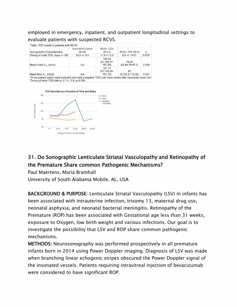
employed in emergency, inpatient, and outpatient longitudinal settings to
evaluate patients with suspected RCVS.
31. Do Sonographic Lenticulate Striatal Vasculopathy and Retinopathy of
the Premature Share common Pathogenic Mechanisms?
Paul Maertens, Maria Bramhall
University of South Alabama Mobile, AL, USA
BACKGROUND & PURPOSE: Lenticulate Striatal Vasculopathy (LSV) in infants has
been associated with intrauterine infection, trisomy 13, maternal drug use,
neonatal asphyxia, and neonatal bacterial meningitis. Retinopathy of the
Premature (ROP) has been associated with Gestational age less than 31 weeks,
exposure to Oxygen, low birth weight and various infections. Our goal is to
investigate the possibility that LSV and ROP share common pathogenic
mechanisms.
METHODS: Neurosonography was performed prospectively in all premature
infants born in 2014 using Power Doppler imaging. Diagnosis of LSV was made
when branching linear echogenic stripes obscured the Power Doppler signal of
the insonated vessels. Patients requiring intravitreal injection of bevacizumab
were considered to have significant ROP.

RESULTS: Out of 177 infants less than 31 weeks, 20 infants required intravitreal
injection of bevacizumab to treat ROP. Six infants less than 31 weeks were
diagnosed with LSV. Diagnosis of LSV was made at 1 month and 6 weeks of age
in the 2 infants with gestational of 24 and 25 weeks respectively: these infants
did not develop ROP. Diagnosis of LSV was made at 4 months of age in 4
infants with mean gestational age of 22 weeks (Between 21 and 23 weeks).:
these infants developed ROP
CONCLUSIONS: Our preliminary results suggest that severe prematurity is an
additional risk factor for LSV. It is postulated that LSV and ROP share common
pathogenic mechanisms.
32. Microembolus detection in cryptogenic stroke: Rationale and design
of the MEDICS study
Anthony Noto, Manjunath Markandaya, Bogachan Sahin
University of Rochester, Strong Memorial Hospital, Department of Neurology
Rochester, NY, USA
BACKGROUND & PURPOSE: Cryptogenic stroke accounts for a substantial
proportion of all ischemic strokes. The detection of cerebral microembolic
signal (MES) by transcranial Doppler (TCD) has the potential to clarify stroke
mechanism and predict recurrent stroke risk in this subpopulation. While a
proximal embolic source or systemic process is often suspected in bilateral
cryptogenic strokes, unilateral cryptogenic strokes affecting one major arterial
territory may result from an occult ipsilateral large vessel thromboembolic
source. Several imaging studies suggest such a mechanism, but no real-time
physiologic evidence is currently available to further substantiate this
possibility. The purpose of this study is two-fold: to determine 1) whether there
is a difference in the pattern of MES (unilateral vs. bilateral) in single- vs. multi-
territory cryptogenic strokes; and 2) whether single-territory cryptogenic
strokes are more likely to be associated with ipsilateral MES, as opposed to no
MES, or contralateral or bilateral MES.
METHODS: This is a single-center prospective cohort study with a planned
enrollment of 40 consecutive patients presenting with an acute cryptogenic

stroke. Patients will be evaluated with 150 minutes of bilateral TCD monitoring
before hospital discharge and followed for 6 months.
RESULTS: The primary endpoint will be unilateral or bilateral MES detection
during the acute hospital admission. The secondary endpoint will be recurrent
stroke or transient ischemic attack within 6 months.
CONCLUSIONS: MES detection by TCD has the potential to clarify stroke
mechanism in patients with cryptogenic stroke. This may in turn help define
new subsets of ischemic stroke patients for future clinical studies.
33. Transcranial Doppler Emboli monitoring artifact due to possible
longer than suggested half-life of definity contrast used in
echocardiogram
Ramnath-Santosh Ramanathan, Vela-Duarte Daniel, Natalie Organek, Lileth Joy
Mondok, Leasa Baus, Maureen McGeary, Yelena Brooks, Ashley Beltran, Mei Lu
Cleveland Clinic/Neurology Cleveland, OH, USA
BACKGROUND & PURPOSE: Definity (perflutren lipid microsphere-composed of
octafluopropane (OFP) encapsulated in an outer lipid shell) is non-blood based
ultrasound contrast agent approved by US FDA for use in patients with
suboptimal echocardiograms to improve the delineation of the left ventricular
endocardial border. OFP was not detectable after 10 minutes in most subjects
in clinical trials either in the blood or in expired air and concentrations in blood
declined in a mono-exponential fashion with a mean half-life of 1.3 minutes.
Definity contrast might have a longer half-life than suggested and may
interfere with transcranial doppler (TCD) emboli monitoring.
METHODS: Case Report
RESULTS: 38-year-old female with HTN and postpartum cardiomyopathy
presented with acute right MCA infarct. She underwent transthoracic
echocardiogram (TTE) and received definity contrast for left ventricular
endocardial border detection. 2 hours after TTE, TCD (figure 1) emboli
monitoring done initially on the left side showed 535 microembolic events in
the left MCA while the right side done 20 minutes later showed 4 microembolic
events in the right MCA while TCD bubble study did not show right to left

shunting. These TCD microemboli were felt to be artifactual due to possible
contamination from definity contrast performed earlier. Repeat TCD (figure 2)
emboli monitoring 4 hours after initial test did not show any microemboli.
CONCLUSIONS: To our knowledge we report first case of definity contrast
causing delayed TCD emboli monitoring artifact in absence of right to left
shunting which may indicate a longer wash out time of the contrast than
estimated.
34. An fNIRS Study of Phonotactic Elements Contributing to English and
Spanish Word Identification
Alejandro Brice, Christina Salnaitis, Molly Quinn, Rachel Gormley, Jacqueline
Barrett, Antwon Frazier, Zachary McNiece, Robert Gray, Ana Luna, Briana Attalla,

Carlos Barbour, Barbara Marques, Diana Rafailova, Rain Christi, Devin Plant,
Ander Baranda
University of South Florida, St. Petersburg St. Petersburg, FL, USA
BACKGROUND & PURPOSE: The purpose of the present study was to identify the
phonotactic elements that affect speed and activation level of word recognition
for monolingual and bilingual speakers. It was expected that due to no lax
vowels in Spanish the bilinguals would be slower and require more activation
than monolinguals who are exposed to tense and lax vowels in English. It was
expected that speakers would require time and more activation for voiceless
consonants due to voicing features.
METHODS: Twenty-six monolingual and 10 bilingual speakers participated.
Sentences were presented in either English or Spanish followed by target words
in either English or Spanish in 70ms increments. After each presentation,
participants indicated whether or not they knew the word and if they were 100%
confident. Participants wore a 16-optode functional near infrared spectroscopy
(fNIRS) device to measure oxygenation changes during the experiment.
RESULTS: Broca’s area was more active in monolinguals than bilinguals across
word type. Across the prefrontal cortex lax vowels produced higher activity
than tense vowels for both monolinguals and bilinguals. The ventromedial
prefrontal cortex showed more activity in bilingual speakers for lax vowels than
monolingual speakers.
CONCLUSIONS: The results confirmed the hypotheses than lax vowels required
more activation due to the lower energy expenditure in producing these
sounds. The hypothesis for bilingual activation being greater on lax vowels was
also supported, but was restricted to the ventromedial area. This area is
generally involved in conflict monitoring and working memory as people regard
different choices.
35. Prefrontal Activation During Word Identification for Bilinguals and
Monolinguals: An fNIRS Study
Christina, L Salnaitis, Alejandro Brice, Molly Quinn, Rachel Barrett, Antwon
Frazier, Zachary McNiece, Ana Luna, Briana Attalla, Carlos Barbour, Diana

Ravailova, Rain Christi, Devin Plant, Ander Baranda, Aaron Huba, Danica Tan,
Alexis Diaz
University of South Florida, St. Petersburg St. Petersburg, FL, USA
BACKGROUND & PURPOSE: The purpose of this investigation was to explore
whether bilinguals and monolinguals engage in different processing in the
prefrontal cortex while recognizing English and Spanish words. Due to learning
more than one language, it is expected that bilinguals will have greater
flexibility in recognizing words.
METHODS: Twenty-six monolingual English-speakers and 10 bilingual English-
and Spanish-speakers participated. Sentences were presented in either English
or Spanish followed by target words in either English or Spanish in 70ms
increments. After each presentation, participants indicated whether or not they
knew the word and if they were 100% confident. Participants wore a 16-optode
functional near infrared spectroscopy (fNIRS) device to measure oxygenation
changes during the experiment.
RESULTS: Bilinguals were faster at recognizing the Spanish words than
monolinguals and were equivalent at word recognition for English words.
Ventromedial prefrontal cortex was more active in monolinguals as they
identified Spanish words followed by English sentences. Right
dorsolateral prefrontal cortex was more active in bilinguals speakers
identifying English words followed by English sentences.
Dorsomedial prefrontal areas were activated in bilinguals recognizing Spanish
words followed by Spanish sentences.
CONCLUSIONS: The right side activation for Spanish speaking bilinguals is
consistent with current hypotheses of bilateral language localization amongst
these second language learners. For English monolingual speakers, the medial
activation suggests the speaker is monitoring the sounds for error detection
and expending additional working memory resources on unfamiliar sounds.
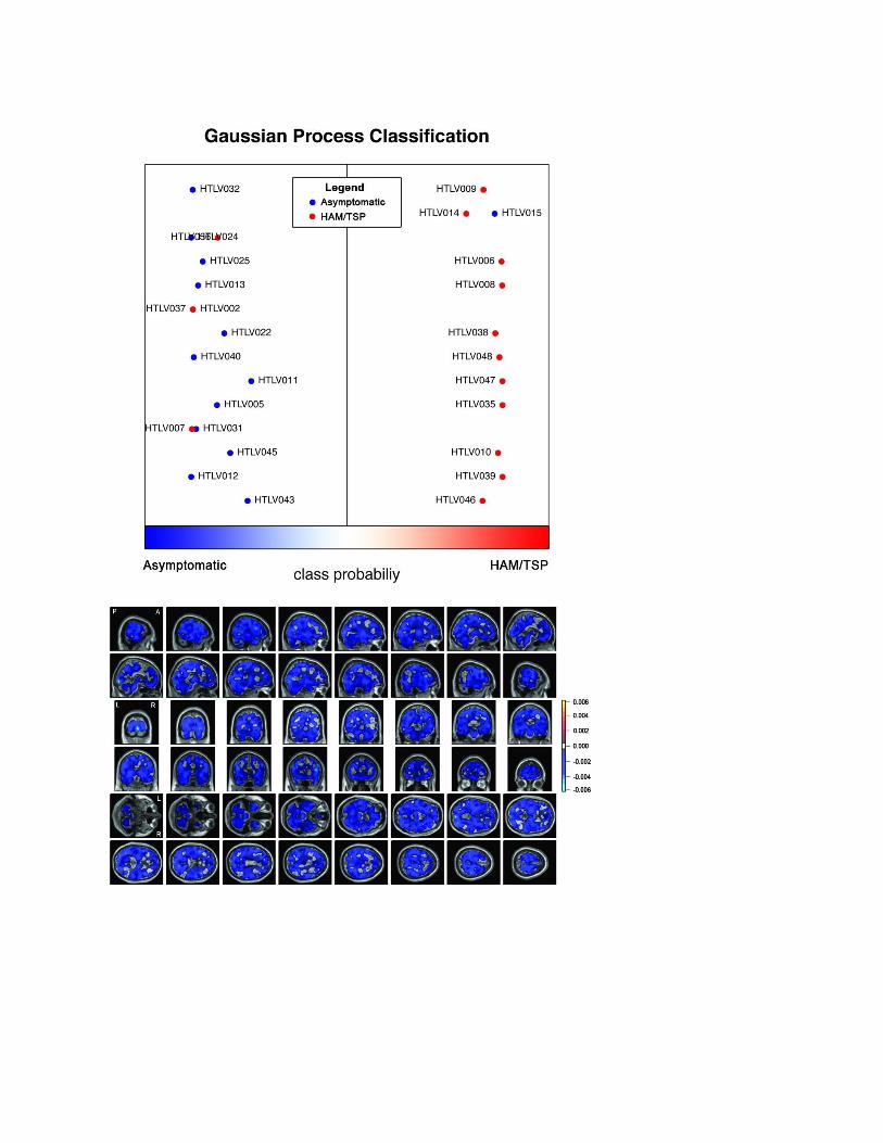
36. Metabolic changes in the brain of patients with HTLV-1-associated
myelopathy/tropical spastic paraparesis
Manuel Schutze1, Luiz C. F. Romanelli1, Herika M. M. Vasconcelos1, Carlos

Malamut2, Debora M. Miranda1, Marco A. Romano-Silva1, Michael Brammer3
1Universidade Federal de Minas Gerais Belo Horizonte, Brazil, 2Centro de
Desenvolvimento da Tecnologia Nuclear Belo Horizonte, Brazil, 3Kings College
London / Institute of Psychiatry London, United Kingdom
BACKGROUND & PURPOSE: Human T-lymphotropic virus type 1 (HTLV-1)
infection is asymptomatic for the major part of seropositive patients, but
approximately 0.25-3.8% present with HTLV-1-associated myelopathy/tropical
spastic paraparesis (HAM/TSP). This clinical condition is characterized mostly
by chronic progressive spastic paresis. Pathophysiology of HAM/TSP includes
T-cell mediated inflammatory response and degeneration in the spinal cord and
in the central nervous system. Little is known about the metabolic correlates of
this process in the brain of subjects with HAM/TSP, particularly when compared
to asymptomatic patients. In this study, we proposed to investigate metabolic
changes in the brain of HTLV-1 seropositive patients with and without
HAM/TSP.
METHODS: We acquired resting state 18F Fluorodeoxyglucose Positron Emission
Tomography (18F FDG-PET) brain images of 28 consented patients seropositive
for HTLV-1. Using the Expanded Disability Status Scale (EDSS), half of the
patients were classified as asymptomatic carriers (EDSS=0) and the other half as
HAM/TSP (EDSS>2). A machine learning approach based on Gaussian Process
Classification (GPC) was used to classify patients according to their images. The
accuracy of the method was assessed through leave-one-out cross-validation.
RESULTS: The algorithm was able to classify patients with 86% accuracy (p<
0.001), which suggests a discriminating pattern of metabolic activity between
the groups (figure1). The weight map for GPC shows a diffuse and generalized
hypermetabolism in the HAM/TSP group (figure2).
CONCLUSIONS: Our results suggest a generalized hypermetabolism in brains of
the HAM/TSP group, which might be related to the inflammatory response.
Further studies are necessary to investigate the cause of the hypermetabolism.