the all-on-4® treatment concept for the rehabilitation of ... · readapted and sutured back into...
TRANSCRIPT

OR I G I N A L A R T I C L E
The All-on-4 treatment concept for the rehabilitation of thecompletely edentulous mandible: A longitudinal study with10 to 18 years of follow-up
Paulo Maló PhD1 | Miguel de Araújo Nobre MSc2 | Armando Lopes MSc1 |
Ana Ferro DDS1 | João Botto DDS1
1Surgeon, Oral Surgery Department, Maló
Clinic, Lisbon, Portugal
2Research and Development Department,
Maló Clinic, Lisbon, Portugal
Correspondence
Miguel de Araújo Nobre, Research and
Development Department, Maló Clinic,
Avenida dos Combatentes, 43, piso 11,
Lisboa 1600-042, Portugal.
Email: [email protected]
Funding information
Nobel Biocare Services AG, Grant/Award
Number: 2017-1534
AbstractBackground: There is a need for studies evaluating the long term outcomes of the All-on-4
treatment concept.
Purpose: To evaluate the long term clinical and radiographic outcomes of the All-on-4 treatment
concept in the mandible.
Materials and Methods: This retrospective longitudinal case series study included 471 patients
(women: 286, men: 185, average age = 57.7 years) rehabilitated with 1884 implants in imme-
diate function supporting 471 fixed full-arch mandibular prostheses and followed for 10 to
18 years. Primary outcome measures were prosthetic survival and implant success and sur-
vival (estimated using life tables). Secondary outcome measures were marginal bone loss
(MBL) at 10 and 15 years, biological and mechanical complications. Multivariable analysis was
used to estimate potential risk indicators for implant failure (Cox regression to estimate hazard
ratios and 95% confidence intervals (95%CI)), MBL > 3 mm at 10 and 15 years, biological and
mechanical complications (binary logistic regression to estimate odds ratios [ORs] with
95%CI).
Results: Twenty-seven patients deceased (5.7%) and 149 patients (31.6%) were lost to follow-
up. The cumulative prosthetic survival rate was 98.8%; the implant cumulative survival and suc-
cess rate was 93% and 91.7%, respectively up to 18 years of follow-up. Previous biological com-
plications (HR = 4.43) were significantly associated with implant failure. Average (95% CI) MBL
at 10- and 15-years were 1.72 mm (95%CI: 1.59, 1.85) and 2.32 mm (95% CI: 1.98, 2.66).
Smoking (OR = 2.72), previous failure of a contiguous implant (OR = 3.89) and biological
complication (OR = 8.11) were associated with MBL > 3 mm. The incidence of biological com-
plications was 11.8% at implant level, with previous failure of a contiguous implant (OR = 5.56),
smoking (OR = 1.75), and systemic condition (OR = 1.65) were significantly associated.
The incidence of mechanical complications was 36.7% with male gender (OR = 1.67) and type
of prosthetic material used in the restoration significantly associated (metal-acrylic OR = 0.30;
metal-ceramic OR = 0.22)).
Conclusions: Considering the implant, prosthetic and MBL outcomes it is concluded that the
All-on-4 is a viable treatment option validated in the long term. Nevertheless, biological and
mechanical complications can occur.
KEYWORDS
All-on-4, complete edentulous, dental implants, immediate function, long term, mandible
Received: 26 October 2018 Revised: 8 March 2019 Accepted: 12 March 2019
DOI: 10.1111/cid.12769
Clin Implant Dent Relat Res. 2019;1–13. wileyonlinelibrary.com/journal/cid © 2019 Wiley Periodicals, Inc. 1

1 | INTRODUCTION
Over 275 million people are globally affected by severe tooth loss,
with a 27% increase in the prevalence in the last decade between
2005 and 2015.1
Even though various treatment approaches are available for the
restoration of partially edentulous patients, the restoration of fully
edentulous jaws has been revolutionized, thanks to the introduction
of implant-supported restorations.2
The use of osseointegrated dental implants in clinical practice
began more than five decades ago with the novel work of
Brånemark.3 Giving the combination of factors: age-related tooth loss,
anatomical condition of edentulous ridges, decreased performance of
removable prosthesis, and predictable long-term results of implant
therapy, demand for implant treatment is increasing,4 specifically
immediate function/loading protocols5 Immediate loading of all-
acrylic, implant-supported prostheses for maxillary and mandibular
arches has been shown to provide advantages to patients (psychologi-
cal and economic benefits)6,7 and dentists (clinical and economic ben-
efits)7,8 with its predictability9 associated with a high level of patient
satisfaction.2
The rehabilitation of severely atrophic mandible using implant-
supported prosthesis is often challenging because of the poor quality
and quantity of residual jawbone posterior to the inferior alveolar
nerve, especially in patients with long-term edentulism. Furthermore,
progressive bone loss in the posterior mandible may lead to a super-
ficialization of the alveolar nerve, which may cause pain to denture
wearers during function.4 At the beginning of the millennium, distally
tilted implants were proposed for these situations, providing a signifi-
cant alternative for the maxillary and mandibular posterior segments
without bone grafting, with posterior tilting of distal implants enabling
the use of denser bone located in the anterior aspects of maxilla and
mandible, reduce cantilever lengths, broaden the prosthetic base, and
improve implant-to-bone surface areas because longer implants can
be used.6,10 The optimal number of four implants in edentulous jaws
was previously reported in the literature with favorable 5 to 10 years
results.11–13 The use of tilted implants together with the optimal num-
ber of four implants to rehabilitate the completely edentulous mandi-
ble with a full-arch fixed prosthesis was further adjusted by using
immediate-function protocols with the connection of the prosthesis
on the day of surgery (the All-on-4 concept is one of such concepts).6
The development to fewer implants (n = 4) was justified provided
they were placed as “cornerstones”: two posterior implants tilted dis-
tally and two anterior implants placed in an axial position, all well
spread; which with optimal implant anchorage could render a high
probability for success.6
The All-on-4 treatment concept demonstrated to be a predictable
treatment modality for full-arch rehabilitation with good results in the
short-, medium- and long-term outcomes (up to 10 years).6,14,15 An
important aspect in the evaluation of implant-supported restorations
concerns the evaluation not only of implant survival and success rates,
but also the occurrence of biological and technical complications.
Despite the occurrence of biological and technical complications, the
same were considered minor and resolved chairside as previously
reported in a follow-up up to 7 years.16 However, evidence is lacking
in the literature for outcomes exceeding 10 years in full-arch rehabili-
tation through this treatment modality using larger samples. The aim
of this study is to report the long-term outcomes of full-arch mandibu-
lar rehabilitations through the All-on-4 concept (with Malo Clinic
Protocol).
2 | MATERIALS AND METHODS
This article was written following the STROBE guidelines for observa-
tional studies.17 This retrospective case series study included patients
treated at a private rehabilitation center (Maló Clinic, Lisbon, Portugal)
and it was approved by an independent ethics committee (Ethical
Committee for Health, Lisbon, Portugal; Authorization n�04/2017). A
chart review of patients rehabilitated with All-on-4 concept in the
mandible was performed. The All-on-4 treatment concept consists in
a full-arch implant-supported rehabilitation for complete edentulous
jaws dependent of the position and anatomy of the mental nerve.
Considering the guideline, the following decision tree was applied as
inclusion criteria for the rehabilitation through the All-on-4 treatment
concept: patients in need of a full-arch restoration through immediate
function in the presence of a complete edentulous mandible with a
residual bone height and width that enabled the insertion of implants
with at least 7 mm of length and 3.75 mm of diameter; in the pres-
ence of periodontally or hopeless teeth that needed to be extracted
while assuring the minimum bone height/width conditions for implant
insertion after performing bone regularization; in the presence of
potentially viable teeth in the anterior segment and absence of teeth
in the posterior segments forcing bone grafting or nerve transposition
before inserting dental implants, teeth were extracted and the All-on-
4 treatment concept was applied aiming for a reduction in morbidity,
treatment time, complexity and financial cost for the patient. The
exclusion criteria for treating a patient were active chemotherapy or
radiotherapy, enough bone height/width in the posterior segments to
insert dental implants for partial restoration, the presence of healthy
teeth in the posterior segments; patients who did not complete the
rehabilitation protocol at the rehabilitation center (being referred
exclusively for the implant insertion with or without a provisional res-
toration: n = 183 patients). Inclusion criterion to be selected for the
study were patients with full-arch rehabilitation in the mandible
through the All-on-4 concept that completed the rehabilitation proto-
col at the rehabilitation center. A total of 654 patients were treated in
the mandible according to the All-on-4 concept. Patients were treated
between April 1998 and December 2006:324 patients from an initial
cohort12 and 147 patients included between February and December
of 2006; routine follow-up was performed between April 1998 and
December 2016; data collection and analysis was performed between
January and June 2018.
2.1 | Surgical protocol
The medical history of each patient was reviewed and the diseases were
coded using the International Classification of Diseases, version 11 (ICD-
11).18 Each patient received a clinical examination and complementary
2 MALÓ ET AL.

radiographic examinations, with an orthopantomography to assess bone
height and computerized tomography scan to assess bone volume and
anatomical structures such as the dental nerve. The surgical procedures
were described in previous reports of the All-on-4 treatment concept
surgical protocol.6,14,15 In brief, insertion of the implants used in this
study (Brånemark System Mk II, Mk III, Mk IV, and NobelSpeedy; Nobel
Biocare AB, Göteborg, Sweden) followed standard procedures. The
implant site preparation was performed according to the bone density in
order to achieve the necessary primary stability (aiming to achieve a final
torque >30 Ncm and maximum of 50 Ncm). For low density bone only
the first drill (2 mm twist drill; Nobel Biocare AB) was used the full length
of the implant site; for medium bone density the 2 mm and the 2.8 mm
twist drills were used full length of the implant site (or later the twist step
drill 2.4/2.8 mm; Nobel Biocare AB); while for dense bone all three drills
were used (2 mm, 2.4/2.8 mm and 3.2/3.6 mm; Nobel Biocare AB). The
length of the implants (all anterior to the foramina) ranged from 8.5 to
18 mm. The two most anterior implants followed jaw anatomy in direc-
tion (lingual tilting in cases of severe mandibular resorption). The two
posterior implants were inserted just anterior to the foramina and tilted
distally ~45� relative to the occlusal plane, aiming for good implant
anchorage, short cantilever length, and large interimplant distance14,15 as
the posterior implants typically emerged at the second premolar position.
The implant platform was positioned 0.8 mm above the bone crest for
MkII, MkIII, and MkIV implants (corresponding to the lower corner of the
cylindrical part of the implant flange was placed flush to bone crest) or
flush to the bone crest for NobelSpeedy implants. The soft tissues were
readapted and sutured back into position with 4-0 nonresorbable silk
sutures (B Braun Silkam, Aesculap Inc, Center Valley, Pennsylvania).
Estheticone abutments (Nobel Biocare AB) were used between April
1998 and June 2002. Straight or 17� angulated multiunit abutments
(Nobel Biocare AB) were placed for anterior implants, and angulated 30�
multiunit abutments (Nobel Biocare AB) were placed for posterior
implants. The abutment angulations were chosen so that the prosthetic
screw access holes were in occlusal or lingual locations, to keep the pros-
thesis with an acceptable thickness and to allow the prosthesis to have
passive fit (the connection of the prosthesis without placing any stress
on the supporting implants as evaluated by the one-screw test and
clinical/radiographic analysis).
2.2 | Immediate provisional prosthetic protocol
Implant-supported fixed prostheses of high-density acrylic resin
(PalaXpress Ultra; Heraeus Kulzer GmbH, Germany) with titanium cylin-
ders (Nobel Biocare AB) were manufactured at the dental laboratory and
inserted on the same day. Anterior occlusal contacts and canine guidance
during lateral movements were preferred in the provisional prosthesis.
No cantilevers were used in the provisional bridges. Emergence positions
of the screw-access holes at the posterior implants of the prostheses
were normally at the level of the second premolar. Posterior tilting of the
distal implants allowed the prostheses to hold a minimum of 10 teeth.
2.3 | Final prosthetic protocol
Considering the patients' financial capability, the provisional prosthe-
sis was replaced by a metal ceramic implant-supported fixed prosthe-
sis with a titanium framework and all-ceramic crowns (Procera
titanium framework, Procera Alumina crowns, NobelRondo ceramics;
Nobel Biocare AB), a metal-acrylic resin implant-supported fixed pros-
thesis with a titanium framework (Procera titanium framework; Nobel
Biocare AB) and acrylic resin prosthetic teeth (Heraeus Kulzer GmbH),
or a high-density acrylic resin (PalaXpress Ultra; Heraeus Kulzer
GmbH, Germany) with titanium cylinders (Nobel Biocare AB). In this
final prosthesis, the occlusion mimicked natural dentition. The final
prosthesis was typically delivered 6 months after the surgery. A clini-
cal long-term example is illustrated in Figures 1–3.
2.4 | Follow-up visits and maintenance protocol
The patients were instructed for soft food diet in the first months. A
postoperative maintenance protocol was indicated to each patient
including oral hygiene instructions.19 Follow-up clinical appointments
were performed at 10 days, 2, 4, and 6 months, 1 year and every
6 months thereafter, consisting in the assessment of clinical parame-
ters, prophylaxis and dental hygiene instructions.
2.5 | Outcome measures
Primary outcome measures were prosthetic survival and implant suc-
cess. Prosthetic survival was based on function, with the necessity of
FIGURE 1 Representative intraoral photograph of a patient
rehabilitated with a mandibular All-on-4 currently with 18 years offollow-up
FIGURE 2 Representative extra-oral photograph of the same patient
as in Figure 1 with the patient smiling
MALÓ ET AL. 3
replacing the prosthesis classified as failure. Implant success was
based on the Maló Clinic success criteria:20 (a) implant fulfilled its pur-
ported function as support for reconstruction (the potential existence
of a sleeping implant was considered a failure); (b) implant was stable
when individually and manually tested; (c) no signs of persistent infec-
tion that could jeopardize the implant outcome; (d) no radiolucent
areas around the implants; (e) good esthetic outcome in the rehabilita-
tion (classified as the absence of esthetic complains from the patient
or Prosthodontist); and (f) allowed construction of an implant-
supported fixed prosthesis, which provided patient comfort and good
hygienic maintenance (classified as the absence of comfort and
hygiene complains from the patient). Implants not complying with the
criteria were considered survivals. Implant removal was classified as
failure.
The secondary outcome measures were marginal bone loss (MBL)
at the 10- and 15-years follow-up and the incidence of biological and
mechanical complications. Periapical radiographs were taken on the
day of surgery, 10- and 15-years using the parallel technique with a
film holder (Super-Bite, Hawe Neos Dental, Bioggio, Switzerland), and
its position was adjusted manually for an estimated orthogonal film
position. An operator blinded to patient information examined radio-
graphs of the implants to determine marginal bone level at each evalu-
ation point. Each periapical radiograph was scanned at 300 dpi
(HP Scanjet 4890; HP Portugal, Paço de Arcos, Portugal), and marginal
bone level was assessed with image analysis software (Image J version
1.40 g for Windows, National Institutes of Health, Bethesda,
Maryland). The reference point for the reading was the implant plat-
form (horizontal interface for the axial implants and the orthogonal
interface for tilted implants between implant and abutment), and mar-
ginal bone level was measured from this reference point to the first
contact between implant and bone. The measurements were per-
formed on the mesial and distal sites, and average values were calcu-
lated. The marginal bone level at 10- and 15-years were compared
with the measurement at the day of surgery and MBL was calculated.
The radiographs were accepted or rejected for evaluation based on
the clarity of the implant threads: a clear thread guarantees both
sharpness and an orthogonal direction of the radiographic beam
towards the implant axis. The radiographs were calibrated using the
implants' interthread length.
Biological complications concerning abscess, fistulae formation
and peri-implant disease (the presence of peri-implant pockets
≥5 mm, bleeding on probing, with concurrent presence of MBL and
clinical attachment loss) performed using a plastic periodontal
probe calibrated to 0.25 N (Hawe Click Probe, Hawe Neos, Bioggio,
Switzerland); and mechanical complications concerning loosening or
fracture of any prosthetic component were assessed throughout the
study follow-up and registered as present or absent.
2.6 | Statistical analysis
Descriptive statistics were computed for prosthetic survival (using the
prosthesis as unit of analysis), implant survival and success (using the
implants as unit of analysis), and MBL (using the implant as unit of
analysis). The cumulative prosthesis survival, cumulative implant suc-
cess, and cumulative implant survival were estimated through life
tables. The Cox proportional hazards regression model was used to
evaluate possible predictors of implant failure at 10 years using the
patient as unit of analysis (patients presenting at least one implant fail-
ure compared to patients without any implant failures). The hazard is
a measure of risk where the hazard function gives the instantaneous
potential per unit time for the event to occur, providing an insight into
the conditional failure rates, being the vehicle by which mathematical
modeling of survival data is used in order to identify a specific
model form.
Considering the statistical assumptions of this method, right cen-
soring was applied at 10 years of follow-up and the failures occurring
after 10th year of follow-up were not accounted in the multivariable
analysis. We used univariable analyses to identify covariates associ-
ated with implant failure: age, gender, systemic condition (absence/
presence), smoking status (smoker/nonsmoker), type of opposing den-
tition (implant-supported fixed prosthesis, natural tooth-supported
fixed prosthesis, natural teeth, miscellaneous, removable denture),
type of prosthetic material used in the restoration (metal-ceramic,
metal-acrylic, acrylic resin), cantilever size (no cantilever, one unit can-
tilever, two units cantilever), mechanical complications (presence/
absence), biological complications (presence/absence). Covariates
(P < 0.20 in univariable analyses) and biologically relevant variables
were then entered into a multivariable Cox proportional hazards
regression model; regression coefficients were estimated with
corresponding SEs.21
For the outcome variables “marginal bone loss >3 mm” (the pres-
ence of at least one implant with this criteria), “biological
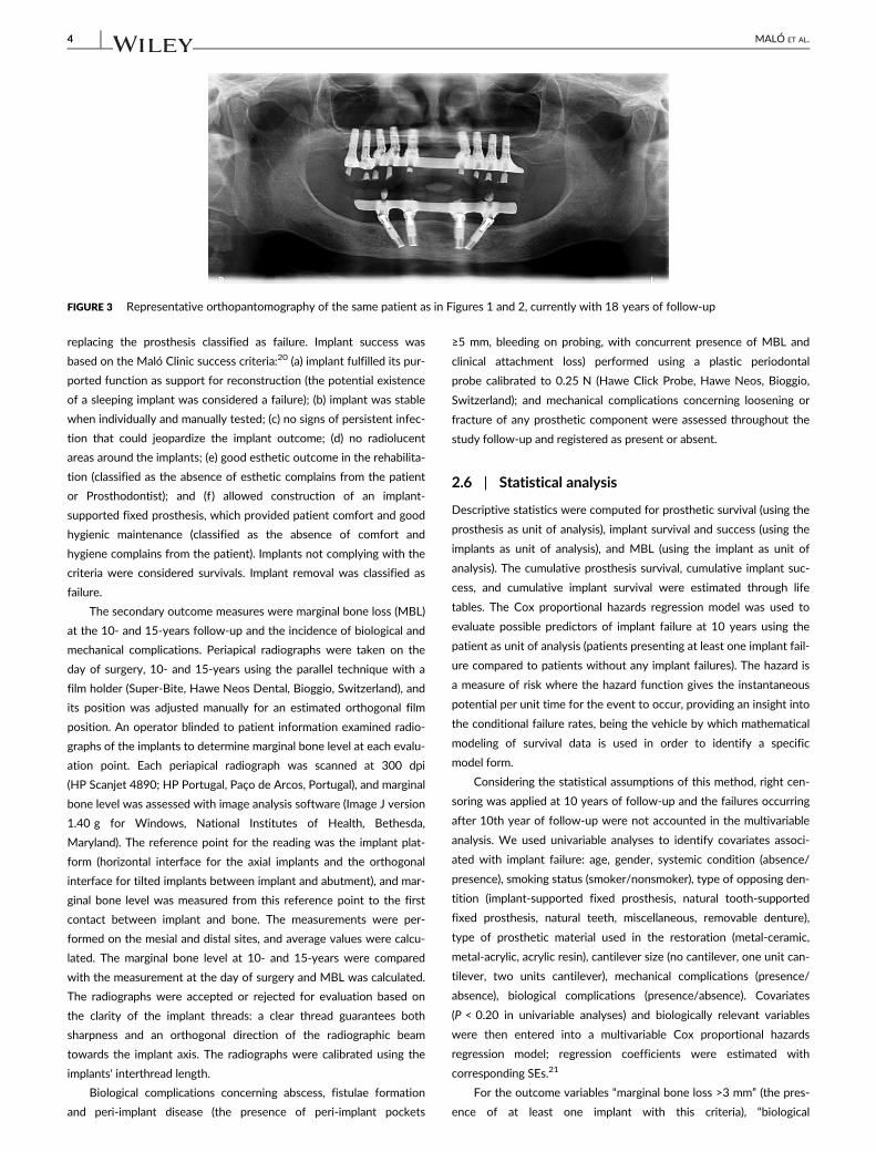
FIGURE 3 Representative orthopantomography of the same patient as in Figures 1 and 2, currently with 18 years of follow-up
4 MALÓ ET AL.

complications” (the presence of at least one implant with this criteria)
and “mechanical complications” a binary logistic regression model was
computed to estimate the odds ratios (ORs) and corresponding 95%
confidence intervals (CIs) for the explanatory variables using the
patient as unit of analysis. The OR represents a measure of associa-
tion between exposure and outcome, representing the odds that an
outcome will occur given a particular exposure, compared to the odds
of the outcome occurring in the absence of exposure. We used
univariable analyses to identify covariates associated with the out-
come variables (all covariates described in the Cox regression analysis
together with the variable “previous failure of a contiguous implant
within the rehabilitation”). Covariates potentially significant (P < 0.20
in univariable analyses) were entered into a multivariable logistic
regression model. The level of significance was 0.05. Statistics were
computed using SPSS 17.0 (IBM, Rochester, New York).
3 | RESULTS
3.1 | Patient characteristics and drop-out rate
Between April 1998 and December 2006, 471 patients (286 women
and 185 men; average age = 57.7 years old, range: 20-85 years old)
received a full-arch rehabilitation in the mandible supported by four
immediately loaded dental implants placed anterior to the mental
foramina, in a total of 1884 implants (n = 219 machined surface
implants inserted from April 1998; n = 1665 anodically oxidized sur-
face implants-TiUnite, Nobel Biocare; inserted since April 2001;
Table 1). A total of 224 patients were positive for ICD-11 with
117 patients who were smokers (24.8%) and 133 patients (28.2%)
who had one systemic condition (Table 2). Regarding opposing denti-
tion, 210 patients had an implant-supported fixed prosthesis,
54 patients had natural teeth, 28 patients had fixed prosthetics over
natural teeth, 68 patients had a combination of natural teeth and
implant-supported fixed prosthetics (miscellaneous), and 111 patients
had a removable prosthesis.
A total of 166 patients was lost to follow-up (with 27 patients
deceased due to causes unrelated to the implant treatment and
149 patients missing the control appointments) with the distribution
according to follow-up time illustrated in Table 2.
3.2 | Prosthetic survival and implant success
Four prostheses were lost after 22, 113, 127, and 149 months in four
patients who lost all four implants, rendering a 98.8% cumulative
prosthetic survival rate.
Considering the evaluation of the implants success criteria, a total
of 70 implants were removed and failed in 41 patients (8.7% failure
rate at patient level), rendering a cumulative implant survival rate of
96.9% at 10 years and 93% up to 18 years of follow-up (life table
analysis, Table 3); In addition, 19 implants in 19 patients were classi-
fied as survivals due to presenting signs of persistent infection that
could jeopardize the implant outcome with MBL extending beyond
50% of the implants' length, rendering a cumulative implant success
rate of 95.9% at 10 years and 91.7% up to 18 years of follow-up (life
table analysis, Table 4; Figure 4). The distribution of survival according
to the health status of the patients (healthy or systemic com-
promised/smoker) is illustrated in Figure 5. The remaining 37 patients
with implant failures but without prosthetic failure maintained the
prosthesis in function through reimplantation of maintaining the pros-
thesis supported on the remaining implants. The insertion of new
implants with immediate loading for complementing the prosthetic
support occurred immediately on the day of failure (n = 7 patients) or
4 to 6 months after implant failure (n = 20 patients) and was success-
ful in all patients, nevertheless one patient that lost implants a second
time and new implants were inserted a second time. The new implants
were not accounted in the analysis for this study. During the healing
time between implant failure and implant insertion, the prosthesis
remained in function supported by the remaining implants, while after
the healing time, the prosthesis was adapted to the new implant posi-
tion. In 10 patients that refused further surgery, no implants were
inserted and the prosthesis remained in function supported on three
implants (n = 9 patients) and two implants (n = 1 patient) during the
remaining follow-up of the study.
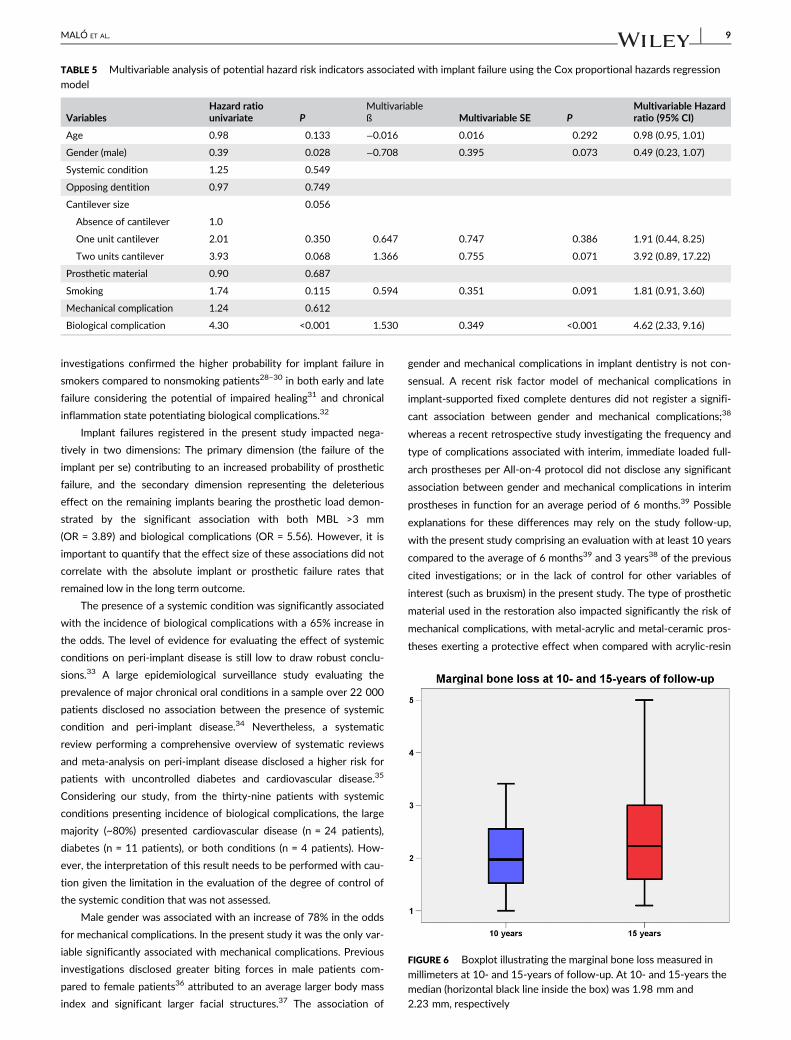
3.3 | Cox proportional hazards regression analysis ofimplant failure
Implant failure in the first decade of follow-up occurred in 36 patients.
The Cox proportional hazards regression model was used to evaluate
the relationship between implant failure and potential risk indicators
with biological complications (HR = 4.62) significantly associated with
implant failure after controlling for the presence of age, gender,
smoking, and cantilever size (Table 5).
3.4 | Marginal bone loss
At the 10-year follow-up, 281 of the 320 patients had readable radio-
graphs (87.8%). The average marginal bone level below the implant-
abutment interface at baseline was −0.02 mm for NobelSpeedy
implants and −0.82 mm for MkII, MkIII, and MkIV implants. The aver-
age (95% confidence interval) MBL was 1.7 mm (95% CI: 1.6, 1.9;
range: 0.7-4.4) (Figure 6). At the 15-year follow-up, all 21 patients had
readable radiographs (100%). The average MBL was 2.3 mm (95% CI:
2.0, 2.7; range: 1.1-6.0) (Figure 6).
TABLE 1 Implant distribution by design
Type of implantTotal number of implants(implant failures)
Brånemark System Mk II implants 42 (5)
Brånemark System Mk III implants 657 (25)
Machined surface 167 (8)
TiUnite surface 488 (17)
Brånemark System Mk IV implants 416 (14)
Machined surface 12 (0)
TiUnite surface 404 (14)
NobelSpeedy Groovy 769 (26)
Total 1884 (70)
MALÓ ET AL. 5

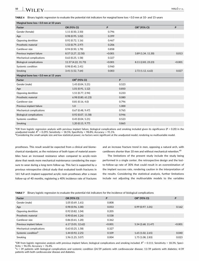
3.5 | Binary logistic regression analysis forMBL >3 mm
There were 27 patients with implants exceeding a MBL of 3 mm. In
the multivariable logistic regression model, the previous failure of a
contiguous implant within the rehabilitation (OR = 3.89), biological
complications (OR = 8.11) and smoking (OR = 2.72) remained signifi-
cantly associated with MBL >3 mm (Table 6). No risk indicators were
associated with MBL >3 mm at 15 years of follow-up (Table 6).
3.6 | Incidence and risk indicators of biologicalcomplications
Biological complications occurred at 223 of the implants (11.8%) in
120 patients (25.5%) consisting of abscess/suppuration (n = 16
implants in 12 patients) and peri-implant disease (n = 207 implants in
108 patients). In the multivariable logistic regression model, previous
failure of a contiguous implant within the rehabilitation (OR = 5.56),
the presence of a systemic condition (OR = 1.65) and smoking
(OR = 1.75) remained significantly associated with the incidence of
biological complications after adjusting for age (Table 7).
3.7 | Incidence and risk indicators of mechanicalcomplications
Mechanical complications occurred in 139 patients in the provisional
prostheses (29.5%) and 173 patients in the definitive prostheses
(36.7%), with 28 patients accumulating mechanical complications in
both the provisional and definitive prostheses (Table 8). In the multi-
variable logistic regression model, male gender (OR = 1.78; P = 0.005)
and the type of prosthetic material used in the restoration (metal-
acrylic resin: OR = 0.30, P < 0.001; metal-ceramic material: OR =
0.22, P < 0.001) remained significantly associated with the incidence
of mechanical complications (Table 9).
TABLE 2 Overall medical status distribution according to the International Classification of Disease, version 11 (ICD-11); Distribution of patients
deceased and lost to follow-up in the sample
ICD-11classification ICD-11 group description Examples
Number ofpatients
Number ofimplants
1 Certain infectious or parasitic diseases (HIV, hepatitis) 11 44
2 Neoplasms (Cancer) 4 16
3 Diseases of the blood or blood forming organs (Coagulation problems) 1 4
5 Endocrine, nutritional of metabolic diseases (Diabetes, Hypercholesterolemia,Hyperthyroidism)
17 68
6 Mental, behavioral or neurodevelopmentaldisorders
(Anxiety) 3 12
8 Diseases of the nervous system (Alzheimer, Epilepsy) 6 24
11 Diseases of the circulatory system (Hypertension, Arrhythmia, Angina) 77 308
12 Diseases of the respiratory system (Emphysema) 3 12
13 Diseases of the digestive system (Heavy bruxer) 5 20
14 Diseases of the skin (Epidermolysis bullosa) 1 4
15 Diseases of the musculoskeletal system orconnective tissue
(Osteoporosis, Rheumatoid arthritis,Sjögren syndrome)
13 52
24 Factors influencing health status or contactwith health services
(Smoking) 117 468
Totals 224a 1665
Distribution of the patients deceased and lost to follow-up according to follow-up time
Follow-up time Patients deceased Patients unreachable Total lost to follow-up
First year – 11 patients 11 patients
1-2 years 2 patients 9 patients 11 patients
2-3 years 4 patients 8 patients 12 patients
3-4 years 2 patients 7 patients 9 patients
4-5 years 3 patients 5 patients 8 patients
5-6 years 1 patient 11 patients 12 patients
6-7 years 2 patients 18 patients 20 patients
7-8 years 7 patients 19 patients 26 patients
8-9 years 3 patients 17 patients 20 patients
9-10 years 2 patients 13 patients 15 patients
10-11 years – 12 patients 12 patients
11-12 years 1 patient 7 patients 8 patients
12-13 years – 1 patient 1 patient
13-14 years – 1 patient 1 patient
aA total of 133 patients had more than one condition.
6 MALÓ ET AL.

4 | DISCUSSION
This study evaluated the long term outcomes of the All-on-4 concept
for full-arch rehabilitation of the mandible, registering a 99.6% pros-
thetic survival rate and a 91.9% cumulative implant success rate up to
18 years of follow-up. To the best of the authors' knowledge these
results represent the longest follow-up recorded for implant-
supported immediate function restorations. The results of this study
follow a pattern considering the previous publication from Maló
et al14 that evaluated the outcome of the All-on-4 concept in the
mandible (with 245 patients from the sample included in the present
study) with up to 10 years of follow-up and registered a 94.8% cumu-
lative implant survival rate. Furthermore, our study included a sample
of 471 patients with a follow-up beginning at 10 years and registered
a cumulative implant survival rate of 96.9% at 10 years of follow-up
and 93% in a follow-up up to 18 years. It is important to notice that
the results of the present study comprise a sample of patients
included irrespective of age, habits, or systemic compromised
TABLE 3 Implant cumulative survival distribution for implants supporting full-arch mandibular rehabilitations through the All-on-4 concept
Time periodTotal numberof implants
Number ofimplants lost Lost to follow-up
Follow-up notcompleted
Cumulativesurvival rate
0-1 year 1884 7 44 0 99.6%
1-2 years 1833 5 44 0 99.3%
2-3 years 1784 5 48 0 99.1%
3-4 years 1731 4 35 0 98.8%
4-5 years 1692 1 31 0 98.8%
5-6 years 1660 4 48 0 98.5%
6-7 years 1608 3 80 0 98.3%
7-8 years 1525 3 104 0 98.1%
8-9 years 1418 2 79 0 98.0%
9-10 years 1337 15 56 0 96.9%
10-11 years 1266 9 46 269 96.1%
11-12 years 942 2 32 219 95.9%
12-13 years 689 6 4 247 94.8%
13-14 years 432 3 2 277 93.9%
14-15 years 150 1 0 81 93.0%
15-16 years 68 0 0 42 93.0%
16-17 years 26 0 0 12 93.0%
17-18 years 14 0 0 4 93.0%
TABLE 4 Implant cumulative success distribution for implants supporting full-arch mandibular rehabilitations through the All-on-4 concept
Timeperiod
Total number ofimplants
Number of unsuccessfulimplantsa
Lost tofollow-up
Follow-up notcompleted
Cumulativesuccess rate
0-1 year 1884 8 44 0 99.6%
1-2 years 1832 6 44 0 99.2%
2-3 years 1782 5 48 0 99.0%
3-4 years 1729 5 35 0 98.7%
4-5 years 1689 3 31 0 98.5%
5-6 years 1655 4 47 0 98.3%
6-7 years 1604 7 80 0 97.8%
7-8 years 1517 9 104 0 97.2%
8-9 years 1404 2 79 0 97.1%
9-10 years 1323 16 56 0 95.9%
10-11 years 1251 11 46 264 94.9%
11-12 years 930 3 32 214 94.6%
12-13 years 681 6 4 245 93.5%
13-14 years 426 3 2 274 92.6%
14-15 years 147 1 0 79 91.7%
15-16 years 67 0 0 42 91.7%
16-17 years 25 0 0 12 91.7%
17-18 years 13 0 0 4 91.7%
aThe sum of implant failures and implant survivals (with marginal bone loss that could jeoperdize the successful outcome as defined by the success criteria).
MALÓ ET AL. 7

conditions. Considering only the compromised status (systemic or
smoking) vs the healthy status on the implants' cumulative success
rate evaluation, the results yielded a 4.2% difference at 10 years and
2.8% up to 18 years in favor of the healthy status. The implant failure
distribution was characterized by an increased density in the first year
of follow-up (n = 12 implants lost) and a late density at 9 to 10 years
of follow-up (n = 24 implants lost). Nevertheless, the late failure den-
sity can be explained by the number of implants that were classified
as unsuccessful prior to the removal date (exhibiting signs of peri-
implant disease with MBL surpassing the implants' middle third) but
were kept in palliative treatment due to the patients' refusal to submit
to surgery for reimplantation. The deleterious influence of biological
complications (namely peri-implant disease) was illustrated by the
regression analysis, where a fourfold increase in the hazard ratio of
implant failure was registered. This result is supported by previous
studies that investigated factors associated with late implant failure
where peri-implant disease was considered as a major risk indica-
tor.22,23 In addition, other factors can partially help explain the late
failure density such as the patient population with a relatively young
at the time of implant placement (average of 57 years of age), and the
broad inclusion criteria with the presence of smokers in the sample, a
situation that was reflected on the proportional hazards analysis for
implant failure. Nevertheless, nearly 70% of the late density failures
(16/24 implants) occurred in peri-implant disease situations that were
unresolved given the patients refusal for further surgical intervention.
The 1.7 mm of average MBL registered in the present study after
10 years of follow-up is within the reported values of other long term
publications on immediate function: Ostman et al24 reported 1.6 mm
of MBL; Glauser25 reported 1.65 mm of MBL; Degidi et al26 reported
a range between 1.93 mm and 1.98 mm (for healed sites and post-
extractive sites, respectively) and Maló et al27 reported 2.0 mm mar-
ginal bone level (specifically for the mandible). However, it is
challenging to draw direct comparisons given that the studies
reported the aggregated data of immediate function and 2-stage
surgery;24 of single teeth, partial and full-arch rehabilitations24–27 and
maxilla and mandible,24–26 being this fact the illustration of the scarce
existence of long term studies.
The average MBL at 15 years measured in 21 patients was
2.32 mm. Twenty-one patients represented a small sample size that
given the low statistical power potentially prevented a more robust
statistical evaluation, yielding no significant risk indicators retrieved
from the multivariable model at 15 years. Nevertheless, these
21 patients represent the large majority of the development group
from the 2003 publication6 with an average MBL of 1.2 mm at 1 year
of follow-up, rendering a 1.12 mm of MBL occurred between the first
and the 15th year of follow-up at a level of 0.08 mm/year. However,
27 patients registered MBL >3 mm on their implants, an occurrence
that was associated with the presence of biological complications
(with an eightfold increase in the odds), smoking habits (with almost
threefold increased odds compared to nonsmokers) and previous
implant failure (with almost fourfold increased odds compared to
patients without implant failures). Biological complications impact sig-
nificantly on the success of implant-supported restorations either
through MBL or by forcing implant removal and consequent failure.
Moreover, the prevalence tends to increase with follow-up time. A
recent systematic review investigating the prevalence of peri-implant
disease disclosed a significant relation between follow-up time and
the prevalence of peri-implant disease,22 a result that previously pro-
jected almost two decades ago.23
The deleterious effect of smoking habits on MBL was previously
reported in a study investigating the long-term outcome of implant
supported restorations24 that registered all implants with MBL at
10 years >3 mm were placed in smokers. In addition, subsequent
FIGURE 4 Cumulative success rate for the implants supporting All-
on-4 mandibular rehabilitations: A 95.9% cumulative success rate wasregistered at 10 years of follow-up while a 91.7% cumulative successrate was registered up to 18 years of follow-up
FIGURE 5 Cumulative success rate for the implants supporting All-
on-4 mandibular rehabilitations. Illustrative comparison for thedistribution of implant success between healthy and systemiccompromised patients at 10 years of follow-up and up to 18 years offollow-up
8 MALÓ ET AL.
investigations confirmed the higher probability for implant failure in
smokers compared to nonsmoking patients28–30 in both early and late
failure considering the potential of impaired healing31 and chronical
inflammation state potentiating biological complications.32
Implant failures registered in the present study impacted nega-
tively in two dimensions: The primary dimension (the failure of the
implant per se) contributing to an increased probability of prosthetic
failure, and the secondary dimension representing the deleterious
effect on the remaining implants bearing the prosthetic load demon-
strated by the significant association with both MBL >3 mm
(OR = 3.89) and biological complications (OR = 5.56). However, it is
important to quantify that the effect size of these associations did not
correlate with the absolute implant or prosthetic failure rates that
remained low in the long term outcome.
The presence of a systemic condition was significantly associated
with the incidence of biological complications with a 65% increase in
the odds. The level of evidence for evaluating the effect of systemic
conditions on peri-implant disease is still low to draw robust conclu-
sions.33 A large epidemiological surveillance study evaluating the
prevalence of major chronical oral conditions in a sample over 22 000
patients disclosed no association between the presence of systemic
condition and peri-implant disease.34 Nevertheless, a systematic
review performing a comprehensive overview of systematic reviews
and meta-analysis on peri-implant disease disclosed a higher risk for
patients with uncontrolled diabetes and cardiovascular disease.35
Considering our study, from the thirty-nine patients with systemic
conditions presenting incidence of biological complications, the large
majority (~80%) presented cardiovascular disease (n = 24 patients),
diabetes (n = 11 patients), or both conditions (n = 4 patients). How-
ever, the interpretation of this result needs to be performed with cau-
tion given the limitation in the evaluation of the degree of control of
the systemic condition that was not assessed.
Male gender was associated with an increase of 78% in the odds
for mechanical complications. In the present study it was the only var-
iable significantly associated with mechanical complications. Previous
investigations disclosed greater biting forces in male patients com-
pared to female patients36 attributed to an average larger body mass
index and significant larger facial structures.37 The association of
gender and mechanical complications in implant dentistry is not con-
sensual. A recent risk factor model of mechanical complications in
implant-supported fixed complete dentures did not register a signifi-
cant association between gender and mechanical complications;38
whereas a recent retrospective study investigating the frequency and
type of complications associated with interim, immediate loaded full-
arch prostheses per All-on-4 protocol did not disclose any significant
association between gender and mechanical complications in interim
prostheses in function for an average period of 6 months.39 Possible
explanations for these differences may rely on the study follow-up,
with the present study comprising an evaluation with at least 10 years
compared to the average of 6 months39 and 3 years38 of the previous
cited investigations; or in the lack of control for other variables of
interest (such as bruxism) in the present study. The type of prosthetic
material used in the restoration also impacted significantly the risk of
mechanical complications, with metal-acrylic and metal-ceramic pros-
theses exerting a protective effect when compared with acrylic-resin
TABLE 5 Multivariable analysis of potential hazard risk indicators associated with implant failure using the Cox proportional hazards regression
model
VariablesHazard ratiounivariate P
Multivariableß Multivariable SE P
Multivariable Hazardratio (95% CI)
Age 0.98 0.133 −0.016 0.016 0.292 0.98 (0.95, 1.01)
Gender (male) 0.39 0.028 −0.708 0.395 0.073 0.49 (0.23, 1.07)
Systemic condition 1.25 0.549
Opposing dentition 0.97 0.749
Cantilever size 0.056
Absence of cantilever 1.0
One unit cantilever 2.01 0.350 0.647 0.747 0.386 1.91 (0.44, 8.25)
Two units cantilever 3.93 0.068 1.366 0.755 0.071 3.92 (0.89, 17.22)
Prosthetic material 0.90 0.687
Smoking 1.74 0.115 0.594 0.351 0.091 1.81 (0.91, 3.60)
Mechanical complication 1.24 0.612
Biological complication 4.30 <0.001 1.530 0.349 <0.001 4.62 (2.33, 9.16)
FIGURE 6 Boxplot illustrating the marginal bone loss measured in
millimeters at 10- and 15-years of follow-up. At 10- and 15-years themedian (horizontal black line inside the box) was 1.98 mm and2.23 mm, respectively
MALÓ ET AL. 9
prostheses. This result would be expected from a clinical and biome-
chanical standpoint, as the resistance of both types of material assem-
blies have an increased resistance when compared to acrylic-resin
alone that needs more mechanical maintenance considering the expo-
sure to wear during a long-term follow-up. This fact is supported by a
previous retrospective clinical study that evaluated tooth fractures in
161 full-arch implant-supported acrylic resin prostheses after a mean
follow-up of 40 months, registering a 40% incidence rate of fractures
and an increase fracture trend in men, opposing a natural arch, with
cantilevers shorter than 10 mm and without mechanical retention.40
The limitations of the present study include the study being
performed in a single center, the retrospective design and the lost-
to-follow-up rate of 30% that could result in an overestimation of
the implant success rate, rendering caution in the interpretation of
the results. Considering the statistical analysis, further limitations
include not adjusting the multivariable models to the variables
TABLE 6 Binary logistic regression to evaluate the potential risk indicators for marginal bone loss >3.0 mm at 10- and 15-years
Marginal bone loss >3.0 mm at 10 years
Factor OR (95% CI) P ORa (95% CI) P
Gender (female) 1.11 (0.50, 2.50) 0.796
Age 0.98 (0.95, 1.02) 0.399
Opposing dentition 0.92 (0.72, 1.16) 0.467
Prosthetic material 1.53 (0.79, 2.97) 0.206
Cantilever size 0.94 (0.50, 1.78) 0.858
Previous implant failure 8.57 (3.27, 22.50) <0.001 3.89 (1.34, 11.30) 0.013
Mechanical complications 0.63 (0.25, 1.58) 0.327
Biological complications 11.57 (4.22, 31.73) <0.001 8.11 (2.83, 23.23) <0.001
Systemic condition 0.98 (0.40, 2.41) 0.960
Smoking 3.41 (1.52, 7.64) 0.003 2.72 (1.12, 6.63) 0.027
Marginal bone loss >3.0 mm at 15 years
Factor ORb (95% CI) P
Gender (male) 1.45 (0.04, 5.21) 0.523
Age 1.01 (0.91, 1.12) 0.850
Opposing dentition 1.51 (0.77, 2.94) 0.233
Prosthetic material 6.98 (0.80, 61.23) 0.080
Cantilever size 0.81 (0.16, 4.0) 0.796
Previous implant failure 1.0 1.000
Mechanical complications 0.67 (0.48, 9.47) 0.765
Biological complications 0.92 (0.07, 11.58) 0.946
Systemic condition 0.45 (0.04, 5.21) 0.523
Smoking 1.20 (0.15, 9.77) 0.865
aOR from logistic regression analysis with pervious implant failure, biological complications and smoking included given its significance (P < 0.20) in theunadjusted model. R2 = 0.295; Sensitivity = 18.5%; Specificity = 98.8%; Accuracy = 91.1%.bConsidering the small sample size and low statistical power, no factors were significant at the unadjusted model, rendering no multivariable model.
TABLE 7 Binary logistic regression to evaluate the potential risk indicators for the incidence of biological complications
Factor OR (95% CI) P ORa (95% CI) P
Gender (male) 1.05 (0.69, 1.61) 0.808
Age 0.98 (0.96, 1.00) 0.053 0.99 (0.97, 1.01) 0.162
Opposing dentition 0.92 (0.82, 1.04) 0.205
Prosthetic material 0.90 (0.64, 1.26) 0.538
Cantilever size 0.86 (0.61, 1.20) 0.362
Previous implant failure 6.17 (3.01, 12.62) <0.001 5.54 (2.68, 11.47) <0.001
Mechanical complications 0.63 (0.25, 1.58) 0.327
Systemic conditionb 1.44 (0.92, 2.25) 0.109 1.65 (1.02, 2.65) 0.040
Smoking 1.96 (1.25, 3.07) 0.004 1.75 (1.08, 2.83) 0.023
aOR from logistic regression analysis with previous implant failure, biological complications and smoking included. R2 = 0.111; Sensitivity = 18.3%; Speci-ficity = 96.3%; Accuracy = 76.4%.bn = 39 patients with biological complications and systemic condition (24/39 patients with cardiovascular disease; 11/39 patients with diabetes; 4/39patients with both cardiovascular disease and diabetes.
10 MALÓ ET AL.

Cawood and Howell classification of bone atrophy and bruxism. It
is important to underline that in long term studies it is expected
that the lost-to-follow-up rate to increase (~20% of the lost-to-
follow-up patients deceased), but nevertheless the sample
encompassed a significant number of implants with more than
15 years of follow-up.
Future research should focus on the report of more long term
follow-up studies evaluating the outcome of full-arch implant-
supported rehabilitations ad modum All-on-4 with emphasis on the
definitive prostheses lifetime.
5 | CONCLUSIONS
Considering the implant and prosthetic survival and success rates, MBL
and incidence of biological and mechanical complications it is concluded
that the full-arch rehabilitation of the edentulous mandible ad modum
All-on-4 is a viable treatment option validated in the long term
outcome. However, biological and mechanical complications can occur.
Implant failure, biological complications, MBL >3 mm, smoking and the
presence of a systemic condition impacted directly and/or indirectly on
the success outcome of the implant-supported rehabilitations.
CONFLICT OF INTEREST
This study was supported by Nobel Biocare Services AG, grant
2017-1534. Paulo Maló is currently a consultant for Nobel Biocare;
Received previous educational fees from Nobel Biocare Services AG;
Received previous scientific Grant support from Nobel Biocare Ser-
vices AG; Miguel de Araújo Nobre: Received previous educational
fees from Nobel Biocare Services AG; Received previous scientific
Grant support from Nobel Biocare Services AG; Armando Lopes:
Received previous educational fees from Nobel Biocare Services AG;
Received previous scientific Grant support from Nobel Biocare Ser-
vices AG; Ana Ferro: Received previous educational fees from Nobel
Biocare Services AG; Received previous scientific Grant support from
Nobel Biocare Services AG; João Botto: No conflict of interest.
AUTHOR CONTRIBUTIONS
Paulo Maló: conception and design of the work; interpretation of data
for the work; drafting the article or revising it critically; final approval
of the version to be published; agreement to be accountable for all
aspects of the work in ensuring that questions related to the accuracy
or integrity of any parts of the work are appropriately investigated
and resolved. Miguel de Araújo Nobre: the acquisition, analysis, and
interpretation of data for the work; drafting the article or revising it
critically; final approval of the version to be published; agreement to
be accountable for all aspects of the work in ensuring that questions
related to the accuracy or integrity of any parts of the work are appro-
priately investigated and resolved. Armando Lopes: design of the
work; interpretation of data for the work; drafting the article or revis-
ing it critically; final approval of the version to be published; agree-
ment to be accountable for all aspects of the work in ensuring that
questions related to the accuracy or integrity of any parts of the work
are appropriately investigated and resolved. Ana Ferro: design of the
TABLE 9 Binary logistic regression to evaluate the potential risk indicators for the incidence of mechanical complications
Factor OR (95% CI) P ORa (95% CI) P
Gender (male) 1.72 (1.17, 2.54) 0.006 1.78 (1.20, 2.69) 0.005
Age 0.99 (0.98, 1.01) 0.403
Opposing dentition 1.12 (0.99, 1.25) 0.219
Prosthetic material <0.001 <0.001
Acrylic resin 1.0 (reference)
Metal-acrylic resin 0.31 (0.20, 0.48) <0.001 0.30 (0.20, 0.47) <0.001
Metal-ceramic 0.22 (0.11, 0.44) <0.001 0.22b (0.11, 0.44) <0.001
Cantilever size 1.08 (0.80, 1.45) 0.618
Previous implant failure 1.52 (0.73, 3.16) 0.268
Biological complications 1.00 0.998
aOdds ratio from logistic regression analysis with gender and prosthetic material included given its significance (P < 0.20) in the unadjusted model.R2 = 0.122; Sensitivity = 71.3%; Specificity = 53%; Accuracy = 64.1%.bAn odds ratio = 0.22 for metal-ceramic material renders an odds ratio = 4.55 for acrylic resin material (odds ratio = 1/0.22 , odds ratio = 4.55).
TABLE 8 Incidence of mechanical complications in the provisional
and definitive prostheses
Complications Number Percentage
Provisional prostheses
Prosthesis fracture 86 18.3%
Abutment fracture 1 0.2%
Abutment screw loosening 75 15.9%
Prosthetic screw fracture 1 0.2%
Prosthetic screw loosening 3 0.6%
Definitive prostheses
Prosthesis fracture (acrylic resin) 107 22.7%
Ceramic crown fracture 15 3.2%
Acrylic crown fracture 75 15.9%
Cylinder fracture 5 1.1%
Artificial gingiva fracture 12 2.6%
Abutment fracture 5 1.1%
Abutment screw loosening 70 14.9%
Prosthetic screw fracture 9 1.9%
Prosthetic screw loosening 23 4.9%
MALÓ ET AL. 11

work; interpretation of data for the work; drafting the article or revis-
ing it critically; final approval of the version to be published; agree-
ment to be accountable for all aspects of the work in ensuring that
questions related to the accuracy or integrity of any parts of the work
are appropriately investigated and resolved. João Botto: design of the
work; interpretation of data for the work; drafting the article or revis-
ing it critically; final approval of the version to be published; agree-
ment to be accountable for all aspects of the work in ensuring that
questions related to the accuracy or integrity of any parts of the work
are appropriately investigated and resolved.
ORCID
Miguel de Araújo Nobre https://orcid.org/0000-0002-7084-8301
REFERENCES
1. GBD 2015 Disease and Injury Incidence and Prevalence Collaborators.Global, regional, and national incidence, prevalence, and years livedwith disability for 310 diseases and injuries, 1990–2015: a systematicanalysis for the Global Burden of Disease Study 2015.
2. Babbush CA. Posttreatment quantification of patient experiences withfull-arch implant treatment using a modification of the OHIP-14 ques-tionnaire. J Oral Implantol. 2012;38:251-260.
3. Brånemark PI. Vital microscopy of bone marrow in rabbit. Scand J ClinLab Invest. 1959;11:S1-S82.
4. Weinstein R, Agliardi E, Fabbro MD, Romeo D, Francetti L. Immediaterehabilitation of the extremely atrophic mandible with fixed full-prosthesis supported by four implants. Clin Implant Dent Relat Res.2012;14:434-441. https://doi.org/10.1111/j.1708-8208.2009.00265.x.
5. Agliardi E, Panigatti S, Clericó M, Villa C, Maló P. Immediate rehabilita-tion of the edentulous jaws with full fixed prostheses supported byfour implants: interim results of a single cohort prospective study. ClinOral Implants Res. 2010;21:459-465.
6. Maló P, Rangert B, Nobre M. "all-on-four" immediate-function conceptwith Brånemark system implants for completely edentulous mandibles:a retrospective clinical study. Clin Implant Dent Relat Res. 2003;5:S2-S9.
7. Babbush CA, Kanawati A, Kotsakis GA, Hinrichs JE. Patient-relatedand financial outcomes analysis of conventional full-arch rehabilitationversus the All-on-4 concept: a cohort study. Implant Dent. 2014;23:218-224.
8. Balshi TJ, Wolfinger GJ. Teeth in a day. Implant Dent. 2001;10:231-233.
9. Esposito M, Grusovin MG, Maghaireh H, Worthington HV. Interven-tions for replacing missing teeth: different times for loading dentalimplants. Cochrane Database Syst Rev. 2013;3:CD003878. https://doi.org/10.1002/14651858.CD003878.pub5.
10. Krekmanov L, Kahn M, Rangert B, Lindström H. Tilting of posteriormandibular and maxillary implants for improved prosthesis support. IntJ Oral Maxillofac Implants. 2000;15:405-414.
11. Brånemark PI, Svensson B, van Steenberghe D. Ten-year survival ratesof fixed prostheses on four or six implants ad modum Brånemark infull edentulism. Clin Oral Implants Res. 1995;6:227-231.
12. Friberg B, Gröndahl K, Lekholm U, Brånemark PI. Long-term follow-upof severely atrophic edentulous mandibles reconstructed with shortBrånemark implants. Clin Implant Dent Relat Res. 2000;2:184-189.
13. Jemt T. Implant survival in the edentulous jaw: 30 years of experience.Part II: a retro-prospective multivariate regression analysis related totreated arch and implant surface roughness. Int J Prosthodont. 2018;31:531-539. https://doi.org/10.11607/ijp.5883.
14. Malo P, de Araújo NM, Lopes A, Moss SM, Molina GJ. A longitudinalstudy of the survival of All-on-4 implants in the mandible with up to10 years of follow-up. J Am Dent Assoc. 2011;142:310-320.
15. Maló P, de Araújo NM, Lopes A, Ferro A, Gravito I. All-on-4® treat-ment concept for the rehabilitation of the completely edentulousmandible: a 7-year clinical and 5-year radiographic retrospective caseseries with risk assessment for implant failure and marginal bone level.
Clin Implant Dent Relat Res. 2015;17:e531-e541. https://doi.org/10.1111/cid.12282 Epub 2014 Dec 23.
16. Tallarico M, Canullo L, Pisano M, Penarrocha-Oltra D, Peñarrocha-Diago M, Meloni SM. An up to 7-year retrospective analysis of bio-logic and technical complication with the All-on-4 concept. J OralImplantol. 2016;42:265-271.
17. von Elm E, Altman DG, Egger M, et al. The strengthening the reportingof observational studies in epidemiology (STROBE) statement: guide-lines for reporting observational studies. J Clin Epidemiol. 2008;61:344-349.
18. World Health Organization. International Classification of Disease, ver-sion 11. https://icd.who.int/browse11/l-m/en. Accessed September2018.
19. Nobre DA et al. Peri-implant maintenance of immediate functionimplants: a pilot study comparing hyaluronic acid and chlorhexidine.Int J Dent Hyg. 2007;5:87-94.
20. Maló PS, de Araújo Nobre MA, Ferro AS, Parreira GG. Five-year out-come of a retrospective cohort study comparing smokersvs. nonsmokers with full-arch mandibular implant-supported rehabili-tation using the All-on-4 concept. J Oral Sci. 2018;60:177-186.https://doi.org/10.2334/josnusd.16-0890 Epub May 10, 2018.
21. Chuang SK, Cai T, Douglass CW, Wei LJ, Dodson TB. Frailty approachfor the analysis of clustered failure time observations in dentalresearch. J Dent Res. 2005;84:54-58.
22. Lee CT, Huang YW, Zhu L, Weltman R. Prevalences of peri-implantitisand peri-implant mucositis: systematic review and meta-analysis.J Dent. 2017;62:1-12. https://doi.org/10.1016/j.jdent.2017.04.011.
23. Tonetti MS. Determination of the success and failure of root-formosseointegrated dental implants. Adv Dent Res. 1999 Jun;13:173-180.
24. Östman PO, Hellman M, Sennerby L. Ten years later. Results from aprospective single-Centre clinical study on 121 oxidized (TiUnite™)Brånemark implants in 46 patients. Clin Implant Dent Relat Res. 2012;14:852-860.
25. Glauser R. Implants with oxidized surface placed predominantly in softbone quality and subjected to immediate occlusal loading: results froman 11-year clinical follow-up. Clin Implant Dent Relat Res. 2016;18:429–38. https://doi.org/10.1111/cid.12327.
26. Degidi M, Nardi D, Piattelli A. 10-year follow-up of immediatelyloaded implants with TiUnite porous anodized surface. Clin ImplantDent Relat Res. 2012;14:828-838.
27. Maló P, de Araújo NM, Gonçalves Y, Lopes A, Ferro A. Immediate func-tion of Anodically oxidized surface implants (TiUnite™) for fixed prostheticrehabilitation: retrospective study with 10 years of follow-up. Biomed ResInt. 2016;2061237:1-11. https://doi.org/10.1155/2016/2061237.
28. Elsubeihi ES, Zarb GA. Implant prosthodontics in medically challengedpatients: the University of Toronto experience. J Can Dent Assoc.2002;68:103-108.
29. Strietzel FP, Reichart PA, Kale A, Kulkarni M, Wegner B, Küchler I.Smoking interferes with the prognosis of dental implant treatment: asystematic review and meta-analysis. J Clin Periodontol. 2007;34:523-544.
30. Chen H, Liu N, Xu X, Qu X, Lu E. Smoking, radiotherapy, diabetes andosteoporosis as risk factors for dental implant failure: a meta-analysis.PLoS One. 2013;8:e71955. https://doi.org/10.1371/journal.pone.0071955 Print 2013.
31. Chrcanovic BR, Albrektsson T, Wennerberg A. Smoking and dentalimplants: a systematic review and meta-analysis. J Dent. 2015;43:487-498. https://doi.org/10.1016/j.jdent.2015.03.003.
32. Al-Bashaireh AM, Haddad LG, Weaver M, Kelly DL, Chengguo X,Yoon S. The effect of tobacco smoking on musculoskeletal health: asystematic review. J Environ Public Health. 2018;2018:4184190-4184106. https://doi.org/10.1155/2018/4184190. eCollection 2018.
33. Clementini M, Rossetti PH, Penarrocha D, Micarelli C, Bonachela WC,Canullo L. Systemic risk factors for peri-implant bone loss: a system-atic review and meta-analysis. Int J Oral Maxillofac Surg. 2014;43:323-334. https://doi.org/10.1016/j.ijom.2013.11.012.
34. de Araújo NM, Maló P. Prevalence of periodontitis, dental caries, andperi-implant pathology and their relation with systemic status andsmoking habits: results of an open-cohort study with 22009 patientsin a private rehabilitation center. J Dent. 2017;67:36-42. https://doi.org/10.1016/j.jdent.2017.07.013.
12 MALÓ ET AL.

35. Ting M, Craig J, Balkin BE, Suzuki JB. Peri-implantitis: a comprehen-sive overview of systematic reviews. J Oral Implantol. 2018;44:225-247. https://doi.org/10.1563/aaid-joi-D-16-00122.
36. Ferrario VF, Sforza C, Serrao G, Dellavia C, Tartaglia GM. Single toothbite forces in healthy young adults. J Oral Rehabil. 2004;31:18-22.
37. Ferrario VF, Sforza C, Poggio CE, Schmitz JH. Facial volume changesduring normal human growth and development. Anat Rec. 1998;250:480-487.
38. Coltro MPL, Ozkomur A, Villarinho EA, Teixeira ER, Vigo A, Shinkai RSA.Risk factor model of mechanical complications in implant-supportedfixed complete dentures: a prospective cohort study. Clin Oral ImplantsRes. 2018;29:915-921. https://doi.org/10.1111/clr.13344.
39. Drago C. Frequency and type of prosthetic complications associatedwith interim, immediately loaded full-arch prostheses: a 2-year retro-spective chart review. J Prosthodont. 2016;25:433-439. https://doi.org/10.1111/jopr.12343.
40. Ventura Ventura J, Jiménez-Castellanos E, Romero J, Enrile F. Toothfractures in fixed full-arch implant-supported acrylic resin prostheses:a retrospective clinical study. Int J Prosthodont. 2016;29:161-165.https://doi.org/10.11607/ijp.4400.
How to cite this article: Maló P, de Araújo Nobre M,
Lopes A, Ferro A, Botto J. The All-on-4 treatment concept for
the rehabilitation of the completely edentulous mandible: A
longitudinal study with 10 to 18 years of follow-up. Clin
Implant Dent Relat Res. 2019;1–13. https://doi.org/10.1111/
cid.12769
MALÓ ET AL. 13