thank you!
DESCRIPTION
The Introduction of the Regional Anatomy Xiaoming Zhang Department of Human Anatomy Medicine school Zhejiang University. - PowerPoint PPT PresentationTRANSCRIPT
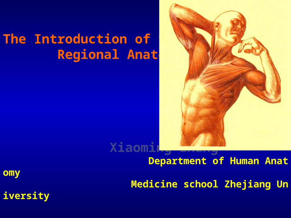
The Introduction of the Regional Anatomy
Xiaoming Zhang Department of Human Anatomy Medicine school Zhejiang University
Definition of the Regional Anatomy The regional anatomy is the science dealing with the form, position and relationship of the structures of the body in a given region. Every chapter of this book includes the four sections, that is the surface landmarks, dissection and observation, main contents, clinical notes.

Surface Landmarks
As a doctor examines a patient, he must firstly inspect the surface anatomy (surface landmarks) and ask what he sees, feels, and hears what he knows existing beneath the skin. In every section of this book there is a paragraph displaying the more important surface landmarks of normal human body. Students should recognize them by looking at the dissected cadaver and then
examining a living body.

Main Contents
In order to learn the basal knowledge of the section, every section presents the main contents to the students. Before dissecting cadaver, the students firstly read the main contents.
Dissection and Observation
In the last semester, the students have learned the anatomical knowledge in the systematic anatomy by observing the specimens. In this term, you dissect cadaver and observe the dissected structures, specially note the variation of the dissected structures.
Clinical Notes
A student should not only grasp the knowledge of anatomy seen on the cadaver or specimens but also learn how to combine his anatomical knowledge with the clinical practice. A series of clinical notes is placed at the end of every section.

I Terms of position During dissection, the cadaver lies horizontally on the table either in the supine position (lying on the back) or in the prone position (lying with the face downward), but the dissector must remember that all of the descriptive terms are based on the anatomical position, e.g.:1. 3 planes The median plane, coronal plane, transverse plane.2. orientation Superior, inferior; anterior, posterior; internal, external These descriptive anatomical terms are also used by the clinician

II. The Construction of the Body
The human body is essentially constructed in layers, From outside inwards, the structures are as follows:
1. The skin(the outermost layer) It covers the body surface and is composed of an epithelial layer of ectodermal origin, the epidermis, and a layer of connective tissue of mesodermal origin, the dermis. It varies greatly in its thickness.
2. The superficial fascia It is beneath the skin and a fibrous, fatty connective tissue, so it is also called the subcutaneous tissue or subcutaneous fat. The parts of the nipple and areola of the breast and some parts of the external genital organs lack the fat. There are the superficial artery, vein, lymphatic vessel, lymph nodes and cutaneous nerve in it.
3. The deep fascia (or proper fascia) It lies under cover of the superficial fascia. It is thin but dense and strong collagenous membrane.Formation: the sheath of the muscle the neurovascular sheath the intermuscular septa the retinaculum
4. The muscle and the deep artery, vein, lymphatic vessel, lymph nodes and nerve most of muscle are the skeletal muscle (flesh belly and dense tendon or aponeurosis).

5. The innermost layer
In the limbs: the bones and joints,
In the trunk: the organs or viscera.
III . The dissecting laboratory1. The instruments of the dissecting cadaver scalpel, forceps with hook, forceps without hook, scissor2. The method of learning the regional anatomy It is the dissection of the cadaver. The medical student must cherish the opportunity to dissect the human body.

3. The preservation of the cadaver
During the dissection, the cadaver may dry out by evaporation, so only dissected part is exposed and other parts are properly wrapped in wet cloth and covered by the a plastic sheet. All scraps(wastes) of anatomical material should be placed in the provided container. After the class, the cadaver must be wrapped by the plastic sheet.

4. To clean the laboratory up
After the class, monitor send a small group to clean the laboratory, the contents of duty include to wipe the blackboard, the table, the stools, and to mop class floor, and to shut off the electric lamps and the taps, and to close the doors and windows.

Ⅳ. The methods of study1. The aim to study regional anatomy Through the dissection, the medical students not only grasp the basal knowledge and theory of human anatomy but also exercise the patience and relative skill of the operation.

2. To have to obey school discipline All students have to go to class and dismiss the class on schedule (time), and be not late and leave early for class.
In every class, the teacher or monitor will call the roll with the classbook and register daily grade.

3. The grouping study Your grade can be grouped into 2 classes;
Each class is subdivided into 3 greater groups, each greater group dissects a cadaver; Each greater group is subdivided into 2 teams and each team (4 students) dissects one side of the cadaver.

During the study, two members of each dissecting team should dissect, and another two members should read aloud the dissecting instructions, supplements the instruction with information from textbooks. It is important that the dissecting team work efficiently and well together and that each member does his share of the work and does not waste time. Dissection should be done among every member of a small team to assure that each student has the opportunity to dissect portion of the cadaver.

4. The preparation before the class and the review after class
A student should not start to dissect the cadaver without carefully reading the paragraph “Dissection and Observation” and “Main Contents” of corresponding section of this book. All of the students must prepare relating lesson before the class and review the last time lesson after class.
V. The methods of dissection
1. The use of anatomical instruments In the first class, the teacher will show students how to use the dissecting instruments.

2. The exercise of dissecting skill In the first class, the teacher will show students how to cut the skin, separate the skin from the superficial fascia, seek (look for) the superficial artery, vein, lymphatic vessel, lymph nodes and cutaneous nerve in the superficial fascia, examine the characters of the deep fascia, clean the muscles, the deep nerves and vessels.
3. To obey of the request of “Dissection and Observation”
The students read the guide of dissection and identify the surface landmark at first, then can start the work. During the dissecting process, the dissecting students should do what the students read.