thaiszia - j - univerzita pavla jozefa Šafárika v košiciach€“ thaiszia – j. bot. 14: 1-22....
TRANSCRIPT
Thaiszia - J. Bot., Košice, 14: 1-22, 2004http://www.upjs.sk/bz/thaiszia/index.html
T H A I S Z I AJOURNAL OF
BOTANY
Contribution to the knowledge of theProtomycetaceae in Slovakia
KAMILA BACIGÁLOVÁ
Department of Cryptogams, Institute of Botany, Dúbravská cesta 14, SK-845 23Bratislava, Slovakia; e-mail: [email protected]
Bacigálová K. (2004): Contribution to the knowledge of theProtomycetaceae in Slovakia. – Thaiszia – J. Bot. 14: 1-22. – ISSN1210-0420.
Abstract: The phytopathogenic fungi of the genera Protomyces and Protomycopsis (Archiascomycetes) are included in the presentpaper. They are biotrophic on leaves and stems, usually gall-forming. Mycelium is apparently diploid, grows intercellularly, formsthick-walled smooth or ornamented “resting-spores” - ascogenouscells. Detailed symptoms of descriptions on the hosts, anatomical-morphological and biometric characteristics of ascogenous cells,nomenclature, chorological data and notes on the ecology of thethree taxa of Protomyces and two of Protomycopsis, associatedwith 10 taxa of vascular plants - Protomyces macrosporus (6);Protomyces pachydermus (1), Protomyces kriegerianus (1);Protomycopsis leontodontis (1), Protomycopsis leucanthemi (1) -are given as well. The new host-fungus combination and newlocations of fungi specimens and their host plants in Slovakia arepublished.
Keywords: Archiascomycetes, Protomyces, Protomycopsis,biotrophic fungi, biology, ecology, chorology, Apiaceae, Asteraceae.
IntroductionMembers of the Protomycetaceae are dimorphic parasites of flowering plants
in Apiaceae and Asteraceae, and represent natural components ofphytocenoses. The fungi cause morphological malformations - galls and lesionson stems, leaves and petioles, often with colour changes of infected host tissues.Since these species do not infect important agricultural or horticultural cropplants, the fungi are poorly known. Also the systematic and taxonomic position offungi has been often discussed. More recent molecular studies confirmed that
1
Protomycetales are very closely related to Taphrinales (SJAMSURIDZAL et al. 1997,SUGIYAMA 1998, PRILLINGER et al. 1990). The authors used inferences from 18SrDNA sequence divergence to set up a new group of Ascomycetes,Archiascomycetes, containing both specimen of the order Taphrinales, generaTaphrina, Protomyces and Protomycopsis (NISHIDA & SUGIYAMA 1994, NISHIDA et al.1995).
The family of Protomycetaceae includes genera: Burenia, Protomyces,Protomycopsis, Taphridium and Volkartia (BÜREN 1915, 1922, REDDY & KRAMER
1975) and Mixia (SAŁATA 1979, KRAMER 1987). Taphridium and Volkartia differmorphologically from Burenia, Protomyces and Protomycopsis in formation oftheir ascogenous cells in a layer just below the upper epidermis of the hostleaves. The species of Burenia, Protomyces and Protomycopsis are form theascogenous cells irregularly in the host tissue. In the case of the genus Mixiabased on morphological and molecular phylogenetic analyses of Mixiaosmundae on Osmunda japonica questions concerning the developement ofendospores of M. osmundae and transfer of this species from Ascomycota to theBasidiomycota were answered (NISHIDA et al. 1995).
The species of Protomyces and Protomycopsis are very similar in forming itsascogenous cells irregularly in the host tissue as well as disorder symptoms onhost plants. Mature ascogenous cells of Protomyces and Protomycopsis showno visible differences. Immature ascogenous cells of Protomycopsis are more orless pear-shaped and the walls are papillous. The walls of ascogenous cells ofProtomyces are smooth.
In Slovakia Protomycetaceae is represented only by genera - ProtomycesUNGER and Protomycopsis MAGNUS. The history and distribution ofProtomycetaceae fungi in Slovak territory were briefly reported by BACIGÁLOVÁ
(1991, 1995). The species Taphridium algeriense from Slovakia cited byBACIGÁLOVÁ (1995) was proved as Protomyces macrosporus on Carum carvi. Thepresent paper summarizes new results of the mycofloristic research and reportson some new aspects of biology, ecology, infection symptoms and chorologicalobservations of fungi in ecological conditions of the Slovak territory. The newspecimens of Protomycetaceae and their host plants for Slovak mycoflora arealso presented.
Material and methodsThe results are based mainly on the study of materials obtained from own
mycofloristic research in Slovakia during the last years (1980-2001), and on theexamination of herbarium specimens deposited in the following institutes(acronyms according to HOLMGREN et al. 1990): BRA, TNP, PRM, PRC,BRNM, W.
All collecting site names cited (“specimens studied”) are local names withinthe studied area. For details compare tourist maps of Slovakia 100 – 142,1:50 000 (published by Vojenský kartografický ústav, Harmanec, Slovakia, 1992-1996).
For identification of the species of the order Protomycetales both visualsymptoms of infected plants and anatomical-morphological characteristics of the
2
fungi were used. Taken cross and longitudinal sections from naturally infectedleaves or stems were observed in drop of 50% lactic acid. An evaluation wasmade by means of Zeiss “Amplival” light microscope with microphotographicequipment. The specimens of Protomyces and Protomycopsis were identifiedaccording to BÜREN (1915, 1922), REDY & KRAMER (1975) and SAŁATA (1979);nomenclature of vascular plants followed MARHOLD & HINDÁK (1998). A list of fungilocations grouped according to their phytogeographical classification (FUTÁK
1966) was compiled. The maps of fungi extension in Slovakia were madefollowed according to JASIČOVÁ ZAHRADNÍKOVÁ (1976). Voucher specimens aredeposited in the mycological herbarium of the Institute of Botany, Bratislava,Slovakia (SAV).
PROTOMYCESThe species of the genus Protomyces are obligate parasites of plants within
the Apiaceae and Asteraceae, causing galls on stems, leaves, flowers and fruits.On the leaves galls are restricted to petiole, midrib, veins and veinlets (Fig. 1, 8).
Spherical to subspherical ascogenous cells with thick and smooth walls areformed intercalaraly in the intercellular mycelium throughout the infected tissue.
Members of the Protomyces parasitizing on Apiaceae (P. macrosporus) areknown from Europe, South Asia and North Africa. The other species(P. pachydermus, P. kriegerianus) parasitizing on Asteraceae (Leontodon,Taraxacum) occur only in Europe (KRAMER, 1975, SAŁATA 1979).
Protomyces macrosporus UNGER
Syn.: Physoderma gibbosum WALLR., Protomyces cari B. BLYTT
Symptoms: The fungus causes white-green hart galls or round callosities withinstem, leaves and fruits tissues (on leaves galls are usually restricted alongpetiole, veins and veinlets) of Apiaceae (Aegopodium, Antriscus, Carum,Chaerophyllum, Heracleum, Ligusticum) (Fig. 1). In these galls, ascogenouscells are present. The ascogenous cells are formed intercalarily in theintercellular mycelium commonly concentrated along the vascular bundles of thehost plant. The cells of surrounding host tissues shown hypertrophy andhyperplasia malformations.
The fact, that species Protomyces macrosporus caused infections of varioushost species of Apiaceae evoked numerous discussions about taxonomy of thisspecimen, (BÜREN 1915, 1922, GJAERUM 1964). Only genotypic identification ofisolates from various host plants will prove taxonomical differences of thesefungi.
The members of Protomyces, predominated by Protomyces macrosporusaccount the major Slovak records of the Protomycetales fungi. It occurs on 6species of host plants (Map 1).
Host: Aegopodium podagraria The fungus induced easily visible whitish galls on the very long green petioles
and on the veins of the leaves. Spherical to roughly spherical ascogenous cellsare affirmed intercalarily in the intercellular mycelium closely associated with the
3
conducting tissues within the veins stems and leaves. The size of theascogenous cells varies between 42-77 µm in diameter (most frequently 64-67µm), with smooth wall, 3-6µm thick (Fig. 2). The fungus locations and their ecological characteristics: Aegopodiumpodagraria is a common species of synantropic plant communities in variousvegetation ranges in Slovakia. Protomyces macrosporus was collected onAegopodium podagraria as early as in the last century by KMEŤ at Prenčov in1978, 1886 (BRA), by HAZSLINSZKY at Prešov (Eperjes), (MOESZ 1939) and byBÄUMLER at Bratislava (Poszony) in 1890 (MOESZ 1939). Our mycofloristicobservations confirmed the fungus occurrence mainly on the humid places alongthe brooks and streams during the whole vegetation season (spring, summerand autumn). List of locations of studied species: (Map 1) 4. Záhorská nížina lowlands:Bratislava – Devínska Nová Ves, 15. km. at the left riverside Morava (1993,SAV); 6. Podunajská nížina lowlands: Hurbanovo (1983, SAV), Bratislava –Petržalka (1985, SAV), Mlyňany in the park (1988, SAV); 10. Malé Karpaty Mts.:Bratislava – Železná studnička (1987, SAV), Píla (1989, SAV), Borinka at themargin of the forest (1992, SAV), Smolenice in the park (1999, SAV); 14e.Štiavnické vrchy hills: Prenčov - Babí potok (KMEŤ 1886, BRA), Prenčov – Medzivršky (KMEŤ 1878, BRA), Banská Štiavnica – surround of water reservoir (2002,SAV); 15. Slovenské rudohorie Mts.: Kokava nad Rimavicou at the riversideRimavica (1987, SAV), Revúca (1989, SAV); 21b. Krivánska Malá Fatra Mts.:Štefanová – Zázrivá by the brook (1998, SAV); 21c. Veľká Fatra Mts.: Vlkolinec(1989, SAV); 23a. Západné Tatry Mts.: Roháčska dolina valley (1988, SAV);23b. Vysoké Tatry Mts.: Tri studničky along the path (1990, SAV), Hrebienok -along the old cableway (2000, SAV), Tatranská Lomnica, in the park (1988,1999, 2000, 2001, omnia SAV), Starý Smokovec along the path (1988, SAV),Tatranská Polianka along the path (1990, 2000, 2001, SAV); 23c Belianske TatryMts.: Dolina Kežmarskej Bielej vody valley (1987, 1999, 2001, SAV), Kežmarskéžľaby - blue tourist mark, (2001, SAV), Monkova dolina valley, (2001, SAV); 26a.Liptovská kotlina hollow: Východná, in the village by the brook (1987, SAV); 28.Západné Beskydy Mts.: Kysuce lazy Čierne (1987, 1989, (SAV), omnia (SAV)leg. et det. K. BACIGÁLOVÁ.
Host: Anthriscus sylvestrisGalls of Protomyces macrosporus are very difficult to find out on flowering
plants of Anthriscus sylvestris, but during the spring (in March), young newleaves emerge, often distorted by galls of Protomyces macrosporus. Thesesymptoms can be seen before the young leaves turn green or greenish purple(Fig. 1).
Entire galls contain very thick walled ascogenous cells, closely associated with thevascular tissues of the host plant. Ascogenous cells are formed intercalarily in theintercellular mycelium. They are spherical to roughly spherical with size of 50-70m(the most frequently 65 µm), with light yellow-brown smooth wall, 4-5 µm thick(Fig. 3). The ascogenous cells are known to produce vesicles and ascospores, butnone have been seen to germinate on natural material of Anthriscus sylvestris.
4
The fungus locations and their ecological characteristics: Anthriscussylvestris is a common species of synantropic plant communities in variousvegetations ranges of the Slovak territory until now, but Protomycesmacrosporus on Anthriscus sylvestris has not been collected in ecologicalconditions of Slovakia. The first one and new locations were found on Anthriscussylvestris along the roadside Dúbravská cesta street (March 31, 2003), Rusovce(April 13, 2003) and along the path in Devínska Kobyla (April 20, 2003), omnialeg. et det. K. BACIGÁLOVÁ (SAV) (Map1).
We assume, the fungus species occurs also in another part of the Slovakterritory and has been overlooked investigated until now on Anthriscus sylvestris.
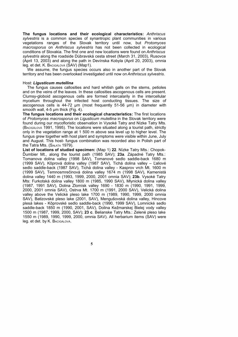
Host: Ligusticum mutellina The fungus causes callosities and hard whitish galls on the stems, petioles
and on the veins of the leaves. In these callosities ascogenous cells are present.Clumsy-globoid ascogenous cells are formed intercalarily in the intercellularmycelium throughout the infected host conducting tissues. The size ofascogenous cells is 44-72 µm (most frequently 51-56 µm) in diameter withsmooth wall, 4-5 µm thick (Fig. 4). The fungus locations and their ecological characteristics: The first locationsof Protomyces macrosporus on Ligusticum mutellina in the Slovak territory werefound during our mycofloristic observation in Vysoké Tatry and Nízke Tatry Mts.(BACIGÁLOVÁ 1991, 1995). The locations were situated along a tourist path, strictlyonly in the vegetation range at 1 500 m above sea level up to higher level. Thefungus grew together with host plant and symptoms were visible within June, Julyand August. This host- fungus combination was recorded also in Polish part ofthe Tatra Mts. (SAŁATA 1979). List of locations of studied specimen: (Map 1) 22. Nízke Tatry Mts.: Chopok-Ďumbier Mt., along the tourist path (1985 SAV); 23a. Západné Tatry Mts.:Tomanova dolina valley (1998 SAV), Tomanové sedlo saddle-back 1680 m(1999 SAV), Kôprová dolina valley (1987 SAV), Tichá dolina valley – Ľaliovésedlo saddle-back (1987 SAV), Tichá dolina valley - Kasprov vrch Mt. 1600 m(1999 SAV), Temnosmrečinová dolina valley 1674 m (1998 SAV), Kamenistádolina valley 1440 m (1993, 1999, 2000, 2001 omnia SAV); 23b. Vysoké TatryMts: Furkotská dolina valley 1800 m (1985, 1990 SAV), Mlynická dolina valley(1987, 1991 SAV), Dolina Zlomísk valley 1690 - 1830 m (1990, 1991, 1999,2000, 2001 omnia SAV), Ostrva Mt. 1700 m (1991, 2000 SAV), Velická dolinavalley above the Velické pleso lake 1700 m (1989, 1990, 1999, 2000 omniaSAV), Batizovské pleso lake (2001, SAV), Mengušovská dolina valley, Hincoveplesá lakes - Kôprovské sedlo saddle-back (1990, 1999 SAV), Lomnické sedlosaddle-back 1850 m (1990, 2001, SAV), Dolina Kežmarskej Bielej vody valley1500 m (1987, 1999, 2000, SAV); 23 c. Belianske Tatry Mts.: Zelené pleso lake1550 m (1989, 1990, 1999, 2000, omnia SAV). All herbarium items (SAV) wereleg. et det. by K. BACIGÁLOVÁ.
5
Host: Heracleum sphondylium The fungus causes hard whitish galls on the long and bulky petioles and on
the veins of the leaves. In these galls ascogenous cells are present. Clumsy-round ascogenous cells are formed intercalary in the intercellular
mycelium throughout the infected conducting host tissue. The ascogenous cellsare 39-77 µm (the most frequently 61-64 µm) in diameter, with smooth wall 5-6µm thick. Frequently, two ascogenous cells in pair are formed (Fig. 5).The fungus locations and their ecological characteristics: The Heracleumsphondylium is a common species of plant comunities in various vegetationsranges of Slovak territory, but the host-fungus combination occurred very rarely.The fungus was not found in the location detected by KMEŤ (1886, BRA), but newlocations were found during mycofloristic observation situated only in mountainvegetation range at altitude of 1180 m. The fungus grew with the host thoroughlyand visual symptoms were presented mainly during June and July. We assumedthat ecological conditions of fungus location (humidity, temperature and altitude)were very important ecological factors for its live cycle on Heracleumsphondylium. List of locations of studied species: (Map 1) 14e. Štiavnické vrchy hills:Prenčov – Babí potok (KMEŤ 1886, BRA), 21b. Malá Fatra (Krivánska Fatra) Mts.:Štefanová – Skalné mesto Mt., 1180 m (1998, SAV); 23a. Západné Tatry Mts.:Javorový žľab valley 1300 m (1998, 1999 SAV); 23c. Belianske Tatry Mts.:Dolina Siedmich prameňov valley (1999, SAV); omnia (SAV) leg. et det. K.BACIGÁLOVÁ.
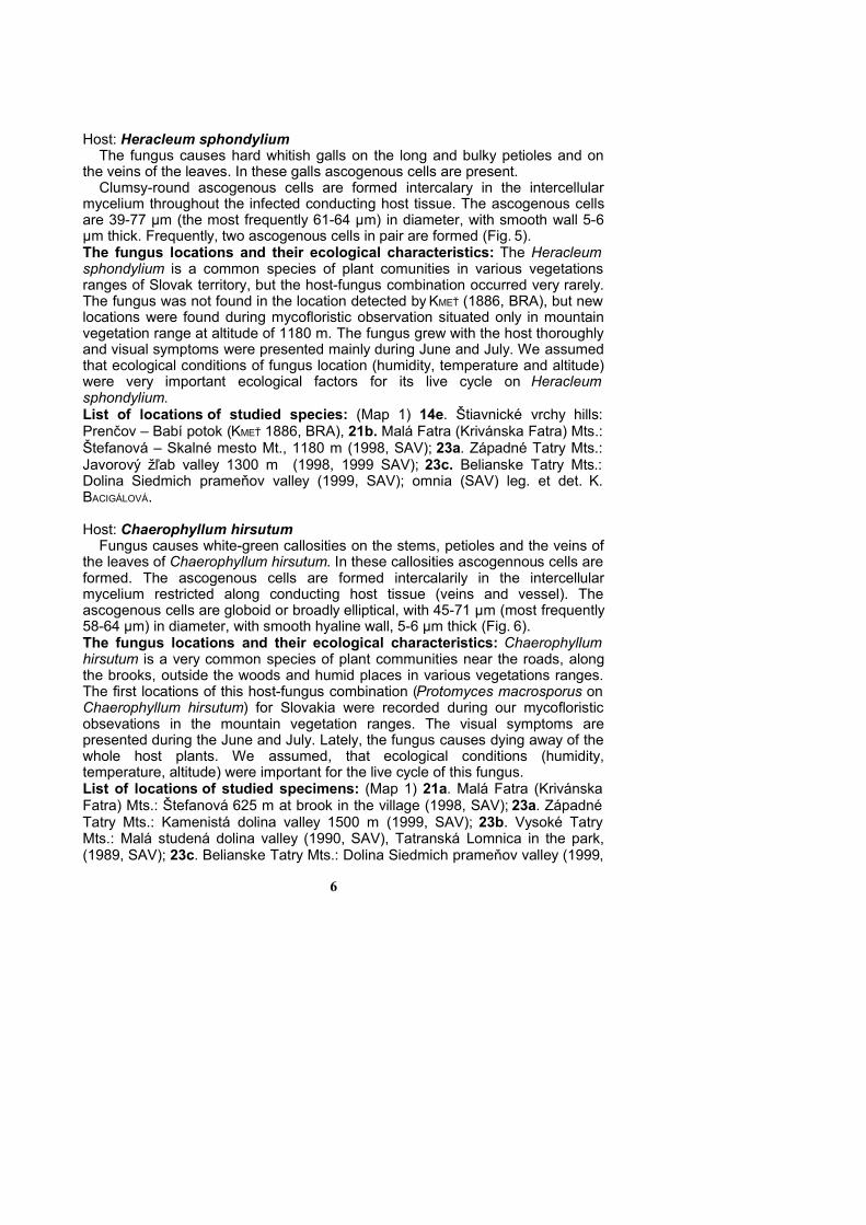
Host: Chaerophyllum hirsutum Fungus causes white-green callosities on the stems, petioles and the veins of
the leaves of Chaerophyllum hirsutum. In these callosities ascogennous cells areformed. The ascogenous cells are formed intercalarily in the intercellularmycelium restricted along conducting host tissue (veins and vessel). Theascogenous cells are globoid or broadly elliptical, with 45-71 µm (most frequently58-64 µm) in diameter, with smooth hyaline wall, 5-6 µm thick (Fig. 6).The fungus locations and their ecological characteristics: Chaerophyllumhirsutum is a very common species of plant communities near the roads, alongthe brooks, outside the woods and humid places in various vegetations ranges.The first locations of this host-fungus combination (Protomyces macrosporus onChaerophyllum hirsutum) for Slovakia were recorded during our mycofloristicobsevations in the mountain vegetation ranges. The visual symptoms arepresented during the June and July. Lately, the fungus causes dying away of thewhole host plants. We assumed, that ecological conditions (humidity,temperature, altitude) were important for the live cycle of this fungus.List of locations of studied specimens: (Map 1) 21a. Malá Fatra (KrivánskaFatra) Mts.: Štefanová 625 m at brook in the village (1998, SAV); 23a. ZápadnéTatry Mts.: Kamenistá dolina valley 1500 m (1999, SAV); 23b. Vysoké TatryMts.: Malá studená dolina valley (1990, SAV), Tatranská Lomnica in the park,(1989, SAV); 23c. Belianske Tatry Mts.: Dolina Siedmich prameňov valley (1999,
6
SAV), Monkova dolina valley (1998, 2000, SAV); 28. Západné Beskydy Mts.:Kysuce, lazy Čierne (1995, SAV); omnia (SAV) leg. et det. K. BACIGÁLOVÁ.
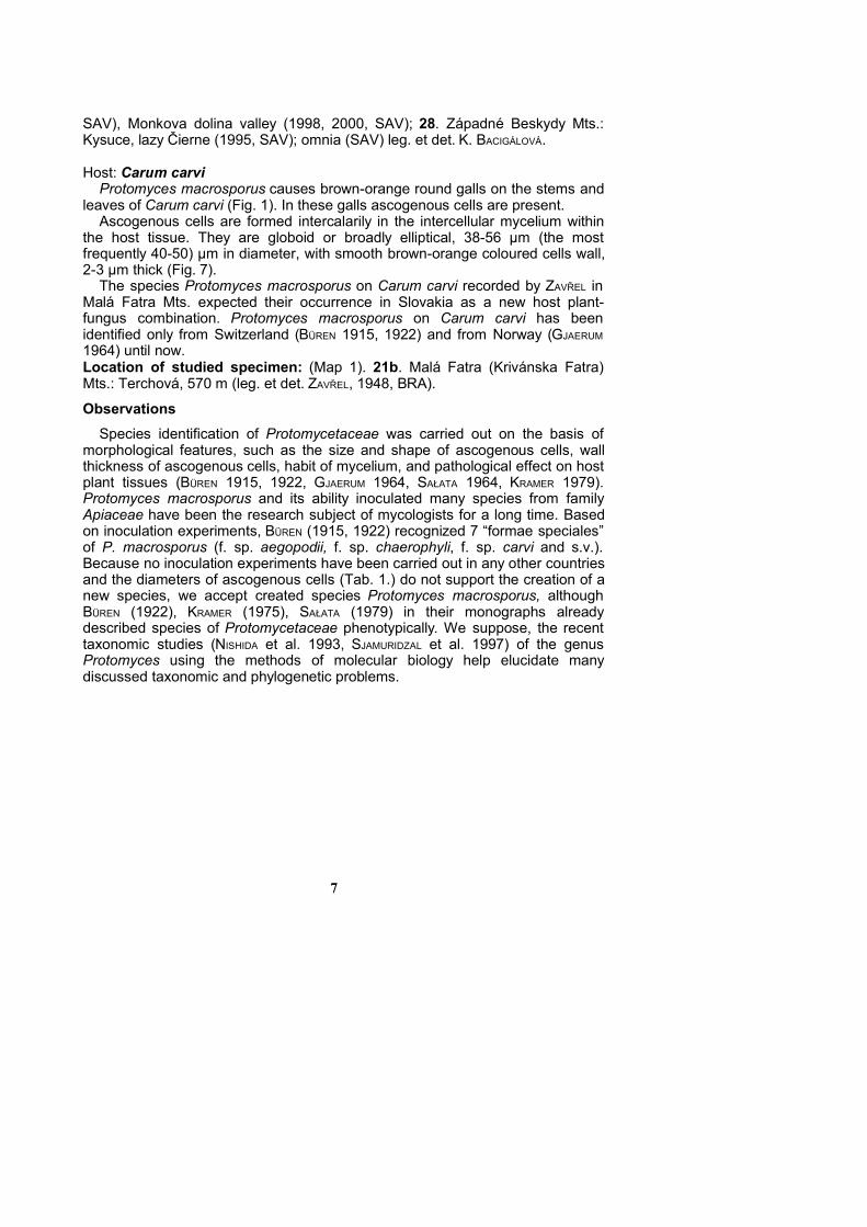
Host: Carum carvi Protomyces macrosporus causes brown-orange round galls on the stems and
leaves of Carum carvi (Fig. 1). In these galls ascogenous cells are present.Ascogenous cells are formed intercalarily in the intercellular mycelium within
the host tissue. They are globoid or broadly elliptical, 38-56 µm (the mostfrequently 40-50) µm in diameter, with smooth brown-orange coloured cells wall,2-3 µm thick (Fig. 7).
The species Protomyces macrosporus on Carum carvi recorded by ZAVŘEL inMalá Fatra Mts. expected their occurrence in Slovakia as a new host plant-fungus combination. Protomyces macrosporus on Carum carvi has beenidentified only from Switzerland (BÜREN 1915, 1922) and from Norway (GJAERUM
1964) until now.Location of studied specimen: (Map 1). 21b. Malá Fatra (Krivánska Fatra)Mts.: Terchová, 570 m (leg. et det. ZAVŘEL, 1948, BRA).
ObservationsSpecies identification of Protomycetaceae was carried out on the basis of
morphological features, such as the size and shape of ascogenous cells, wallthickness of ascogenous cells, habit of mycelium, and pathological effect on hostplant tissues (BÜREN 1915, 1922, GJAERUM 1964, SAŁATA 1964, KRAMER 1979).Protomyces macrosporus and its ability inoculated many species from familyApiaceae have been the research subject of mycologists for a long time. Basedon inoculation experiments, BÜREN (1915, 1922) recognized 7 “formae speciales”of P. macrosporus (f. sp. aegopodii, f. sp. chaerophyli, f. sp. carvi and s.v.).Because no inoculation experiments have been carried out in any other countriesand the diameters of ascogenous cells (Tab. 1.) do not support the creation of anew species, we accept created species Protomyces macrosporus, althoughBÜREN (1922), KRAMER (1975), SAŁATA (1979) in their monographs alreadydescribed species of Protomycetaceae phenotypically. We suppose, the recenttaxonomic studies (NISHIDA et al. 1993, SJAMURIDZAL et al. 1997) of the genusProtomyces using the methods of molecular biology help elucidate manydiscussed taxonomic and phylogenetic problems.
7

Tab. 1. Protomyces macrosporus and their host plants in some Europeancountries.
Host SlovakiaSwitzerland
BÜREN
(1922)
PolandSAŁATA
(1979)
NorwayGJAERUM
(1964)
Great BritainPREECE
HICK (2001)
Size of the ascogenous cells in μmAegopodium podagraria
Anthriscus sylvestris
Chaerophyllum hirsutum
Heracleum sphondylium
Ligusticum mutellina
Carum carvi
42-72(64-67)50-70(65)
45-71(58-64)37-77
(61-64)44-72
(51-56)38-56
(40-50)
50-70
+
+
+
+
+
35-80(50-60)
+
+
+
+
-
31-66
31-61
-
-
-
31-66
+
+
-
-
-
-
+ = host / fungus combination is present-= host / fungus combination was not found
Protomyces pachydermus THÜM.Host: Taraxacum officinale Symptoms: The fungus causes swellings and hard galls on peduncle and on theleaves along the main vein, often 2-15 mm long in diameter. The galls are limpid,white green to yellow or violet. Intensive fungus infections on the leaves areeasily recognizable by the violet-brown or yellow coloured spots with a network ofswollen veins and veinlets (Fig. 8).
The multinucleate mycelium with septa invades the host tissue intercellularlypenetrating throughout all tissues and concentrates along the vascular bundles.The ascogenous cells are formed intercalarily in the intercellular mycelium. Theyare spherical to roughly spherical or elliptic 23-42 µm, (most frequently 25-35µm)in diameter, with smooth, yellow coloured wall, 2-6 µm thick (Fig. 9).The fungus locations and their ecological characteristics: The host plantTaraxacum officinale is very often infected with Protomyces pachydermus mostlyduring humid vegetation season in various vegetation ranges. The first funguslocations founded by KMEŤ 1887 (BRA), were proved in the Štiavnické vrchy hills.Their occurrence in the Slovak territory is being confirmed during mycofloristicresearch every year (Map 2). The “white places” in the map of Slovakia (mainlythe east part) here been poorly studied until now.List of locations of studied species: 5. Devínska Kobyla hill: Bratislava in thegarden of Institute of Botany SAV (1990, 1991, 1992, 1993, 1999, 2000, 2001);6. Podunajská nížina lowland: Bratislava-Rusovce in the park (1991), Bratislava-Petržalka at the Danube riverside (1991), Veľký Grob in the garden (1991); 10.Malé Karpaty Mts.: Dobrá voda (1989); 14e. Štiavnické vrchy hills: Prenčov (KMEŤ
1887 BRA); 15. Slovenské rudohorie: Revúca, at the brook (1989); 21a. Malá8
Fatra Mts.: Štefanová near the tourist path in Kreminná dolina valley (1998),Štefanová on the meadow 644 m (1998); 22. Nízke Tatry Mts.: Krížna-TureckáMt., along the tourist path (1987); 23b. Štrbské pleso lake 1400 m (1990, 1991),Velická dolina valley (1989), along the tourist path to Popradské pleso lake(2000), Dolina Zlomísk valley (1991), Ostrva Mt. along the tourist path (1991),Starý Smokovec (1988); omnia (SAV) leg. et det. K. BACIGÁLOVÁ.
Protomyces kriegerianus BÜREN
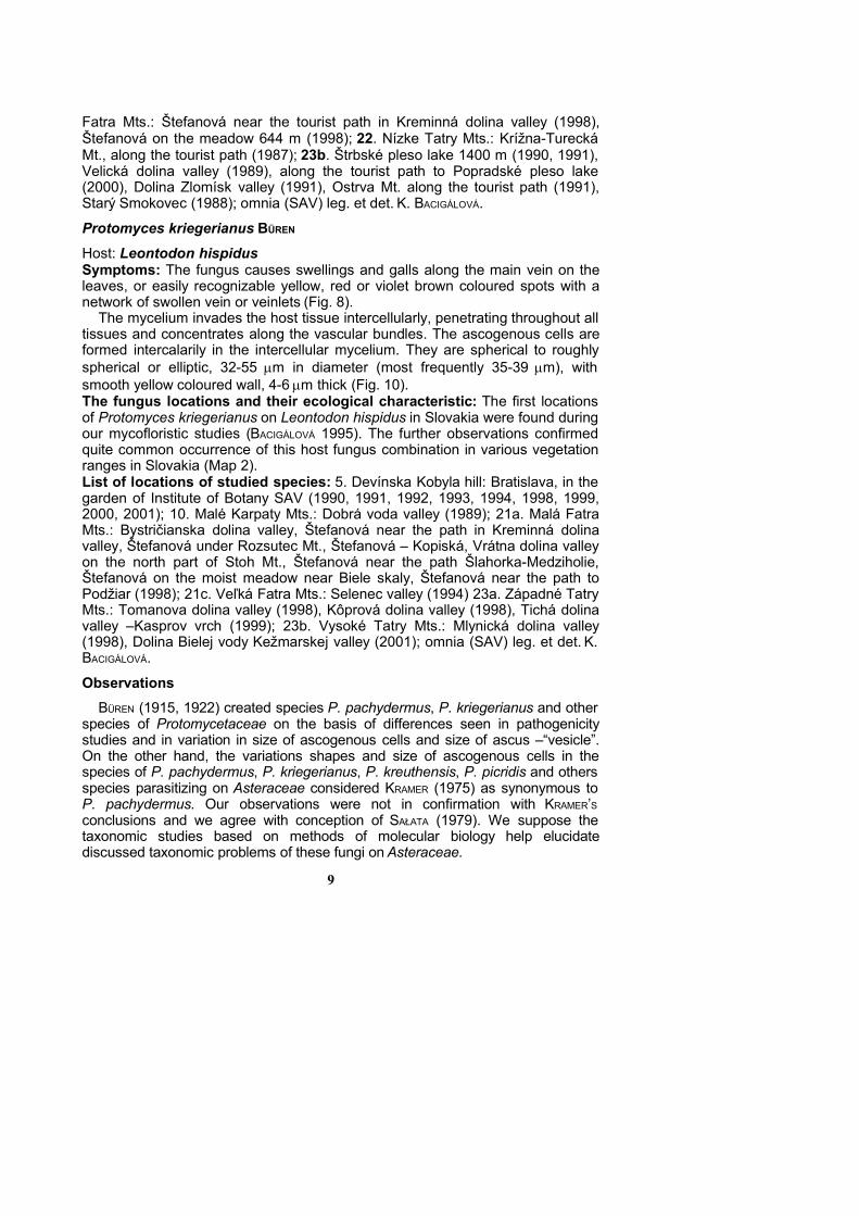
Host: Leontodon hispidusSymptoms: The fungus causes swellings and galls along the main vein on theleaves, or easily recognizable yellow, red or violet brown coloured spots with anetwork of swollen vein or veinlets (Fig. 8).
The mycelium invades the host tissue intercellularly, penetrating throughout alltissues and concentrates along the vascular bundles. The ascogenous cells areformed intercalarily in the intercellular mycelium. They are spherical to roughlyspherical or elliptic, 32-55 m in diameter (most frequently 35-39 m), withsmooth yellow coloured wall, 4-6 m thick (Fig. 10).The fungus locations and their ecological characteristic: The first locationsof Protomyces kriegerianus on Leontodon hispidus in Slovakia were found duringour mycofloristic studies (BACIGÁLOVÁ 1995). The further observations confirmedquite common occurrence of this host fungus combination in various vegetationranges in Slovakia (Map 2).List of locations of studied species: 5. Devínska Kobyla hill: Bratislava, in thegarden of Institute of Botany SAV (1990, 1991, 1992, 1993, 1994, 1998, 1999,2000, 2001); 10. Malé Karpaty Mts.: Dobrá voda valley (1989); 21a. Malá FatraMts.: Bystričianska dolina valley, Štefanová near the path in Kreminná dolinavalley, Štefanová under Rozsutec Mt., Štefanová – Kopiská, Vrátna dolina valleyon the north part of Stoh Mt., Štefanová near the path Šlahorka-Medziholie,Štefanová on the moist meadow near Biele skaly, Štefanová near the path toPodžiar (1998); 21c. Veľká Fatra Mts.: Selenec valley (1994) 23a. Západné TatryMts.: Tomanova dolina valley (1998), Kôprová dolina valley (1998), Tichá dolinavalley –Kasprov vrch (1999); 23b. Vysoké Tatry Mts.: Mlynická dolina valley(1998), Dolina Bielej vody Kežmarskej valley (2001); omnia (SAV) leg. et det. K.BACIGÁLOVÁ.
ObservationsBÜREN (1915, 1922) created species P. pachydermus, P. kriegerianus and other
species of Protomycetaceae on the basis of differences seen in pathogenicitystudies and in variation in size of ascogenous cells and size of ascus –“vesicle”.On the other hand, the variations shapes and size of ascogenous cells in thespecies of P. pachydermus, P. kriegerianus, P. kreuthensis, P. picridis and othersspecies parasitizing on Asteraceae considered KRAMER (1975) as synonymous toP. pachydermus. Our observations were not in confirmation with KRAMER’Sconclusions and we agree with conception of SAŁATA (1979). We suppose thetaxonomic studies based on methods of molecular biology help elucidatediscussed taxonomic problems of these fungi on Asteraceae.
9
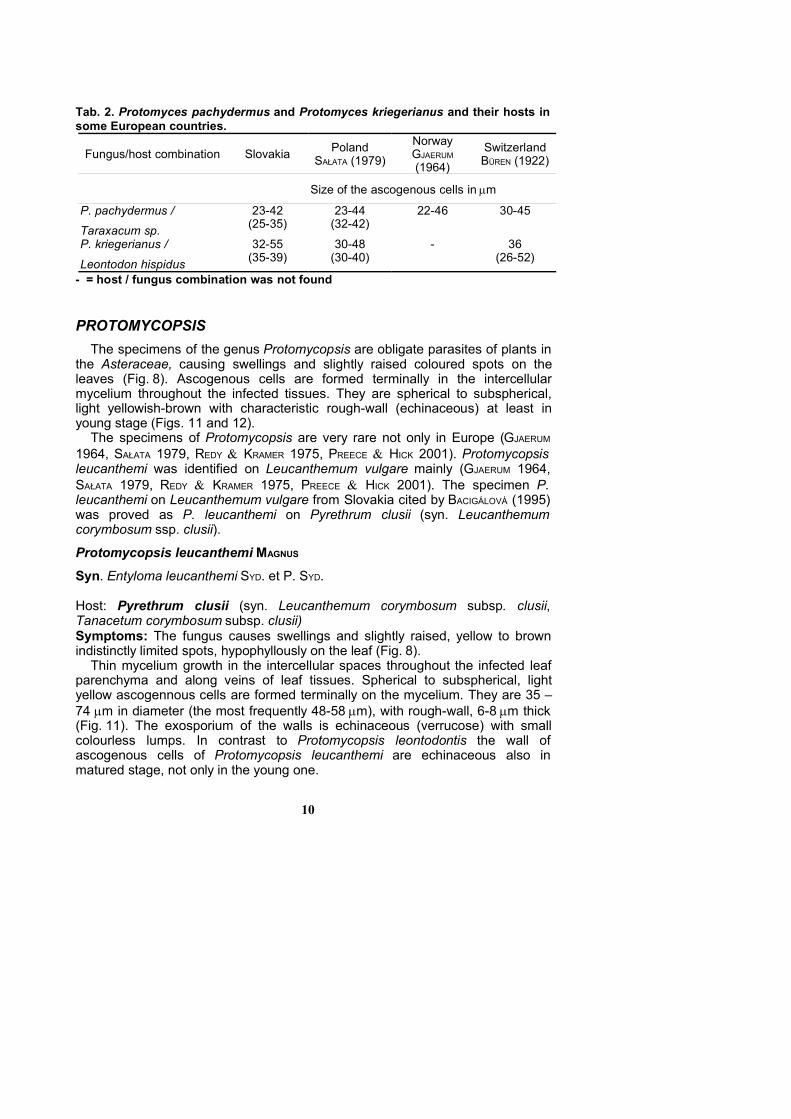
Tab. 2. Protomyces pachydermus and Protomyces kriegerianus and their hosts insome European countries.
Fungus/host combination Slovakia PolandSAŁATA (1979)
NorwayGJAERUM
(1964)
SwitzerlandBÜREN (1922)
Size of the ascogenous cells in m
P. pachydermus /Taraxacum sp.
23-42(25-35)
23-44(32-42)
22-46 30-45
P. kriegerianus /
Leontodon hispidus
32-55(35-39)
30-48(30-40)
- 36(26-52)
- = host / fungus combination was not found
PROTOMYCOPSISThe specimens of the genus Protomycopsis are obligate parasites of plants in
the Asteraceae, causing swellings and slightly raised coloured spots on theleaves (Fig. 8). Ascogenous cells are formed terminally in the intercellularmycelium throughout the infected tissues. They are spherical to subspherical,light yellowish-brown with characteristic rough-wall (echinaceous) at least inyoung stage (Figs. 11 and 12).
The specimens of Protomycopsis are very rare not only in Europe (GJAERUM
1964, SAŁATA 1979, REDY KRAMER 1975, PREECE HICK 2001). Protomycopsisleucanthemi was identified on Leucanthemum vulgare mainly (GJAERUM 1964,SAŁATA 1979, REDY KRAMER 1975, PREECE HICK 2001). The specimen P.leucanthemi on Leucanthemum vulgare from Slovakia cited by BACIGÁLOVÁ (1995)was proved as P. leucanthemi on Pyrethrum clusii (syn. Leucanthemumcorymbosum ssp. clusii).
Protomycopsis leucanthemi MAGNUS
Syn. Entyloma leucanthemi SYD. et P. SYD.
Host: Pyrethrum clusii (syn. Leucanthemum corymbosum subsp. clusii,Tanacetum corymbosum subsp. clusii)Symptoms: The fungus causes swellings and slightly raised, yellow to brownindistinctly limited spots, hypophyllously on the leaf (Fig. 8).
Thin mycelium growth in the intercellular spaces throughout the infected leafparenchyma and along veins of leaf tissues. Spherical to subspherical, lightyellow ascogennous cells are formed terminally on the mycelium. They are 35 –74 m in diameter (the most frequently 48-58 m), with rough-wall, 6-8 m thick(Fig. 11). The exosporium of the walls is echinaceous (verrucose) with smallcolourless lumps. In contrast to Protomycopsis leontodontis the wall ofascogenous cells of Protomycopsis leucanthemi are echinaceous also inmatured stage, not only in the young one.
10
The fungus locations and their ecological characteristics: Host speciesPyrethrum clusii grow only on locations from submontane slopes to higheraltitudes on carbonate and silicate rocks as well as the limestone in the Vysokéand Západné Tatry Mts. Protomycopsis leucanthemi was found on Pyrethrumclusii near the tourist path in two valleys with cold and humid condition during thespring (June – early July). List of locations of studied specimens: (Map 3) 23a. Západné Tatry Mts.:Kamenistá dolina valley 1240 m (1988, 1993, 2003) (SAV); 23b. Vysoké TatryMts.: Dolina Bielej vody Kežmarskej valley 1200 m (1995) (SAV), leg. et det. K.BACIGÁLOVÁ.
Protomycopsis leontodontis BÜREN
Host. Leontodon hispidus subsp. danubialis Symptoms: The fungus causes small galls on veins with yellowish-grey slightlyraised spots on the leaves (Fig. 8).
Ascogenous cells are formed terminally on the intercellular myceliumoccurring throughout the veins of the leaf tissue. They are round to oval 29-48µmin diameter, but most frequently 32-42 µm, and wall 3-5µm thick. On youngascospores the small papillae on the walls are formed (Fig. 12).The fungus locations and their ecological characteristics: Host plant of thisfungus (Leontodon hispidus subsp. danubialis) is one of the most widespreadplants in various vegetation ranges (lowlands, hills, mountains) in Slovakia, butthe host-fungus combination were found only on locations in subalpine or alpinemountain range. Similarly as for the species Protomycopsis leucanthemi andsome other specimens of Protomycetaceae, ecological condition of the highmountains (extreme cold during the winter, cold and humid conditions during thespring, May, June), are favourable for growth and development of this fungusspecimen (Protomycopsis leontodontis) on Leontodon hispidus subsp.danubialis only in this region.List of locations of studied specimen: (Map 3) 23a. Západné Tatry Mts.: Tichádolina valley (1988), Kamenistá dolina valley, 1240 m along the tourist path(2003) (SAV); 23b. Vysoké Tatry Mts.: Štrbské pleso, vodopád Skok (1990),Mlynická dolina valley (1991), Štrbské pleso, 1400 m (2000); omnia (SAV) leg. etdet. K. BACIGÁLOVÁ.
This host-fungus combination is new not only for mycoflora of Slovakia, butalso for the whole West Carpathian Mts. region. The fungus has been knownonly from Switzerland on Leontodon montanus until now (BÜREN 1922).
11
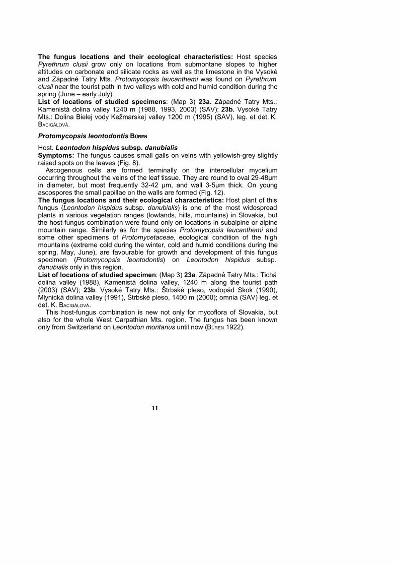
Tab. 3. Species of Protomycopsis and their host plants in some Europeancountries.
Host/fungus combination SlovakiaPolandSAŁATA
(1979)
NorwayGJAERUM(1964)
SwitzerlandBÜREN (1922)
Size of the ascogenous cells in mP. leucanthemi/Pyrethrum clusii 35-74
(48-58)- - -
P. leucanthemi / Leucanthemumvulgare /Chrysanthemumleucanthemum
- 24-61(36-45)
27-51 35(22-48)
P. leontodontis / Leontodonhispidus subsp. danubialis
29-48(32-42)
- - +
P. leontodontis /Leontodonautumnalis
- 34(30-41)
- -
+ = host fungus combination was found- = host fungus combination was not found
AcknowledgementsI would like to express my thanks to RNDr. Pavel Lizoň for comments of the
manuscript, Mgr. Taňa Miháliková for helping with constructions of maps, RNDr.Iva Hodálová for revision of host plants and Gabriela Vosátková for her technicalassistance. This study was supported by grant agency VEGA Slovakia (No.2/1069/21).
References BACIGÁLOVÁ K. (1991): Príspevok k poznaniu druhov radu Taphrinales vo Vysokých
Tatrách. – Zborník prác o Tatranskom národnom parku 31: 35-43.BACIGÁLOVÁ K. (1995): Rozšírenie zástupcov čeľade Protomycetaceae (rad Taphrinales)
na Slovensku. Bull. Slov. Bot. Spol., Bratislava, 17:39-43.BACIGÁLOVÁ K. (1995): Ekologické poznámky k výskytu druhov radu Taphrinales na
Slovensku. – In: TOPERCER J. (ed.), Diverzita rastlinstva Slovenska. pp. 98-102. –Zborník zo VI. Zjazdu SBS pri SAV, Blatnica, 6-10. Júna 1994.
BACIGÁLOVÁ K. (1999): Fytopatogénne mikroskopické huby Vysokých Tatier I. – Štúdie oTatranskom Národnom Parku. 4 (37): 41-70.
BÄUMLER J. A. (1890): Beiträge zur Cryptogamen – Flora des Presburger Comitates II. –Vehr. Vereins Natur-Heilk. Pressburg. 6: 62-126.
BÜREN G. (1915): Die schweizerischen Protomycetaceen mit besondererBerücksichtigung ihrer Entwicklungsgeschichte und Biologie. – Beitr. Kryptogamenfl.Schweiz 5.1: 1-95.
BÜREN G. (1922): Weitere Untersuchungen über die Entwicklungsgeschichte und Biologieder Protomycetaceen. – Beitr. Kryptogamenfl. Schweiz, 5. 3: 1-94.
FUTÁK J. (1966): Fytogeografické členenie Slovenska. In: DOSTÁL J., NOVÁK F.A. (eds.)Flóra Slovenska 1. – Veda, Vyd. SAV Bratislava. 602 pp.
GJAERUM H.B. (1964): Protomycetes, Protomycopsis and Taphridium in Norway. – NyttMagasin for Botanikk. 12: 19-28.
12
HOLMGREN P.K., HOLMGREN N. H. BARNETT L.C. (1990): Index herbariorum. Part I: Theherbaria of the world. ed. 8. – Regnum Veg. 120: 1-693.
JASIČOVÁ M. ZAHRADNÍKOVÁ K. (1976): Organizácia a metodika mapovania rozšíreniarastlinných druhov v západnej tretine Slovenska. – Biológia (Bratislava), 31:74-80.
MARHOLD K. & HINDÁK F. (eds.) (1998): Zoznam nižších a vyšších rastlín Slovenska.Checklist of non-vascular and vascular plants of Slovakia. – Veda, Bratislava, 687pp.
MOESZ G. (1935): Fungi hungariae, III. Ascomycetes, Pars l, XXXII. Annales Mus. Nat.Hungarici, – Pars Botanica, 9.
MUŁENKO W. & BACIGÁLOVÁ K. (2001): Parasitic microfungi of the Tatra Mts. 52 Zjazd PTBPoznan, 2001, 190p.
NISHIDA H. BLANZ P. A. SUGIYAMA J. (1993): The higher fungus Protomyces inouyei hastwo Group 1 introns in the 18S rRNA gene. Journal of Molecular Evolution 37: 25-28.
NISHIDA H. SUGIYAMA J. (1994): Archiascomycetes: detection of a new lineage within theAscomycota. Mycoscience 35: 361-366.
NISHIDA H. ANDO K. ANDO Y. HIRATA A. SUGIYAMA J. (1995): Mixia osmundae: transfer from theAscomycota to the Basidiomycota based on evidence from molecules andmorphology. Can. J. Bot. 73: 660-666.
PREECE T.F. HICK A.J. (2001): An introduction to the Protomycetales: Burenia inundataon Apium nodiflorum and Protomyces macrosporus on Anthriscus sylvestris.Mycologist 15: 118-125.
PRILLINGER H. DÖRFLER C. LAASER G. ECKERLEIN B. LEHLE K. (1990): Ein Beitrag zurSystematik und Entwicklungsbiologie Höherer Pilze: Hefe-Typen derBasidiomyceten. Teil I. Schizosaccharomycetales, Protomyces-Typ. - Zeitschrift fürMycologie 56: 219-250.
REDDY M.S. & KRAMER CH.L. (1975): A taxonomic revision of the Protomycetales,Mycotaxon, 3: 1-50.
SAŁATA B. (1979): Grzyby. Tom XII. Workowce (Ascomycetes) Pierwogrzybowe(Protomycetales). Warszawa-Kraków, 10-44 pp.
SJAMSURIDZAL W. TAJIRI Y. NISHIDA H. THUAN T.B. KAWASAKI H. HIRATA A. YOKOTA A. SUGIYAMA
J. (1997): Evolutionary relationships of members of the genera Taphrina,Protomyces, Schizosaccharomyces and related taxa within the Archiascomycetes.Integrated analysis of genotypic and phenotypic characters. Mycoscience 38: 267-280.
SUGIYAMA J. (1998): Relatedness, phylogeny, and evolution of the fungi. Mycosciences 39.487-511.
SUGIYAMA J. NISHIDA H. (1995): The higher fungi: their evolutionary relationsips andimplications for fungal systematics. In: AAI R., KATO M. DOI Y. (Eds.) Biodiversityand Evolution National Science Museum Foundation, Tokyo. pp. 177-195.
Received: 26 September 2003Revised: 11 November 2003Accepted: 11 Novenber 2003
13

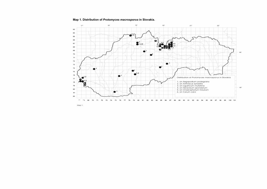
Map 1. Distribution of Protomyces macrosporus in Slovakia.
18° 19° 20° 21° 22°17°
49°
48°
Distribution of in Slovakia
1. on Aegopodium podagraria2. on Anthriscus sylvestris3. on Ligust icum mutellina4. on Heracleum spondylium5. on Chaerophyllum hi rsutum6. on Carum carvi
Protomyces macrosporus
6
Map 1.

Map 2. Distribution of Protomyces pachydermus and Protomyces kriegerianus in Slovakia.
Distribut ion of and in Slovak ia
on
on
P. pachydermus P. kriegerianus
P. pachydermus Taraxacum spp. P. kriegerianus Leontodon hispidus
Map 2.
18° 19° 20° 21° 22°17°
49°
48°
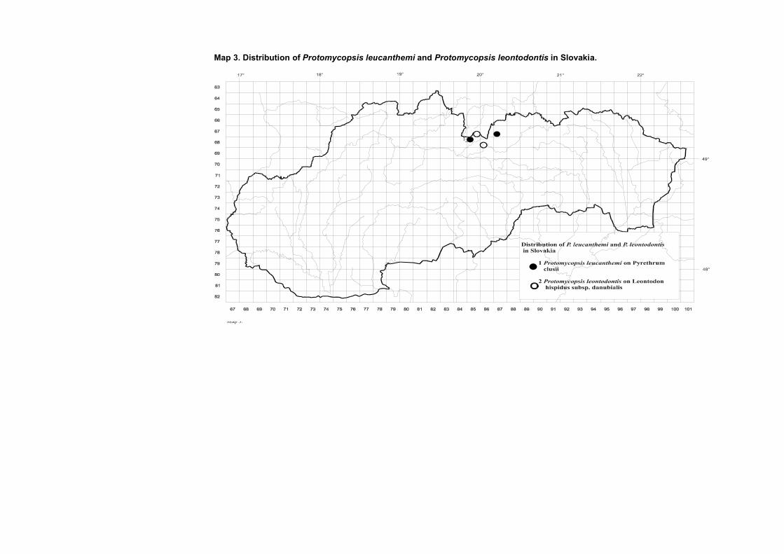
Map 3. Distribution of Protomycopsis leucanthemi and Protomycopsis leontodontis in Slovakia.

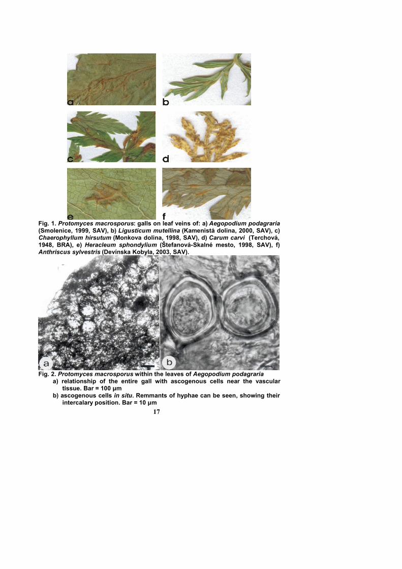
Fig. 1. Protomyces macrosporus: galls on leaf veins of: a) Aegopodium podagraria(Smolenice, 1999, SAV), b) Ligusticum mutellina (Kamenistá dolina, 2000, SAV), c)Chaerophyllum hirsutum (Monkova dolina, 1998, SAV), d) Carum carvi (Terchová,1948, BRA), e) Heracleum sphondylium (Štefanová-Skalné mesto, 1998, SAV), f)Anthriscus sylvestris (Devínska Kobyla, 2003, SAV).
Fig. 2. Protomyces macrosporus within the leaves of Aegopodium podagraria a) relationship of the entire gall with ascogenous cells near the vascular
tissue. Bar = 100 μmb) ascogenous cells in situ. Remmants of hyphae can be seen, showing their
intercalary position. Bar = 10 μm17

Fig. 3. Protomyces macrosporus within the leaves of Anthriscus sylvestrisa) relationship of the entire gall with ascogenous cells near vascular tissue.
Bar =100 μmb) ascogenous cells. Bar =10 μm
Fig. 4. Protomyces macrosporus within the leaves of Ligusticum mutellina a) cross section of the gall. Bar = 100 μmb)ascogenous cells. Bar =10 μm
18
Fig. 5. Protomyces macrosporus within the leaves of Heracleum sphondylium a) relationship of the entire gall with ascogenous cells near vascular tissue.
Bar = 100μmb) ascogenous cell in situ. Remmants of hyphae can be seen showing their
intercalary position. Bar = 10μm
Fig. 6. Protomyces macrosporus within the leaves of Chaerophyllum hirsutuma) relationship of the entire gall with ascogenous cells near the vascular
tissue. Bar = 100μmb) ascogenous cell in the stage of ”vesicule – ascus” emergence. Bar = 10μm
19

Fig. 7. Protomyces macrosporus within the leaves of Carum carvia) cross section of the galls. Bar = 100 μmb) ascogenous cells. Bar = 10μm
Fig. 8. a) Protomyces pachydermus: galls on leaf veins of Taraxacum officinale, b)Protomyces kriegerianus: galls on leaf veins of Leontodon hispidus, (Bratislava,garden of Inst. of Botany, 2000), c) Protomycopsis leucanthemi: galls on leaf veinsand steams of Pyretrum clusii (Kamenistá dolina, 2003, SAV), d) Protomycopsisleontodontis: galls on leaf veins of Leontodon hispidus subsp. danubialis(Štrbské pleso, 2000, SAV).
20
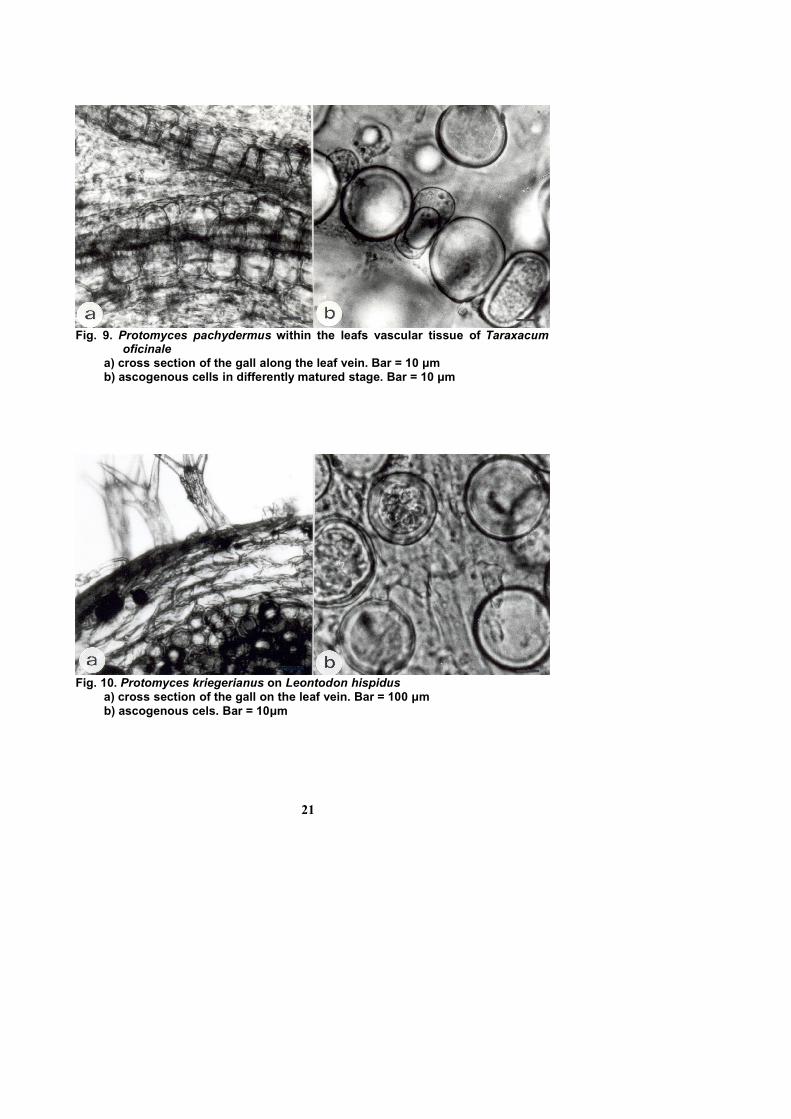
Fig. 9. Protomyces pachydermus within the leafs vascular tissue of Taraxacumoficinale
a) cross section of the gall along the leaf vein. Bar = 10 μmb) ascogenous cells in differently matured stage. Bar = 10 μm
Fig. 10. Protomyces kriegerianus on Leontodon hispidusa) cross section of the gall on the leaf vein. Bar = 100 μmb) ascogenous cels. Bar = 10μm
21
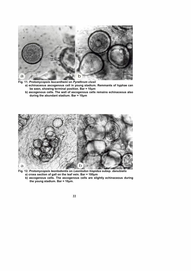
Fig. 11. Protomycopsis leucanthemi on Pyrethrum clusiia) echinaceous ascogenous cell in young stadium. Remmants of hyphae can
be seen, showing terminal position. Bar = 10μmb) ascogenous cells. The wall of ascogenous cells remains echinaceous also
during the abundant stadium. Bar = 10μm
Fig. 12. Protomycopsis leontodontis on Leontodon hispidus subsp. danubialis a) cross section of gall on the leaf vein. Bar = 100μmb) ascogenous cells. The ascogenous cells are slightly echinaceous during
the young stadium. Bar = 10μm.
22