testicular tumour
TRANSCRIPT

Testicular tumors
PROF DR PANNA LAL SAHAPROFESSOR OF SURGERY & HOD
BGC TRUST MEDICAL COLLEGECHITTAGONG
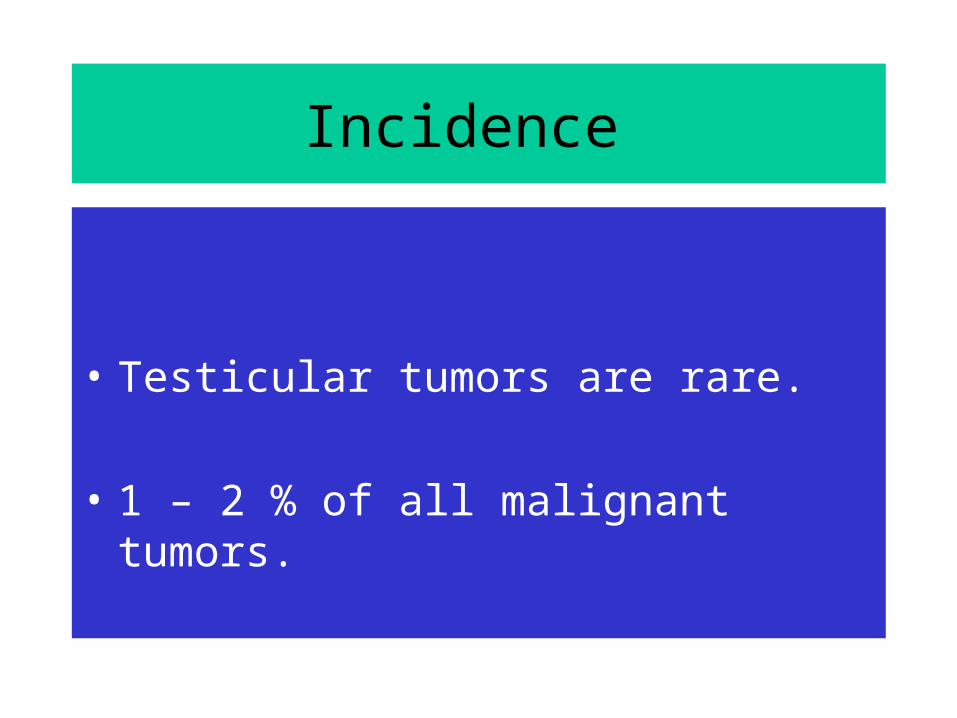
Incidence
• Testicular tumors are rare.
• 1 – 2 % of all malignant tumors.
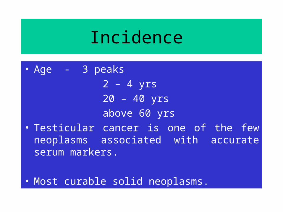
• Age - 3 peaks 2 – 4 yrs 20 – 40 yrs above 60 yrs• Testicular cancer is one of the few neoplasms
associated with accurate serum markers.
• Most curable solid neoplasms.
Incidence
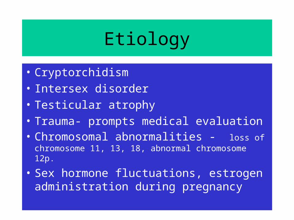
Etiology
• Cryptorchidism • Intersex disorder• Testicular atrophy• Trauma- prompts medical evaluation • Chromosomal abnormalities - loss of
chromosome 11, 13, 18, abnormal chromosome 12p.
• Sex hormone fluctuations, estrogen administration during pregnancy
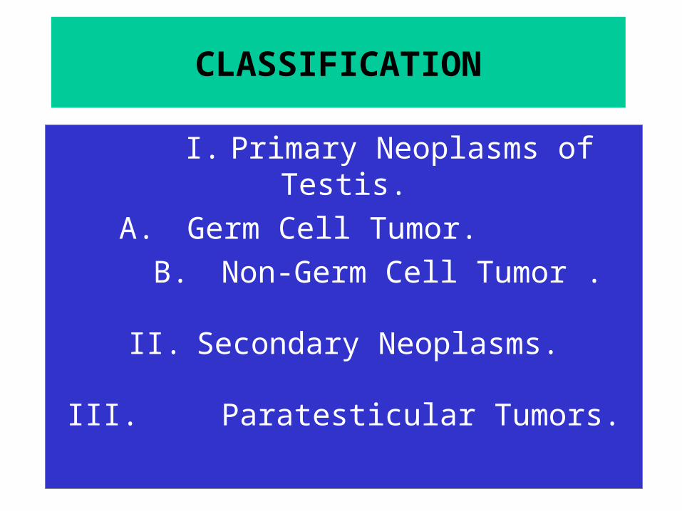
CLASSIFICATION
I. Primary Neoplasms of Testis.A. Germ Cell Tumor.
B. Non-Germ Cell Tumor .
II. Secondary Neoplasms.
III. Paratesticular Tumors.
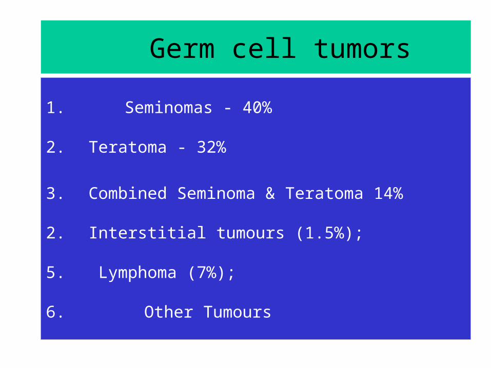
Germ cell tumors
1. Seminomas - 40%
2. Teratoma - 32%
3. Combined Seminoma & Teratoma 14%
2. Interstitial tumours (1.5%);
5. Lymphoma (7%);
6. Other Tumours

Lymphatic drainage
• The primary drainage of the right testis is within the inter aorto caval region.
• Left testis drainage , the para-aortic region in the compartment bounded by the left ureter, the left renal vein, the aorta, and the origin of the inferior mesenteric artery.
• Cross over from right to left is possible.
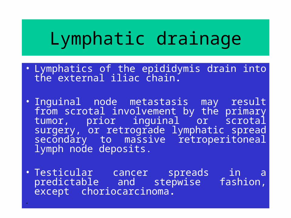
Lymphatic drainage• Lymphatics of the epididymis drain into the
external iliac chain.
• Inguinal node metastasis may result from scrotal involvement by the primary tumor, prior inguinal or scrotal surgery, or retrograde lymphatic spread secondary to massive retroperitoneal lymph node deposits.
• Testicular cancer spreads in a predictable and stepwise fashion, except choriocarcinoma.
.

Clinical features
• Painless Swelling of One testis• Dull Ache or Heaviness in Lower Abdomen• 10% - Acute Scrotal Pain• 10% - Present with Metatstasis
- Neck Mass / Cough / Anorexia / Vomiting / Back Ache/ Lower limb swelling
• 5% - Gynecomastia• Rarely - Infertility
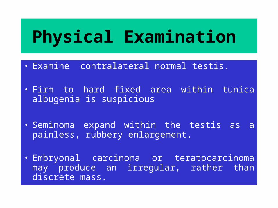
Physical Examination • Examine contralateral normal testis.
• Firm to hard fixed area within tunica albugenia is suspicious
• Seminoma expand within the testis as a painless, rubbery enlargement.
• Embryonal carcinoma or teratocarcinoma may produce an irregular, rather than discrete mass.

Differential Diagnosis
• Testicular torsion• Epididymitis, or epididymo-orchitis• Hydrocele, • Hernia, • Hematoma, • Spermatocele, • Syphilitic gumma .

DICTUM FOR ANY SOLID SCROTAL SWELLINGS
• All patients with a solid, firm intra testicular mass that cannot be trans illuminated should be regarded as Malignant unless otherwise proved.

Scrotal ultrasound
• Ultrasonography of the scrotum is a rapid, reliable technique to exclude hydrocele or epididymitis.
• Ultrasonography of the scrotum is basically an
extension of the physical examination.
• Hypoechoic area within the tunica albuginea is markedly suspicious for testicular cancer.

Tumor markers
TWO MAIN CLASSES• Onco-fetal Substances : AFP & HCG• Cellular Enzymes : LDH & PLAP AFP - Trophoblastic Cells
HCG - Syncytiotrophoblastic Cells
( PLAP- placental alkaline phosphatase, & LDH lactic acid dehydrogenase)

AFP –( Alfafetoprotein)
NORMAL VALUE: Below 16 ngm / mlHALF LIFE OF AFP – 5 and 7 days
Raised AFP : • Pure embryonal carcinoma• Teratocarcinoma • Yolk sac Tumor • Combined tumors,• AFP not raised in pure choriocarcinoma , & in pure
seminoma
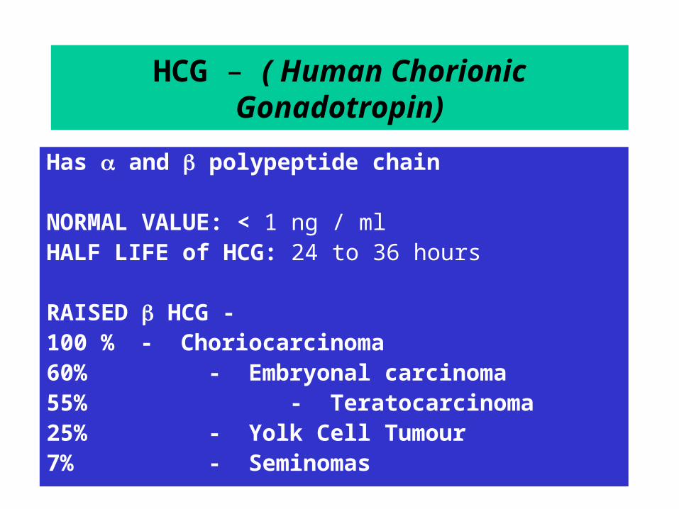
HCG – ( Human Chorionic Gonadotropin)
Has and polypeptide chain
NORMAL VALUE: < 1 ng / ml HALF LIFE of HCG: 24 to 36 hours
RAISED HCG - 100 % - Choriocarcinoma 60% - Embryonal carcinoma 55% - Teratocarcinoma25% - Yolk Cell Tumour7% - Seminomas

ROLE OF TUMOUR MARKERS
• Helps in Diagnosis - 80 to 85% of Testicular Tumours have Positive Markers
• Most of Non-Seminomas have raised markers• Only 10 to 15% Non-Seminomas have normal marker
level • After Orchidectomy if Markers Elevated means Residual
Disease .• Elevation of Markers after Lymphadenectomy means a
STAGE III Disease
ROLE OF TUMOUR MARKERS
• Degree of Marker Elevation Appears to be Directly Proportional to Tumor Burden
• Markers indicate Histology of Tumor: If AFP elevated in Seminoma - Means Tumor has Non-Seminomatous elements
• Negative Tumor Markers becoming positive on follow up usually indicates - Recurrence of Tumor
• Markers become Positive earlier than X-Ray studies

Imaging studies
• Chest X ray
• CT Scan
• PET (Positron Emission Tomography)- No apparent advantage over CT
• MRI - No apparent advantage over CT
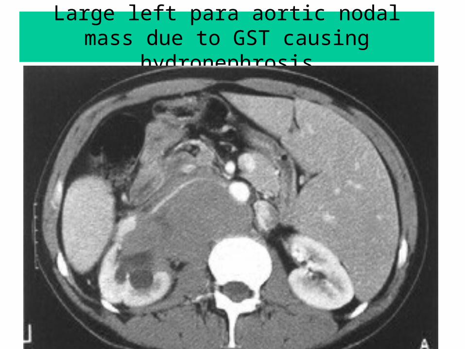
Large left para aortic nodal mass due to GST causing hydronephrosis
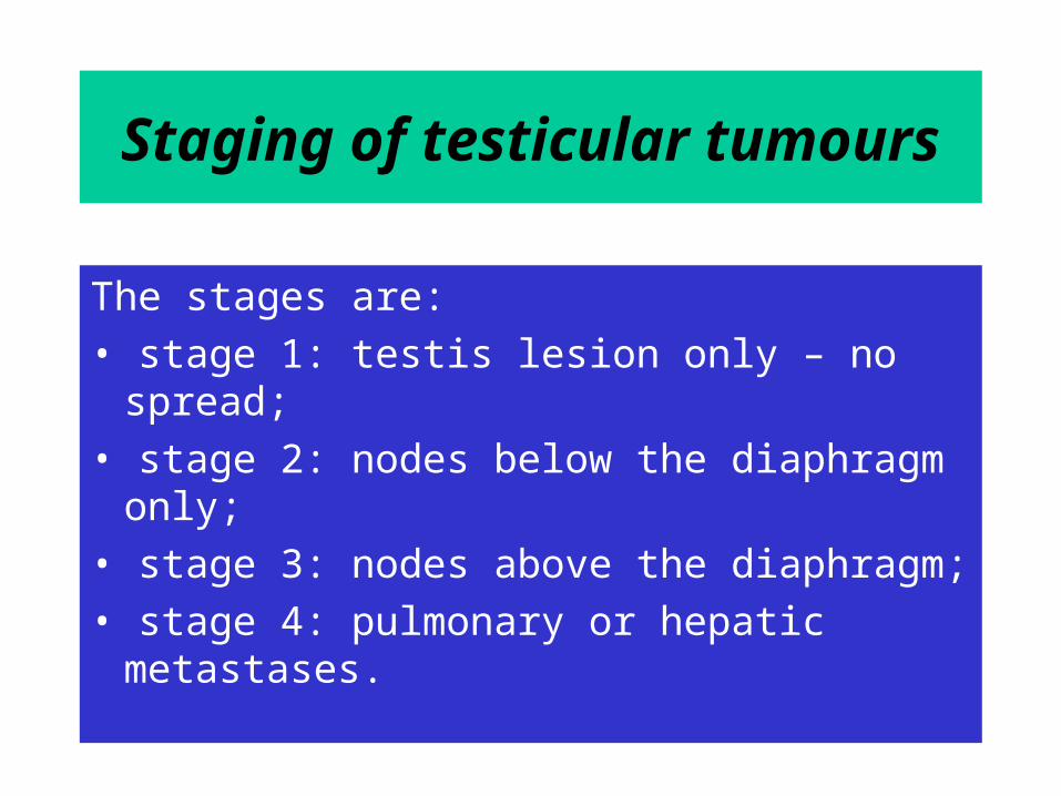
Staging of testicular tumours
The stages are:• stage 1: testis lesion only – no spread;• stage 2: nodes below the diaphragm only;• stage 3: nodes above the diaphragm;• stage 4: pulmonary or hepatic metastases.

Serum tumor markersLDH HCG
Miu/mlAFPNg/ml
S0 _< N <N <N
S1 <1.5 x N < 5000 < 1000
S2 1.5-10x N 5000 to 50000
1000 to 10000
S3 >10x N > 50000 >10000
PRINCIPLES OF TREATMENT
• Treatment should be aimed at one stage above the clinical stage
• Seminomas - Radio-Sensitive. Treat with Radiotherapy.
• Non-Seminomas are Radio-Resistant and best treated by Surgery
• Advanced Disease or Metastasis - Responds well to Chemotherapy
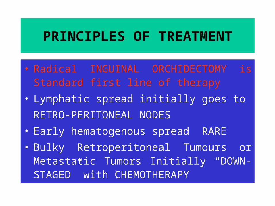
PRINCIPLES OF TREATMENT
• Radical INGUINAL ORCHIDECTOMY is Standard first line of therapy
• Lymphatic spread initially goes to RETRO-PERITONEAL NODES
• Early hematogenous spread RARE• Bulky Retroperitoneal Tumours or Metastatic
Tumors Initially “DOWN-STAGED” with CHEMOTHERAPY
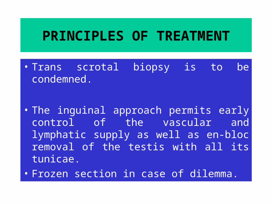
PRINCIPLES OF TREATMENT
• Trans scrotal biopsy is to be condemned.
• The inguinal approach permits early control of the vascular and lymphatic supply as well as en-bloc removal of the testis with all its tunicae.
• Frozen section in case of dilemma.
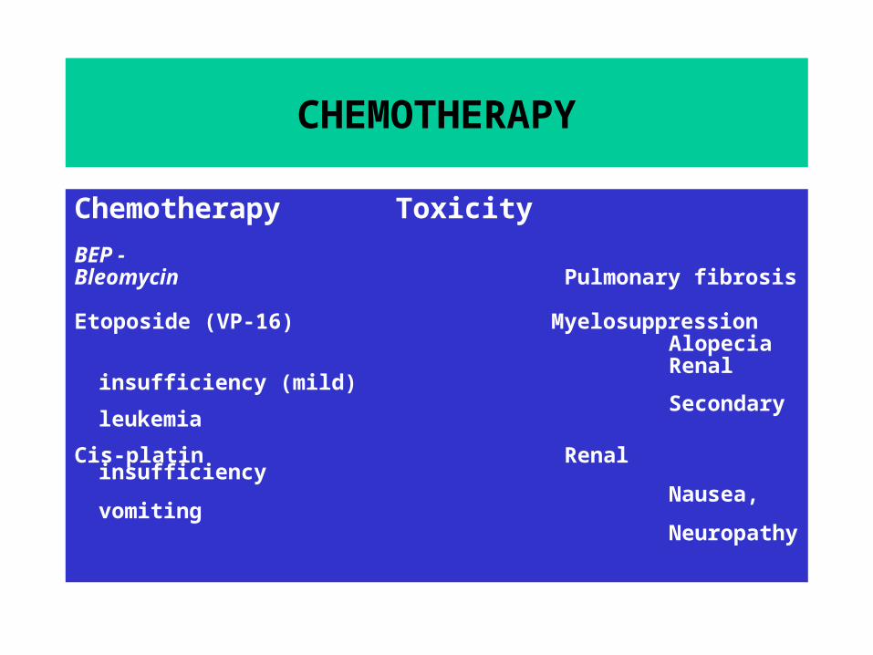
CHEMOTHERAPY
Chemotherapy ToxicityBEP -Bleomycin Pulmonary fibrosis
Etoposide (VP-16) Myelosuppression Alopecia Renal insufficiency (mild) Secondary leukemia
Cis-platin Renal insufficiency Nausea, vomiting Neuropathy

Lymph Nodes Dissection For Right & Left Sided Testicular Tumours
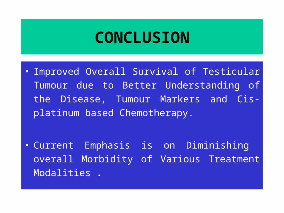
CONCLUSION
• Improved Overall Survival of Testicular Tumour due to Better Understanding of the Disease, Tumour Markers and Cis-platinum based Chemotherapy.
• Current Emphasis is on Diminishing overall Morbidity of Various Treatment Modalities .

THANK YOU