test review exam 1 chapters 1 & 4 spring 2013 9/19/20151
TRANSCRIPT

Test Review
Exam 1Chapters 1 & 4
Spring 2013
04/19/23 1

Cardiovascularsystem
OrganelleMoleculeAtoms
Chemical levelAtoms combine to form molecules.
Cellular levelCells are made up ofmolecules.
Tissue levelTissues consist of similartypes of cells.
Organ levelOrgans are made up of different typesof tissues.
Organ system levelOrgan systems consist of differentorgans that work together closely.
Organismal levelThe human organism is made upof many organ systems.
Smooth muscle cell
Smooth muscle tissue
Connective tissue
Blood vessel (organ)
HeartBloodvessels
Epithelialtissue
Smooth muscle tissue
12
3
4
56
Figure 1.1204/19/23

Figure 1.3a
NailsSkin
Hair
(a) Integumentary System Forms the external body covering, and protects deeper tissues from injury. Synthesizes vitamin D, and houses cutaneous (pain, pressure, etc.) receptors and sweat and oil glands.
304/19/23

Figure 1.3b
Bones
Joint
(b) Skeletal System Protects and supports body organs, and provides a framework the muscles use to cause movement. Blood cells are formed within bones. Bones store minerals.
404/19/23

Figure 1.3c
Skeletalmuscles
(c) Muscular System Allows manipulation of the environment, locomotion, and facial expression. Main- tains posture, and produces heat.
504/19/23

Figure 1.3d
Brain
NervesSpinalcord
(d) Nervous System As the fast-acting control system of the body, it responds to internal and external changes by activating appropriate muscles and glands.
604/19/23

Figure 1.3e
Pineal gland
PituitaryglandThyroid
glandThymus
AdrenalglandPancreas
Testis
Ovary
(e) Endocrine System Glands secrete hormones that regulate processes such as growth, reproduction, and nutrient use (metabolism) by body cells.
704/19/23

Figure 1.3f
(f) Cardiovascular System Blood vessels transport blood, which carries oxygen, carbon dioxide, nutrients, wastes, etc. The heart pumps blood.
Heart
Bloodvessels
804/19/23

Figure 1.3g
Lymphaticvessels
Red bonemarrow
Thoracicduct
Thymus
Spleen
Lymphnodes
(g) Lymphatic System/Immunity Picks up fluid leaked from blood vessels and returns it to blood. Disposes of debris in the lymphatic stream. Houses white blood cells (lymphocytes) involved in immunity. The immune response mounts the attack against foreign substances within the body.
904/19/23

Figure 1.3h
Nasalcavity
Bronchus
Pharynx
Larynx
Trachea
Lung
(h) Respiratory System Keeps blood constantly supplied with oxygen and removes carbon dioxide. The gaseous exchanges occur through the walls of the air sacs of the lungs.
1004/19/23

Figure 1.3i
Liver
Oral cavity
Esophagus
Largeintestine
StomachSmallintestine
RectumAnus
(i) Digestive System Breaks down food into absorbable units that enter the blood for distribution to body cells. Indigestible foodstuffs are eliminated as feces. 1104/19/23

Figure 1.3j
Kidney
Ureter
UrinarybladderUrethra
(j) Urinary System Eliminates nitrogenous wastes from the body. Regulates water, electrolyte and acid-base balance of the blood.
1204/19/23

Figure 1.3k-l
Prostategland
Ductusdeferens
Penis
Testis
Scrotum
Ovary
Uterinetube
Mammaryglands (inbreasts)
Uterus
Vagina
Overall function is production of offspring. Testes produce sperm and male sexhormone, and male ducts and glands aid in delivery of sperm to the femalereproductive tract. Ovaries produce eggs and female sex hormones. The remainingfemale structures serve as sites for fertilization and development of the fetus.Mammary glands of female breasts produce milk to nourish the newborn.
(k) Male Reproductive System (l) Female Reproductive System
1304/19/23

Copyright © 2010 Pearson Education, Inc.
Necessary Life Functions
1. Maintaining boundaries between internal and external environments
• Plasma membranes
• Skin
2. Movement (contractility)
• Of body parts (skeletal muscle)
• Of substances (cardiac and smooth muscle)

Copyright © 2010 Pearson Education, Inc.
Necessary Life Functions
3. Responsiveness: The ability to sense and respond to stimuli (“irritability”)
• Withdrawal reflex
• Control of breathing rate
4. Digestion
• Breakdown of ingested foodstuffs
• Absorption of simple molecules into blood

Copyright © 2010 Pearson Education, Inc.
Necessary Life Functions
5. Metabolism: All chemical reactions that occur in body cells
• Catabolism and anabolism
6. Excretion: The removal of wastes from metabolism and digestion
• Urea, carbon dioxide, feces

Copyright © 2010 Pearson Education, Inc.
Necessary Life Functions
7. Reproduction
• Cellular division for growth or repair
• Production of offspring
8. Growth: Increase in size of a body part or of organism

Copyright © 2010 Pearson Education, Inc.
Survival Needs
1. Nutrients
• Chemicals for energy and cell building
• Carbohydrates, fats, proteins, minerals, vitamins
2. Oxygen
• Essential for energy release (ATP production)

Copyright © 2010 Pearson Education, Inc.
Survival Needs
3. Water
• Most abundant chemical in the body
• Site of chemical reactions
4. Normal body temperature
• Affects rate of chemical reactions
5. Appropriate atmospheric pressure
• For adequate breathing and gas exchange in the lungs

Copyright © 2010 Pearson Education, Inc.
Homeostasis
• Maintenance of a relatively stable internal environment despite continuous outside changes
• A dynamic state of equilibrium

Copyright © 2010 Pearson Education, Inc.
Homeostatic Control Mechanisms
• Involve continuous monitoring and regulation of many factors (variables)
• Nervous and endocrine systems accomplish the communication via nerve impulses and hormones

Copyright © 2010 Pearson Education, Inc.
Components of a Control Mechanism
1. Receptor (sensor)
• Monitors the environment
• Responds to stimuli (changes in controlled variables)
2. Control center
• Determines the set point at which the variable is maintained
• Receives input from receptor
• Determines appropriate response

Copyright © 2010 Pearson Education, Inc.
Components of a Control Mechanism
3. Effector
• Receives output from control center
• Provides the means to respond
• Response acts to reduce or enhance the stimulus (feedback)

Copyright © 2010 Pearson Education, Inc.
Negative Feedback
• The response reduces or shuts off the original stimulus
• Examples:
• Regulation of body temperature (a nervous mechanism)
• Regulation of blood volume by ADH (an endocrine mechanism)

Figure 1.5
Sweat glands activated
Shiveringbegins
StimulusBody temperaturerises BALANCE
Information sentalong the afferentpathway to controlcenter
Information sentalong the afferentpathway to controlcenter
Afferentpathway
Afferentpathway
Efferentpathway
Efferentpathway
Information sentalong the efferentpathway toeffectors
Information sentalong the efferentpathway to effectors
StimulusBody temperature falls
ReceptorsTemperature-sensitivecells in skin and brain
ReceptorsTemperature-sensitivecells in skin and brain
EffectorsSweat glands
EffectorsSkeletal muscles
Control Center(thermoregulatory
center in brain)
Control Center(thermoregulatory
center in brain)
ResponseEvaporation of sweatBody temperature falls;stimulus ends
ResponseBody temperature rises;stimulus ends
2504/19/23

Copyright © 2010 Pearson Education, Inc.
Negative Feedback: Regulation of Blood Volume by ADH
• Receptors sense decreased blood volume
• Control center in hypothalamus stimulates pituitary gland to release antidiuretic hormone (ADH)
• ADH causes the kidneys (effectors) to return more water to the blood

Copyright © 2010 Pearson Education, Inc.
Positive Feedback
• The response enhances or exaggerates the original stimulus
• May exhibit a cascade or amplifying effect
• Usually controls infrequent events e.g.:
• Enhancement of labor contractions by oxytocin (Chapter 28)
• Platelet plug formation and blood clotting (Chapter 17)

Feedback cycle endswhen plug is formed.
Positive feedbackcycle is initiated.
Positivefeedbackloop
Break or tearoccurs in bloodvessel wall.
Plateletsadhere to siteand releasechemicals.
Releasedchemicalsattract moreplatelets.
Platelet plugforms.
1
23
4
Figure 1.62804/19/23

Copyright © 2010 Pearson Education, Inc.
Homeostatic Imbalance
• Disturbance of homeostasis
• Increases risk of disease
• Contributes to changes associated with aging
• May allow destructive positive feedback mechanisms to take over (e.g., heart failure)

Copyright © 2010 Pearson Education, Inc.
Anatomical Position
• Standard anatomical body position:
• Body erect
• Feet slightly apart
• Palms facing forward

Copyright © 2010 Pearson Education, Inc. Figure 1.7a
Cervical
(a) Anterior/Ventral
Pubic(genital)
CephalicFrontalOrbitalNasalOralMental
ThoracicAxillaryMammarySternalAbdominalUmbilicalPelvicInguinal(groin)
Upper limbAcromialBrachial (arm)AntecubitalAntebrachial (forearm)Carpal (wrist)Manus (hand)PalmarPollexDigital
Lower limbCoxal (hip)Femoral (thigh)PatellarCrural (leg)Fibular or peronealPedal (foot)Tarsal (ankle)MetatarsalDigitalHallux
ThoraxAbdomenBack (Dorsum)

Copyright © 2010 Pearson Education, Inc. Figure 1.7b
Cervical Back (dorsal)
(b) Posterior/Dorsal
Scapular Vertebral Lumbar Sacral Gluteal Perineal (between anus and external genitalia)
Upper limb AcromialBrachial (arm) Olecranal Antebrachial (forearm)Manus (hand) Metacarpal DigitalLower limb Femoral (thigh) Popliteal Sural (calf) Fibular or peronealPedal (foot) Calcaneal Plantar
Cephalic Otic Occipital (back of head)
ThoraxAbdomenBack (Dorsum)

Copyright © 2010 Pearson Education, Inc. Table 1.1

Copyright © 2010 Pearson Education, Inc. Table 1.1

Copyright © 2010 Pearson Education, Inc. Table 1.1

Copyright © 2010 Pearson Education, Inc. Table 1.1

Copyright © 2010 Pearson Education, Inc.
Body Cavities
• Dorsal cavity
• Protects nervous system
• Two subdivisions:
• Cranial cavity
• Encases brain
• Vertebral cavity
• Encases spinal cord

Copyright © 2010 Pearson Education, Inc.
Body Cavities
• Ventral cavity
• Houses internal organs (viscera)
• Two subdivisions (separated by diaphragm):
• Thoracic cavity
• Abdominopelvic cavity

Copyright © 2010 Pearson Education, Inc. Figure 1.9a-b
Cranialcavity(contains brain)
Dorsalbodycavity
Vertebralcavity(contains spinal cord)
Cranialcavity
Superiormediastinum
Pericardialcavity withinthe mediastinum
Pleuralcavity
Vertebralcavity
Abdomino-pelviccavity
Ventral bodycavity(thoracic andabdominopelviccavities)
Abdominal cavity(contains digestiveviscera)
Diaphragm
Pelvic cavity(contains urinary bladder, reproductive organs, and rectum)
Thoraciccavity(containsheart andlungs)
(a) Lateral view (b) Anterior view
Dorsal body cavityVentral body cavity

Copyright © 2010 Pearson Education, Inc.
Ventral Body Cavities
• Thoracic cavity subdivisions:
• Two pleural cavities
• Each houses a lung
• Mediastinum
• Contains pericardial cavity
• Surrounds thoracic organs
• Pericardial cavity
• Encloses heart

Copyright © 2010 Pearson Education, Inc.
Serous Membrane (Serosa)
• Thin, double-layered membrane separated by serous fluid
• Parietal serosa lines internal body walls
• Visceral serosa covers the internal organs

Copyright © 2010 Pearson Education, Inc.
Epithelial Membranes
• Serous Membranes
• Serosae—membranes (mesothelium + areolar tissue) in a closed ventral body cavity
• Parietal serosae line internal body walls
• Visceral serosae cover internal organs

Copyright © 2010 Pearson Education, Inc. Figure 4.11c
Parietalpericardium
Visceralpericardium
(c) Serous membranes line body cavitiesclosed to the exterior.
Parietalperitoneum
Visceralperitoneum
ParietalpleuraVisceralpleura

Copyright © 2010 Pearson Education, Inc.
Epithelial Membranes
• Mucous membranes
• Mucosae
• Line body cavities open to the exterior (e.g., digestive and respiratory tracts)

Copyright © 2010 Pearson Education, Inc. Figure 4.11b
Mucosa ofnasal cavity
Mucosa oflung bronchi
Mucosa ofmouth
Esophaguslining
(b) Mucous membranes line body cavitiesopen to the exterior.
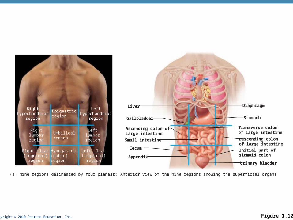
Copyright © 2010 Pearson Education, Inc. Figure 1.12
Epigastricregion
Umbilicalregion
Rightlumbarregion
Leftlumbarregion
Righthypochondriac
region
Lefthypochondriac
region
Hypogastric(pubic)region
Right iliac(inguinal)
region
Left iliac(inguinal)
region
Liver
Gallbladder
Ascending colon oflarge intestine
Small intestine
Appendix
Cecum
Diaphragm
Stomach
Descending colonof large intestine
Transverse colonof large intestine
Initial part ofsigmoid colon
Urinary bladder
(a) Nine regions delineated by four planes (b) Anterior view of the nine regions showing the superficial organs

Copyright © 2010 Pearson Education, Inc.
Epithelial Membranes
• Cutaneous membrane (skin) (More detail with the Integumentary System, Chapter 5)

Copyright © 2010 Pearson Education, Inc.
Tissues
• Groups of cells similar in structure and function
• Types of tissues
• Epithelial tissue
• Connective tissue
• Muscle tissue
• Nerve tissue

Copyright © 2010 Pearson Education, Inc.
Epithelial Tissue (Epithelium)
• Two main types (by location):
1. Covering and lining epithelia
• On external and internal surfaces
2. Glandular epithelia
• Secretory tissue in glands

Copyright © 2010 Pearson Education, Inc.
Characteristics of Epithelial Tissue
1. Cells have polarity—apical (upper, free) and basal (lower, attached) surfaces
• Apical surfaces may bear microvilli (e.g., brush border of intestinal lining) or cilia (e.g., lining of trachea)
• Noncellular basal lamina of glycoprotein and collagen lies adjacent to basal surface

Copyright © 2010 Pearson Education, Inc.
Characteristics of Epithelial Tissue
2. Are composed of closely packed cells
• Continuous sheets held together by tight junctions and desmosomes
3. Supported by a connective tissue reticular lamina (under the basal lamina)
4. Avascular but innervated
5. High rate of regeneration

Copyright © 2010 Pearson Education, Inc.
Classification of Epithelia
• Ask two questions:
1. How many layers?
1 = simple epithelium
>1 = stratified epithelium
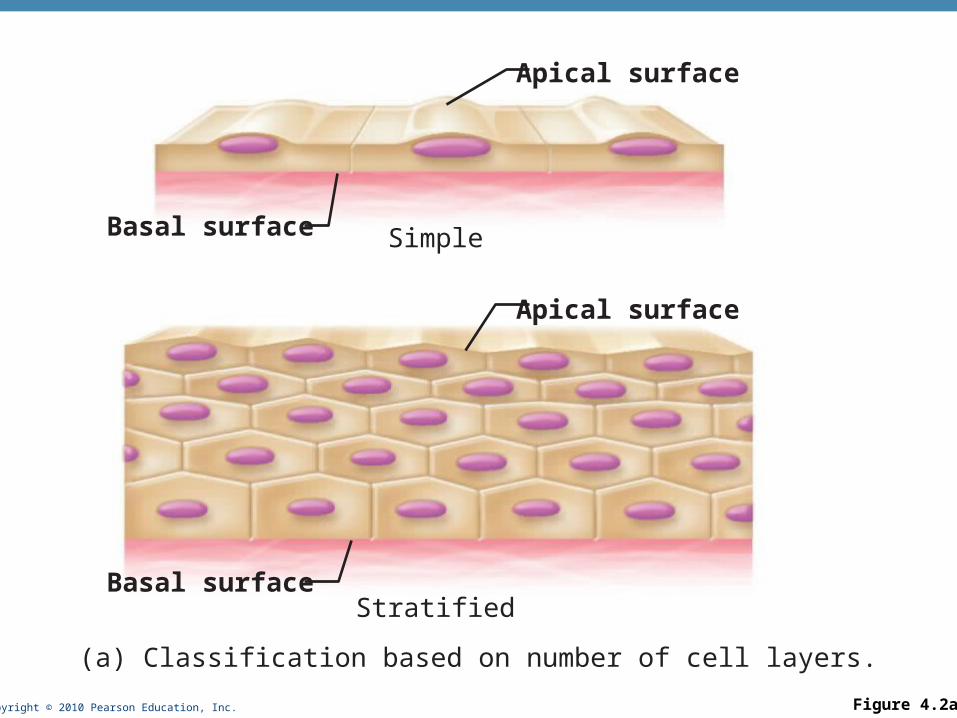
Copyright © 2010 Pearson Education, Inc. Figure 4.2a
Stratified
Simple
Apical surface
Basal surface
Apical surface
Basal surface
(a) Classification based on number of cell layers.

Copyright © 2010 Pearson Education, Inc.
Classification of Epithelia
2. What type of cell?
• Squamous
• Cuboidal
• Columnar
• (If stratified, name according to apical layer of cells)

Copyright © 2010 Pearson Education, Inc. Figure 4.2b
Squamous
Cuboidal
Columnar(b) Classification based on cell shape.

Copyright © 2010 Pearson Education, Inc.
Overview of Epithelial Tissues
• For each of the following types of epithelia, note:
• Description
• Function
• Location
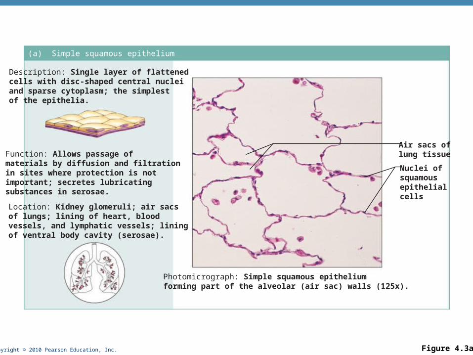
Copyright © 2010 Pearson Education, Inc. Figure 4.3a
(a) Simple squamous epithelium
Description: Single layer of flattenedcells with disc-shaped central nucleiand sparse cytoplasm; the simplestof the epithelia.
Function: Allows passage ofmaterials by diffusion and filtrationin sites where protection is notimportant; secretes lubricatingsubstances in serosae.
Location: Kidney glomeruli; air sacsof lungs; lining of heart, bloodvessels, and lymphatic vessels; liningof ventral body cavity (serosae).
Photomicrograph: Simple squamous epitheliumforming part of the alveolar (air sac) walls (125x).
Air sacs oflung tissue
Nuclei ofsquamousepithelialcells

Copyright © 2010 Pearson Education, Inc.
Epithelia: Simple Squamous
• Two other locations
• Endothelium
• The lining of lymphatic vessels, blood vessels, and heart
• Mesothelium
• The epithelium of serous membranes in the ventral body cavity

Copyright © 2010 Pearson Education, Inc. Figure 4.3b
(b) Simple cuboidal epithelium
Description: Single layer ofcubelike cells with large,spherical central nuclei.
Function: Secretion andabsorption.
Location: Kidney tubules;ducts and secretory portionsof small glands; ovary surface.
Photomicrograph: Simple cuboidalepithelium in kidney tubules (430x).
Basementmembrane
Connectivetissue
Simplecuboidalepithelialcells

Copyright © 2010 Pearson Education, Inc. Figure 4.3c
(c) Simple columnar epithelium
Description: Single layer of tall cells with round to oval nuclei; some cells bear cilia; layer may contain mucus-secreting unicellular glands (goblet cells).
Function: Absorption; secretion of mucus, enzymes, and other substances; ciliated type propels mucus (or reproductive cells) by ciliary action.
Location: Nonciliated type lines most of the digestive tract (stomach to anal canal),gallbladder, and excretory ducts of someglands; ciliated variety lines small bronchi, uterine tubes, and some regionsof the uterus.
Photomicrograph: Simple columnar epitheliumof the stomach mucosa (860X).
Simplecolumnarepithelialcell
Basementmembrane

Copyright © 2010 Pearson Education, Inc. Figure 4.3d
(d) Pseudostratified columnar epithelium
Description: Single layer of cells ofdiffering heights, some not reachingthe free surface; nuclei seen atdifferent levels; may contain mucus-secreting cells and bear cilia.
Function: Secretion, particularly ofmucus; propulsion of mucus byciliary action.
Location: Nonciliated type in male’ssperm-carrying ducts and ducts oflarge glands; ciliated variety linesthe trachea, most of the upperrespiratory tract.
Photomicrograph: Pseudostratified ciliatedcolumnar epithelium lining the human trachea (570x).
Trachea
Cilia
Pseudo-stratifiedepitheliallayer
Basementmembrane
Mucus ofmucous cell

Copyright © 2010 Pearson Education, Inc. Figure 4.3e
(e) Stratified squamous epithelium
Description: Thick membranecomposed of several cell layers;basal cells are cuboidal or columnarand metabolically active; surfacecells are flattened (squamous); in thekeratinized type, the surface cells arefull of keratin and dead; basal cellsare active in mitosis and produce thecells of the more superficial layers.
Function: Protects underlyingtissues in areas subjected to abrasion.
Location: Nonkeratinized type formsthe moist linings of the esophagus,mouth, and vagina; keratinized varietyforms the epidermis of the skin, a drymembrane.
Photomicrograph: Stratified squamous epitheliumlining the esophagus (285x).
Stratifiedsquamousepithelium
Nuclei
Basementmembrane
Connectivetissue

Copyright © 2010 Pearson Education, Inc. Figure 4.3f
(f) Transitional epithelium
Description: Resembles both stratified squamous and stratified cuboidal; basal cells cuboidal or columnar; surface cells domeshaped or squamouslike, depending on degree of organ stretch.
Function: Stretches readily and permits distension of urinary organ by contained urine.
Location: Lines the ureters, urinary bladder, and part of the urethra.
Photomicrograph: Transitional epithelium lining the urinary bladder, relaxed state (360X); note the bulbous, or rounded, appearance of the cells at the surface; these cells flatten and become elongated when the bladder is filled with urine.
BasementmembraneConnectivetissue
Transitionalepithelium

Copyright © 2010 Pearson Education, Inc.
Glandular Epithelia
• A gland is one or more cells that makes and secretes an aqueous fluid
• Classified by:
• Site of product release—endocrine or exocrine
• Relative number of cells forming the gland—unicellular (e.g., goblet cells) or multicellular
Copyright © 2010 Pearson Education, Inc.
Endocrine Glands
• Ductless glands
• Secrete hormones that travel through lymph or blood to target organs

Copyright © 2010 Pearson Education, Inc.
Exocrine Glands
• More numerous than endocrine glands
• Secrete products into ducts
• Secretions released onto body surfaces (skin) or into body cavities
• Examples include mucous, sweat, oil, and salivary glands

Copyright © 2010 Pearson Education, Inc.
Unicellular Exocrine Glands
• The only important unicellular gland is the goblet cell

Copyright © 2010 Pearson Education, Inc.
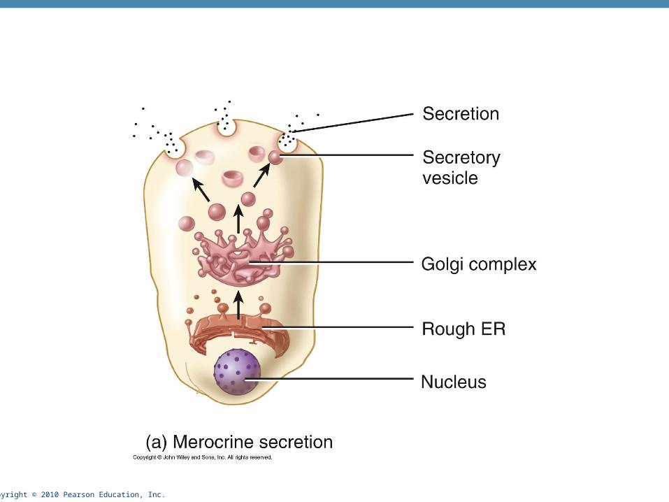
Modes of Secretion
• Merocrine
• Products are secreted by exocytosis (e.g., pancreas, sweat and salivary glands)
• Holocrine
• Products accumulated in cell and then secreted by rupture of gland cells (e.g., sebaceous glands)
• Entire cell is broken down
• Apocrine
• Product accumulated in cell and then pinched off apical surface
• Axillary and inguinal regions (body odor)
• Modified apocrine glands – mammary and ceruminous
• Some question their existence in humans
Copyright © 2010 Pearson Education, Inc.

Copyright © 2010 Pearson Education, Inc.

Copyright © 2010 Pearson Education, Inc.

Copyright © 2010 Pearson Education, Inc.
Connective Tissue
• Most abundant and widely distributed tissue type
• Four classes
• Connective tissue proper
• Cartilage
• Bone tissue
• Blood

Copyright © 2010 Pearson Education, Inc. Table 4.1

Copyright © 2010 Pearson Education, Inc.
Major Functions of Connective Tissue
• Binding and support
• Protection
• Insulation
• Transportation (blood)

Copyright © 2010 Pearson Education, Inc.
Characteristics of Connective Tissue
• Connective tissues have:
• Mesenchyme as their common tissue of origin
• Varying degrees of vascularity
• Cells separated by nonliving extracellular matrix (ground substance and fibers)

Copyright © 2010 Pearson Education, Inc.
Structural Elements of Connective Tissue
• Ground substance
• Medium through which solutes diffuse between blood capillaries and cells
• Components:
• Interstitial fluid
• Adhesion proteins (“glue”)
• Proteoglycans
• Protein core + large polysaccharides (chondroitin sulfate and hyaluronic acid)
• Trap water in varying amounts, affecting the viscosity of the ground substance (major part of matrix)

Copyright © 2010 Pearson Education, Inc.
Structural Elements of Connective Tissue
• Three types of fibers
• Collagen (white fibers)
• Strongest and most abundant type
• Provides high tensile strength
• Elastic
• Networks of long, thin, elastin fibers that allow for stretch
• Reticular
• Short, fine, highly branched collagenous fibers

Copyright © 2010 Pearson Education, Inc.
Structural Elements of Connective Tissue
• Cells
• Mitotically active and secretory cells = “blasts”
• Mature cells = “cytes”
• Fibroblasts in connective tissue proper
• Chondroblasts and chondrocytes in cartilage
• Osteoblasts and osteocytes in bone
• Hematopoietic stem cells in bone marrow
• Fat cells, white blood cells, mast cells, and macrophages

Copyright © 2010 Pearson Education, Inc. Figure 4.7
Macrophage
Fibroblast
Lymphocyte
Fat cell
Mast cell
Neutrophil
Capillary
Cell types Extracellularmatrix
Fibers• Collagen fiber• Elastic fiber• Reticular fiber
Ground substance

Copyright © 2010 Pearson Education, Inc.
Connective Tissue: Embryonic
• Mesenchyme — embryonic connective tissue
• Gives rise to all other connective tissues
• Gel-like ground substance with fibers and star-shaped mesenchymal cells

Copyright © 2010 Pearson Education, Inc.
(a) Connective tissue proper: loose connective tissue, areolar
Description: Gel-like matrix with allthree fiber types; cells: fibroblasts,macrophages, mast cells, and somewhite blood cells.
Function: Wraps and cushionsorgans; its macrophages phagocytizebacteria; plays important role ininflammation; holds and conveystissue fluid.
Location: Widely distributed underepithelia of body, e.g., forms laminapropria of mucous membranes;packages organs; surroundscapillaries.
Photomicrograph: Areolar connective tissue, asoft packaging tissue of the body (300x).
Epithelium
Laminapropria
Fibroblastnuclei
Elasticfibers
Collagenfibers
Figure 4.8a

Copyright © 2010 Pearson Education, Inc. Figure 4.8b
(b) Connective tissue proper: loose connective tissue, adipose
Description: Matrix as in areolar,but very sparse; closely packedadipocytes, or fat cells, havenucleus pushed to the side by largefat droplet.
Function: Provides reserve foodfuel; insulates against heat loss;supports and protects organs.
Location: Under skin in thehypodermis; around kidneys andeyeballs; within abdomen; in breasts.
Photomicrograph: Adipose tissue from thesubcutaneous layer under the skin (350x).
Nucleus offat cell
Vacuolecontainingfat droplet
Adiposetissue
Mammaryglands

Copyright © 2010 Pearson Education, Inc. Figure 4.8c
(c) Connective tissue proper: loose connective tissue, reticular
Description: Network of reticularfibers in a typical loose groundsubstance; reticular cells lie on thenetwork.
Function: Fibers form a soft internalskeleton (stroma) that supports othercell types including white blood cells,mast cells, and macrophages.
Location: Lymphoid organs (lymphnodes, bone marrow, and spleen).
Photomicrograph: Dark-staining network of reticularconnective tissue fibers forming the internal skeletonof the spleen (350x).
Spleen
White bloodcell(lymphocyte)
Reticularfibers

Copyright © 2010 Pearson Education, Inc. Figure 4.8d
(d) Connective tissue proper: dense connective tissue, dense regular
Description: Primarily parallelcollagen fibers; a few elastic fibers;major cell type is the fibroblast.
Function: Attaches muscles tobones or to muscles; attaches bonesto bones; withstands great tensilestress when pulling force is appliedin one direction.
Location: Tendons, mostligaments, aponeuroses.
Photomicrograph: Dense regular connectivetissue from a tendon (500x).
Shoulderjoint
Ligament
Tendon
Collagenfibers
Nuclei offibroblasts

Copyright © 2010 Pearson Education, Inc. Figure 4.8e
(e) Connective tissue proper: dense connective tissue, dense irregular
Description: Primarilyirregularly arranged collagenfibers; some elastic fibers;major cell type is the fibroblast.
Function: Able to withstandtension exerted in manydirections; provides structuralstrength.
Location: Fibrous capsules oforgans and of joints; dermis ofthe skin; submucosa ofdigestive tract.
Photomicrograph: Dense irregularconnective tissue from the dermis of theskin (400x).
Collagenfibers
Nuclei offibroblasts
Fibrousjointcapsule

Copyright © 2010 Pearson Education, Inc. Figure 4.8f
(f) Connective tissue proper: dense connective tissue, elastic
Description: Dense regularconnective tissue containing a highproportion of elastic fibers.
Function: Allows recoil of tissuefollowing stretching; maintainspulsatile flow of blood througharteries; aids passive recoil of lungsfollowing inspiration.
Location: Walls of large arteries;within certain ligaments associatedwith the vertebral column; within thewalls of the bronchial tubes.
Elastic fibers
Aorta
HeartPhotomicrograph: Elastic connective tissue inthe wall of the aorta (250x).

Copyright © 2010 Pearson Education, Inc.
Connective Tissue: Cartilage
• Three types of cartilage:
• Hyaline cartilage
• Elastic cartilage
• Fibrocartilage

Copyright © 2010 Pearson Education, Inc. Figure 4.8g
(g) Cartilage: hyaline
Description: Amorphous but firmmatrix; collagen fibers form animperceptible network; chondroblastsproduce the matrix and when mature(chondrocytes) lie in lacunae.
Function: Supports and reinforces;has resilient cushioning properties;resists compressive stress.
Location: Forms most of theembryonic skeleton; covers the endsof long bones in joint cavities; formscostal cartilages of the ribs; cartilagesof the nose, trachea, and larynx.
Photomicrograph: Hyaline cartilage from thetrachea (750x).
Costalcartilages
Chondrocytein lacuna
Matrix

Copyright © 2010 Pearson Education, Inc. Figure 4.8h
(h) Cartilage: elastic
Description: Similar to hyalinecartilage, but more elastic fibersin matrix.
Function: Maintains the shapeof a structure while allowinggreat flexibility.
Location: Supports the externalear (pinna); epiglottis.
Photomicrograph: Elastic cartilage fromthe human ear pinna; forms the flexibleskeleton of the ear (800x).
Chondrocytein lacuna
Matrix

Copyright © 2010 Pearson Education, Inc. Figure 4.8i
(i) Cartilage: fibrocartilage
Description: Matrix similar tobut less firm than that in hyalinecartilage; thick collagen fiberspredominate.
Function: Tensile strengthwith the ability to absorbcompressive shock.
Location: Intervertebral discs;pubic symphysis; discs of kneejoint.
Photomicrograph: Fibrocartilage of anintervertebral disc (125x). Special stainingproduced the blue color seen.
Intervertebraldiscs
Chondrocytesin lacunae
Collagenfiber

Copyright © 2010 Pearson Education, Inc. Figure 4.8j
(j) Others: bone (osseous tissue)
Description: Hard, calcifiedmatrix containing many collagenfibers; osteocytes lie in lacunae.Very well vascularized.
Function: Bone supports andprotects (by enclosing);provides levers for the musclesto act on; stores calcium andother minerals and fat; marrowinside bones is the site for bloodcell formation (hematopoiesis).Location: Bones
Photomicrograph: Cross-sectional viewof bone (125x).
Lacunae
Lamella
Centralcanal

Copyright © 2010 Pearson Education, Inc. Figure 4.8k
(k) Others: blood
Description: Red and whiteblood cells in a fluid matrix(plasma).
Function: Transport ofrespiratory gases, nutrients,wastes, and other substances.
Location: Contained withinblood vessels.
Photomicrograph: Smear of human blood (1860x); twowhite blood cells (neutrophil in upper left and lymphocytein lower right) are seen surrounded by red blood cells.
Neutrophil
Red bloodcells
Lymphocyte
Plasma

Copyright © 2010 Pearson Education, Inc. Figure 4.9
Photomicrograph: Neurons (350x)
Function: Transmit electricalsignals from sensory receptorsand to effectors (muscles andglands) which control their activity.
Location: Brain, spinalcord, and nerves.
Description: Neurons arebranching cells; cell processesthat may be quite long extend fromthe nucleus-containing cell body;also contributing to nervous tissueare nonirritable supporting cells(not illustrated).
Dendrites
Neuron processes Cell body
Axon
Nuclei ofsupportingcells
Cell bodyof a neuron
Neuronprocesses
Nervous tissue

Copyright © 2010 Pearson Education, Inc. Figure 4.10a
(a) Skeletal muscle
Description: Long, cylindrical,multinucleate cells; obviousstriations.
Function: Voluntary movement;locomotion; manipulation of theenvironment; facial expression;voluntary control.
Location: In skeletal musclesattached to bones oroccasionally to skin.
Photomicrograph: Skeletal muscle (approx. 460x).Notice the obvious banding pattern and thefact that these large cells are multinucleate.
Nuclei
Striations
Part ofmuscle fiber (cell)

Copyright © 2010 Pearson Education, Inc. Figure 4.10b
(b) Cardiac muscle
Description: Branching, striated, generally uninucleate cells that interdigitate atspecialized junctions (intercalated discs).
Function: As it contracts, it propels blood into the circulation; involuntary control.Location: The walls of the heart.
Photomicrograph: Cardiac muscle (500X);notice the striations, branching of cells, andthe intercalated discs.
Intercalateddiscs
Striations
Nucleus

Copyright © 2010 Pearson Education, Inc. Figure 4.10c
(c) Smooth muscle
Description: Spindle-shapedcells with central nuclei; nostriations; cells arranged closely to form sheets.
Function: Propels substancesor objects (foodstuffs, urine,a baby) along internal passage-ways; involuntary control.Location: Mostly in the wallsof hollow organs.
Photomicrograph: Sheet of smooth muscle (200x).
Smoothmusclecell
Nuclei

Copyright © 2010 Pearson Education, Inc.
Epithelial Membranes
• Cutaneous membrane (skin) (More detail with the Integumentary System, Chapter 5)

Copyright © 2010 Pearson Education, Inc. Figure 4.11a
Cutaneousmembrane(skin)
(a) Cutaneous membrane (the skin)covers the body surface.

Copyright © 2010 Pearson Education, Inc.
Epithelial Membranes
• Mucous membranes
• Mucosae
• Line body cavities open to the exterior (e.g., digestive and respiratory tracts)

Copyright © 2010 Pearson Education, Inc. Figure 4.11b
Mucosa ofnasal cavity
Mucosa oflung bronchi
Mucosa ofmouth
Esophaguslining
(b) Mucous membranes line body cavitiesopen to the exterior.

Copyright © 2010 Pearson Education, Inc.
Epithelial Membranes
• Serous Membranes
• Serosae—membranes (mesothelium + areolar tissue) in a closed ventral body cavity
• Parietal serosae line internal body walls
• Visceral serosae cover internal organs
• Name according to regions
• Pleura
• Pericardium
• Peritoneum

Copyright © 2010 Pearson Education, Inc. Figure 4.11c
Parietalpericardium
Visceralpericardium
(c) Serous membranes line body cavitiesclosed to the exterior.
Parietalperitoneum
Visceralperitoneum
ParietalpleuraVisceralpleura

Copyright © 2010 Pearson Education, Inc.
Steps in Tissue Repair
• Inflammation
1. Release of inflammatory chemicals
2. Dilation of blood vessels
3. Increase in vessel permeability
4. Clotting occurs

Copyright © 2010 Pearson Education, Inc. Figure 4.12, step 1
Scab
Blood clot inincised wound
Epidermis
Vein
Inflammatorychemicals
Inflammation sets the stage:• Severed blood vessels bleed and inflammatory chemicals are
released.• Local blood vessels become more permeable, allowing white
blood cells, fluid, clotting proteins and other plasma proteinsto seep into the injured area.
• Clotting occurs; surface dries and forms a scab.
Migrating whiteblood cell
Artery
1

Copyright © 2010 Pearson Education, Inc.
Steps in Tissue Repair
• Organization and restored blood supply
• The blood clot is replaced with granulation tissue
• Epithelium begins to regenerate
• Fibroblasts produce collagen fibers to bridge the gap
• Debris is phagocytized

Copyright © 2010 Pearson Education, Inc. Figure 4.12, step 2
Regeneratingepithelium
Area ofgranulationtissueingrowth
FibroblastMacrophage
Organization restores the blood supply:• The clot is replaced by granulation tissue, which restores
the vascular supply.• Fibroblasts produce collagen fibers that bridge the gap.• Macrophages phagocytize cell debris.• Surface epithelial cells multiply and migrate over the
granulation tissue.
2

Copyright © 2010 Pearson Education, Inc.
Steps in Tissue Repair
• Regeneration and fibrosis
• The scab detaches
• Fibrous tissue matures; epithelium thickens and begins to resemble adjacent tissue
• Results in a fully regenerated epithelium with underlying scar tissue

Copyright © 2010 Pearson Education, Inc. Figure 4.12, step 3
Regeneratedepithelium
Fibrosedarea
Regeneration and fibrosis effect permanent repair:• The fibrosed area matures and contracts; the epitheliumthickens.• A fully regenerated epithelium with an underlying area ofscar tissue results.
3

Copyright © 2010 Pearson Education, Inc.
Developmental Aspects
• Primary germ layers: ectoderm, mesoderm, and endoderm
• Formed early in embryonic development
• Specialize to form the four primary tissues
• Nerve tissue arises from ectoderm
• Muscle and connective tissues arise from mesoderm
• Epithelial tissues arise from all three germ layers

Copyright © 2010 Pearson Education, Inc. Figure 4.13
MesodermEndoderm
16-day-old embryo(dorsal surface view)
Epithelium
Nervous tissue(from ectoderm)
Muscle and connectivetissue (mostly frommesoderm)
Ectoderm