temporal sequence of neurotransmitter expression by developing neurons of fetal monkey visual cortex
TRANSCRIPT

Developmental Brain Research, 43 (1988) 69-96 69 Elsevier
BRD 507%
Temporal sequence of neurotransmitter expression by developing neurons of fetal monkey visual cortex
G.W. Huntley 1, S.H.C. Hendry 1, H.P. Killackey 1'2, L.M. Chalupa 3 and E .G. Jones 1
Departments of 1Anatomy and Neurobiology and 2psychobiology, University of California at Irvine, Irvine, CA 92717 (U.S.A.) and 3Department of Psychology and The California Primate Research Center,
University of California at Davis, Davis, CA 95616 (U.S.A.)
(Accepted 26 April 1988)
Key words: Visual cortex; Development; Immunocytochemistry; 7-Aminobutyric acid; Substance P; Neuropeptide
The developing fetal monkey visual cortex was studied immunocytochemically from 110-155 days post-conception in order to loca- lize cell populations immunoreactive (ir) for y-aminobutyric acid, Substance P, cholecystokinin-octapeptide, somatostatin, neuro- peptide Y, and proenkephalin A peptide (BAM-18). The area 17/18 border and all cortical laminae identified in the adult visual cortex were discernible from the youngest age examined. All ir-cell populations studied were present at each fetal age. However, despite a relatively adult-like cytoarchitecture, all ir-cell populations studied displayed patterns of immunostaining which were unlike those de- scribed in adult visual cortex, and showed significant changes in laminar distribution, morphology, and numbers over the time course of gestation examined. Despite the differences in the patterns of immunostaining between the fetal and adult visual cortex, ir-cell pop- ulations intrinsic to the developing visual cortex exhibited adult-like combinations of co-localized transmitters and peptides. The de- veloping monkey cortex also contains ir-cell populations, particularly BAM-18-ir cells, which have not been detected immunocyto- chemically in the adult monkey cortex. Differences between the fetal and the adult ir-cell populations might be accounted for by cell death, morphological transformation, secondary migration or changes in gene expression for neurotransmitters and neuropeptides.
INTRODUCTION
Development of function in the cerebral cortex is
associated with the development of connectivity and
of transmitter expression. Both are subject to plastic
phenomena. Visual deprivation in young monkeys
can lead to changes in the distribution of geniculocor-
tical axons within area 17, disrupting the connectivity underlying the ocular dominance of cortical neu- rons 27, and can lead to changes in the expression of 7-
aminobutyric acid ( G A B A ) and substance P by cells intrinsic to primary visual cortex 22'25. The laminar or-
ganization of G A B A - and most peptide-immuno- reactive (ir) cell populations intrinsic to adult mon-
key visual cortex have been described previous- ly 6A5'18-20'23'25"30. However , little is known about the
ontogeny and developmental regulation of the immu- noreactive cell populations during fetal life, especial- ly in relation to cytoarchitectonic differentiation and
the establishment of cortical connectivity.
In the present study we have examined the mor-
phology and laminar distributions of GABA- immu-
noreactive cells and of a number of neuropeptide-im-
munoreactive cell populations intrinsic to fetal mon-
key visual cortex during the last third of gestation
(165 days). During this period of cortical develop- ment thalamo-cortical connections are becoming es- tablished 3z'49'51, but it is unclear whether during this
time there are concomitant changes in the cell pop- ulations intrinsic to the cortex, many of which in the
adult synthesize G A B A and neuropeptides. Previous
investigators have examined immunoreactive cell populations in fetal non-primates 8'39'66, but few pre-
vious systematic studies have been conducted in fetal primatesZ4.2s, 41
MATERIALS AND METHODS
Six fetal rhesus monkey brains (Macaca mulatta)
were used in this study. In each monkey, fluorescent
Correspondence: E.G. Jones, Dept. of Anatomy and Neurobiology, University of California at Irvine, Irvine, CA 92717, U.S.A.
0165-3806/88/$03.50 ~ 1988 Elsevier Science Publishers B.V. (Biomedical Division)

7O
dyes had been injected into the post-central gyrus through a Hamilton microliter syringe 2-14 days prior to sacrifice as part of another study 7. Animals
were delivered by Caesarean section on embryonic (E) days 110, 121, 131, 135, 150, and 155. Gestation in this species is normally 165-170 days. Animals were deeply anesthetized and perfused transcardially
with normal saline followed by 2% paraformalde-
hyde and 0.1% glutaraldehyde in 0.1 M phosphate buffer (pH 7.4). The brains were removed immedi- ately, blocked, and immersed in phosphate-buffered 30% sucrose at 4 °C. After all blocks had sunk, they were frozen on dry ice (some blocks were flattened in a plane parallel to the pial surface between two slides while freezing) and sectioned at 15 or 20/xm on a slid-
ing microtome either in the frontal plane or tangen- tial to the pial surface (flattened blocks).
Sections were pre-incubated in phosphate buffer containing 3% normal serum and 0.25% Triton X- 100 for 2 h at 4 °C, then transferred to an identical so- lution which contained, in addition, one of the fol- lowing primary antisera: rabbit anti-Neuropeptide Y (NPY; Amersham, 1:1000); rabbit anti-somatostatin ( S R I F ; DAKO, 1:1000); rabbit anti-BAM-18 (pro- vided by C. Evans, 1:1000), or monoclonal antibod- ies: rat anti-Substance P (SP; Sera Labs, 1:500); rat anti-GABA (Eugene Tech., l:1000); mouse anti- cholecystokinin (CCK; provided by E. Katt and J. Walsh, 1:20). After overnight incubation at 4 °C fol- lowed by washes in phosphate buffer, sections incu- bated in rat or mouse primary antisera were pro- cessed by the avidin-biotin-peroxidase method 26
using Vectastain ABC kits (Vector Labs, Inc.). All
other sections were processed by the peroxidasc anti- peroxidase (PAP) method 63 using an unlabeled swine anti-rabbit antiserum and rabbit PAP (Dako Corp. ).
All sections were then reacted with 3,3'-diammoben- zidine tetrahydrochloride (DAB; 50 rag/100 ml phos- phate buffer) and 0.01% hydrogen peroxide, mounted onto gelatin-subbed slides, dehydrated and cleared in xylene and coverslipped. Sections adjacent to those
processed for immunocytochemistry were stained with thionin.
Sections used for double-labeling were also cut at 15 or 20/xm on a sliding microtome in either the fron- tal plane or tangential to the pial surface. After pre- incubation identical to that described above, sections were incubated simultaneously in different combina- tions of two primary antisera. Primary antisera used were the same as those above, except for the addition of rabbit anti-GABA antisera (immunoNuclear; 1 : 1000), which was used in lieu of the rat monoclonal anti-GABA antibody (Eugene Tech) when incu- bated simultaneously with the rat monoclonal anti- SP antibody (Sera Labs). After overnight incubation at 4 °C, sections were washed in phosphate buffer,
then incubated with the two appropriate, affinity pu- rified FITC- or RITC-conjugated secondary antibod- ies (CalTag, Inc.) for 2 h at 4 °C. The sections were viewed and photographed with a Leitz Dialux epi- fluorescence microscope equipped with fluorescein and rhodamine filter packs.
For all immunocytochemical procedures, control sections were processed as described above, except
for the replacement of the primary antiserum with normal serum at a concentration twice that of the pri-
GABA
"
** " * ~ i IVa l i b
:. .: . . ' . . .". : . v]
NPY ,
SP CCK
IVa IVb
. * * • IVc
, V
Fig, 1. Camera lucida drawings showing the laminar distributions of GABA-, NPY-, SP-, and CCK-ir somata in adult monkey area 17. Each drawing shows the density of ir-somata in columns through the thickness of the cortex in a 15-#xm thick section. Bars = 200/xm.

71
mary. Additionally, synthetic NPY, SRIF, CCK-oc- tapeptide, SP, Substance K and neuromedin K were added where appropriate to the diluted antisera or monoclonal antibodies to control for cross-reactivity. The peptides were added at concentrations of 5, 10 and 50 #g/ml. Staining with each anti-peptide anti- body solution was abolished by pre-incubation with the corresponding peptide at the lowest concentra- tion. Staining was not affected by incubation with heterologous peptides at even the highest concentra- tions, except in the case of the anti-SP antibody which was adsorbed by substance K and neuromedin K at concentrations of 10/~g/ml, indicating that it re- cognizes these other tachykinins as well. Immuno- reactive staining for GABA was blocked by pre-incu- bation with a GABA-bovine serum albumin conju- gate (15 ktg/ml) but was unaffected by pre-incubation with any of the above peptides.
The distributions of immunocytochemically labeled somata in alternate sections were plotted with the aid of a camera lucida. Laminar and areal boundaries were determined by comparison with im- mediately adjacent thionin-stained sections. Cell counts were made with the aid of a grid reticule on sections cut tangential to the pial surface. The no- menclature used in the present study for the identifi- cation of cortical laminae is that of Brodmann 5.
RESULTS
Laminar distribution and morphology of GABA- and peptide-ir neurons in adult monkey visual cortex
The laminar distributions and morphology of GABA- and most peptide-containing cell popula- tions intrinsic to adult monkey visual cortex have been described previously 6a5,18-2°'23,25'3°. A review
of these data is provided here since they represent the endpoint of the developmental sequences exam- ined in this study.
NPY and SRIF SRIF- and NPY-ir somata are present in all layers
but are concentrated in a superficial band which oc- cupies layer II and the upper half of layer III, and in a deep band which includes layer VI and the subcorti- cal white matter 6'2° (Fig. 1). Very few immunoreac- tive cells are present in layer I. In general, a greater
number of NPY-ir neurons are present in the white matter than in the overlying cortex in comparison with the distribution of SRIF-ir neurons.
Minor SRIF-ir plexuses are present in layers I and II, and in layer VI. In the middle layers of cortex, two plexuses are present; one occupies the upper part of layer V, the other occupies the deep half of layer III, layer IVA, and most of layer IVB (see Figs. 4 and 6D in ref. 20).
NPY-ir plexuses are similar in their laminar distri- bution compared to SRIF-ir plexuses, but are denser. Minor plexuses are present in layers I and II, and in layer VI. Two middle plexuses bracket layer IVC. Oneplexus spans the deeper half of layer III down to layer IVB. The other plexus spans layer V (see Figs. 4 and 6E in ref. 20).
Neurons immunoreactive for NPY or SRIF have round or fusiform cell bodies which vary in size from 8 x 8/~m in diameter to 20 x 10~m. Immunoreactive neurons present in the white matter are fusiform with long axes usually parallel to the layer VI/white mat- ter border. Most somata in the cortex or in the white matter are homogeneously stained.
Both NPY- and SRIF-ir somata give rise to 2-3 or more smooth processes, which divide to yield thin, regularly beaded processes. From immunoreactive somata in the white matter, processes can be fol- lowed into the overlying cortex, contributing to the plexuses in layers V and VI. The processes of NPY- and SRIF-ir neurons have no consistent pattern of branching.
CCK CCK-ir somata are present in all layers but are
concentrated in the supragranular layers of cortex ~9 (Fig. 1). Typically, immunoreactive neurons located in the superficial layers of cortex have small (8-12 /~m long), spindle-shaped somata. From such cells,
one or two long, thin processes emerge from each pole and either ascend towards the pial surface, or descend towards the white matter.
Of the few immunoreactive neurons in the deep layers of cortex, the majority are multipolar or bi- tufted. Processes of such neurons are usually thin and beaded.
BAM-18 BAM-18 is one of several opioid peptides which

72
Fig. 2 Photomicrograph of a thionin-stained section through areas 17 and 18 at Ell0. The area 17/18 border tarrowl antl atl laminac identified in the adult visual cortex are present, but, unlike the cytoarchitecture of the adult, a thin line of darkly stained cells occupic. layer II at this age. Bar = 5001~m.
are derived from the p ropep t ide precursor proen-
kephalin A. Al though BAM-18 and o ther proen-
kephalin A pept ide derivat ives have been found to
exist in high concentra t ions in rodent brain 13'4°, no
systematic study has examined the areal or laminar
distr ibutions of BAM-18- i r cell popula t ions within
primate cortex. Our own observat ions (unpubl ished)
indicate that BAM-18- i r cells are not de tec table in
adult monkey visual cortex, but they are de tec ted in
the developing monkey visual cortex (see below).
SP
Two popula t ions of SP-ir neurons have been de-
scribed, based on their morphology , intensity of
staining, laminar dis tr ibut ion and co-localizat ion
with G A B A or NPY 3°.
A minori ty of the SP-ir cells have large, ovoid so-
mata (15-21/~m maximum diameter ) that are darkly
stained. Such cells are present in all layers but are
concentra ted in a superficial band which includes lay-
er II and the upper half of layer III , and a deep band
which includes the bo t tom half of layer VI and the
subcortical white mat te r (Fig. 1). Only a very small
number of the large, darkly stained somata are also
immunoreact ive for G A B A but most are immuno-
reactive for NPY.
Processes of such cells are long, beaded , and ex-
tend for considerable distances (300-400 ~tm) before
adopting a vertical or ienta t ion and crossing several
layers.
The major i ty of immunoreac t ive neurons have
small, round somata (8-10/~m in d iameter ) , that are
lightly stained and show few stained processes. Such
cells are concent ra ted in a middle cortical band that
includes the upper half of layer V in area 18 and layer
IVC in area 17. A small number of this SP-ir cell type
is also found in layers II and II125. Al l small, lightly
stained SP-ir neurons are also immunoreact ive for
G A B A .
Dense immunoreact ive plexuses are present in the
upper half of layer I and in the deep half of layer VI
and in the subjacent white matter . A less dense plex-
us is present in layer II and the upper half of layer III.
Fig. 3. Photomicrographs from sections through area 17 at E110 stained for NPY. A: NPY-ir somata are concentrated in deep layer VI and the subjacent white matter. Fewer cells are found superficially, in layers II and III. Arrows indicate ir-cells shown at higher magni- fication in (B) and (C). Bars = 150/~m (A) and 25/~m (B and C).


74
Layer IVC of area 17 is filled with immunoreactive puncta (see Figs. 1 and 6 in ref. 30).
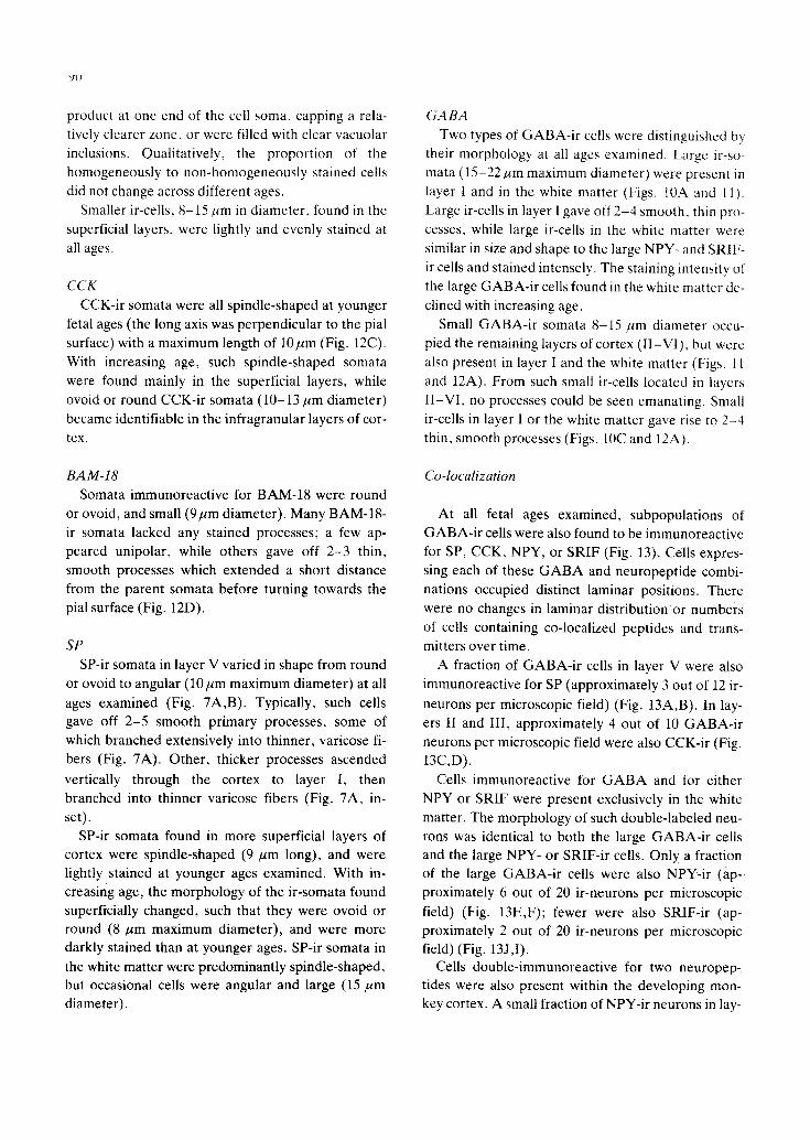
GABA GABA-containing cells are present in all cortical
layers and in the subjacent white matter ~5,1s'23, but
are densest in the principal thalamo-cortical recipient layers of cortex (Fig. 1). In area 17, this corresponds to layers IVA and IVC; in area 18, this corresponds to layer IV.
Somata vary in size from 9 to 30/~m. In general,
smaller cells 8-10ktm in diameter are present in layer I, and in layer VI and the subjacent white matter (see Fig. 2B in ref. 23).
Cytoarchitecture of fetal monkey visual cortex
EllO and El21 The area 17/18 border and all cortical laminae de-
scribed in the adult visual cortex were identifiable at E l l 0 (Fig. 2). Three general features distinguish the
visual cortex at these ages in comparison with the adult, and apply equally to areas 17 and 18. First, the width of the cortex, from pia mater to white matter, was narrower at these ages (approximately 950 ~m) than in the adult (approximately 1500 j~m). Second, in the fetuses many large cells were present within the white matter, primarily concentrated immedi- ately subjacent to layer VI. Some of these large Nissl-stained cells were extremely darkly stained, while the majority were lightly stained. Third, a thin line of darkly stained cells occupied layer 1I in the fe-
tuses. Such cells were predominantly ovoid in shape and probably represent the remaining cells of the cor-
tical plate.
El31 and E135 The width of both areas of the cortex from pia mat-
er to white matter had increased to approximately 1200/~m. Large cells (both darkly and lightly stained) were still evident within the white matter. The line of darkly stained, ovoid cells situated in layer II de- scribed at younger ages had disappeared altogether,
leaving a layer I/II border which appeared identical to that in the adult.
E150 and E155 The width of both areas of the cortex was approxi-
mately 1450 um at these ages. Large cells (of both staining intensities) were still present within the su- perficial white matter.
Distribution of GABA and peptide immunoreactive
somata and fibers
NPY
EllO
Area 17. Neurons immunoreactive for NPY occu- pied all layers and were also present in large numbers in the subcortical white matter. The largest numbers of NPY-ir cells were found in two bands, deep and su- perficial (Figs. 3 and 4).
NPY-ir somata were densely packed in a deep band which included the deeper half of layer VI and the subjacent white matter (approximately 25 Jr-cells per 1000 /~mZ); their numbers became less dense deeper in the white matter. The cells were large (15- 22/~m) and gave rise to processes which could be fol- lowed for considerable distances within the white matter and up into the overlying cortex (Fig. 3B, C). These processes formed a dense fiber plexus in the superficial part of the white matter and in layer VI (Figs. 3A and 4, top left). The density of the fiber plexus declined in layer V, and layer IVC was rela- tively fiber-free. However, many radially oriented fi- bers could be followed from cells within the deep band as they ascended through all layers and branched in layer I.
A second band of NPY-ir somata was found super- ficially (Fig. 4, top left). These cells were sparsely scattered in layers II and III (approximately 8 ir-celts per 1000/~m2). Fewer NPY-ir cells were found in the intervening layers, between the superficial and deep bands, and only one NPY-ir cell was found in layer I. NPY-ir cells found superficial to the deep band had generally smaller somata (8-15 #m), but gave rise to processes which formed a broad fiber plexus which occupied layers I - I I I (Fig. 4, top left). The density of this plexus was greatest in the upper half of layer I, and declined in density in deep layer i, layer II and
layer III. Sections cut tangential to the pial surface revealed a dense plexus of NPY-ir fibers running horizontally in the upper half of layer I, to which the vertical fibers from the deep cells also contributed.
Area 18. The laminar distribution of NPY-ir soma- ta and fibers was similar in area 18 to that seen in area

75
NPY
E I I O
IVab
C
s ;41"?-
.r': ...~'~- /]....'.?- :,.
/ ' ~
v
V t . * * , * • , . - , - , - . - ' : "~".--", .. • , . ° ~ . . _ L . . . .
W M * * . * .
E I 3 5
°~ . ~ • . . : _ :z
\ J
i
\
~ . . '
. , ,.:::,,:. . . . . . . . . _ _ %
* j "
SRIF
I
II
E I 5 5
;>"
,°;
I
IVab
c '~:
V
.. .o.
I
III
EIIO
i " ' " : " , " - . .
D
E I ~
C
V
/
i
. / !
. . . - ' . . °
Vl . * . . . . . . . . , * . - . , - - *
W M . * * i :..y. . . . . . . . . . . . . . . . . •
• * , , , ¢,J"
E I ~
II .," "X.
. \ , t
lit ,
C
V
Vl * * . . . . . . .
Fig. 4. Camera lucida drawings through area 17 at E110, E135 and E155, showing the temporal changes in the distribution of so mata and fibers immunoreactive for NPY (top) or SRIF (bottom), plotted from single 15-#tm thick sections. Bar = 100#m.

7~
17. NPY-ir cells occupied all layers except layer IV
and were present in the subjacent white matter.
Large cells densely packed in the deep half of layer VI and the subcortical white matter formed a deep
band of cells which appeared continuous with the
deep band of cells in area 17. Fibers given off by
these cells in area 18 formed a deep plexus similar in laminar position to that described for area 17.
Smaller cells were found superficially and ap-
peared continuous with the superficial band of NPY-
ir cells in area 17. The laminar positions of the fibers
given off by these cells were similar to those de-
scribed for the superficial NPY-ir cells in area 17.
Although the density of NPY-ir somata appeared
equal across the two areas, a disparity in immuno- reactive fiber density was evident between areas 17
and 18 at this fetal age. The density of fibers found in
the deeper plexus (layer VI and the subcortical white
matter) and found superficially (layers I - I l i ) was
greater in area 18 compared to the density of NPY-ir
fibers in area 17.
El21
Area 17. The laminar distribution and density of
NPY-ir somata and fibers at E121 was similar to that
described in E110, with the exception that no NPY-ir
cells were found in layer I at this fetal age.
Area 18. The pattern of NPY-immunostaining in area 18 was similar to that described for area 18 at
Ell0. No NPY-ir cells were found in layer I. The
greater fiber density in area 18 compared to area 17
described at E l l 0 was still evident at this fetal age.
El31 and E135
Area 17. There was a decrease in the density of NPY-ir somata in deep layer VI and the subcortical
white matter at these fetal ages compared to El21 (approximately 15 if-cells per 1000/,tm2). Moreover,
NPY-ir somata found superficially had also declined in number in comparison with E121 (approximately 4 ir-cells per 1000 #m2). No NPY-ir cells were present
in layer I. However, across all layers there was an overall decrease in the density of NPY-ir fibers. There
were no vertical fibers which spanned the thickness of the cortex seen at these ages (Fig. 4, top middle).
Area 18. A similar reduction in density of somata and fibers across all layers was evident in area 18 compared to area 17. The areal difference in fiber
density between area 17 and area 18 described for
younger fetal ages was still apparent at El31 and
E135.
El50 and E155
Area 17. Fewer NPY-ir somata were present in
deep layer VI and the subcortical white matter at
these fetal ages compared to E135 (approximately 10
ir-cells per 1000/,tm2). There was no change in the number of NPY-ir somata found superficially in com-
parison with E135 (Fig. 4, top right). No cells were
present in layer 1. The fiber plexuses formed by the two bands of cells had further decreased in density
compared to younger fetal ages. Although hori-
zontally running fibers were still present in the upper half of layer I, the fibers in layers lI and Ill were ex-
tremely sparse (Fig. 4, top right). No vertical fibers
which spanned the thickness of the cortex were seen at these ages.
Area 18. A reduction in the density of NPY-ir cells
in the deep band of area 18 compared to younger
ages was evident at these ages. The density of cells in
more superficial parts of the cortex was unchanged
from E135. At these late fetal ages, the areal differ-
ences in plexus density between areas 17 and 18 de-
scribed at younger fetal ages were no longer detect- able.
A summary of the temporal changes in the lami-
nation of ir-somata is shown in Fig. 4.
SRIF
The laminar positions of SRIF-ir somata and fibers
paralleled that of NPY-ir cells and processes very closely, although at any given age SRIF-ir cells were less numerous than NPY-ir cells.
El lO
Area 17. Cells immunoreactive for SRIF were present in all layers except layer I and sublayer IVC. Somata were concentrated in two cell-dense bands (Fig. 4, bottom left). Large numbers of SRIF-ir cells
(15-22 ~m) were present in deep layer VI and the subjacent white matter (approximately 20 ir-cells per 1000 ktm2). Processes given off by these cells could be followed for long distances within the white matter, or could be followed into the overlying cortex, form- ing a plexus in layer VI and the subcortical white mat-

ter. No radially-oriented fibers could be seen span-
ning the thickness of the cortex, as was seen for NPY- ir fibers at E l l0 .
Superficially, smaller (8-15 pm) cells were found scattered in the upper half of layer II (approximately
5 ir-cells per 1000 pm2). Processes from these cells gave rise to a sparse plexus located in layers I - I I I . The density of this plexus was greater in layer I than in layers II and III (Fig. 4, bottom left).
Area 18. The density and laminar position of SRIF- ir cells and fibers were similar to those described for area 17.
El21
Area 17. The laminar position and density of SRIF- ir somata and fibers at El21 were similar to that at E l l0 . There appeared to be a slight increase in the density of the superficial fiber plexus in layers I - I I I .
Area 18. A similar increase in the density of the su- perficial SRIF-ir fiber plexus in layers I - I I I was evi- dent in area 18. There were no other changes in SRIF immunostaining evident compared to E l l0 . No dif- ferences were apparent across areas at this fetal age.
E131 and E135
Area 17. The density of SRIF-ir cells in layer VI and the subcortical white matter had decreased com- pared to younger ages (approximately 10 ir-cells per 1000 pm2). There was no change in the density of SRIF-ir somata found in the superficial layers of the cortex (Fig. 4, bottom middle). The density of fibers in the deep (layer VI and the subcortical white mat- ter) and superficial (layers I - I I I ) fiber plexuses had also decreased relative to El21.
Area 18. A similar decrease in the density of fibers and somata was evident in area 18 compared to El21, such that there were no differences in density or distribution of SRIF immunostaining across areas at these fetal ages.
El50 and E155
Area 17. Although SRIF-ir cells occupied similar
laminar positions to those described for younger fetal ages, the density of the deep band of cells was re- duced even further from that at E135 (to approxi- mately 7 ir-cells per 1000/xmZ). There was no change in the density of SRIF-ir somata found in the superfi- cial layers in comparison with E135. The fiber plex-
77
uses formed by these two bands of SRIF-ir cells had
also decreased in density compared to E135 (Fig. 4, bottom right).
Area 18. SRIF-ir somata and fiber density had also decreased substantially in area 18 compared to E135, in similar proportions to the decreased density noted for area 17. Thus, no areal differences in the patterns of SRIF immunostaining between areas 17 and 18 were evident at these late fetal ages.
CCK
CCK-ir somata were present in all cell-rich layers ( I I -VI ) in both areas 17 and 18. CCK-ir cells were not present in layer I at any of the ages examined. The relative density of CCK-ir cells and fibers be- tween laminae and across areas changed substantial- ly with increasing fetal ages (Fig. 5).
EllO
Area 17. Very few CCK-ir somata were detectable at El10 (approximately 1-2 ir-cells per 15-pro thick section). The only immunoreactive cells present were sparsely distributed in layers II and III. CCK-ir cells were not found in the white matter.
Sparse, radially oriented fibers were present in the middle cortical layers, but there were no discernible fiber plexuses at this fetal age (Fig. 5, top left).
Area 18. The patterns of CCK immunostaining in area 18 resembled area 17 at this age. No differences in lamination or density were evident across areas.
El21
Area 17. Changes in the laminar distribution and density of CCK-ir somata and fibers were evident at this age compared to E l l0 . The greatest density of CCK-ir somata was found in a broad, superficial band corresponding to layers II and III (approxi-
mately 12 if-cells per 1000 pm2), and in a thin deep band corresponding to layer V (approximately 8 ir- cells per 1000/~m2). No cells were present in the white matter.
Long radially oriented CCK-ir fibers were present in the subcortical white matter; many of them could be seen entering the overlying cortex and forming a dense plexus in layers VI and V. In addition, ascend- ing fibers could be traced from cells which occupied the superficial band into layer I, where they

7g
branched to form a diffuse layer I plexus.
Area 18. The density of CCK-ir somata and fibers
was lower in area 18 than in area 17 at this age. Soma-
ta were present in a broad, superficial band which
spanned layers II and I l l (approximate ly 8 Jr-cells per
1000 Hm:), and in a broad deep band which corre-
I
II *
III
IVab
C
V
VI
WM
EIIO
i ,:,
CCK
EI35
11"
EI55
" ". -" i II * * .
L I
/ I
:?,,,.,
',.',.:.'\..- _ /
,'::~;,:::
BAM-18 EI55
EIIO EI35 ~ .---... - . , . . _ . -
I I * *
I l l
C
I ~ b v
- - C
VL - - V
W M " ; . . . . . . . . . . . . . . . . . . . . . . . . . . . . . . Vl * . . . . . " I~P;~:"
Fig. 5. Camera lucida drawings through area 17 at El l0 , E135 and E155 showing the temporal changes in the distribution of somata and fibers immunoreactive for CCK (top) or BAM-18 (bottom), plotted from single 15-/~m thick sections. Bar = 100/~m.

sponded to layers V and VI (approximately 4 ir-cells
per 1000/~m2). The deep band of cells in area 18 was
less restricted in its laminar extent relative to the
79
deep band in area 17. No immunoreact ive cells were
present in the white matter.
Long CCK-ir fibers were detected in the subjacent
Fig. 6. A: darkfield photomicrograph from section through areas 17 and 18 stained for CCK at E155. The density of CCK-ir fibers is greater in area 18 than in area 17 at this fetal age. Arrow shows position of the area 17/18 border. B: photomicrograph from section ad- jacent to one in (A), stained with thionin. Arrow shows position of the area 17/18 border. Bar = 250am.

white matter, many of which entered the overlying
cortex and formed a plexus in layers VI and V. This
plexus was less dense than the comparable deep fiber
plexus in area 17. Fibers from superficial CCK-ir
cells could be traced into layer I. There was no differ-
ence across areas in the density of fibers forming the
plexus in layer I.
El31 and E135
Area 17. The laminar distribution of CCK-ir soma-
ta and fibers was unchanged at this fetal age com- pared to El21, although an increase in the density of
cells comprising the superficial band (approximately
25 ir-cells per 1000#m 2) and the deep band (approxi-
mately 18 ir-cells per 1000 #m 2) was evident (Fig. 5,
top middle).
Area 18. The laminar pattern of CCK immuno-
staining in area 18 described at E121 was similar at these fetal ages. The density of cells which occupied
the deep and superficial bands had increased in com-
parison with E121, and in similar proportions to that
described for area 17. Thus, disparities in cell and fi- ber density across areas 17 and 18 described at El21
were still evident at these ages. Deep CCK-ir somata
and the plexus in layers V and VI were denser in area
17 than in area 18.
El50 and E155
Area 17. The thin cell-dense band which occupied
layer V described at younger ages was no longer de-
tectable at these fetal ages. CCK-ir somata were in- stead sparsely scattered in all of the infragranular
layers. Occasional cells were present in the superfi- cial white matter. The density of CCK-ir somata found in superficial layers did not change from E135
(Fig. 5, top right).
Changes in the density and laminar spread of the
deep fiber plexus were evident at these ages com-
pared to E135. The plexus was now restricted to lay- er V1, and was less dense than at E135. A sparse fiber plexus was still present in layer I (Fig. 5, top right).
Area 18. There was a similar decline m dcnsil) of the deep band of CCK-ir cells in area 18. 111 contrast
to area 17, however, the density of the supeHicial
CCK-ir cells was substantially greater than at t'~ 135
(approximately 35 ir-cells per 1000/~m=).
The deep fiber plexus which occupied layer VI and the subcortical white matter was very dense at these
ages. Thus, the disparity in plexus and cell density across areas at these ages was the reverse of that ob-
served earlier (Fig. 6). Long, radially oriented fibers
could be followed from the deep plexus through the middle layers of cortex. Fibers ascending from the su-
perficial band of cells could be seen entering layer 1,
forming a plexus of comparable density to that in area 17.
BAM-18
The density and distribution of cells immunoreac- tive for BAM-18 was similar across both visual areas
at all fetal ages examined. Thus, the description of
BAM-18 immunoreactivity for each fetal age per- tains equally to area 17 and to area 18.
E l i & BAM-18-ir somata were found in a sparse
thin band which occupied the upper half of layer If
(approximately 5 ir-cells per 500 pro2). BAM-18-ir
cells were not observed in other layers or in the sub- jacent white matter.
Occasional short, beaded fibers were present in
layer I and in the white matter (Fig. 5, bottom left).
El21. Three changes were evident at El21 com-
pared to E l l0 . First, there was an increase in the
density of BAM-18-ir cells which occupied the upper half of layer II (approximately 10 ir-cells per 500 pro2). Second, occasional BAM-18-ir cells were
found in the white matter immediately subjacent to layer VI. BAM-18-ir cells were not present in other
layers. Third, a sparse fiber plexus was evident in lay-
er I. Occasional short, beaded fibers were still pres- ent in the white matter.
El31 and E135. There was a further increase in the
Fig. 7. Photomicrographs of sections through area 17 stained for SP illustrating the morphology and laminar distribution of ir-somata and fibers found at fetal ages. The age of the animal from which the photomicrographs were taken was E 135. A: SP-ir somata are con- centrated in layer V and give rise to coarse fibers which ascend radially through the cortex to branch and form a fiber plexus in layer I (inset, darkfield). B: SP-ir cell shown at higher magnification. Arrows indicate ascending process. Bars = 5 0 , m (A); 1 0 0 , m (insetl; 25 ,urn (B).

~|
....
...
,!
t
:i:i iiiiiiii!!
ili!i!il,i
l;7!@!
ii iiiii 711 iiiii
iiii!
~ii~ilM
T!~!~i:
:~71~
:i~,%~
:~,~i~
ii:i ~i~i II' ~
~ ~
~ "
~
....
i i~
':~
ik~
y~'z
J ~¸
¸¸¸
~r
k
~ ¸¸
~
mM
~
A~
+,l
, ~
¢<
i~
u~
:~
i~
!!
~!
~,
+~
:~
;l
tt
~i
V
~ ~
7~
'~-~
~ i~
i~
!!i~i~
ii~i~
iii~!ii!
~ ..
...
~ii
i~'i
i~iili ~
•
~i~
~ ..
.. ~
'~
;~
~i~ ~"
~
~i~{
~ ~
~if!
i~?:
i!~d
i~i~
..
....
....
..
~ -,~
~
~717
~i~17
~!7~ ~
~' .~,
~,
, ~d
i%~i~
i~ili~ii
:~:i:£
1Dibi~
,
~ ii!~
;~i~ '~ ,~
. ~<~
.ii
ii~d.
7'~
" ~i
~!~i
i~i~
~i~
i~ ~
~ ~
~ ~ ,
~,~
:i~
~
~'
~-
-
~,~,
~q~q
~i~,
!~r~
i~i@
~;i~
~
~ ~
~!ii~i
~ ~i:
~;~,~,
~;:~:~
,;:!~:~
,~:~:~
::,~:~u
~,~ ...
......
......
......
......
......
......
i!ili
!~iiii~
i~ii!if
i!~i!i~
? ~
~ ?;~
:~'~!~!~
:~,L77
~',~i
!~i~,~7
~i~ ~7~;~
:~17!!
!i~1%
~,~ ...
.... ~;
~;;~
~d~!7
1!i~;!
i!~!~i
i~i~!!
ii~i~!
L~!~i
ii~!~!
~i~;id
;i!~!i~
i~!~i~
7; ~?~i~
i!~ii!!i
~7: ~
~x:~
:~ ~
~:
~!i~
7~!~
!!~77
#~!~
?~7~
7!~i
7~!~
::::~
:~ii~
xq~
¢~i~:
~ ~%
~;~7
1~iii~
i!!!fi~
i~!~!
~i~ii~
;~i!i~
:~I~L
~ ~:
~ ~:
~ ~
qi~i
~i'
::~:i~
~
~M~,
~ ~
~q~i
~i:~

~2
density of BAM-18-ir cells found within the upper
half of layer II compared to El21 (approximately 21
Jr-cells per 500 !~m:). An occasional cell could be
found within the subjacent white matter. BAM-l g-Jr
cells were not present in other layers at these fetal
ages (Fig. 5, bottom middle).
The density of the fiber plexus in layer 1 had in-
creased substantially relative to El21. Additionally,
the plexus spanned layer I through the upper half of
layer II. Processes in the white matter were longer,
and could be seen entering the overlying cortex.
El50 and E155. At El50 there was no change in
the laminar distribution or density of BAM-18-ir cells
relative to younger fetal ages (El31 and E135). Cells
remained largely confined to the upper half of layer
11 but a few were found occasionally in the white mat-
ter. Processes from cells in layer ii could be followed
into layer I. The density of the fiber plexus in layers
I - I I was unchanged relative to E 135.
By contrast, at E155 there was a decline m both the
density of cells found in the upper half of layer li (to
approximately 15 ir-cells per 500 am2)~ and in the fi-
ber plexus in layers I and I1. In addition, a greater
number of cells was found in layers V and VI, as well
as in the subjacent white matter (Fig. 5, bottom
right). There did not appear to be an increase in the
density of processes in the deep layers of cortex or in
the subjacent white matter.
Fig. 8. Darkfield photomicrographs from sections through area 17 (A) and area 18 (B) at E135, stained for SP. A: dense ir-fiber plex- uses are present in layer I, layer III and the deep half of layer V. Coarser, radially oriented fibers are also present. B: the Jr-fiber plex- uses in area 18 are similar in position to those in area 17, but are much less dense. The lower half of layer IV in both (A) and (B) is iden- tifiable by the refringent background caused by high cell density. Bar = 100 urn.

SP
EllO
Area 17. Somata immunoreactive for SP were con-
centrated in a thin band which corresponded to layer V (approximately 15 ir-cells per 500/xm2). These cells gave rise to processes which formed a sparse plexus within layer V; many other thick, radially
oriented fibers could be followed from their parent somata in layer V as they ascended through the thick- ness of the cortex and branched in layer I (Figs. 7 and 9). Sections cut tangential to the pial surface through layer I revealed a dense SP-ir fiber plexus. No SP-ir somata were present in layer I.
Far fewer SP-ir cells were found scattered in layer
VI and the subjacent white matter. Although the processes given off by individual cells could be fol- lowed only a short distance, from their vicinity, long SP-ir fibers were seen running deep into the white matter.
Occasional SP-ir cells were found superficially in layers II and III. No processes could be seen emerg- ing from these cells.
Area 18. The laminar distribution and density of SP-ir somata and fibers was similar in area 18 to that in area 17. SP-ir somata were concentrated in layer V. Prominent, ascending processes similar to those described in area 17 were present in area 18, and branched in layer I yielding a plexus equal in density to that of area 17. No SP-ir somata were present in layer I.
Occasional SP-ir cells were present in layers II, III and VI. Within the subcortical white matter, sparsely scattered SP-ir cells and fibers were present.
E121
Area 17. The pattern of SP immunostaining at El21 was similar to E l l0 . No changes in density or
lamination were evident. Area 18. The laminar positions occupied by SP-ir
somata were similar at E121 to that at E l l0 . There
were no differences in somata or fiber density evi- dent at this age compared to E l l0 .
E131 and E135
Area 17. The density of somata which occupied layer V was greater at these ages in comparison with
younger fetal ages (approximately 20 ir-cells per 500
83
~tmZ). Moreover, processes given off by these cells
now formed a dense plexus in the deeper sublayer VB, decreasing in density in the more superficial sub- layer VA (Fig. 9, top middle). Many thick, radially oriented processes ascended from the cells and branched in layer I, forming a fiber plexus which was denser at these ages compared to that at younger
ages. No somata were present in layer I. A greater number of cells were found superficially
in comparison with younger fetal ages (approximate- ly 7 ir-cells per 500/~m2). These cells were concen- trated in the upper half of layer II and gave rise to fi- bers which descended, forming a plexus in layer III (Fig. 8).
SP-ir cells were present in layer VI and the subja-
cent white matter. Fibers from these cells gave rise to a sparse plexus in deep layer VI and the superficial white matter. Occasional fibers were present deeper in the white matter.
Area 18. The density and distribution of SP-ir so- mata in area 18 was comparable to that in area 17. Fi- ber plexuses similar in laminar position to those de- scribed for area 17 were present. The density of fi- bers forming plexuses in layers III , deep V and VI was less in area 18 than those in comparable laminar positions in area 17 (Fig. 8B).
E150 and E155
Area 17. The density of SP-ir cells which occupied layer V was unchanged in comparison with younger ages. The fiber plexus these cells formed in sublayer VB was also denser than at E135. Thick, ascending processes were seen arising from the cells and span- ning the thickness of the cortex, as described above. The number of these prominent fibers appeared con-
stant throughout this period of gestation. A dense fi- ber plexus was evident in layer I. No SP-ir somata were present in layer I.
The number of SP-ir cells found superficially had increased from E135 (approximately 12 ir-cells per 500/~me). These cells formed a broad band occupying layers II and III (Fig. 9, top right). Processes given off by cells within the superficial band formed a fiber plexus in layer III of equal density to the layer III plexus described at E135.
A greater number of SP-ir cells were found in layer VI and the subcortical white matter than had been found in these positions at E135. These cells gave rise

"(H) tur/00 E pue (D pu~ V) tur/00I = sae~I "(AXO1Oq) gla.I.11UaA IE-1OlP.1 oql Oa auaawrpg ouoz ole!patuJalu ! oq~, tuoaj s! UA~OqS plo!j s!qL '.~olletu 01!q~ aql U.t luosa.~d o.~e s.~aqD .~!-V~IVO '0I [~ lg :D "xal
-leua ot!q,~ Iea!laooqns oql pug 'DAI ' I I I - I s-~o~el u! lsasuap aae eleUlOS a!-V~/VrD "so~e ao~uno,( tuoaj poseoaou! gAl~q ell3ttlOS-J! JO S.laq -mnu oql '0gI 3 le :~t 'aolletu 01!qA~ le3!l.tooqns aql pue IA :to,{eI daop pue I aa~el u! palealu~ouoo oae lnq 'ao~e I qoeo u! luasoad oae ~letu -os a!-V~[VO "0II~l le :V ' (~) 0g I~ pue (~D pu~ V) 0[[~t le V~XcD aoj pou!els L[ eoae q~noJql suo!loas jo sqdea~oaa!tuoloqd "0I "~!:I <.--
tur/00[ = 2~t "suo!loos ~to!ql tuH-gI oI~U!S tuoaj poliold '(tuolloq) V f l V D ~o (do1) dS aoj aA!lOeg.lounlllul! s~oq! j pue eletuos jo uonnq!.ns!p aql u! so~ueqo l~odtual atI1 ~u!~oqs ggI~t pue g~ i~ '0113 lg L[ eo.~e qgno.~qi S~U!A~e.~p ep!0n I e~otue D "6 '~!d
', ^ , " --" .. . . . . . . - . . . . . .
- ~ IA
..'.,
i,
" : " qeAI
• ~ : L , " III III ' ' "
• ° I I - •
::~.....-'~7-~' ~
ggl 3 g~13 0 1 1 3
VEIVZ)
• / . . . . . . L
J
, ...(" ~1_ I
\ " , . . . / i ~
t '
H ~ E-"
~2"!!~.:i' ,' in
' i i} ,., , •
t.,i" ' . . . . " ""-
9glN
/
• .. t i , , L
.2 ! ..
/ " c
"? qA'.
, I f
/ 3
_ t - /
, f
Ii
• V ~ M
- - IA
• " '7 - - [
i
' I
- - / ~ - ; , ,
• II1
A
9
qeAI
• II
I
g~13 011 3
d 9

0O
LJI

8(3
t " ll~ " i
.i1
Fig. 11. Photomicrograph of section through layer I at E150 cut tangential to the pial surface and stained for G A B A . Both large (ar- rowhead) and small (arrow) ir-cells tire present. Bar = 3(~ u m
to a fiber plexus similar in density and laminar posi- tion to that described at earlier ages.
Area 18. SP immunostaining in area 18 appeared
similar to area 17 at these ages, with the exception
that the plexuses occupying layer III, the deep half of
layer V, and layer VI were less dense than those in
comparable laminar positions in area 17.
GA BA
Cells immunoreactive for GABA were present in
all layers and the subcortical white matter in both
visual areas at all ages examined.
EllO
Area 17. GABA-ir somata were concentrated in
two cell-dense bands at El l0 . A superficial band of GABA-ir cells occupied layer I; GABA-ir cells form- ing a deep band were packed in the subcortical white matter (Fig. 10).
Sections cut tangential to the pial surface through
layer I revealed both large (15-20 ym) and small
(8-10 ym) GABA-ir cells (Fig. 11). There were ap- proximately 25 ir-cells of both sizes per 500 ym ~.
Cells of both sizes gave rise to processes which arbo- rized extensively within layer I. Numerous GABA-ir
punctate profiles were also evident within layer I. Both large (15-20 ~tm) and small (8-10 ktm)
GABA-ir cells were densely packed in large numbers in the subcortical white matter. Processes given off
by white matter GABA-ir cells of either size formed
a dense plexus within the white matter. This was densest in the intermediate zone adjacent to the lat-
eral ventricle (Fig. 10C). Other fibers could be fol-
lowed from the parent somata into the overlying cor-
tex. No GABA-ir fibers which spanned the entire thickness of the cortex were seen.
GABA-ir cells were sparsely distributed in layers I I -VI (Fig. 9, bottom left). These cells were similar in size to the smaller GABA-ir cells found in layer I.
Additionally, some large GABA-ir cells were pres- ent in layer VI, but were not found in layers I I -V.
Fig. 12. A: small G A B A - i r cells present in the white matter . Somata are usually round and give rise to 2 - 4 thin, smooth processes. B: SRIF-ir cells present in the subcortical white mat ter at E l l 0 . Somata are ovoid or angular. C: CCK-ir somata in layer II at E155 dis- play spindle-shaped somata which give off thin ascending and descending processes. D: BAM-18-ir cells in layer II at El31. Some ir- cells appear unipolar, while others give off 2 - 3 thin, smooth processes. Bars = 2 0 u r n

f
Ell
---I

SS
Area 18. G A B A immunostaining in area 18 was
similar in density and laminar distr ibution to thai de-
scribed for area 17 at this age. No differences in im-
nmnostaining were evident across the two areas.
El21
Area 17. There were no changes detec table in the
laminar posit ion or densi ty of G A B A - i r cells and fi-
bers compared to E110.
Area 18. G A B A immunostaining in area 18 did not
change from that at E 110.
E131 and E135
Area 17. Changes in the density of G A B A - i r soma-
ta were evident at these ages compared to younger
ages (Fig. 9, bo t tom middle) . G A B A - i r somata occu-
pied a superficial band which spanned layers 1-11I
and a deep band, cor responding to the subcortical
white matter , G A B A - i r somata and fibers were less
dense in the subcortical white mat ter , al though both
large and small G A B A - i r cells were present and ap-
peared to be in the same propor t ions as at younger
ages.
The number of small G A B A - i r cells present in lay-
ers II and III had increased from El21. No large
G A B A - i r cells were found in layers II and II l . In lay-
er I, both large and small G A B A - i r cells were pres-
ent in density comparable to that descr ibed for
younger ages.
At El31 and E135, then, G A B A - i r somata occu-
pied a superficial band which spanned layers I - I I l
and a deep band, corresponding to the subcortical
white matter .
Area 18. A similar decrease in the density of sub-
cortical white mat ter G A B A - i r cells and an increase
in small G A B A - i r cells present in layers Ii and 1II de-
scribed for area 17 was evident in area 18.
El50 and E155 Area 17. In addi t ion to superficial and deep bands
of s tained somata , a prominent middle band of
G A B A - i r cells which corresponded to sublayer IV( '
was now evident (Figs. 9, bot tom right and I(~Bt. The
cells comprising this middle band were small G A B A -
ir cells, similar in size to those described at eaHier
ages.
A further decrease in the density of fibers and in
the number of G A B A - i r cells found within the white
mat ter was evident at these late fetal ages, although
both large and small G A B A - i r cells were still found.
There was a decrease in the density of large and
small G A B A - i r cells which occupied layer I (approxi-
mately 15 ir-cells per 500/~m:). There was no change
in the density of the fiber plexus located in laver i.
Area 18. The number of small G A B A - i r cells occu-
pying layer IV increased compared to younger ages
and 3 cell-dense bands were also evident in area 18:
layers I - I l l , layer IV and the subcortical white mat-
ter. The number of cells in the white mat ter decl ined,
as was described for area 17 at these ages. There was
a decrease in the density of the large and small
G A B A - i r cells occupying layer I similar to that de-
scribed for area 17.
Morphology of immunoreactive somata and fibers
G A B A - and pept ide- i r cells and fibers had similar
morphologies in areas 17 and 18 at each fetal age. No
G A B A - or pept ide- i r cells had the appearance of py-
ramidal cells. Certain morphological changes were
detected over time.
NPY and SR1F
Somata immunoreact ive for NPY or S R I F were
ovoid or angular at all ages examined (Figs. 3B and
12). Typically, large (15-22 p m diameter ) , deeply
stained somata were found in the deep band at all
ages, but the staining intensity of somata and fibers
declined with increasing age. The major i ty of these
deep ir-somata were homogeneous ly stained; howev-
er, a small p ropor t ion of these somata at each age
ei ther had a dense aggregat ion of D A B reaction
Fig. 13. Pairs of fluorescence micrographs taken from single sections, showing co-localization of GABA and neuropeptides in fetal monkey cortex. A, B: section through area 17, layer V at El l0 showing cells stained for GABA (A), one of which is also immunoreac- tive for SP (B). C, D: section through area 17, layer II at El21, showing co-localization of GABA (C) and CCK (D). E, F: section through white matter subjacent to area 17 at E110 showing NPY-ir cell (E) also immunoreactive for GABA (F). G, H: section through area 17, layer V at E110 showing cell immunoreactive for SP (G) and CCK (H). I, J: section through white matter subjacent to area 18 at E155 showing cell immunoreactive for SRIF (I) and GABA (J). Bars = 20#m (A-F, I, J) and 15um (G and H).

product at one end of the cell soma, capping a rela- tively clearer zone, or were filled with clear vacuolar inclusions. Qualitatively, the proportion of the homogeneously to non-homogeneously stained cells did not change across different ages.
Smaller if-cells, 8-15 j~m in diameter, found in the superficial layers, were lightly and evenly stained at all ages.
CCK
CCK-ir somata were all spindle-shaped at younger fetal ages (the long axis was perpendicular to the pial surface) with a maximum length of 10~m (Fig. 12C). With increasing age, such spindle-shaped somata were found mainly in the superficial layers, while ovoid or round CCK-ir somata (10-13 k~m diameter) became identifiable in the infragranular layers of cor- tex.
BAM-18
Somata immunoreactive for BAM-18 were round or ovoid, and small (9/lm diameter). Many BAM-18- ir somata lacked any stained processes; a few ap- peared unipolar, while others gave off 2-3 thin, smooth processes which extended a short distance from the parent somata before turning towards the pial surface (Fig. 12D).
SP
SP-ir somata in layer V varied in shape from round or ovoid to angular (10 j~m maximum diameter) at all ages examined (Fig. 7A,B). Typically, such cells gave off 2-5 smooth primary processes, some of which branched extensively into thinner, varicose fi- bers (Fig. 7A). Other, thicker processes ascended
vertically through the cortex to layer I~ then branched into thinner varicose fibers (Fig. 7A, in- set).
SP-ir somata found in more superficial layers of cortex were spindle-shaped (9 j~m long), and were lightly stained at younger ages examined. With in- creasing age, the morphology of the ir-somata found superficially changed, such that they were ovoid or round (8 ~m maximum diameter), and were more darkly stained than at younger ages. SP-ir somata in the white matter were predominantly spindle-shaped, but occasional cells were angular and large (15/~m diameter).
GA BA
Two types of GABA-ir cells were distinguished by their morphology at all ages examined. Large ir-so- mata (15-22 #m maximum diameter) were present in layer I and in the white matter (Figs. t0A and 11). Large Jr-cells in layer I gave off 2-4 smooth, thin pro- cesses, while large if-cells in the white matter were similar in size and shape to the large NPY- and SRIF- ir cells and stained intensely. The staining intensity of the large GABA-ir cells found in the white matter de- clined with increasing age.
Small GABA-ir somata 8-15 ,urn diameter occu- pied the remaining layers of cortex ( I I -VI ) , but were also present in layer I and the white matter (Figs. 11 and 12A). From such small Jr-cells located in layers I I -VI , no processes could be seen emanating. Small ir-cells in layer I or the white matter gave rise to 2-4 thin, smooth processes (Figs. 10C and 12A).
Co-localization
At all fetal ages examined, subpopulations of GABA-ir cells were also found to be immunoreactive for SP, CCK, NPY, or SRIF (Fig. 13). Cells expres- sing each of these GABA and neuropeptide combi- nations occupied distinct laminar positions. There were no changes in laminar distribution or numbers of cells containing co-localized peptides and trans- mitters over time.
A fraction of GABA-ir cells in layer V were also immunoreactive for SP (approximately 3 out of 12 ir-
neurons per microscopic field) (Fig. 13A,B). In lay- ers II and IIi, approximately 4 out of 10 GABA-ir neurons per microscopic field were also CCK-ir (Fig. 13C,D).
Cells immunoreactive for G A B A and for either NPY or SRIF were present exclusively in the white matter. The morphology of such double-labeled neu- rons was identical to both the large GABA-ir cells and the large NPY- or SRIF-ir cells. Only a fraction of the large GABA-ir cells were also NPY-ir (ap- proximately 6 out of 20 Jr-neurons per microscopic
field) (Fig. 13E,F); fewer were also SRIF-ir (ap- proximately 2 out of 20 if-neurons per microscopic field) (Fig. 13J,l).
Cells double-immunoreactive for two neuropep- tides were also present within the developing mon- key cortex. A small fraction of NPY-ir neurons in lay-

er II and in the white matter also contained SP (ap- proximately 2 out of 20 ir-neurons per microscopic field). In layers II and V, small numbers of SP-ir cells were found to be double-ir for CCK (approximately 2 out of 20 ir-neurons per microscopic field) (Fig. 13G,H).
DISCUSSION
In the present study, light microscopic immuno- cytochemical methods were used to identify GABA-, SP-, BAM-18-, CCK-, SRIF-, and NPY-ir cell pop- ulations intrinsic to the fetal monkey visual cortex, and to study the morphology and laminar distribu- tions of such cell populations during the latter third of gestation. Previous investigators have examined some of these immunoreactive cell populations pri- marily in postnatal development. Such studies in- clude those on mouse 14, rat 39'66, and cat 8'64'65 cortex.
The present study is a systematic survey of the onto- geny of such ir-cell populations in the developing monkey cortex. The following points emerge for dis- cussion: (1) the cytoarchitecture of monkey visual cortex is established very early; (2) considerable changes occur subsequent to this time in the distribu- tion and morphology of immunocytochemically iden- tified cell populations; (3) these changes are likely accounted for by significant changes in transmitter and neuropeptide expression.
Cytoarchitecture of fetal v&ual cortex The cytoarchitecture of fetal monkey visual cortex
is remarkably adult-like during the last third of gesta- tion. The area 17/18 border and all cortical laminae, including the cytoarchitectonic subdivisions of layer IV seen in adult monkey area 17, are easily distin- guishable at El10. By contrast with the adult, howev- er, a line of darkly Nissl-stained, predominantly ovoid-shaped cells occupies layer II in the El10 and E121 fetus. Such cells probably represent undifferen- tiated neurons, or neurons still migrating to their fi- nal position in the cortex 1'~7, since such cells destined for the most superficial part of layer II are the last to be born (around E102) 5°, and finish migration around E12448.
The width of the cortex, from pia mater to white matter, is considerably narrower in the younger fe- tuses than in the older fetuses or the adult. Presum-
91
ably the expansion in cortical width from younger to older fetuses represents the expansion of the neuro- pil - - both the maturation of dendritic and axonal processes of intrinsic cortical cell populations 34, and the ingrowth of various afferent axonal systems. For example, geniculo-cortical afferent axons invade the cortex and begin to establish their future laminar pat- terns of termination during the period of gestation studied 49'51. Because cells destined for area 17 have
completed neurogenesis by El02, and those for area 18 slightly earlier 5°, it is unlikely that the expansion in the width of visual cortex throughout this fetal period represents the ongoing addition of proliferating neu- rons.
Significant changes in expression Despite the relatively adult-like cytoarchitecture
of the fetal visual cortex during the period of devel- opment examined, significant changes in the mor- phology, the laminar distributions and the numbers of immunoreactive cells studied were evident, both over the gestational period examined and in compari- son with the adult.
Ir-cells stained in the fetus but not in the adult. Cer- tain immunoreactive cell populations are detectable only in the fetal monkey cortex 24. In the present study, transiently detectable ir-cells were found
which either exhibited morphologies not found in the adult, or were only detectable immunocytochem- ically in the fetus. An example of the former is the SP-ir cells which occupy layer V. Such cells, by their morphology, closely resemble Martinotti cells as de- scribed in Golgi studies of the developing cortex in the cat and human 36'37'53. Such cells were character-
ized as possessing an axonal process which ascended vertically through the thickness of the cortex to reach layer I, where they branched to form a horizontal fi- ber plexus within this layer. This process, however, has not been firmly established as an axon. Although the earlier studies identified similar Martinotti cells as occupying layer VI, the Martinotti-like SP-ir cells of layer V in both area 17 and area 18 clearly possess a similar ascending process, which also branches con- siderably to form a horizontal fiber plexus in layer I. The difference in laminar position between the Mar- tinotti cell type described in Golgi material of cat and human cortex, and the Martinotti-like SP-ir cell type of layer V in monkey cortex could reflect species dif-

fcrences or the presence of more than one population
of Martinotti cells, only one of which is immunoreac-
tire for SP. Whether such ascending SP-ir processes
represent axons is not resolvable with light micro-
scopic, immunocytochemical techniques, for the rea-
son that all processes given off by the SP-ir cells of
layer V, despite initial differences m fiber width and
appearance, become uniformly thin and varicose.
Thus, the possibility exists that the ascending process
which reaches layer 1 is an ascending dendritic pro-
cess. In either case, such morphologically distinct SP-
it cells are not detected in the adult.
Large NPY-ir cells, whose somata occupied the
deep half of layer V1 and the subcortical white mat-
ter, also possessed processes that could be followed through the cortex to layer I. These could either re-
present a Martinotti-like cell or a type of interstitial neuron of the white matter 31"3~''37'53. The latter sug-
gestion has already been made for deeply situated
NPY-ir cells in the cat cortex s. The possible reasons
for the lack of detectability of immunoreactive cells of the Martinotti type in the adult monkey cortex are
discussed below.
The co-existence of SP- and CCK-immunoreactiv-
ity in the same cell, which was demonstrated for a small number of cells in layers 1I and V, represents an
immunocytochemically detectable combination found only in the fetal cortex. BAM-18-ir cells are
also only detectable immunocytochemically in the fe-
tal monkey cortex. The significance of these various
forms of transiently detectable ir-cell types is dis-
cussed below.
Changes in numbers o f it-cells over time. The num-
ber of immunoreactive cells located in the white mat- ter is considerably higher in fetal monkey visual cor-
tex than in adult monkey visual cortex. This feature of cortical development has been recognized pre-
viously in cat visual cortex 8"64'65. and in monkey sen- sorimotor 2s and visual cortex 24. Previous Golgi stud- ies 3s and [3H]thymidine birthdating experiments 35 in
the cat cortex have suggested that at least some of these white matter neurons represent a population of neurons generated prior to the formation of the corti- cal plate. The observation in the present study that the NPY-, SRIF-, and GABA-ir white matter neu- rons in the monkey have morphologically mature processes and are intensely immunoreactive from the youngest ages examined, suggests that such cells in
the monkey may also represent an earlier generated
population of neurons and, as reported prcviously ~'.
may be a feature common to mammalian neocortical development. The immunoreaclive white matter cell
populations are the most numerous and intensely
stained at El 10, both of these features declining with
increasing age. A number of possible reasons for the
decreasing detectability are possible (see below), but
clearly this loss of detectability with increasing age is similar to that observed in cat cortex ~64"~.
By contrast with the large numbers and well-differ- entiated appearance of the immunoreactive white
matter cells, other Jr-cell populations in the monkey
visual cortex increased their numbers and morpho-
logical complexity with increasing age. Such it-cell populations were located predominantly within the
overlying gray matter. Changes in numbers and ma-
turation of processes probably reflect the increasing
differentiation of cell structure and function in the cortex.
Changes in distribution over time. All ir-cell pop- ulations examined in this study displayed changes in
the laminar distribution of their somata or processes
during the period of development examined. In some cases, such changes were confined either to increases
or decreases in the numbers of ir-somata or the den-
sity of fiber plexuses, while in other cases, there were
actual laminar redistributions over time. The impor-
tance of changes in morphology, number, and lami-
nar distribution to the normal formation of cortical
structure and function is not known at the present
time. Such changes could be due to several possible
mechanisms which include cell death, changes in
gene expression, morphological transformation, or secondary migrations.
The elimination of neurons by selective cell death is a process thought to occur in the formation of all
neural centers 1116. Such cells usually die before be-
coming fully differentiated, suggesting that cell death in this case is involved in coordinating the size of par- ticular neural centers with the innervation of their targets w. In some cases, however, cell death occurs in populations of neurons which appear only tran- siently during development and do not persist into adulthood, for example, the Rohon-Beard cells of the developing amphibian spinal cord 62.
It is probable that the NPY-, SRIF-, and large GABA-ir neurons present in deep layer VI and the

subcortical white matter in the present study decline in number with increasing fetal age due to cell death. Because such cell types are intensely immunoreac- tive and morphologically well-differentiated from very early fetal ages, it is possible that they die fol- lowing the completion of a temporally limited func- tion during development (see below), rather than by failure to establish connections, such as discussed above 11"16A7. Tritiated thymidine birthdating experi- ments done in the cat cortex 35 suggest that similar, early generated, morphologically well-differentiated subplate neurons - - some of which are immunoreac- tive for NPY, SRIF, and GAD 8 - - have mostly disap- peared by two months postnatally. Such cells are pre- sumed to die, rather than being 'diluted out' by the expansive growth of the brain 55, because [3H]thymi- dine-labeled cells occupying layer VI, and thus in close spatial proximity to the subplate cells, were not dispersed by two months postnatally. Many SP-, NPY-, and SRIF-ir cells, however, do persist in the white matter of adult monkeys 25'3°.
Immunocytochemical studies in the cat 64'65 have
also suggested that some of the NPY-, SRIF-, and GAD-ir neurons located in the white matter are elim- inated by cell death on account of somal vacuolar in- clusions and broken primary processes, thought to be indicative of neuronal degeneration 9,46'56. In the
present study, such morphological features were ap- parent for a minority of the NPY-, SRIF- and large GABA-ir cells located in the white matter, although qualitatively such cells did not appear to vary in num- ber relative to homogeneously stained ir-cells during the period of development studied.
Cell death may also be responsible for the disap- pearance of most of the large GABA-ir neurons from layer I. In the same [3H]thymidine birthdating ex- periments in the cat cited above 35, the majority of labeled cells present in the marginal zone - - con- sidered as a population of neurons co-generated with the early-forming cell population of the subplate - - also largely disappeared by two months postnatally. Additionally, Cajal-Retzius neurons, a morpho- logically distinct class of cell found in layer I of devel- oping cortex 36'~4'52'54, are also thought by some to dis-
'appear largely by cell death 4. The morphological similarity between the large GABA-ir cells and Ca- jal-Retzius cells suggests that the two cell popula- tions may be identical.
93
For most of the immunoreactive cell types found in other layers of the cortex, cell death during the period of development studied seems unlikely as a mechanism accounting for changes in number and laminar distribution. Although cell death within the cortical plate of developing monkeys has been sug- gested to occur prenatally 45, in the present study, the numbers of the ir-cells found within layers I I -VI , in general, appeared to increase with increasing fetal age, not decrease, as would be likely if the ir-ceUs were dying.
At E155, the latest fetal age examined, SP-, BAM- 18-, CCK-, and GABA-immunoreactivity more closely resembles fetal patterns than adult patterns of distribution. Hence, redistributions leading to the adult appearance must occur either between E155 and birth (E165-E170), or postnatally. Because our material does not cover these time periods, it cannot be excluded that death of the immunoreactive cells is occurring after E155. However, before E155, such an explanation is unlikely.
Differentiating neurons in the peripheral nervous system can exhibit a phenotypic plasticity. For exam- ple, isolated neurons from the ciliary ganglia change from a cholinergic to an adrenergic phenotype upon transplantation into presumptive adrenergic regions of embryonic chicks 1°. Moreover, adrenal medullary
chromaffin cell progenitors, upon completing their migration to the adrenal anlage, also undergo changes in transmitter phenotype, converting from a noradrenergic to an adrenergic phenotype e. This al- teration in phenotype involves the de novo appear- ance of the synthesizing enzyme phenylethanol- amine-N-methyltransferase 3. The rise in enzyme ex- pression is associated with a concomitant rise in the mRNA coding for this peptide, suggesting that the regulation of phenotype in certain systems occurs at the transcriptional level 58. Hence, similar changes in gene expression could underlie some of the observed changes in the patterns of immunostaining in the cor- tex during the period of development studied. For example, down regulation of preprotachykinin could cause SP-ir, Martinotti-like neurons of layer V to be- come non-detectable in the adult visual cortex. An alteration in the particular epitope recognized by the anti-SP antisera could also be occurring, resulting in an undetectable ir-cell population in the adult. Final- ly, cells could undergo a morphological transforma-

tion into another cell type, perhaps still immunoreac- tire for SP, as suggested by Marin-Padilla 37 for cer-
tain pyramidal-like neurons of fetal cat cortex.
The fate of cells double-Jr for SP and CCK in the
fetus may also be due to changes in the expression of
either the SP or the CCK gene, since such double-
labeled cells are undetectable in the adult ~°. Because
CCK- and SP-ir cell populations exist separately in
the adult, double-ir cells of the fetus may become predominantly a single peptide expressing cell type,
or remain potentially double-ir, but one peptide may
not remain sufficiently abundant to be detected im-
munocytochemically in the adult. The appearance of CCK-ir cells in the white matter
at E155 could also represent changing gene expres-
sion. It is unlikely that these, and other ir-cell types
examined, undergo a secondary migration, since neuronal migration in the cortex is thought to be
completed by these ages as.
Dif ferences across areas
Areas 17 and 18 in the adult monkey visual cortex
are distinguishable from one another by differences
in cytoarchitecture and in afferent and efferent con-
nections. The basis for these anatomical differences is laid down early in the formation of the monkey
neocortex. For example, at E l I0 the cytoarchitec-
ture of both area 17 and area 18 is readily recognized as adult-like, with a clear cytoarchitectonic border.
Additionally, subcortically projecting efferent sys-
tems (such as corticotectal and corticogeniculate
pathways) are established during a one-month period
between E63 and E97 <. Finally, the axons of the principal afferent projection system - - the geniculo- cortical axons - - have been shown to enter area 17
and establish their future segregated terminations
during the period of gestation studied ag. Despite the relatively early appearance of connectivity and cy-
toarchitectonic organization, very few differences in the distributions of peptide-ir cells are apparent be- tween the two visual areas during the last third of ges- tation. Moreover, when the patterns of peptide-im- munostaining in the two visual cortical areas are com- pared to those in sensorimotor cortex during the
same time course of development, surprismgtx few
differences are apparent :~,
Little is known about the function of neuropep-
tides in the cerebral cortex. Previous evidence indi-
cates that some neuropeptides exhibit a high inci-
dence of co-existence with classical, fast-acting
neurotransmitters such as G A B A :>>~'", and may act
physiologically over longer time courses than do clas- sical neurotransmitters >. The observation of GABA
and neuropeptide co-existence in the fetus similar to
that in the adult may indicate similar functional roles. Neuropeptides in the developing monkey cortex
may also be exerting long-lasting trophic effects upon
surrounding cells. Substance P and other peptides
have been shown to stimulate DNA synthesis, and
promote growth and cell division of mesodermally derived cells in culture 29"435"7.
The laminar distribution of GABA-ir cells and the
progressive increases in their numbers may be corre-
lated with the formation of cortical connections. For example, previous studies have shown that in the rat
and cat cortex, synapses appear initially within the marginal zone and beneath the cortical plate 1242. In
the present study, this correlates spatially with the
appearance of two cell-dense bands in the younger
fetuses: one band in layer I and the other band in the
subcortical white matter. Furthermore, the appear-
ance of the middle band of GABA-ir cells corre- sponding to layer IVC at El50 may be related to the
appearance and subsequent connectivity of the geni-
culocortical axons which underlie ocular dominance
columns, since Rakic has shown 49 that by E144, oc-
ular dominance columns can be detected autoradio- graphically following [3H]proline injections into one eye. Of course, examination of fetuses closer to the
E144 time-point would be necessary before drawing further conclusions.
ACKNOWLEDGEMENTS
Supported by Grants EY06432, NS21377 and RR00169 from the National Institutes of Health. S.H.C.H. is an Alfred P. Sloan Foundation Fellow.

REFERENCES
1 Berry, M. and Rogers, A.W., The migration of neuroblasts in the developing cerebral cortex, J. Anat., 99 (1965) 691-709.
2 Black, I.B., Adler, J.E., Dreyfuss, C.F., Jonakait, G.M., Katz, D.M., LaGamma, E.F. and Markey, K.M., Neuro- transmitter plasticity at the molecular level, Science, 225 (1984) 1266-1270.
3 Bohn, M.C., Goldstein, M. and Black, I.B., Role of gluco- corticoids in expression of the adrenergic phenotype in rat embryonic adrenal gland, Dev. Biol., 82 (1981) 1-10.
4 Bradford, R., Parnavelas, J.G. and Lieberman, A.R., Neurons in layer I of the developing occipital cortex of the rat, J. Comp. Neurol., 176 (1977) 121-132.
5 Brodmann, K., Beitrage zur histologischen Lokalisation der Grosshirnrinde. Dritte Mitteilung: die Rindenfelder der niederen Affen, J. Psychol. Neurol. Leipzig, 54 (1905) 177-226.
6 Campbell, M.J., Lewis, D.A., Benoit, R. and Morrison, J.H., Regional heterogeneity in the distribution of somato- statin-28 and somatostatin-28~_12 immunoreactive profiles in monkey neocortex, J. Neurosci., 7 (1987) 1133-1144.
7 Chalupa, L.M. and KiUackey, H.P., Double-labeled neu- rons in primary somatosensory cortex (area 3B) of the fetal rhesus monkey, Soc. Neurosci. Abstr., 13 (1987) 76.
8 Chun, J.J., Nakamura, M.J. and Shatz, C.J., Transient cells of the developing mammalian telencephalon are pep- tide-immunoreactive neurons, Nature (Lond.), 325 (1987) 617-620.
9 Chu-Wang, I.-W. and Oppenheim, R.W., Cell death of motoneurons in the chick embryo spinal cord, J. Comp. Neurol., 177 (1977) 33-58.
10 Coulombe, J.N. and Bronner-Fraser, M., Cholinergic neu- rones acquire adrenergic neurotransmitters when trans- planted into an embryo, Nature (Lond.), 324 (1986) 569-572.
11 Cowan, W.M., Neuronal death as a regulative mechanism in control of cell number in the nervous system, In M. Rockstein (Ed.), Development and Aging in the Nervous System, Academic, New York, 1973, pp. 19-41.
12 Cragg, B.G., The development of synapses in the visual system of the cat, J. Comp. Neurol., 16 (1975) 147-166.
13 Evans, C.J., Erdelyi, E., Makk, G. and Barchas, J.D., Identification of a novel proenkephalin derived opioid in brain and adrenal, Fed. Proc., 44 (1985) 422.
14 Fair6n, A., Cobas, A. and Fonseca, M., Times of gener- ation of glutamic acid decarboxylase immunoreactive neu- rons in mouse somatosensory cortex, J. Comp. Neurol., 251 (1986) 67-83.
15 Fitzpatrick, D., Lund, J.S., Schmechel, D.E. and Towles, A.C., Distribution of GABAergic neurons and axon termi- nals in the macaque striate cortex, J. Comp. Neurol., 264 (1987) 73-91.
16 Hamburger, V., Cell death in the development of the later- al motor column of the chick embryo, J. Comp. Neurol., 160 (1976) 535-546.
17 Hamburger, V. and Oppenheim, R.W., Naturally occur- ring neuronal death in vertebrates, Neurosci. Comment, 1 (1982) 39-55.
18 Hendrickson, A.E., Hunt, S.P. and Wu, J.-Y., Immuno- cytochemical localization of glutamic acid decarboxylase in monkey striate cortex, Nature (Lond.), 292 (1981) 605-607.
95
19 Hendry, S.H.C., Jones, E.G. and Beinfeld, M.C., Chole- cystokinin-immunoreactive neurons in rat and monkey ce- rebral cortex make symmetric synapses and have intimate associations with blood vessels, Proc. Natl. Acad. Sci. U.S.A., 80 (1983) 2400-2404.
20 Hendry, S.H.C., Jones, E.G. and Emson, P.C., Morpholo- gy, distribution and synaptic relations of somatostatin- and neuropeptide Y-immunoreactive neurons in rat and mon- key neocortex, J. Neurosci., 4 (1984) 2497-2517.
21 Hendry, S.H.C., Jones, E.G., DeFelipe, J., Schmechel, D.E., Brandon, C. and Emson, P.C., Neuropeptide-con- taining neurons of the cerebral cortex are also GABAergic, Proc. Natl. Acad. Sci. U.S.A., 81 (1984) 6526-6530.
22 Hendry, S.H.C. and Jones, E.G., Reduction in number of immunostained GABAergic neurons in deprived-eye col- umns of monkey area 17, Nature (Lond.), 320 (1986) 750-753.
23 Hendry, S.H.C., Schwark, H.D., Jones, E.G. and Yan, J., Numbers and proportions of GABA-immunoreactive neu- rons in fetal monkey cerebral cortex, J. Neurosci., 7 (1987) 1503-1519.
24 Hendry, S.H.C., Jones, E.G., Killackey, H.P. and Chalu- pa, L.M., Choline acetyltransferase-immunoreactive neu- rons in fetal monkey cerebral cortex, Dev. Brain Res., 37 (1987) 313-317.
25 Hendry, S.H.C., Jones, E.G. and Burstein, N., Activity- dependent regulation of tachykinin-like immunoreactivity in neurons of monkey visual cortex, J, Neurosci., 8 (1988) 1225-1239.
26 Hsu, S.M., Raine, L. and Fanger, H., Use of avidin-biotin- peroxidase complex (ABC) in immunoperoxidase tech- niques: a comparison between ABC and unlabeled anti- body (PAP) procedures, J. Histochem. Cytochem., 29 (1981) 577-580.
27 Hubel, D.H., Wiesel, T.N. and LeVay, S., Plasticity of oc- ular dominance columns in monkey striate cortex, Phil. Trans. Roy. Soc. Lond. Ser. B, 278 (1977) 377-409.
28 Huntley, G.W., Hendry, S.H.C., Jones, E.G., Chalupa, L.M. and Killackey, H.P., GABA, neuropeptide and chat expression in neurons of the fetal monkey sensory-motor cortex, Soc. Neurosci. Abstr., 13 (1987) 76.
29 James, R. and Bradshaw, R.A., Polypeptide growth fac- tors, Annu. Rev. Biochem., 53 (1984)259-292.
30 Jones, E.G., DeFelipe, J., Hendry, S.H.C. and Maggio, J.E., A study of tachykinin immunoreactive neurons in monkey cerebral cortex, J. Neurosci., 8 (1988) 1206-1225.
31 Kostovi6, I. and Rakic, P., Cytology and time of origin of interstitial neurons in the white matter in infant and adult human and monkey telencephalon, J. Neurocytol., 9 (1980) 219-242.
32 Kostovi6, I. and Rakic, P., Development of prestriate visual projections in the monkey and human fetal cerebrum revealed by transient cholinesterase staining, J. Neurosci., 4 (1984) 25-42.
33 Krieger, D.T. and Martin, J.B., Brain peptides, New Eng. J. Med., 304 (1981) 876-951.
34 Lund, J.S., Booth, R'G" and.Lund~ R.D., Development of neurons in the Visual cortex (area 17) of the monkey (Maca- ca nemestrina): a Golgi study from fetal day 127 to postnatal maturity, J. Comp. Neurol., 176 (1977) 149-188.
35 Luskin, M.B. and Shatz, C.J., Studies of the earliest gener- ated cells of the cat's visual cortex: cogeneration of subplate and marginal zones, J. Neurosci., 5 (1985) 1062-1075.
36 Marin-Padilla, M., Early prenatal ontogenesis of the cere-

bral cortex (neocortex) of the cat {Felis domestica). A Golgi study. I. The primordial neocortical organization, Z. Amlt. Entwickl., 134 (1971) 117 145.
37 Marin-Padilla, M., Prenatal ontogenic history of the princi- pal neurons of the neocortex of the cat (Felis domestica). A Golgi study. II. Developmental differences and their signi- ficances, Z. Anat. Entwickl., 136 (1972) 125- 142.
38 Marin-Padilla, M., Dual origin of the mammalian neocor- tex and evolution of the cortical plate, Anat. Embryol., 152 (1978) 109-126.
39 McDonald, J.K., Parnavelas, J.G., Karamanlidis, A.W., Brecha, N. and Koenig, J.I., The morphology and distribu- tion of peptide-containing neurons in the adult and devel- oping visual cortex of the rat. I. Somatostatin, J. Neurocy- tol., 11 (1982) 809-824.
40 McGinty, J.F., Van der Kooy, D. and Bloom, F.E., The distribution and morphology of opioid peptide immuno- reactive neurons in the cerebral cortex of rats, J. Neurosci., 4 (1984) 1104-1117.
41 Mehra, R. and Hendrickson, A., Developmental studies of substance P and neuropeptide Y neurons in monkey visual cortex, Soc. Neurosci. Abstr., 13 (1987) 358.
42 Molliver, M.E. and Van der Loos, H., The ontogenesis of cortical circuitry: the spatial distribution of synapses in somesthetic cortex of newborn dog, Ergebn. Anat. Ent- wickl.. 42 (1970) 1-54.
43 Nilsson, J., yon Euler, A.M. and Dalsgaard, C.-J., Stimula- tion of connective tissue cell growth by substance P and sub- stance K, Nature (Lond.), 315 (1985) 61-63.
44 Noback, C.R. and Purpura, D.P., Postnatal ontogenesis of neurons in cat neocortex, J. Comp. Neurol., 117 (1961) 291-307.
45 O'Kusky, J. and Colonnier, M., Postnatal changes in the number of neurons and synapses in the visual cortex (area 17) of the macaque monkey: a stereological analysis in nor- mal and monocularly deprived animals, J. Comp. Neurol., 210 (1982) 291-306.
46 Peters, A., Palay, S.L. and Webster, H., The Fine Structure of the Nervous System, Saunders, Philadelphia (1976).
47 Rakic, P., Mode of cell migration to the superficial layers of fetal monkey neocortex, J. Comp. Neurol., 145 (1972) 61-84.
48 Rakic, P., Neurons in rhesus monkey visual cortex: system- atic relation between time of origin and eventual disposi- tion, Science, 183 (1974) 425-427.
49 Rakic, P., Prenatal genesis of connections subserving oc- ular dominance in the rhesus monkey, Nature (Lond.), 261 (1976) 467-471.
50 Rakic, P., Differences in the time of origin and in eventual distribution of neurons in areas 17 and 18 of the visual cor- tex in rhesus monkey, Exp. Brain Res. Suppl., 1 (1976) 244-248.
51 Rakic, P., Prenatal development of the visual system in rhe- sus monkey, Phil. Trans. R. Soc. Lond. Ser. B, 278 (1977) 245-260.
52 Ramon Y Cajal, S., Sobre ia cxistencia dc cclula~ ncrviosas especiales en la primera capa tic las circunvohlcioncs ccrc- brales, Gac. Mfd. Catalana, 3 (189(I) 737-73!).
53 Ramdn Y Caial, S., Histologie du S.vs'teme Nerveu~ &" l'Homme et des VertObrOs, Vol. 2, Maloine, Paris, 1911.
54 Ramdn Y Cajal, S., Studies on Vertebrate Neurogenesis, (l,. Guth, Transl.), Charles C. Thomas. Springfield, IL, 1929.
55 Rickmann, M., Chronwall, B.M. and Wolff, J.R., On the development of non-pyramidal neurons and axons outside the cortical plate: the early marginal zone as a patlial an- lage, Anat. Embryol., 15l (1977) 285-307.
56 Romijn, H.J., Van Huizen, F. and Wolters, S.P., Towards an improved serum-free, chemically defined medium for long-term culturing of cerebral cortex tissue, Neurosci. Be- hay. Rev., 8 (1984) 301-334.
57 Rozengurt, E. and Sinner-Smith, J., Bombesin stimulation of DNA synthesis and cell division in cultures of Swiss 3T3 cells, Proc. Natl. Acad. Sci. U.S.A., 80 (1983) 2936-2940.
58 Sabban, E., Goldstein, M., Bohn, M.C. and Black, I.B., Development of the adrenergic phenotype: increase in the adrenal messenger RNA coding for phenylethanolamine- N-methyltransferase, Proc. Natl. Acad. Sci. U.S.A., 79 (1982) 4823-4827.
59 Schmechel, D.E., Vickrey, B.G., Fitzpatrick, D. and Elde, R.P., GABAergic neurons of mammalian cerebral cortex: widespread subclass defined by somatostatin content. Neu- rosci. Lett., 47 (1984) 227-232.
60 Somogyi, P., Hodgson, A.J., Smith, A.D., Nunzi, M.G., Gorio, A. and Wu, J.-Y., Different populations of GABA- ergic neurons in the visual cortex and hippocampus of cat contain somatostatin- or cholecystokinin-immunoreactive material, J. Neurosci., 4 (1984) 2590-2603.
61 Shatz, C.J. and Rakic, P., The genesis of efferent connec- tions from the visual cortex of the fetal rhesus monkey, J. Comp. Neurol., 196 (1981) 287-307.
62 Spitzer, N.C. and Lamborghini, J.E., Programs of early neuronal development, In W.M. Cowan (Ed.), Studies" in Developmental Neurobiology, Oxford University Press, New York, 1981, pp. 261-287.
63 Sternberger, L.A., lmmunoclvtochemistry. 2nd edn., Wi- ley, New York (1979).
64 Wahle, P. and Meyer, G., Morphology and quantitative changes of transient NPY-ir neuronal populations during early postnatal development of the cat visual cortex, Y. Comp. Neurol., 261 (1987) 165-192.
65 Wahle, P., Meyer, G., Wu, J.-Y. and Albus, K., Morphol- ogy and axon terminal pattern of glutamate decarboxylase- immunoreactive cell types in the white matter of the cat oc- cipital cortex during early postnatal development. Dev. Brain Res., 36 (1987) 53-61.
66 Wolff, J.R., Bottcher, H., Zetzsche, T., Oertel, W.H. and Chronwall, B.M., Development of GABAergic neurons in rat visual cortex as identified by glutamate decarboxylase- like immunoreactivity, Neurosci. Lett., 47 (1984) 207-212.