temporal and spatial expression of amygdalin hydrolase and (r
TRANSCRIPT
Plant Physiol. (1995) 109: 31-39
Temporal and Spatial Expression of Amygdalin Hydrolase and (R)-(+)-Mandelonitrile Lyase in Black Cherry Seeds'
Liansheng Zheng' and Jonathan E. Poulton*
Department of Biological Sciences, The University of lowa, lowa City, lowa 52242
In black cherry (Prunus serotina Ehrh.) macerates, the cyano- genic diglucoside (R)-amygdalin undergoes stepwise degradation to HCN catalyzed by amygdalin hydrolase (AH), prunasin hydrolase, and (R)-(+)-mandelonitrile lyase (MDL). A near full-length AH cDNA clone (pAHl), whose insert encodes the isozyme AH I, has been isolated and sequenced. AH I exhibits severa1 features char- acteristic of P-glucosidases of the BCA family, including their likely nucleophile center (isoleucine-threonine-glutamic acid-asparagine- glycine) and acid catalyst (asparagine-glutamic acid-proline/isoleu- cine) motifs. The temporal expression of AH and MDL in ripening fruit was analyzed by northern blotting. Neither mRNA was detect- able until approximately 40 days after flowering (DAF), when em- bryos first became visible to the naked eye. Both mRNAs peaked at approximately 49 DAF before declining to negligible levels when the fruit matured (82 DAF). Taken together with enzyme activity data, these time courses suggest that AH and MDL expression may be under transcriptional control during fruit maturation. In situ hybridization analysis indicated that AH transcripts are restricted to the procambium, whereas MDL transcripts are localized within cotyledonary parenchyma cells. These tissue-specific distributions are consistent with the major locations of AH and MDL protein in mature seeds previously determined by immunocytochemistry (E. Swain, C.P. Li, and J.E. Poulton 119921 Plant Physiol 100: 291-300).
Approximately 3000 species of higher plants, including such agronomically important crops as cassava, sorghum, and rosaceous stone fruits, exhibit cyanogenesis (HCN re- lesse). In most cases, this HCN is generated during the degradation of cyanogenic glycosides, a group of p-glyco- sylated a-hydroxynitriles, by specific 6-glycosidases and a-hydroxynitrile lyases. A general feature of cyanogenic species is that tissue disruption or infection is required to initiate the large-scale catabolism of these glycosides to HCN. Consequently, it is generally accepted that undam- aged plants avoid premature, and possibly suicidal, cya- nogenesis by some critica1 compartmentation of cyanogly- cosides and their catabolic enzymes at either tissue or subcellular levels (Kojima et al., 1979; Poulton, 1988; Pan- coro and Hughes, 1992).
Long regarded as highly cyanogenic, the kernels of
' This work was supported by National Science Foundation
Present address: Monsanto Agrochemical Co., St. Louis, MO
* Corresponding author; e-mail jepoultn8vaxa.uiowa.edu; fax
grant No. IBN 9218929.
63198.
1-319-335-3620.
Prunus species (Rosaceae) are a rich source of the cyano- genic diglucoside (RI-amygdalin [the @-gentiobioside of (R)-mandelonitrile] and its catabolic enzymes. In black cherry (Prunus serotina) seed macerates, three glycoproteins cooperate in cyanogenesis (Poulton, 1993). AH cleaves the p(l+6)-glycosidic bond of amygdalin, yielding the mono- glucoside (R)-prunasin, which is subsequently hydrolyzed to (RI-mandelonitrile by PH. The dissociation of mande- lonitrile to benzaldehyde and HCN may proceed nonenzy- mically, but this reaction is greatly accelerated by MDL (EC 4.1.2.10), which constitutes approximately 10% of the sol- uble protein of black cherry seeds. These enzymes were purified to homogeneity and characterized (Poulton, 1993). Monospecific polyclonal antisera raised against each of the deglycosylated proteins allowed us to gain some insights into the temporal and spatial regulation of cyanogenesis in maturing cherry fruits. The three catabolic enzymes, which first appeared within developing seeds about 6 weeks after flowering (Swain et al., 1992a), were localized at the tissue and subcellular levels by colloidal gold immunocytochem- istry. AH and PH are restricted to protein bodies of the procambium, whereas MDL occurs primarily within pro- tein bodies of the cotyledonary parenchyma cells (Swain et al., 199213). When tissue printing subsequently localized amygdalin to the cotyledonary parenchyma cells, it became clear that premature cyanogenesis was precluded in intact black cherry and plum (Prunus domestica) seeds by segre- gation of AH and amygdalin in different tissues (Poulton and Li, 1994).
To begin exploring at the molecular leve1 how cyanogen- esis is temporally and spatially regulated within develop- ing stone fruits, we first constructed a hgtll cDNA expres- sion library using poly(A)' RNA isolated from mid- maturation black cherry seeds. Screening this library yielded a full-length MDL cDNA clone, designated pMDL1, and two partial-length putative AH clones (Cheng and Poulton, 1993; Li, 1993). In the present paper, we report the isolation and characterization of a near full-length AH cDNA clone (designated pAH1) whose insert encodes AH I, one of the four known AH isozymes (Li et al., 1992). Furthermore, we describe how pAHl and pMDLl have been used to study the temporal and spatial expression of AH and MDL in maturing black cherry fruits.
31
Abbreviations: AH, amygdalin hydrolase; DIG, digoxigenin; MDL, (R)-( +)-mandelonitrile lyase; PH, prunasin hydrolase.
3 2 Zheng and Poulton Plant Physiol. Vol. 109, 1995
MATERIALS AND METHODS
Plant Material
Developing fruits (29-82 DAF) were collected from a single black cherry (Prunus serotina Ehrh.) tree growing locally, immediately frozen in liquid N,, and stored at - 70°C.
RNA lsolation and Analysis
Total RNA was isolated from developing seeds (n = 40) essentially as described by Sharrock and Quail (19891, sep- arated by electrophoresis (10 pg/lane) on denaturing 1.2% (w/v) agarose gels containing 1.2 M formaldehyde, and blotted onto nylon membranes (Micron Separations Inc., Westboro, MA) (Sambrook et al., 1989). After the samples were UV cross-linked, prehybridization and hybridization were undertaken at 65°C in 0.25 M Na,HPO,, pH 7.4, containing 1 mM EDTA, 1% (w/v) BSA, and 7% (w/v) SDS. The membranes were probed with 32P-labeled pAHl and pMDLl inserts generated by random priming (Boehringer Mannheim) and subsequently washed twice (30 min each) at a maximum stringency of 0.1x SSC containing 0.1% (w/v) SDS at 65°C. Autoradiography was performed over- night at -80°C with intensifying screens.
Assay of AH and MDL Protein Levels in Developing Seeds
Immature fruits (n = 40), stored at -70°C since harvest, were halved with a razor blade. The developing seeds were rapidly excised and homogenized in a mortar at 4°C with 0.2 g of polyvinylpolypyrrolidone, 1 g of sand, and 15 mL of 0.1 M His-HC1 buffer, pH 6.0. After the macerate was centrifuged twice for 25 min at 12,10Og, an aliquot (2.5 mL) of the final supernatant liquid was chromatographed on a Sephadex G-25 column (8.3 X 1.5 cm) using 20 mM His-HC1 buffer, pH 6.0. AH and MDL enzyme activities were as- sayed in duplicate as previously described (Swain et al., 1992a).
lsolation and Sequencing of AH cDNA Clones
Poly(A)+ RNA was purified from total RNA using the PolyATract mRNA isolation system (Promega). Following the manufacturer's instructions (Stratagene), we con- structed a cDNA library (3.2 X 10⁵ plaque-forming units) in AZAPII using poly(A)+ RNA isolated from mid-maturation seeds. Two partial-length putative AH cDNA clones, iden- tified in previous work (Li, 1993), were labeled by random priming (Boehringer Mannheim) and utilized to screen approximately 2.5 X 10⁵ plaque-forming units by standard methods (Sambrook et al., 1989). The longest cDNA insert recognized by both probes was subcloned into pBluescript SK(-) by in vivo excision, yielding the clone pAHl. Its insert (designated AH1) was sequenced completely in both directions by the dideoxy chain-termination method (Sanger et al., 1977). Sequencing analysis was performed using the University of Wisconsin Genetics Computer Group software package (Devereux et al., 1984).
In Situ RNA Localization
AH and MDL mRNAs were localized in paraffin-embed- ded seed sections by the nonisotopic DIG-labeling system (Boehringer Mannheim) essentially as described by Cox and Goldberg (1988). Sense and antisense DIG-labeled ri- boprobes were generated from both pAHl and pMDLl using T7 and T3 RNA polymerases and reduced to approx- imately 300 nucleotides in length by alkaline hydrolysis.
Seeds excised from immature fruits (49 DAF) were fixed in Histochoice medium following the manufacturer's in- structions (Amresco, Solon, OH) before being embedded in Paraplast wax (Monoject Scientific, St. Louis, MO). Tissue sections (15 pm thick) were mounted on poly-L-Lys-coated glass slides. After deparaffinization with xylene, the sec- tions were hydrated and treated with proteinase K. Hy- bridization was performed at 42°C for 16 h with the ribo- probe (0.3 pg mL-') in 50 mL of hybridization buffer (50% [v/v] formamide, 5X SSC, 2% blocking reagent [Boehr- inger Mannheim], 0.1 % [w/vl N-lauroylsarcosine, and 0.02% [w/v] SDS). The nonspecifically bound riboprobe was removed by three washings (10 min each) with 4X SSC containing 1 mM DTT followed by digestion for 30 min at 37°C with RNase A (20 pg mL-l) in 10 mM Tris-HC1, pH 7.5, containing 0.5 M NaCl and 1 mM EDTA. The sections were finally washed following the method of Cox and Goldberg (1988). Immunological detection was performed using 5-bromo-4-chloro-3-indolyl-phosphate (30 pg mL-') and nitroblue tetrazolium (60 pg mL-') as chromogens. After 10 h, color development was terminated by adding 10 mM Tris-HC1, pH 8.0, containing 1 mM EDTA. Where in- dicated, sections were stained with Fast Green FCF (Sig- ma). The slides were permanently mounted in Cytoseal 60 (Stephens Scientific, Riverdale, NJ), examined under an Olympus model BH-2 microscope, and photographed us- ing Kodak Ektar 100 film.
RESULTS AND DISCUSSION
lsolation and Characterization of AH and MDL cDNA Clones
0-Glycosidases and a-hydroxynitrile lyases involved in cyanogenesis in rosaceous stone fruits were among the earliest enzymes to be described in the scientific literature (Liebig and Wohler, 1837; Rosenthaler, 1908). During the past 3 decades, these catabolic enzymes have been highly purified from many species, allowing the characterization of their major kinetic and physical properties (for review, see Poulton, 1993). More recently, their tissue and subcel- lular localizations have been determined by immunocyto- chemistry (Swain et al., 1992b; Swain and Poulton, 1994a, 199413). However, little is known about the molecular biol- ogy of cyanogenesis in rosaceous stone fruits; this consti- tutes the major goal of our current research.
In 1993, we screened a Agtll expression library con- structed from poly(A)+ RNA isolated from mid-maturation black cherry seeds, using polyclonal antibodies monospe- cific for AH and MDL. Although this initial screen yielded a full-length MDL cDNA clone designated pMDLl (Cheng and Poulton, 1993), we were able to identify only two
Temporal and Spatial Regulation of Cyanogenesis in frunus 33
partial-length AH clones (Li, 1993). Their inserts, which were 551 and 294 nucleotides in length, respectively, were 73 and 56% identical with the Trifolium repens linamarase (Hughes, 1993). Using such inserts as probes to rescreen this library failed to yield any longer clones, thereby indi- cating the need for alternative libraries. A new cDNA library was therefore constructed in AZAPII using poly(A)+ RNA isolated from mid-maturation cherries by a method based on that of Sharrock and Quail(1989). Screen- ing this library with the AH partial-length clones yielded 59 putative AH cDNA clones that hybridized to both probes. The longest insert, as revealed by PCR analysis, was subsequently subcloned into pBluescript SK( - ) for double-strand sequencing, yielding a cDNA clone desig- nated pAH1.
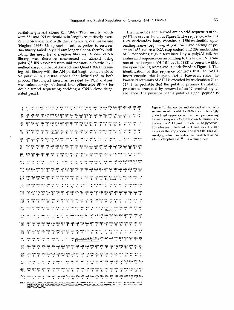
The nucleotide and derived amino acid sequences of the pAHl insert are shown in Figure 1. The sequence, which is 1859 nucleotides long, contains a 1656-nucleotide open reading frame (beginning at position 1 and ending at po- sition 1653 before a TGA stop codon) and 203 nucleotides of 3' noncoding region terminated by a poly(A) tail. An amino acid sequence corresponding to the known N termi- nus of the isozyme AH I (Li et al., 1992) is present within the open reading frame and is underlined in Figure 1. The identification of this sequence confirms that the pAHl insert encodes the isozyme AH I. However, since the known N terminus of AH I is encoded by nucleotides 70 to 117, it is probable that the putative primary translation product is processed by remova1 of an N-terminal signal sequence. The presence of this putative signal peptide is
1 ACG AAG TE f f i C TCT TE CTC TTA E T GCG C T I ClK CTC GCT GGC TM GCA TIG ACA AAT AGC M A GCT GCG Figure 1. Nucleotide and derived amino acid sequences of the pAH1 cDNA insert. The singly
1 T K L G S L L L C A L L L A G F A L T N S K A A - 73 AAA ACA GAT CCA CCC ATT CAC TGT GCT TCT CTC AAC AGG AGC AGT TE GAT GCT CTC GAA CCA GGG TE ATA
1 4 5 TM GGC ACA GCC TCA GCA GCT TAC CAG TK GAA GGT GCT GCA AAA GAA GAT GGT AGA GGA CCA AGT ATA E G
underlined sequence within the open reading frame corresponds to the known N terminus of the mature AH I protein. Putative N-glycosyla- tion sites are underlined by dotted lines. The star
2 5 K T D P P I H C A S L E _.__ _ _ _ _ $ S F D A L E P G F I
4 9 F G T A S A A Y Q F E G A A K E D G R G P S I W
2 1 7 GAT ACC TAC ACC CAC AAC CAT TCA GAA AGG ATC AAA GAT GGC AGT AAT GGA GAT GTC GCT GTT GAT CAA TAT indicates the stop codon. The motif Ile-Thr-Glu- Asn-Glv. which includes the Dredicted active
73 D T Y T H N...Jj _ _ _ _ $ E R I K D G S N G D V A V D Q Y
I .
site nucleophile G I u ~ ~ ' , i s within a box. 2 8 9 CAC CGA TAT AAG GAA GAT G E AGG A W ATG AAG AAA A E Gff i m GAT GCT TAT Aff i TM TCT ATC TCG lK% 9 7 H R Y K E D V R I M K K M G F D A Y R F S I S W
3 6 1 1 2 1 S R V L P N G K V S G G V N E D G I K F Y N N L
TCC AGA GTC "2 CCA AAT ffiA AAG GTA AGT GGG GGC G E AAT GAG GAT GGA ATC AAA TIT TAC AAC AAT C W
4 3 3 1 4 5 I N E I L R N G L K P F V T I Y H W D L P Q A L
ATC AAT GAA ATC CTA CGT AAT GGT CTA AAA CCA TlT G E ACA ATC TAT CAT W G GAT CTT CCC CAA GCT T I A
505 1 6 9 E D E Y G G F L S P N I V D H F R D Y A N L C F
GAG GAC GAA TAC GGT GGT ITt TTA AGC CCT AAT A l T GTC GAT CAC TM AGA GAC TAT GCA AAC CTT TGT TIT
5 7 7 1 9 3 K K F G D R V K H W I T L N E P Y T F S S S G Y
AAG AAA TM GGC GAT CGA GTA AAA CAC E G ATC ACG TE AAT GAG CCA TAT ACC TM AGT AGC AGT GGT TAT
6 4 9 GCA TAC GGG GTC CAT GCA CCA GGA CGA TGC TCT GCT lK% CAA AAA CTA AAT TGC ACT GGT GGG AAT TCG GCA 2 1 7 A Y G V H A P G R C S A W Q K L A .... Ç .... G G N S A
721 ACT GAA CCA TAT TTG G E ACA CAC CAC CAA CTC C W GCT CAT GCA GCG GCT GTA AAA TIG TAC AAA GAT GAA Z U T E P Y L V T H H Q L L A H R A A V K L Y K D E
793 2 6 5 Y Q A S Q N G L I G I T L V S P W F E P A S E A
TAT CAG GCA TCT CAA AAT f f iC TE ATA ff iA ATA ACA TIG G ' E TCA CCT lQ2 TM GAG CCT GCT TCG GAG GCA
865 GAG GAA GAT ATA AAT GCT GCA TM CGA TCT TTG GAT TlT ATP TlT GGA lK% TM ATG GAC CCG TTG ACA AAT 2 8 9 E E D I N A A F R S L D F I F G W F M D P L T N
937 313 G N Y P H L M R S I V G E R L P N . . . K .... T E E Q S K
GGT AAC TAT CCG CAC CTC A E CGA TCA ATT GTT GGG GAA CGA TTA CCA AAT TIK ACG GAA GAA CAA TCC AAG
1 0 0 9 TE CTA AAG GGG TCA "I GAT TM ATT ffiA CTA AAT TAT TAT ACA ACT AGA TAT GCA AGC AAT GCA CCT AAG 3 3 7 L L K G S F D F I G L N Y Y T T R Y A S N A P K
1081 A T I ACT TCT GTA CAT GCA AGC TAC ATA ACA GAT CCT CAA G'IT AAT GCT ACA GCT GAG CTT AAG GGG GTC CCC 3 6 1 I T S V H A S Y I T D P Q V N _ _ _ _ & _ _ _ _ T A E L K G V P
1153 ATT GGT CCA A E GCT GCT TCA GGC lK% TTA TAT G l T TAT CCC AAA GGA ATT CAC GAT CTT GTA CTT TAC ACA 385 I G P M A A S G W L Y V Y P K G I H D L V L Y T
1 2 2 5 AAG GAA AAG TAT AAT GAT CCC CTC ATT TAC GTT GAT GAG TIK AAT GAT CCC AAA T I A 4 0 9 K E K Y N D P L I Y V D E F N D P K L
1 2 9 7 TCA A E GAG GAA GCC CTC AAA GAT ACC M T AGA ATT GAC TM TAT TAT CGT CAC CTT TGT TAC CTT CAA GCA 4 3 3 S M E E A L K D T N R I D F Y Y R H L C Y L Q A
1 3 6 9 GCC ATC AAA AAG GGT TCT AAA G E AAG GGT TAC TM GCA 'KX TCA TM CTA GAC AAC TlT GAA GAT GCA 4 5 7 A I K K G S K V K G Y F A W S F L D N F E W D A
1 4 4 1 GGA TAC ACT GTT CGA TlT GGT ATC AAC TAC G E GAT TAC AAT GAC AAT TTA AAA AGG CAC TCT AAA CTC TCA 4 8 1 G Y T V R F G I N Y V D Y N D N L K R H S K L S
1513 ACG TAC lK% TE ACA AGT TK CTC AAG AAG TAC GAA AGA AGT ACG AAA GAA ATC CAA A I G l T T G I G GAA AGT M S T Y W F T S F L K K Y E R S T K E I Q M F V E S
1585 AAA CTA GAA CAT CAA AAG TlT GAA TCC CAA A ' E A E AAT AAA GTA CAA AGC TCT CTA GCA GTC GTC G ' X E A 5 2 9 K L E H Q K F E S Q M M N K V Q S S L A V V V '

34 Zheng and Poulton Plant Physiol. Vol. 109, 1995
consistent with the protein body location of AH in black cherry seeds (Swain et al., 1992b). The processed protein has a predicted molecular mass of 60.3 kD and a p1 of 6.76, which correlates well with the observed values for the glycosylated protein of 62 kD (by SDS-PAGE) and 6.6, respectively. Consistent with the known glycoprotein na- ture of AH I, five putative N-glycosylation sites (Asn-X- Ser/Thr) are present in the deduced amino acid sequence, although Asn329 may remain unglycosylated because this site has Pro at position +1 of the consensus sequence.
During the past decade, the availability of the primary structures of several hundred glucan hydrolases (EC 3.2.1.x) has allowed these enzymes to be reclassified based on their structural similarities rather than according to their substrate specificities (Henrissat, 1991). Sequence analysis indicates that P-glucosidases fall into two distinct families, designated by Béguin (1990) as families BGA and BGB. The BGA family includes P-glucosidases, phospho-P- glycosidases, thio-p-glucosidases, and P-galactosidases from organisms as diverse as archaebacteria, bacteria, plants, and mammals. The BGB family includes funga1 enzymes and the P-glucosidases of rumen bacteria.
The common structural features shown by BGA enzymes suggest a shared mechanism for the enzymatic hydrolysis of P-glycosidic bonds. The BGA enzymes catalyze glyco- side hydrolysis by a double-displacement mechanism in which an enzymatic nucleophile attacks the substrate form- ing a glucosyl-enzyme intermediate (Sinnott, 1990). The latter is then hydrolyzed, releasing the sugar with overall retention of anomeric configuration. Aglycone group de- parture may be aided by a protonated amino acid side chain with acid catalytic function. Withers et al. (1990) identified the active site nucleophile ( G ~ u ~ ~ ~ ) of the Agrobacterium faecalis P-glucosidase by inactivating the en- zyme with the mechanism-based inhibitor 2’,4’-dinitrophe- nyl-2-deoxy-2-fluoro-~-~-glucopyranoside. The catalytic importance of this amino acid residue, which occurs as part of an Ile/Val-Thr-Glu-Asn-Gly motif that is highly con-
served in members of the BGA family (but not of the BGB family), was subsequently confirmed by site-directed mu- tagenesis (Trimbur et al., 1992). The nature of the acid-base catalytic residue remains less certain. Trimbur et al. (1992) suggested the participation of Asp374, which exists as part of an Asp-X-Arg-X-Tyr sequence that is highly conserved in the BGA family glucosidases. More recently, however, the inactivation of cassava linamarase by N-bromoacetyl- P-D-glucopyranosylamine identified G ~ u ’ ~ ~ as the likely active site carboxylate group with acid catalytic function (Keresztessy et al., 1994b). This residue occurs within a Asn-Glu-Pro/Ile motif that is also highly conserved in BGA family P-glycosidases (with the exception of myrosinases).
Comparison of the deduced amino acid sequence of the mature AH I protein with other known sequences in the data base leads us to conclude that P. serotina AH belongs to the BGA family of P-glycosidases for the following rea- sons: (a) AH I exhibits high homology (37-65% identity) with other members of the BGA family, even including the prokaryotic and mammalian members (Table I). Highest similarity was observed with white clover linamarase (65.5% identity; 78.5% similarity), the white clover noncya- nogenic P-glucosidase of unknown physiological function (59.1 % identity; 73.1 % similarity), and cassava linamarase (51.7% identity; 69.5% similarity). Like AH, these linama- rases hydrolyze both cyanogenic glycosides and the chro- mogenic substrate p-nitrophenyl-P-D-glucoside. Somewhat less homology (4348% identity) was observed with O-P- glucosidases from the monocots maize and oats and with thioglucosidases from the Brassicaceae. (b) Contrasting with the magnitude of the foregoing values, the AH I amino acid sequence exhibits only low homology (16-2370 identity) with six members of the BGB family (data not shown). (c) The deduced AH I sequence includes the highly conserved Ile-Thr-Glu-Asn-Gly motif (putative active site nucleophilic center) at residues 396 to 400 of the mature protein. (d) The sequence also contains the Asn-Glu-Pro
Table 1. ldentity (percentage) of the deduced amino acid sequence o f the mature A H I protein (less signal sequence) with several glucosi- dases of the BGA family, as determined by the FastA program
Except for AH 1, the enzymes are identified by their GenBank accession numbers: AH I, P. serotina (this work); X56733, T. repens linamarase (Hughes, 1993); X56734, T. repens noncyanogenic P-glucosidase (Hughes, 1993); S35175, Manihot esculenta linamarase (Hughes, 1992); X74217, Zea mays (Brzobohaty et al., 1993); X78433, Avena sativa (Gus-Mayer et al., 1994); X59879, Sinapis a b a myrosinase (Xue et al., 1992); X60214, Brassica napus myrosinase (Falk et al., 1992); L11454, Arabidopsis thaliana thioglucosidase (Chadchawan et al., 1993); M96979, Bacillus circulans 0-glucosidase (Paavilainen et al., 1993); M19033, Agrobacterium faecalis (Wakarchuk et al., 1988); M61841, human lactase-phlorizin hydrolase domain 3 (Mantei et al., 1988). Where known, the sequences of mature proteins (lacking signal or transit peptides) were used for analysis.
Enzvme X56733 X56734 S35175 X74217 X78433 X59879 X60214 L11454 M96979 M19033 M61841
AH I 65.5 X56733 X56734 S35175 X74217 X78433 X59879 X602 1 4 L11454 M96979 M19033
~ ~
59.0 51.7 47.4 43.7 46.9 46.1 48.1 43.4 59.3 50.7 47.4 45.6 48.5 48.4 48.6 42.1
42.6 45.9 45.2 46.0 45.2 46.2 39.7 41.6 41.6 40.5 40.9 41.7 38.4
61 .O 40.9 41 .O 41 .O 39.0 39.5 39.1 39.2 38.2
90.8 70.1 39.1 72.5 38.9
35.5
36.8 36.9 36.6 33.1 34.3 33.8 32.1 33.6 30.8 46.3
40.3 42.6 38.3 37.6 37.4 40.0 39.3 38.5 36.7 39.5 35.1

1 1 1 1 1 1 1 1 1 1
6 5 52 56 62
Temporal and Spatial Regulation of Cyanogenesis
T-------------------KLCISLLLCALLLAGF~TNS~T------------DPPI--HUSLNRSSFIIl---IEPDFIKTASMYQPEG~ AH I LSl~.HIH.F.P------------L.IS~DPSD .... C.A V.....S.F.Y....F. X56733
M...........----------LV.FISL.A.TRP,MGTDDDDDN------~~----I.------DDFS.KY.pD~~~~~~D......TS...I..E.TA S35175 M-------------------DFIVAIF~FJISS,TI.STN.VE------~~~-----AST~DIGN.5....P------R.....AG.S.......VN. X56734 M A P L ~ P G L R S H . V G P " E S F S R H H L P S S S P Q S S . R R ~ ~ F T T R ~ ~ ~ S Q - ~ G V Q M . ~ ~ . EI~-PS~.T..A.TS ... I...WN. X74217 MA-LLCSRLSNST-HPSFRSH-IGR".W--HLSADPAQKS.RR~TLS5~RI5S~ESAKQ~~Q~K~P.E.M..A......I...WN. X78433 ................................................................... ~ ~ ~ ~ , ~ . ~ ~ ~ ~ . L . . V . T . S F . I . . S T . ~ "19033 . . . . . . . . . . . . . . . . . . . . . . . . . . . . . . . . . . . . . . . . . . . . . . . . . . . . . . . . . . . . . . . . . . . . . . . . . ~ ~ ~ ~ ~ p ~ ~ , ~ , ~ , ~ , , , , ~ , . , Y N . M96979
~ L L H G - ARSCKROEEIT--CE-------ENE.FTCSNTDI.SSI(.GK------D . . . . V..S...I..GR-- X59819 ~LLn-LIIF-------------------VP...LRTCKGDE-FJ--CE-------ENE.FT~~KLF.SGN.EK------.....V..S...V..GR-- Lll454
D ~ R G P S I W D ~ T H N H S E R I - K S N G D V A ~ ~ ~ E D ~ I ~ ~ G F D A Y R F S I ~ S R ~ ~ G K Y ~ G ~ D G I K F ~ I ~ I L R N G L K P ~ I Y ~ AH I .. K.......F..KYP.K.-..RT.....I.E.......IG...~.NL.........P....K..L... .RE..NY.......V.A..MQ.Y..LF... X56733 K..A..V..IFSKETPD..-L..........F.N..IQ.IIWY.....N.F.M.......I.S.RRRE....E..Q...DV....IS...E.....F... 535175
.. F..KYP.K.-R....A.IT...........G...WNM.S.......P.I..K..L...I.HE...Y.......L.A..IQ....LF... X56734 100 96 3 3
.. K.E.N . . HFC ... P...-L....S.IGANS..M..T...LL.E..M.........P.I..K.TKE..I.P....Y.R....LL.E..IE.Y...F... X74217 G.K ... S. .NFC.S.PD..-M,K..A...MS.Yn......~.EI.M.S.......P.I..K.TLD..I.HE..QY..D.L~LIE..I..YI.LF... X78433 . . . K.....RFCNEI-PGHV-FG~...I.C.H.N.WE..LDLT.E..YE.....LII.P.II.D.F--.PI..K.LO..DR.WOC~.I.TYA.L.... M19033
28 .... M.....FA.T-PGKY-.N.D..N..C.S...VE...QLL.DL.VKV.......P....Q.T--.E..RR.LDY.HR.M.L.A..IE..C.L.... M96979 65 - . . .VNV.. GFS.RYP.KSGS.LK ... TSCE5.T.W.K .. E..GELNATG....FA...IV.K....R..~A.LDY.H...DRL.EI(IT....LF... X59879 63 -...LMI.. SF..RFP.KG~.LG...TTC.S.TLWQK.IW.DELNSTG ..... A...L..K..R.R...PGA..Y..G..DjLV~....LF... LI1454
164 L 4 1 A L E D E Y O O F L S P N - - - I M H F R D Y ~ C F K R F G D R V K H AH I 151 V........R...GR.---...D.....E....E..............WGV.MNA....TF...-...D.L.....--..D.GR....AR.Y........ X56733 155 T....Q.K......RD---..YDYLQ..D.L.ER......P.M.F...SAWGFAHDD..F...-...S.VNRQ.L--A.D......I.A.NL..S.... S35175 161 ... V.........NSG---VIND....TD....E.....RY.S.....WY..N....L.TN...-....SNYAK----P.D.G.G..I...N.I....E. X56734 199 X74217 195 T....AL...W..DR---R..WYT...III..EH...K..N.F.F...HS.CGL..GT.L....A....--~.VIPEEDALRN,.I.G.NL.....ET X78433 129 .. LT .IXjD----FGWASRST.Q~..K~...LDAVA.F...WCIVYILSHL.......E.---------------N..IN...GFG Ml9033 124 ..... Q.Q----GOWOSRITI.A . R E . . E.M .. EL.GK1.Q ... F...WCHAFLSNYL......NK---------------DLQLAID.S..L.V..GR. M96979 164 ... T.Q ... E...DRQ---.IOD.K...D....E..GK..N...I.QL..VPPR...L.TD...-...PKVDTKQRCY....S....I.A.N.......l X59879 162 ... T.Q ... N...NKT---...D.K...D...EL......N...I.QL..VPTR...L.TD...-...PKIDV--RCP....S....I.A.N........ LI1454
258 VRLYKDEYQASQNGLIGITLVSPYIFEPAS~-EDIN~RSLDFIF~PLTNGNYPH~S---IVGERLF~TEEQSKLLKG5FDFIG~Y~R AH I 245 RR...TX....... I. ...... H......KEK-A.M..R.....H...K.R,.E5..Y---L.RK...K.ST.E..E.T.....L.....5SY X56733 249 .HQ.RKY..GT.K.K.....F~.Y..L.DSKY-.VQ..KTA...M..LW.,.M,Y.R..RT.M---~.DK.IG..D.E.Q..R..Y..V..Q...AY 535175 253 .HV..TK... Y.K.K . . . . . . . N.Ln.LDDNSIP..K..E.....Q..L..EQ..T.D.SKS..R---..I(...K.S~E.S.~.......I...SSS X56734 296 .D..NKH.KR-DDTR..LIIFrmMGRV.YGTSFLDK-Q.EE..W.INL...LE.VVR.D..FS...---IIIR....F.KD..KEK.A..YNML......S. X74217 290 .DV.NI(P.KG-DD.Q..MV.DVHAY..YG"FLDQ-Q.QE.AI..HI...LE.~.D..FS...---L..D...F..KSEQEK.VS.Y..V.I....5. X78433 210 .ERSR---HVAPKVW.LV.NAHSRI...DG.-A.LK..E.RFQ.HN.A.F..VFK.E..AE.M---EAL.D.H.~.DLGIISPKL.WW......P- I419033 205 .T.FR---E~IS.E...APNTS.AV.YRRTK-..ME.CL.VNGWSGD.~..IYF.E..KF.LDW~.YKP.IWO-DME.IHQFI....I....SS M96979 260 .D..RTN.-.F...K..PVMITR..L.YD.SDPAC.E..E.~Q.PH..Y.E...X.R..DI..Q---...S.......~.VA..Y..L.....V.Q X59879 256 .OV.RTK.KDD.K.M..PVHITR..L.FDHSQ-.SKD.TE.AKI.FH....G...E.K..DI..E---Y..D...E.S.TERI\.V...Y..L.....V.Q LI1454
354 Y A S N A P K I T S V H A S Y I T D P Q - Y N R T - ~ L K ~ I G P ~ ~ ~ P - - ~ - K G I H D L V L Y T K E K ~ P L I Y - I T ~ G ~ E ~ - - D P K L S M E ~ ~ I AH I
345 . . EP1.PWPKFRR.K .. SG- .... PYD.N . N L . . . Q.Y.S.Q.IF RHFLN...ar....V..-V......NY.--NESOPI....Q.DF.. 535175 350 .I....SHGNAKF..S.N.M-T.IS-F.KH.I.L..R...I.I....~FIQEDFEIFC.ILKINIT~.QFS.....~...--.AT.PV....LN.Y.. X56734 391 FSK.IDISPNYS WISY.. DAYASQ-GPD.K .... PMCNP.1.M E.LK . . Il(1M.N . . GN.P TGDYDTKETP.P..D..N.YK.L X74217 385 F.WIDISPEFIPKIN .. DWS.PEVNDSN.1.. .. DVGWF1.S LYSIIL .RH.... GN.P TADMOGWGNP-P.TDP.D.PL.. X78433 302 ~VMDATPG.EFPATMFA.S ----------- D V R T .. E-----WAPRL.T..ETLY.R.DL.EC.-.....----ACYNnGVE-NGQ~.QP.L M19033 300 MNRYN.GERGGML.SE--AISMG-----------A.KTDI,.E-----IY~.LY..LR..AD..GN.TL.-.....----ACYNDj..LffiRIH.QR.. K96979 356 .. KPK.NPYPSETHTALLrm\O.DL .P"SR.EYP..VF.ED---IINSWYP...WYXD.F.T...N....-.....IST----PGSE.RC..lA.YK.. X59879 352 .. O.N~.VPSDVHTLLIIDSRTTL.SKNI\T .~F..PF--N---MSWYP...WYXD.F.TT.G.....-V....FST----PGDEDF.K.TA.YK.. L11454
341 ..IU(.. R.P-FAIQ .. SL-I F.HN.K.L......S..CI..----Q..RK.L..Y.NH..N.Y..-.....RN...--..T..LQ.S.L..P.. X56733
445 D ~ f f L ( I M I K K - G S K Y F A W S F L ~ ~ G ~ I ~ ~ - ~ R H S ~ S T ~ S F L K ~ E R S T K E I Q ~ S ~ ~ Q K F E S Q ~ AH I 432 .~....,~.~T.,G~.,~,,.,,.,,~,.~,,,~.,..... L------ ................................................. X56733 431 SY .KK. HWNILIDSL .NY. V.L ....... Y......NI...S...LY....KN-..T.YP.K.RH...K..NISVNR"IYELTSKDSRRVO..------- 535175 446 .Y.....Y.IRS..R&-..N...FY......CN..F..F.....L.F.. X56734 486 .YI~..IAT.KES.DLGSN-.Q......L......F..F.E.Y..V...R.N-.CT.Yn.E.I\K.LKQ.NM-KXPS.K.LTP----------------- X74217 179 CIL~.MTAIKE..DLGRRTLR.H.T..LI.....SL..LS....V.I.R..-GC..I~.K.I\K.LKE.NGATKX~.~ASSCCSG~OOG----- X78433 380 .Y.AB..GIVADL.RD-.YPMR......Ln.....AE..RM...LVH...QTQV--.TY.N.GK.YS~SGFPKGNHGVA------------------- M19033 377 .YWI..IQA~R..ED-.INL...ME..Ln.....RE..GM...LYH...DTLV--.TP.D.F..YKGV---IS.GWLDL M96979 448 NYLCS...F.RKV.RER.VNIR.....~..Y.FCX.F.....LS..NWD.L-DD.NL.E.GK.YQR.INGTRKNPVKQOFLRS.LSSQS-Q~~-- X59879 442 .YLCS...F.SKV..EIWVN........LG..Y.FW.F.....LS...FRNITGD.DL.A.GK..QK.I~DEDSTNQDLLRS.VSS~D~5~- L11454
motif (putative acid-base catalyst) at residues 183 to 185 of the mature protein. (e) The sequence includes four His residues that are conserved within the four other se- quenced plant P-glucosidases (Fig. 2). In this context, the presence of a reactive His residue at the active center of cassava linamarase should be noted (Keresztessy et al., 1994a). (f) Three of the five putative N-glycosylation sites within the AH I sequence are shared by white clover linamarase. The high homology between AH I and other BGA family P-glucosidases, not only at the putative active site moieties but also scattered throughout their entire sequences, is clearly illustrated by the multiple sequence alignment in Figure 2.
A H I X56733 535175 X56134 X74217 X78433 M19033 M96979 X59879 L11454
in Prunus 35
Figure 2. Multiple sequence alignment of the deduced amino acid sequence of A H I with sequences of nine other members of the BGA family of P-glucosidases. Amino acid alignment was performed using the DNASTAR (Madison, WI) Megalign Clustal Program (PAM250 residue weight table). The sources of P-glucosidase se- quences (identified by GenBank accession num- ber, except for AH I) used in this comparison are: AH I, P. serotina (this work); X56733, T. repens linamarase (Hughes, 1993); S35175, M. esculenta linamarase (Hughes et al., 1992); X56734, T. repens noncyanogenic p-glucosi- dase (Hughes, 1993); X74217, 2. mays (Brzobo- haty et al., 1993); X78433, Avena sativa (Gus- Mayer et al., 1994); M19033, Agrobacterium faecalis (Wakarchuk et al., 1988); M96979, B. circulans P-glucosidase (Paavilainen et al., 1993); X59879, S. alba myrosinase (Xue et al., 1992); L11454, Arabidopsis thaliana myrosinase (Chadchawan et al., 1993). Residues identical with those of AH I are shown by dots, whereas introduced gaps are represented by dashed lines.
Developmental Expression of A H and MDL in Maturing Fruits
In previous work (Swain et al., 1992a), biochemical changes related to cyanogenesis were monitored during the maturation of black cherry fruits. It was shown that, concomitant with cotyledon development during phase 11, the seeds begin accumulating both amygdalin and the cat- abolic enzymes AH, PH, and MDL and, from that time onward, are therefore highly cyanogenic when disrupted. In contrast, the pericarp remains acyanogenic throughout the entire ripening process because it lacks the catabolic enzymes.

36 Zheng and Poulton Plant Physiol. Vol. 109, 1995
DAF29 36 40 44 49 56 62 70 76 82
1.8kb*-
B120
, i oo-l
H 8(H•<I 60
'g 40-
^ 20-
25 35 45 55 65 75DAF
C DAF29 36 40 44 49 56 62 70 76 82
1.9kb*-
D120
•s 100-J 80-Q
E 60H
i'5 40-
25 35 45 55 65DAF
75 85
Figure 3. Temporal accumulation of AMI and MDL1 mRNAs andproteins during fruit maturation in P. serotina. A and C, Northern blotanalyses. Total RNA (10 ng) isolated from developing seeds at thetimes indicated (DAF) was fractionated on a denaturing agarose geland blotted onto nylon membranes. The blots were hybridized with32P-labeled pAHl insert (A) or pMDLl insert (C) under conditionsdescribed in "Materials and Methods." B and D, Estimation of AH (B)and MDL (D) protein levels by direct enzyme assay. Enzyme activi-ties are given as a percentage of the maximum level observed foreach enzyme (AH, 48.8 /xmol min"1 seed"1; MDL, 10.8 jLimol min~'seed"1). Each data point represents the mean of duplicates, the rangeof which did not exceed the dimensions of the symbol shown.
In the current study, we have now utilized northern blotsto examine the temporal expression of AH1 and MDL1transcripts in ripening fruits. Total RNA was isolated atapproximately weekly intervals from maturing seeds(29-82 DAF) and probed using 32P-labeled pAHl andpMDLl cDNA inserts. As Figure 3, A and C, illustrates,neither mRNA was detectable during phase I of fruit rip-
ening. However, when embryos first became visible to thenaked eye during early phase II (40 DAF), both transcriptsbecame detectable, increasing to reach a maximum at ap-proximately 49 DAF. Subsequently, transcript levels de-clined and were undetectable at full fruit maturity (82DAF). It should be noted that the mRNAs detected by theAH1 and MDL1 probes were approximately 1.8 and 1.9 kbin length, respectively; these values correlate well with theknown sizes of the cDNAs for these enzymes (Cheng andPoulton, 1993).
The levels of AH and MDL proteins were also measuredduring fruit ripening by direct enzyme assay of seed ho-mogenates. Confirming previously published data (Swainet al., 1992a), Figure 3, B and D, illustrates that AH andMDL activities were undetectable until 44 DAF. They thenincreased rapidly during mid-phase II, essentially reachinga plateau at full fruit maturity (82 DAF). Comparison oftheir respective temporal patterns of mRNA and proteinaccumulation during fruit maturation suggests that theexpression of AH and MDL may be regulated at the tran-scriptional level, although run-on transcription studies arerequired to confirm this tentative conclusion.
In Situ Localization of AH1 and MDL1 mRNAs inImmature Embryos
The spatial expression patterns of AH and MDL mRNAswere analyzed by in situ hybridization using antisense andsense DIG-labeled riboprobes transcribed from pAHl andpMDLl, respectively. Immature seeds collected 49 DAFwere selected for analysis because northern analysis hadshown that they exhibit the highest transcript levels (Fig.3). When seed sections were hybridized with the antisenseAH1 riboprobe, intense labeling was observed exclusivelywithin the procambial cells (Fig. 4, A and B). By contrast,control hybridizations using the DIG-labeled sense AH1riboprobe gave no positive signal (Fig. 4C). The tissue-specific localization of AH1 mRNA within the procambialcells is in accordance with our previous immunocytochem-ical data showing that AH protein is restricted to theprotein bodies of that tissue (Swain et al., 1992b).
MDL, which constitutes approximately 10% of the solu-ble protein of black cherry seeds, is believed to be multi-functional, serving both as a storage protein and in cyano-genesis (Swain et al., 1992b). In situ hybridization analysisshowed that the spatial expression of MDL1 mRNA differsgreatly from that of AH1 mRNA, although both transcriptsshow similar temporal expression patterns in developingseeds (Fig. 3). In contrast to the procambial location of AH1transcripts, MDL1 mRNA exhibited a spatial expressionpattern more characteristic of storage proteins, being re-stricted to the cotyledonary parenchyma cells (Fig. 4D).With the antisense MDL1 riboprobe, hybridization signalswere strongest at the interior of the cotyledon and dimin-ished sharply toward the periphery of that organ. No sig-nal was observed when seed sections were exposed to thesense MDL1 riboprobe (Fig. 4E). The presence of MDL1mRNA in the cotyledonary storage parenchyma cells cor-relates well with the known major location of MDL proteinin mature seeds (Swain et al., 1992b). Although previous

Temporal and Spatial Regulation of Cyanogenesis in Prunus 37
Figure 4. Localization oi AH and MUL i-xpifsbiun in immature black cherry seeds (49 DAF) by in situ hybridization. Tissuewas fixed, embedded in paraffin, sectioned, and hybridized in situ with DIC-labeled sense or antisense transcriptssynthesized from pAH1 and pMDLl. The dark blue or purple represents hybridization to target mRNAs, indicating theirtissue distributions. A, Transverse section probed with AH1 antisense riboprobe, stained with Fast Green FCF. Bar, 200 /im.B, Longitudinal section probed with AH1 antisense riboprobe, stained with Fast Green FCF. Bar, 200 /xm. C, Transversesection probed with AH1 sense riboprobe, stained with Fast Green FCF. Bar, 200 /urn. D, Transverse section probed withMDL1 antisense riboprobe. Bar, 500 /xm. E, Transverse section probed with MDL1 sense riboprobe. Bar, 500 jam.

38 Zheng and Poulton Plant Physiol. Vol. 109, 1995
immunocytochemical studies had also detected minor amounts of MDL protein in procambial cells, MDLl mRNA was not detectable in such cells during the present study (Fig. 4, D and E). Assuming that in situ hybridization and immunocytochemistry are equally sensitive to localizing small amounts of their respective target molecules, we offer two explanations that might account for this apparent discrepancy. First, it should be noted that our immunocy- tochemical study was undertaken using fully mature seeds, whereas the in situ hybridization analysis performed here utilized immature (49 DAF) seeds. It is therefore possible that procambial MDL expression occurs after 49 DAF and was therefore not detected by our in situ analysis. Alter- natively, because black cherry has severa1 MDL isozymes (Yemm and Poulton, 1986), it is possible that the procam- bial MDL protein is encoded by a distinct lyase gene whose mRNA hybridizes poorly, if at all, to the MDLl probe used here.
In conclusion, the isolation and sequencing of the AH cDNA clone pAHl constitute important steps toward a better understanding of the molecular biology of cyano- genesis in rosaceous stone fruits. Confirming previous im- munocytochemical data, our in situ analysis has again demonstrated the remarkable tissue-specific expression of AH and MDL in rosaceous stone fruits. Seeking to under- stand the molecular mechanisms that underlie such pat- terns of expression, we are currently characterizing AH and MDL genomic clones with the goal of identifying those promoter regions that confer tissue specificity in black cherry seeds.
ACKNOWLEDCMENTS
The authors wish to thank Dr. Chun Ping Li (University of Arizona) for isolation of the partial-length AH cDNA clones, Drs. Richard Sjolund and Shelley Plattner for assistance with photo- graphic reproductions, the University of Iowa DNA Facility for DNA sequencing, and Dr. Asim Esen (Virginia Polytechnic Insti- tute) for helpful discussions.
Received March 20, 1995; accepted June 7, 1995. Copyright Clearance Center: 0032-0889/95/l09/0031/09. The GenBank accession number for the sequence reported in this
article is U26025.
LITERATURE ClTED
Béguin P (1990) Molecular biology of cellulose degradation. Annu Rev Microbiol 44: 219-248
Brzobohaty B, Moore I, Kristoffersen P, Bako L, Campos N, Schell J, Palme K (1993) Release of active cytokinin by a P-glu- cosidase localized to the maize root meristem. Science 262 1051- 1054
Chadchawan S, Bishop J, Thangstad OP, Bones AM, Mitchell- Olds T, Bradley D (1993) Arabidopsis cDNA sequence encoding myrosinase. Plant Physiol 103: 671-672
Cheng I-P, Poulton JE (1993) Cloning of cDNA of Prunus serotina (R)-(+)-mandelonitrile lyase and identification of a putative FAD-binding site. Plant Cell Physiol 34: 1139-1143
Cox KH, Goldberg RB (1988) Analysis of plant gene expression. In CH Shaw, ed, Plant Molecular Biology: A Practical Approach. IRL Press, Oxford, UK, pp 1-34
Devereux J, Haeberli P, Smithies O (1984) A comprehensive set of sequence analysis programs for the VAX. Nucleic Acids Res 12
Falk A, Xue J, Lenman M, Rask L (1992) Sequence of a cDNA clone encoding the enzyme myrosinase and expression of my- rosinase in different tissues of Brassicu napus. Plant Sci 83:
Gus-Mayer S, Brunner H, Schneider-Poetsch HAW, Rudiger W (1994) Avenacosidase from oat: purification, sequence analysis and biochemical characterization of a new member of the BGA family of P-glucosidases. Plant Mo1 Biol 2 6 909-921
Henrissat B (1991) A classification of glycosyl hydrolases based on amino acid sequence similarities. Biochem J 280: 309-316
Hughes MA (1993) Molecular genetics of plant cyanogenic P-glu- cosidases. In A Esen, ed, Symposium Series 533. American Chemical Society, Washington, DC, pp 153-169
Hughes MA, Brown K, Pancoro A, Murray BS, Oxtoby E, Hughes J (1992) A molecular and biochemical analysis of the structure of the cyanogenic P-glucosidase (linamarase) from cassava (Mani- hot esculenta Cranz). Arch Biochem Biophys 295 273-279
Keresztessy Z, Kiss L, Hughes MA (1994a) Investigation of the active site of the cyanogenic P-D-glucosidase (linamarase) from Manihot esculenta Crantz (cassava). I. Evidence for an essential carboxylate and a reactive histidine residue in a single catalytic center. Arch Biochem Biophys 314 142-152
Keresztessy Z, Kiss L, Hughes MA (199413) Investigation of the active site of the cyanogenic P-o-glucosidase (linamarase) from Manihot esculentu Crantz (cassava). 11. Identification of Glu-198 as an active site carboxylate group with acid catalytic function. Arch Biochem Biophys 315 323-330
Kojima M, Poulton JE, Thayer S S , Conn EE (1979) Tissue distri- butions of dhurrin and of enzymes involved in its metabolism in leaves of Sorghum bicolor. Plant Physiol 63: 1022-1028
Li CP (1993) Cyanogenesis in rosaceous stone fruits: temporal and spatial expression of amygdalin hydrolase in Prunus serotina seeds. PhD thesis. University of Iowa, Iowa City
Li CP, Swain E, Poulton JE (1992) Prunus serotina amygdalin hydrolase and prunasin hydrolase. Purification, N-terminal se- quencing, and antibody production. Plant Physiol 1 0 0 282-290
Liebig J, Wohler F (1837) Ueber die Bildung des Bittermandeloels. Liebigs Ann Chem 2 2 1-24
Mantei N, Villa M, Enzler T, Wacker H, Boll W, James P, Hun- ziker W, Semenza G (1988) Complete primary structure of human and rabbit lactase-phlorizin hydrolase: implications for biosynthesis, membrane anchoring and evolution of the enzyme.
Paavilainen SK, Hellman J, Korpela T (1993) Purification, char- acterization, gene cloning, and sequencing of a new beta-gluco- sidase from Bacillus circulans subsp. alkalophilus. Appl Environ Microbiol 59: 927-932
Pancoro A, Hughes MA (1992) In-situ localization of cyanogenic P-glucosidase (linamarase) gene expression in leaves of cassava (Manihot esculenta Cranz) using non-isotopic riboprobes. Plant J
Poulton JE (1988) Localization and catabolism of cyanogenic gly- cosides. Ciba Found Symp 140: 67-91
Poulton JE (1993) Enzymology of cyanogenesis in rosaceous stone fruits. In A Esen, ed, Symposium Series 533. American Chemical Society, Washington, DC, pp 170-190
Poulton JE, Li CP (1994) Tissue leve1 compartmentation of (R)- amygdalin and amygdalin hydrolase prevents large-xale cya- nogenesis in undamaged Prunus seeds. Plant PhysiollO4: 29-35
Rosenthaler L (1908) Durch Enzyme bewirkte asymmetrische Syn- thesen. Biochem 2 14: 238-253
Sambrook J, Fritsch EF, Maniatis T (1989) Molecular Cloning: A Laboratory Manual. Cold Spring Harbor Laboratory Press, Cold Spring Harbor, NY
Sanger F, Nicklen S , Coulson AR (1977) DNA sequencing with chain-terminating inhibitors. Proc Natl Acad Sci USA 74: 5463- 5467
Sharrock RA, Quail PH (1989) Nove1 phytochrome sequences in
387-395
181-1 86
EMBO J 7: 2705-2713
2 821-827

Temporal and Spatial Regulation of Cyanogenesis in Prunus 39
Arabidopsis thaliana: structure, evolution, and differential expres- sion of a plant regulatory photoreceptor family. Genes Dev 3:
Sinnott ML (1990) Catalytic mechanisms of enzymic glycosyl transfer. Chem Rev 9 0 1171-1202
Swain E, Li CP, Poulton JE (1992a) Development of the potential for cyanogenesis in maturing black cherry (Prunus serotina) fruits. Plant Physiol 98: 1423-1428
Swain E, Li CP, Poulton JE (199213) Tissue and subcellular local- ization of enzymes catabolizing (R)-amygdalin in mature Prunus serotina seeds. Plant Physiol 100 291-300
Swain E, Poulton JE (1994a) Utilization of amygdalin during seedling development of Prunus serotina. Plant Physiol 106 437-445
Swain E, Poulton JE (1994b) Immunocytochemical localization of prunasin hydrolase and mandelonitrile lyase in stems and leaves of Prunus serotina. Plant Physiol 106: 1285-1291
Trimbur DE, Warren RAJ, Withers SG (1992) Region-directed
1745-1757
mutagenesis of residues surrounding the active site nucleophile in /?-glucosidase from Agrobacterium faecalis. J Biol Chem 267:
Wakarchuk WW, Greenberg NM, Kilburn DG, Miller RC Jr, Warren RAJ (1988) Structure and transcription analysis of the gene encoding a cellobiase from Agrobacterium sp. strain ATCC 21400. J Bacteriol 170: 301-307
Withers SG, Warren RAJ, Street IP, Rupitz K, Kempton JB, Aebersold R (1990) Unequivocal demonstration of the involve- ment of a glutamate residue as a nucleophile in the mechanism of a "retaining" glycosidase. J Am Chem SOC 112: 5887-5889
Xue J, Lenman M, Falk A, Rask L (1992) The glucosinolate- degrading enzyme myrosinase in Brassicaceae is encoded by a gene family. Plant Mo1 Biol 18: 387-398
Yemm RS, Poulton JE (1986) Isolation and characterization of multiple forms of mandelonitrile lyase from mature black cherry (Prunus serotina Ehrh.) seeds. Arch Biochem Biophys 247: 440-445
10248-10251