temperature dependence of oxygen diffusion into polymer/carbon nanotube composite films
TRANSCRIPT

Temperature Dependence of Oxygen Diffusion IntoPolymer/Carbon Nanotube Composite Films
Onder Yargı,1 S�aziye Ugur,1 Onder Pekcan21 Department of Physics, Istanbul Technical University, Maslak 34469, Istanbul, Turkey
2 Faculty of Arts and Science, Kadir Has University, Cibali 34320, Istanbul, Turkey
This study examines the transport properties ofpolystyrene (PS)/multiwalled carbon nanotube (MWNT)composite films taking into consideration both MWNTcomposition and temperature, via fluorescence tech-nique. Three different (3, 15, and 40 wt%) MWNTcontentfilms were prepared from PS/MWNT mixtures byannealing them at 1708C, above the glass transitiontemperature of PS for 10 min. The diffusivity of thePS/MWNT composite was determined by performingoxygen (O2) diffusion measurements within a tempera-ture range of 24 to 708C for each film and pyrene (P) wasused as the fluorescent probe. The diffusion coefficients(D) of oxygen were determined by the fluorescencequenching method assuming Fickian transport.Results indicated that D values are strongly dependenton both temperature and the MWNT content in the filmand it was also observed that D coefficients obeyArrhenius behavior, from which diffusion energies wereproduced and increased along with increases of MWNTcontent. POLYM. ENG. SCI., 52:172–179, 2012. ª 2011 Societyof Plastics Engineers
INTRODUCTION
There has been growing interest in producing
new materials by filling polymers with inorganic natural
(minerals) and/or synthetic (carbon black and silica) com-
pounds [1–5] because single component polymer films
have poor mechanical and gas barrier properties. Compos-
ite polymers display improved mechanical and gas barrier
properties and a decrease in flammability in comparison
with simple polymers [6]. The enhancement in barrier
properties in composites depends on several factors, such
as the amount, length, and width of the filler, as well as
the orientation and dispersion of the filler particles. For
more than 50 years, polymer scientists have been inter-
ested in the influence of fillers on gas diffusion through
polymer membranes [7–11], and this study expands on
that research by bringing to light the effects of mineral
fillers on the performance of oxygen sensors. Gorrasi
et al. [8] studied the transport properties of n-pentane and
dichloromethane vapors in polypropylene-organophilic
layered silicate nanocomposites with different clay con-
centrations, and it was found that the permeability of both
solvent vapors was reduced, mainly as a result of the
decreased diffusion, since the solubility was less affected
by the presence of fillers. Lu et al. [9] examined the
influence of 10 nm diameter silica particles on oxygen
diffusion in polydimethylsiloxane (PDMS) polymer film.
A decrease was observed in oxygen diffusion coefficients,
D, with increases of silica content. This reduction in
D was attributed to the tortuous path for diffusing gas
molecules and the reduced molecular mobility of polymer
chains caused by the filler particles.
Carbon nanotubes (CNT) have been identified as fun-
damentally new nanoporous materials that show great
potential for sensors [12, 13], composites [14], catalytic
supports [15], and as membrane materials [16, 17]. Signif-
icant effort has been devoted to the fabrication of mixed
matrix membranes by using carbon nanotubes as filler
with great potential [18]. Many researchers have studied
composites composed of conductive materials/polymers as
candidates for chemical vapor sensors because composite
materials have the advantages of high sensitivity and
reproducibility, good processability, and reasonable costs
[19]. Polymer/conductive material composites used as
sensors have the following properties: (1) numerous possi-
bilities for preparation of various types with a wide range
of selectivity owing to the diversity of the chemical and
physical properties of polymers, (2) sensitive, rapid, and
reversible responses to analytes, and (3) the most interest-
ing materials can transform a chemical signal into an
electric signal [19]. The polymer/conducting material
composites are prepared by blending polymer matrices
with carbon black, metal powder, carbon fiber, graphite,
or carbon nanotubes. These composites as chemical vapor
sensors are expected to be utilized for a broad spectrum
of applications in the detection of environmental gases,
solvent leaks, and identification of polymers, in addition
Correspondence to: S�aziye Ugur; e-mail: [email protected]
DOI 10.1002/pen.22061
Published online in Wiley Online Library (wileyonlinelibrary.com).
VVC 2011 Society of Plastics Engineers
POLYMER ENGINEERING AND SCIENCE—-2012

to other uses as well [20]. Among the various conductive
materials, carbon nanotubes (CNT) have attracted notable
attention because of their unique structure and remarkable
physical properties. These properties make them a strong
candidate for active materials in electronics, field emission
devices, and chemical vapor sensors. The chemical vapor
sensing property of CNT is due to the fact that CNT is a
nanosized material and has a high aspect ratio, resulting in
high sensitivity and rapid chemical vapor adsorption [21].
In early 2000, the potential for CNTs as gas sensors
was first reported in a study that noted an increase in con-
ductivity by several orders of magnitude upon exposure
of CNTs to oxygen [22]. There are two basic types of
CNTs: single-wall carbon nanotubes (SWNT) and multi-
wall carbon nanotubes (MWNT) [23]. In this respect,
more researchers have devoted effort to the fabrication of
mixed matrix membranes by dispersing either single-
walled or multiwalled carbon nanotubes into various poly-
mer matrices. The properties of polymer nanocomposites
containing carbon nanotubes depend on several factors, in
addition to the polymer: the synthetic process used to pro-
duce nanotubes; the nanotube purification process; the
amount and types of impurities in the nanotubes; diame-
ter, length, and aspect ratio of the nanotube’s objects in
the composite (isolated, ropes, and/or bundles); and the
orientation of the nanotubes in the polymer matrix. Kim
et al. [24] studied how incorporating CNTs into the polyi-
midesiloxane matrix impacted gas separation perform-
ance. They observed that the addition of small CNTs to
the copolymer matrix reduced the permeability of helium
reduced and hindered the diffusion of nitrogen as a result
of the impermeable properties of CNTs.
Oxygen is considered to be one of the most important
reactants in the diffusion phenomenon. The control of the
diffusion of oxygen is of particular importance in polymer
oxidative degradation, protective coatings, and in the
design of polymeric membranes for separation processes
in the production of films for the packing industry, as
well as in the development of biocompatible materials
[25]. Oxygen diffusivity in polymers is most commonly
determined by measuring the rate of oxygen permeation
across a membrane by exposing one side of the polymer
film to oxygen at time zero and measuring the flux of ox-
ygen across the film as a function of time. Knowing the
dimensions of the film and the partial pressure of oxygen
on the high-pressure side, allows for a calculation of the
diffusion coefficient from the flux [26]. The diffusion
coefficient of oxygen is determined from the kinetics of
the approach of the O2 flux to its steady-state. Because
oxygen is such a powerful quencher of fluorescence and
phosphorescence, this property is useful for determining
such values. Another approach, developed by Cox and
Dunn, also looks at large samples [27] utilizing a square
cell measuring 1 cm height. The cell is filled with a poly-
mer containing the dye and all oxygen is removed. Cox
and Dunn irradiated a thin (1 mm) middle cross-section
of the cell, and then allowed oxygen to diffuse into the
cell from the top. As the oxygen concentration profile
propagates through the illuminated middle slice, the emis-
sion intensity is quenched.
Several spectroscopic techniques that utilize oxygen
quenching to determine the rate of oxygen diffusion
through polymer films have been reported. Cox [28] and
Dunn [29], and MacCallum and Rudkin [30] measured
oxygen diffusion coefficients by fluorescence quenching
in planar sheets of poly(dimethyl siloxane) [28], filled
poly(dimethyl siloxane) samples [29], and polystyrene
[30]. They monitored oxygen quenching of a fluorophore
as a function of time by assuming that fluorophores dis-
persed homogeneously within the film. The mathematical
determination of D varied, but a single underlying
assumption in all cases was that the time-dependent emis-
sion intensity was measured during the experiment. In
some cases, the intensity versus time curve was converted
to a concentration versus time curve using the Stern-
Volmer relationship [28, 29]. Lu et al. [31–33] have used
time-scan experiments to measure the decay of lumines-
cence intensity as oxygen diffuses into polymer films
under constant illumination and the growth of intensity as
oxygen diffuses out of the film. They interpreted their
data with the aid of theoretical expressions based on
Stern-Volmer quenching kinetics with Fick’s laws of dif-
fusion. In some of our earlier studies, we examined the
effect of temperature [34] and Na-activated bentonite
(MNaLB) clay content [35] on the oxygen diffusion coef-
ficient, D, in PS/MNaLB composite films by using a
steady state fluorescence (SSF) technique. In these stud-
ies, we found that D values increased by increasing the
temperature [34] and MNaLB content [35].
In the present study, the feasibility of MWNT doped P
labeled PS composite films (PS/MWNT) for possible oxy-
gen gas sensing applications was investigated. The gas
sensitivity of composite films was evaluated by measuring
the change in P fluorescence intensity in the presence of
oxygen for different MWNT contents at various tempera-
tures. For this purpose, three different sets of composite
films were prepared from PS latex-MWNT mixtures with
3, 15, and 40 wt% MWNT content by annealing at 1708Cfor 10 min. A steady-state fluorescence technique (SSF)
was used to study oxygen diffusion into these films over
the temperature range of 24 to 708C. The time drive mode
of the SSF spectrometer was employed to monitor the in-
tensity change of excited P during oxygen penetration into
composite films and a model was developed by combin-
ing Stern-Volmer and Fickian models for low quenching
efficiency to measure the oxygen diffusion coefficient, D.
EXPERIMENT
Materials
PS particles were produced via a free emulsion poly-
merization process. The polymerization was performed
DOI 10.1002/pen POLYMER ENGINEERING AND SCIENCE—-2012 173

batch-wisely using a thermostatted reactor equipped with
a condenser, thermocouple, mechanical stirring paddle,
and nitrogen inlet. The agitation rate was 400 rpm and
the polymerization temperature was controlled at 708C.Water (100 ml) and styrene (5 g) were first mixed in the
polymerization reactor where the temperature was kept
constant (at 708C). The potassium peroxodisulfate (KPS)
initiator (0.1 g) dissolved in a small amount of water (2
ml), was then introduced to induce styrene polymeriza-
tion. The polymerization was conducted over a period of
18 h. The polymer had a high glass transition temperature
(Tg ¼ 1058C) and the latex dispersion has an average par-
ticle size of 400 nm. Figure 1a shows the SEM image of
the PS latex produced for this study.
Commercially available MWNTs (Cheap Tubes Inc.,
VT; 10–30 lm long, average inner diameter 5–10 nm,
outer diameter 20–30 nm, density approximately 2.1 g/
cm3, and purity higher than 95 wt%) were used as sup-
plied in black powder form without further purification. A
stock solution of MWNTs was prepared following the
manufacturers regulations: nanotubes were dispersed in
deionized (DI) water with the aid of polyvinyl pyrolidone
(PVP) in the proportions of 10 parts MWNTs; 1 to 2 parts
PVP and 2.000 parts DI water by bath sonication for 3 h.
PVP is a good stabilizing agent for dispersion of carbon
nanotubes, enabling preparation of polystyrene composites
from dispersions of MWNT in a polystyrene solution.
Figure 1b shows the TEM image of MWNTs used in this
study (available at: www.cheaptubesinc.com).
Preparation of PS/MWNT Composite Films
A 15 g/l solution of polystyrene (PS) in water was pre-
pared separately. The dispersion of MWNT in water was
mixed with the solution of PS yielding the required ratio of
MWNT in the PS latex. Three different mixtures were pre-
pared with 3, 15, and 40 wt% MWNT. Each mixture was
stirred separately for 1 h followed by sonication for 30 min
at room temperature. By placing the same number of drops
on a glass plates with similar surface areas (0.8 3 2.5 cm2)
and allowing the water to evaporate at 608C in the oven,
dry films were obtained. After drying, samples were sepa-
rately annealed above Tg of PS (170 6 28C) for 10 min.
Following the annealing process, films were removed from
the oven and cooled down to room temperature. Figure 2
shows SEM micrographs of composite films with 15 and 40
wt% MWNT content before and after annealing at 1708C,respectively. After the annealing treatment, SEM images
clearly present the coalescence of PS particles in which the
PS particle shape has been destroyed and the microstructure
of the latex has completely disappeared. The thickness of
the films was determined from the weight and the density
of samples (�3 lm).
Fluorescence Measurements
In fluorescence measurements for oxygen diffusion
experiments, films were placed in a round quartz tube
filled with nitrogen, in a Perkin Elmer Model LS-50 fluo-
rescence spectrophotometer. Slit widths were kept at 8
nm. Experiments were carried out at 24, 40, 50, 60, and
708C temperatures. A thermistor-based digital temperature
probe was used to monitor temperatures during the diffu-
sion experiments. The temperature in the chamber was
observed to remain constant within 628C during the
course of diffusion measurements. In all experiments, P
was excited at 345 nm and the intensity at the emission
maximum (395 nm) was used for the P intensity (Ip)measurements. Ip was monitored against time at different
temperatures for each MWNT content composite film af-
ter the quartz tube was opened to the air for O2 diffusion
experiments by using a time-drive mode of spectropho-
tometer. Since the diffusion measurements required that
oxygen permeate only from one surface of the film, to
further ensure that lateral diffusion did not affect results,
a small region in the center of the films was masked off
for measurement using black tape on the opposite side of
the window from the samples.
THEORETICAL CONSIDERATIONS
Fluorescence Quenching by Oxygen
The mechanism of oxygen quenching involves a
sequence of spin allowed internal conversion processes
FIG. 1. (a) SEM picture of pure PS latex. (b) SEM picture of MWNT.
174 POLYMER ENGINEERING AND SCIENCE—-2012 DOI 10.1002/pen

which take place within a weakly associated encounter
complex between the probe and oxygen. The product is ei-
ther a singlet ground state or an excited triplet species [36].
Data generated from oxygen quenching studies on fluores-
cence molecules in homogeneous medium are usually ana-
lyzed using the Stern-Volmer relation (Eq. 1), provided that
the oxygen concentration [O2] is not too high [37].
I0I¼ 1þ kqt0½O2�: (1)
where I and I0 are, respectively, the fluorescence intensitiesin the presence and absence of oxygen, kq is the bimolecu-
lar quenching rate constant and t0 is the fluorescence life-
time in the absence of O2. This equation requires that the
decay of fluorescence is single exponential and, moreover,
that quenching interactions occur with a unique rate con-
stant, kq. From the slope of a plot of I0/I versus [O2], kq canbe determined provided that t0 is known.
Diffusion in Plane Sheet
Fick’s second law of diffusion was used to model dif-
fusion phenomena in plane sheet. The following equation
is obtained by assuming a constant diffusion coefficient,
for concentration changes in time [38]
C
C0
¼ x
dþ 2
p
X1n¼1
cos npn
sinnpxd
exp �Dn2p2td2
� �; (2)
where d is the thickness of the slab, D is the diffusion
coefficient of the diffusant, and C0 and C are the concen-
tration of the diffusant at time zero and t, respectively. xcorresponds to the distance at which C is measured. We
can replace the concentration terms directly with the
amount of diffusant, M, by using the following relation:
M ¼Zv
CdV: (3)
When Eq. 3 is considered for a volume element in the
plane sheet and substituted in Eq. 2, the following solu-
tion is obtained [38]:
Mt
M1¼ 1� 8
p2X1n¼0
1
ð2nþ 1Þ2 exp �Dð2nþ 1Þ2p2td2
!: (4)
Here Mt and M! represent the amounts of diffusant
entering the plane sheet at time t and infinity, respectively.
RESULTS AND DISCUSSIONS
Diffusion Coefficients
Normalized pyrene intensity, Ip, curves are presented
in Fig. 3 as a function of time for the 40 wt% MWNT
content film exposed to oxygen at three different tempera-
tures. It is seen that as oxygen diffused through the planar
film, the emission intensity of the pyrene decreased
according to Eq. 1 for each temperature. The rate of
decrease in intensity is higher at higher temperatures
predicting the more rapid quenching of excited pyrene
molecules by O2 molecules diffused into the films. All
FIG. 2. Scanning electron micrographs (SEM) of composite films prepared with 15 and 40 wt% MWNT
content a) before and b) after annealing at 1708C temperature.
DOI 10.1002/pen POLYMER ENGINEERING AND SCIENCE—-2012 175

curves behave almost in the same fashion, as oxygen
diffused through and equilibrated in the film. It is also
seen that the diffusion curves reach their equilibrium
value at shorter times for higher temperatures. In order
to interpret the above findings, Eq. 1 can be used by
expanding in a series for low quenching efficiency, i. e.
kqs0[O2] � 1 which then produces the following useful
result:
I � I0 1� kqt0½O2�� �
: (5)
During diffusion into the films, P molecules are
quenched in the volume which is occupied by O2 mole-
cules at time, t. Then P intensity at time t can be repre-
sented by the volume integration of Eq. 5 as
Ip ¼RIdvRdv
¼ I0 � kqt0I0V
Zdv½O2�; (6)
where dv is the differential volume and V is the total vol-
ume of the film as shown in Fig. 4. In Fig. 4, oxygen dif-
fusion into the film is presented at different time steps,
where pyrene quenching take place at t . 0 and levels
off at t ¼ !. Performing the integration the following
relation is obtained
Ip ¼ I0 1� kqt0VO2ðtÞ
� �; (7)
where O2ðtÞ ¼Rdv½O2� is the amount of oxygen mole-
cules diffused into the film at time t. If it is assumed that
O2(t) corresponds to Mt then Eq. 4 can be combined for
oxygen with Eq. 7 and the following useful relation is
obtained which can be used to interpret the diffusion
curves in Fig. 3
IpI0
¼ Aþ 8C
p2exp �Dp2t
d2
� �; (8)
where d is now present as the film thickness, D is the
oxygen diffusion coefficient, C ¼ kqs0O2ð1ÞV and A ¼ 1� C.
Here O2(!) is the amount of oxygen molecules diffused
into the film at time infinity. The logarithmic form of Eq.8 can be written as follows:
LnIpI0� A
� �¼ Ln
8C
p2
� �� Dp2
d2t (9)
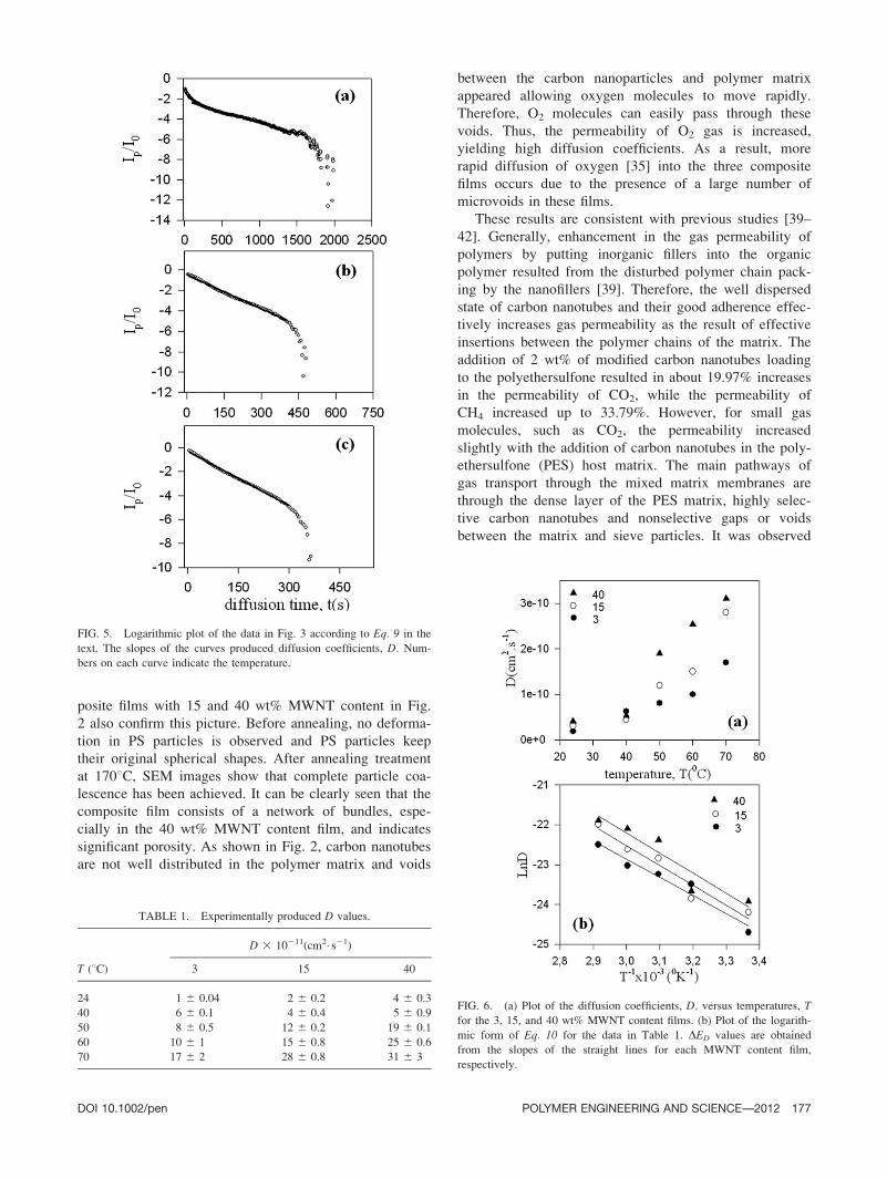
Figure 5 presents a logarithmic plot of the data in Fig.
3 (Ln(Ip/I0 2 A)) versus diffusion time for the 40 wt%
MWNT content film at three different temperatures. Eq. 9is fitted to these data by the linear least square method,
and the oxygen diffusion coefficients, D, at different tem-
peratures were produced from the slopes of the plots.
Similar fittings were done for the other MWNT content
films and D values were obtained for different tempera-
tures and collected in Table 1. The average D values were
determined from three or five measurements on different
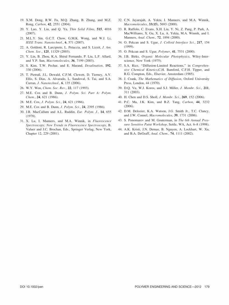
samples in each case and the standard deviations on Dvalues are also given in Table 1. D values versus temper-
ature are plotted for three MWNT content films in Fig.
6a. It is seen that D coefficients are strongly dependent
on both temperature and MWNT content in the film. It is
worthy to note that, as expected, D increases with
increases in temperature for all composite films. The
increasing of D values for the three films is seen clearly
especially above a certain temperature (508C). Increasesin temperature naturally increase the Brownian motion of
oxygen molecules giving them more chance to meet the P
molecules in the composite film.
On the other hand, increasing the MWNT content also
increases the D values. As seen in Fig. 1, the structure of
PS particles is spherical, while MWNT is long and cylin-
drical in structure. This difference in the formation of
defects or voids in the films enhances the diffusion rate of
oxygen along the film by increasing the surface area
against the oxygen molecules. This result is consistent
with microstructural analysis. SEM micrographs of com-
FIG. 3. The time behavior of the pyrene, P, and fluorescence intensity,
I, during oxygen diffusion into the 40 wt% MWNT content film at vari-
ous temperatures. Numbers on each curves indicate the temperature.
FIG. 4. Cartoon representation of oxygen diffusion into the film at ele-
vated time intervals.
176 POLYMER ENGINEERING AND SCIENCE—-2012 DOI 10.1002/pen
posite films with 15 and 40 wt% MWNT content in Fig.
2 also confirm this picture. Before annealing, no deforma-
tion in PS particles is observed and PS particles keep
their original spherical shapes. After annealing treatment
at 1708C, SEM images show that complete particle coa-
lescence has been achieved. It can be clearly seen that the
composite film consists of a network of bundles, espe-
cially in the 40 wt% MWNT content film, and indicates
significant porosity. As shown in Fig. 2, carbon nanotubes
are not well distributed in the polymer matrix and voids
between the carbon nanoparticles and polymer matrix
appeared allowing oxygen molecules to move rapidly.
Therefore, O2 molecules can easily pass through these
voids. Thus, the permeability of O2 gas is increased,
yielding high diffusion coefficients. As a result, more
rapid diffusion of oxygen [35] into the three composite
films occurs due to the presence of a large number of
microvoids in these films.
These results are consistent with previous studies [39–
42]. Generally, enhancement in the gas permeability of
polymers by putting inorganic fillers into the organic
polymer resulted from the disturbed polymer chain pack-
ing by the nanofillers [39]. Therefore, the well dispersed
state of carbon nanotubes and their good adherence effec-
tively increases gas permeability as the result of effective
insertions between the polymer chains of the matrix. The
addition of 2 wt% of modified carbon nanotubes loading
to the polyethersulfone resulted in about 19.97% increases
in the permeability of CO2, while the permeability of
CH4 increased up to 33.79%. However, for small gas
molecules, such as CO2, the permeability increased
slightly with the addition of carbon nanotubes in the poly-
ethersulfone (PES) host matrix. The main pathways of
gas transport through the mixed matrix membranes are
through the dense layer of the PES matrix, highly selec-
tive carbon nanotubes and nonselective gaps or voids
between the matrix and sieve particles. It was observed
FIG. 5. Logarithmic plot of the data in Fig. 3 according to Eq. 9 in the
text. The slopes of the curves produced diffusion coefficients, D. Num-
bers on each curve indicate the temperature.
FIG. 6. (a) Plot of the diffusion coefficients, D, versus temperatures, T
for the 3, 15, and 40 wt% MWNT content films. (b) Plot of the logarith-
mic form of Eq. 10 for the data in Table 1. DED values are obtained
from the slopes of the straight lines for each MWNT content film,
respectively.
TABLE 1. Experimentally produced D values.
D 3 10211(cm2� s21)
T (8C) 3 15 40
24 1 6 0.04 2 6 0.2 4 6 0.3
40 6 6 0.1 4 6 0.4 5 6 0.9
50 8 6 0.5 12 6 0.2 19 6 0.1
60 10 6 1 15 6 0.8 25 6 0.6
70 17 6 2 28 6 0.8 31 6 3
DOI 10.1002/pen POLYMER ENGINEERING AND SCIENCE—-2012 177

that the main factor affecting the increase of CH4 perme-
ability with the addition of carbon nanotubes into the
polymer host resulted from the extremely rapid diffusion
of gas molecules adsorbed inside the carbon nanotubes.
FESEM data also showed that the carbon nanotubes are
well dispersed in the polymer matrix and serve as chan-
nels to transport gas molecules [40–42]. It is known that
the addition of filler into polymer films above a critical
percentage creates voids [43, 44] in the polymer matrix.
Ponomarev and Gouterman [43] have reported that the
addition of high amounts of titanium oxide (TiO2) in
pressure sensitive (PSP) paints cause the presence of a
large fraction of microvoids inside the films. As a result,
air can diffuse very rapidly to the inside of the coating
through these voids.
Diffusion Energies
The diffusion of small molecules through membranes
can be described as a thermally activated process that
obeys Arrhenius behavior. The temperature dependence of
the diffusion coefficient, D, can be written as follows:
D ¼ D0 exp�DED
kBT
� �: (10)
Here kB is the Boltzmann constant, D0 is pre-exponen-
tial factor and DED is the energy as associated with the
oxygen diffusion. The activation energy was determined
from the logarithmic plots of the D coefficient against the
reciprocal of the absolute temperature. In Fig. 6b, Ln(D)was plotted versus 1000/T for the different clay fraction,
respectively. The value of the activation energy associated
with oxygen diffusion (DED) for different MWNT frac-
tions was calculated from the slope of these plots by fit-
ting the data in Fig. 6b to the Eq. 10 by a least square fit.
The results are given in Table 2, where DED increases
with increasing MWNT content. The energy need for oxy-
gen diffusion in the high MWNT medium is much higher
than in a low MWNT environment. Most probably, the
motion of O2 molecules is screened by the large number
of MWNT barriers during their journey in the high
MWNT content medium, in where O2 needs higher
energy to overcome this difficulty.
CONCLUSION
In this article, we examined the use PS/MWNT nano-
composites as fluorescent oxygen sensors and how oxygen
diffusion is affected by both MWNT content and tempera-
ture using the fluorescence technique. Fluorescence
experiments were carried out on composite films contain-
ing pyrene as a sensor dye. We monitored the change in
fluorescence intensity as oxygen was allowed to diffuse
into the film and the presence of oxygen was detected
through measurements of fluorescence quenching. Diffu-
sion experiments demonstrated that the quenching rate
during oxygen diffusion was completely consistent with
Fickian diffusion. The diffusion coefficients increased
drastically with both increases of MWNT content and also
of the temperature and this increase was explained via the
existence of large amounts of pores in composite films
which facilitate oxygen penetration into the structure.
We thus have proposed a simple, rapid, and practical
means to measure the diffusion of oxygen into PS/MWNT
composite films. This study illustrates that PS/MWNT
nanocomposites have useful properties as fluorescent oxy-
gen sensors, and a simple SSF technique can be used to
measure the diffusion coefficient of oxygen molecules
into these films quite accurately.
REFERENCES
1. R.A. Vara, K.D. Jandt, F.J. Kramer, and F.P. Giannelis,
Chem. Mater., 8, 2628 (1996).
2. M.W. Noh and D.C. Lee, Polym. Bull., 42, 619 (1999).
3. H.Z. Friedlander and C.R. Grink, J. Polym. Sci. Part B:Polym. Lett., 2, 475 (1964).
4. C. Kato, K. Kuroda, and H. Takahara, Clays Clay Miner.,29, 294 (1981).
5. J.G. Doh and I. Cho, Polym. Bull., 41, 511 (1998).
6. Y. Li, B. Zhao, S.B. Xie, and S. Zhang, Polym. Int., 52, 892(2003).
7. R.M. Barrer, in Diffusion in Polymers, J. Crank and G.S.
Park, Eds., Academic Press, New York, 164 (1968).
8. G. Gorrasi, M. Tortora, V. Vittoria, D. Kaempfer, and R.
Mulhaupt, Polymer, 44, 3679 (2003).
9. X. Lu, I. Manners, and M.A. Winnik, Macromolecules, 34,1917 (2001).
10. R.K. Bharadwaj, Macromolecules, 34, 9189 (2001).
11. P.G. Villaluenga, M. Khayet, M.A. Lopez-Manchado, J.L.
Valentin, B. Seoane, and J.I. Mengual, Eur. Polym. J.,43(4), 1132 (2007).
12. J. Kong, N.R. Franklin, C. Zhou, M.G. Chapline, S. Peng,
K. Cho, and H. Dai, Science, 28, 622 (2000).
13. P.G. Collins, K. Bradley, M. Ishigami, and A. Zettl, Science,287, 1801 (2000).
14. P. Calvert, Nature, 399, 210 (1999).
15. J.M. Planei, N. Coustel, B. Coq, V. Brotons, P.S. Kumbhar,
R. Dutartre, P. Geneste, P. Bernier, and P.M. Ajayan, J. Am.Chem. Soc., 116, 7935 (1999).
16. B.J. Hinds, N. Chopra, T. Rantell, R. Andrews, V. Gavalas,
and L.G. Bachas, Science, 303, 62 (2003).
17. J.K. Holt, H.G. Park, Y. Wang, M. Stadermann, A.B. Artyu-
khin, C.P. Grigoropoulos, A. Noy, and O. Bakajin, Science,312, 1034 (2006).
18. A.F. Ismail, T.D. Kusworo, and A. Mustafa, J. Membr. Sci.,319, 306 (2008).
TABLE 2. Experimentally produced diffusion energies.
MWNT (wt%) 3 15 40
DED (kJ mol21) 68 76 100
178 POLYMER ENGINEERING AND SCIENCE—-2012 DOI 10.1002/pen
19. X.M. Dong, R.W. Fu, M.Q. Zhang, B. Zhang, and M.Z.
Rong, Carbon, 42, 2551 (2004).
20. Y. Luo, Y. Liu, and Q. Yu, Thin Solid Films, 515, 4016
(2007).
21. M.L.Y. Sin, G.C.T. Chow, G.M.K. Wong, and W.J. Li,
IEEE Trans. Nanotechnol., 6, 571 (2007).
22. A. Goldoni, R. Larciprete, L. Petaccia, and S. Lizzit, J. Am.Chem. Soc., 125, 11329 (2003).
23. Y. Lin, B. Zhou, K.A. Shiral Fernando, P. Liu, L.F. Allard,
and Y.P. Sun, Macromolecules, 36, 7199 (2003).
24. S. Kim, T.W. Pechar, and E. Marand, Desalination, 192,330 (2006).
25. T. Pietraß, J.L. Dewald, C.F.M. Clewett, D. Tierney, A.V.
Ellis, S. Dias, A. Alvarado, L. Sandoval, S. Tai, and S.A.
Curran, J. Nanotechnol., 6, 135 (2006).
26. W.Y. Wen, Chem. Soc. Rev., 22, 117 (1993).
27. M.E. Cox and B. Dunn, J. Polym. Sci. Part A: Polym.Chem., 24, 621 (1986).
28. M.E. Cox, J. Polym. Sci., 24, 621 (1986).
29. M.E. Cox and B. Dunn, J. Polym. Sci., 24, 2395 (1986).
30. J.R. MacCallum and A.L. Ruddin, Eur. Polym. J., 14, 655(1978).
31. X. Lu, I. Manners, and M.A. Winnik, in FluorescenceSpectroscopy; New Trends in Fluorescence Spectroscopy, B.Valuer and J.C. Brochan, Eds., Springer Verlag, New York,
Chapter 12, 229 (2001).
32. C.N. Jayarajah, A. Yekta, I. Manners, and M.A. Winnik,
Macromolecules, 33(15), 5693 (2000).
33. R. Ruffolo, C. Evans, X.H. Liu, Y. Ni, Z. Pang, P. Park, A.
MacWilliams, X. Gu, X. Lu, A. Yekta, M.A. Winnik, and I.
Manners, Anal. Chem., 72, 1894 (2000).
34. O. Pekcan and S. Ugur, J. Colloid Interface Sci., 217, 154(1999).
35. O. Pekcan and S. Ugur, Polymer, 41, 7531 (2000).
36. J.B. Birks, Organic Molecular Photophysics, Wiley-Inter-
science, New York (1975).
37. S.A. Rice, ‘‘Diffusion-Limited Reactions,’’ in Comprehen-sive Chemical Kinetics,C.H. Bamford, C.F.H. Tipper, and
R.G. Compton, Eds., Elsevier, Amsterdam (1985).
38. J. Crank, The Mathematics of Diffusion, Oxford University
Press, London, 44 (1970).
39. D.Q. Vu, W.J. Koros, and S.J. Miller, J. Membr. Sci., 211,311 (2003).
40. H. Chen and D.S. Sholl, J. Membr. Sci., 269, 152 (2006).
41. P.C. Ma, J.K. Kim, and B.Z. Tang, Carbon, 44, 3232
(2006).
42. D.M. Delozier, K.A. Watson, J.G. Smith Jr., T.C. Clancy,
and J.W. Connel, Macromolecules, 39, 1731 (2006).
43. S. Ponomarev and M. Gouterman, in The 6th Annual Pres-sure Sensitive Paint Workshop, Settle, WA, Act. 6–8 (1998).
44. A.K. Kristi, J.N. Demas, B. Nguyen, A. Lockhart, W. Xu,
and B.A. DeGraff, Anal. Chem., 74, 1111 (2002).
DOI 10.1002/pen POLYMER ENGINEERING AND SCIENCE—-2012 179