teks: 130.6 (c)(11)(a) - kelley anne hutchinson -...
TRANSCRIPT
TEKS: 130.6 (c)(11)(a)

Define what a digestive system does;
List the five functions of the digestive
system;
Explain the difference between a Ruminant
and Non-Ruminant;
List the 6 major parts of the digestive
system;
Explain what each part of the digestive
system does;
List the compartments of a ruminant
stomach;

Label the parts of a ruminant stomach;
Write where the absorption sites are for
water and nutrients;
Label the parts of a small intestine;
Label the parts of a large intestine;
Label the digestive system of a chicken;
List the accessory digestive organs;
List the three digestive process actions;
List the mechanical actions of digestion;
Collect pictures of the parts of real
polygastric and monogastric digestive tracts;
Distinguish the parts of the digestive tracts
for each picture their classmates brought;
and
Rearrange an incorrect digestive tract to
make it correct.
Break food into smaller
particles so the body can
absorb and utilize the
nutrients

Ingesting Food
Grinding Food
Digesting Food
Absorbing Nutrients
Eliminating Body Waste

Ruminant animals have a 4-
chamber stomach (polygastric)
that includes a large rumen.
Has large digestive system for
utilizing bulky, forage-type feeds
Non-ruminant animals have a
single compartment stomach
(monogastric).

Cattle
Sheep
Goat
Deer

Swine
Chicken
Human

Mouth
Pharynx
Esophagus
Stomach
Small Intestine
Large Intestine
Digestive tract extends from the lips to the
anus.

Grasp food, grind food, and mix the food
with saliva
Accomplishes these tasks with the use of the
lips, tongue, teeth, cheeks, jaw, and salivary
glands
Horse lips are used to grasp food
Sheep & Cattle’s lips are to simply close the
mouth

Used by most animals to grasp food
Helps in the chewing process & formation of
boluses (cud)
In ruminants, large quantities of ingested food
are regurgitated as boluses (cud) so that it can
be re-chewed.
Papillae cover the top of the surface and
contain the taste buds
Papillae – finger-like projections

Responsible for cutting (incisors) and grinding
(premolars & molars) food.
New born animal develops milk teeth or baby
teeth
Milk teeth are replaced with permanent
teeth as the animal ages

Consist mostly of muscle that is lined with a
mucous membrane
Cheeks line up food with teeth

Common pathway for food & air
Food passes from mouth to pharynx
Pharyngeal muscles force food to the
esophagus
A muscular tube that connects the pharynx
to the stomach
The esophagus passes through the chest cavity
& connects with the stomach just after
passing through the diaphragm

Serves to digest food
Monogastric – 1 true stomach
Polygastric – 4 compartments

Single stomach compartment
“true” stomach
Location: just beyond the diaphragm on the
left side of the body
Has folds in the lining that creates gastric
pits
Glands secrete digestive fluids into the pits:
Hydrochloric acid
Pepsin
Rennin

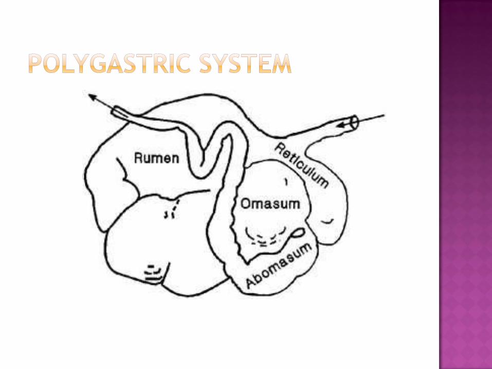
AKA: Ruminants
4 compartments:
Rumen
Reticulum
Omasum
Abomasum

Contain no glands
Soak food
Allow microbial digestion to take place

1st compartment
Fills most of the left side of abdomen
Thick muscular wall
2 sacs that contain many papillae
Dorsal sac
Ventral sac
Food passes into rumen, where it can be
regurgitated as cud.
Site of 60%-90% of digestion in ruminants

Becomes functional at 6-8wks of age in a calf
Makes up 80% of total stomach capacity when
animal reaches maturity

Forward most portion of the stomach
Inner surface has inward folds
Resembles a honeycomb shape

Capable of closing off the rumen and
reticulum
Food passes straight to the omasum
Seen in calves before the rumen is functional

3rd compartment
Contain muscular projections
Covered by mucous membrane
Contain many small papillae
Papillae in omasum is responsible for grinding
roughage
“True” stomach
Only glandular stomach of ruminants
Located under omasum
Lining and glands are the same as those in
the stomach of a non-ruminant
3 part tube
Duodenum
Jejunum
Ileum
Site of some digestion & absorption of
nutrients
Lined with mini villi, which absorb nutrients
Villi – tiny finger-like projections that protrude
from the lining of the intestinal wall


Larger tube of digestive tract
Consists of:
Cecum (a blind pouch)
Colon
Colon – site of water absorption
Excretes waste from the body
Little nutrients other than water are
absorbed here

Cecum is larger in horses and rabbits
Action of microorganims allows for digestion
of fiber (roughage)


Differs from other animals
Do not have teeth
Prehensile structure is the beak
Prehensile – body part adapted for grasping or
holding

Food passes from mouth to esophagus to the
crop
Crop – enlargement of the gullet
Crop temporarily stores food & softens it
Food passes to the glandular stomach
Food passes to the gizzard
Mouth Esophagus Crop Glandular stomach Gizzard

Crushes & grinds coarse feed
Aided by grit & gravel that has accumulated in
the gizzard during the bird’s life
Food then goes to the small intestine


Salivary glands
Pancreas
Liver
Gall bladder
Secretes saliva that softens food, which aids
in swallowing
Contains mostly water
Contains some enzymes that begins the
chemical breakdown
In pairs

Located under the ears
Ducts pass over the rear of the mandible to
near the middle of the cheek
Penetrate mucous membrane of the mouth &
secrete saliva

Mandibular – pertaining to the jaw of an
animal
Location: under & to the rear of the parotid
glands
Ducts pass in the middle of the mandibles &
open into the mouth under the tongue

Located: under mucous membrane around
the outer sides of the tongue
Empty into the floor of the mouth

Serous
Secretes clear, watery fluid
Parotid & Mandibular glands
Mucous
Secretes a thick, cloudy substance
Serves as a protective coating to the mucous
membranes
Mixed
Secretes both serous & mucous
Sublingual glands

An elongated, lobe-shaped organ
Location: beginning of small intestine,
behind the liver
Exocrine functions:
Largest function
Produces digestive juices
Pass through pancreatic duct & empty into the
beginning of the duodenum
Endocrine functions:
Produce insulin, which lowers blood sugar
Insulin goes directly into the bloodstream
A lobe-shaped organ
Location: behind the diaphragm on the right
side of the body
Receives blood from the hepatic artery
Purifies blood it receives from:
Stomach
Spleen
Pancreas
Intestines
Produces bile (waste)

Small, sac-like organ attached to the liver
Collects bile produced by liver
Secretes it into the duodenum
Horses are the ONLY domestic animal that
does not have a gall bladder
Mechanical actions
Mastication (chewing)
Deglutition (swallowing)
Regurgitation
Gastric & Intestinal motility
Defecation
Chemical actions
Actions of enzymes & glandular secretions
Microbial actions
Activities of bacteria & protozoa
Glucose level
Amount of feed in the stomach
Environmental temperature (hot or cold)
Appetite is controlled in the hypothalamus
gland

Digested nutrients pass through the walls of
the digestive tract into the blood
Small intestine – site for nutrient absorption
Villi are responsible for collecting & absorbing
nutrients
Very little nutrients are absorbed in the mouth,
esophagus, or stomach
Colon of large intestine – site for water
absorption
