taxonomy and phylogeny of pheretimoid earthworms
TRANSCRIPT

Instructions for use
Title Taxonomy and phylogeny of pheretimoid earthworms (Clitellata:Megascolecidae) from Mindanao and associatedislands, Philippines
Author(s) Aspe, Nonillon Mante
Citation 北海道大学. 博士(理学) 甲第12240号
Issue Date 2016-03-24
DOI 10.14943/doctoral.k12240
Doc URL http://hdl.handle.net/2115/64845
Type theses (doctoral)
File Information Nonillon_Mante_Aspe.pdf
Hokkaido University Collection of Scholarly and Academic Papers : HUSCAP

i
Taxonomy and phylogeny of pheretimoid earthworms
(Clitellata: Megascolecidae) from Mindanao and
associated islands, Philippines
(フィリピン・ミンダナオ島及び周辺島嶼産フトミミズ類(環帯類:フトミミ
ズ科)の分類と系統)
A Dissertation
Presented to the
Department of Natural History Sciences,
Graduate School of Science,
Hokkaido University,
Sapporo 060-0810, Japan
Nonillon Mante Aspe
2016

ii
ABSTRACT
This dissertation presents a total of 39 new pheretimoid species of the family
Megascolecidae, from Mindanao and associated islands, in the Philippines. Among
these, 29 are in the genus Pheretima, characterized by having nephridia on the
spermathecal ducts, having prominent dome-shaped copulatory bursae, and having a
pair of caeca originating in xxvii. Of these, 27 are in the subgenus Pheretima, while
two species are the first records of the subgenus Parapheretima in the Philippines.
Parapheretima is characterized by having secretory diverticula projecting from the
copulatory bursae, in contrast to members in the subgenus Pheretima, which do not
possess such organ. Also, three species are in Pithemera, characterized by having a
pair of caeca originating in or near xxii, three are in Polypheretima, characterized by
having no caeca, and three are in Amynthas, characterized by having no copulatory
bursae. The three latter genera also do not possess nephridia on the spermathecal ducts,
in contrast to Pheretima. With the new species described, there are now 80 known
Pheretima s. str. species in the Philippines, comprising 76% of the world’s Pheretima,
and there are now 14 species of Pithemera, comprising 47% of the world’s Pithemera.
These figures suggest that the Philippine archipelago may be the center of species
radiation for these groups. Also, there are now 16 species of Amynthas in the
Philippines representing less than 1% of the world’s Amynthas and there are now 10
species of Polypheretima in the Philippines representing 15% of the world’s
Polypheretima. The high diversity of the two latter genera in mainland Asia and
iii
Indonesia, respectively, strongly suggests that Indochina may be the center of species
radiation for these two genera. The known ranges of the Philippine species are
restricted to areas around the type localities. This pattern indicates a remarkable
degree of endemicity, both among local areas, among islands in the Philippines, and in
the Philippines as a whole, and suggesting that many species remain to be detected in
the Philippines.
A molecular phylogenetic study was done in attempt to infer phylogenetic
relationships among the pheretimoid species in Mindanao and associated islands. Gene
markers used include the mitochondrial cytochrome c oxidase subunit I (COI) and 16S
rRNA, and the nuclear 28S rRNA and protein-coding histone H3 genes. Despite
having limited taxa and limited genes included in the analyses, the combined data set
generated a phylogeny more or less consistent with morphology-based expectation.
Results show that taxonomic assignment of the genus Amynthas and the subgenus
Parapheretima do not reflect phylogeny. The species grouping in Pheretima based on
the location of spermathecae is partially reflected in the pheretimoid phylogeny. Also,
results show that loss of spermatheca or fusion of two spermathecae into one can
occur in pheretimoid evolution. In general, several of the nodes of the tree based on
combined data set have support values that are very weak and have formed polytomies,
which is most likely due to insufficient data. The results could have improved if more
data were available. Further molecular work including more taxa is needed to be able
to establish a more robust system of classification of the pheretimoid species and come
up with a better-resolved phylogeny.

iv
TABLE OF CONTENTS
TITLE PAGE ………………………………………………………..…… i
ABSTRACT …………………………………………………………………….. ii
TABLE OF CONTENTS …………………………………………………….. iv
ACKNOWLEDGEMENTS …………………………………………………….. vii
CHAPTER
1 General Introduction …………………….……….…..…. 1
1.1 Biodiversity in the Philippines under threat …………….. 1
1.2 General biology of earthworms ……………………. 2
1.3 Ecology of earthworms ………………………….… 5
1.4 Systematic accounts of the pheretimoid earthworms ……. 9
1.5 Status of the diversity of Philippine earthworms ……..…. 12
2 New species of Pheretima (Clitellata: Megascolecidae) from
the Mt. Malindang Range, Mindanao Island, Philippines …… 15
2.1 Introduction …………………………………………….. 15
2.2 Material and methods …………………………………….. 16
2.3 Results ………………………………………….…. 21
Pheretima maculodorsalis Aspe & James, 2014….. 23
Pheretima tigris Aspe & James, 2014 ………….… 27
Pheretima immanis Aspe & James, 2014 ................. 31
Pheretima lago Aspe & James, 2014 ………….….. 34
Pheretima nunezae Aspe & James, 2014 ….……… 38
Pheretima boniaoi Aspe & James, 2014 ……..…… 41
Pheretima malindangensis Aspe & James, 2014 …. 44
Pheretima misamisensis Aspe & James, 2014 ……. 47
Pheretima wati Aspe & James, 2014 …………….. 50
Pheretima longiprostata Aspe & James, 2014 ……. 54
Pheretima nolani Aspe & James, 2014 …………….57
Pheretima longigula Aspe & James, 2014 …………61
Pheretima adevai Aspe & James, 2014 ………….... 64
Pheretima lluchi Aspe & James, 2014 …………….. 68
Pheretima potonganensis Aspe & James, 2014 …… 72
Pheretima vergrandis Aspe & James, 2014 ………. 76
Pheretima concepcionensis Aspe & James, 2014 …. 79
Pheretima subanensis Aspe & James, 2014 ………. 82
2.4 Discussion ……………………………………………. 87

v
3 New Polypheretima and Pithemera (Clitellata: Megascolecidae)
species from the Mt. Malindang Range, Mindanao Island,
Philippines …………………………………………………… 93
3.1 Introduction …………………………………………… 93
3.2 Material and methods …………………………………… 94
3.3 Results …………………………………..……….. 96
Polypheretima mindanaoensis Aspe & James, 2015 98
Pithemera malindangensis Aspe & James, 2015 … 103
Pithemera duminagati Aspe & James, 2015 …….. 106
Pithemera donvictorianoi Aspe & James, 2015 …. 109
3.4 Discussion …………………………………………….. 111
4 New species of Pheretima, Amynthas, Polypheretima, and Pithemera
(Clitellata: Megascolecidae) from Mindanao and associated islands,
Philippines …………………………………………………...... 114
4.1 Introduction ……………………………………………. 114
4.2 Material and methods …………………………………….. 115
4.3 Results …………………………………………….. 118
Pheretima acia n. sp. …………………………….. 120
Pheretima dinagatensis n. sp. ……………………. 123
Pheretima enormis n. sp. ……………………. 126
Pheretima hamiguitanensis n. sp. ………………… 129
Pheretima lantapanensis n. sp. …………….. 131
Pheretima timpoongensis n. sp. ……………. 134
Pheretima camiguinensis n. sp. ……………. 138
Pheretima sibucalensis n. sp. ……………………. 141
Pheretima apoensis n. sp. ……………………. 144
P. (Parapheretima) pandanensis n. sp. ……. 148
P. (Parapheretima) boaensis n. sp. ……………. 151
Amynthas dinagatensis n. sp. ……………………. 154
Amynthas cagdianaoensis n. sp. ……………. 158
Amynthas talaandigensis n. sp. ……………. 160
Polypheretima bukidnonensis n. sp. ……………. 164
Polypheretima zamboangensis n. sp. ……………. 167
Pithemera nolani n. sp. ……………………. 171
4.4 Discussion …………………………………………….. 173
5 A molecular phylogenetic study of pheretimoid species (Clitellata:
Megascolecidae) in Mindanao and associated islands,
Philippines …………………………………………………….. 181
5.1 Introduction …………………………………………….. 181
5.2 Material and methods …………………………………….. 184
5.3 Results ……………………………………………. 187
5.4 Discussion ……………………………………………. 189
SUMMARY, CONCLUSION, AND RECOMMENDATIONS ……………. 195

vi
REFERENCES ……………………………………………………………. 198
TABLES ………………………………………………………………….… 219
FIGURES …………………………………………………………….……… 239
APPENDIX ……………………………………………………………………. 260
CURRICULUM VITAE ……………………………………………………. 263

vii
ACKNOWLEDGEMENTS
It is with pride, great honor and gratitude that I mention the following entities
and individuals who have shared ideas, support, and contributed in various ways for
the completion of this dissertation. Their suggestion, comments, and inspiring words
play major roles in accomplishing the task.
First of all, to the committee members of the Hokkaido University Special
Grant Program, for giving me the privilege to pursue doctoral degree in this
prestigious University, and for the financial provision until the completion of my
doctoral course;
To Assoc. Prof. Hiroshi Kajihara and Prof. Matthew Dick, my supervisors, for
their invaluable suggestions, comments, guidance, moral and financial support;
To Assoc. Prof. Sam James, my mentor for earthworm systematics, for sharing
his expertise and for reviewing the papers related to this dissertation;
To Prof. Takeo Horiguchi and Prof. Ryuichi Masuda, the esteemed committee
members of the dissertation panel, for their time, effort, and their invaluable comments
and suggestions for the improvement of this dissertation;
To Asst. Prof. Toru Katoh, Lecturer Keiichi Kakui and Dr. Shimpei F. Hiruta
for their assistance during the analyses of data and for their invaluable comments and
suggestions;

viii
To my labmates at Biodiversity I, especially to Takumi Onishi, for their
assistance when I was doing the molecular procedures, and for their friendship;
To my parents, especially my mom, for their love, moral support, prayers, and
words of encouragement, even when we are many miles apart;
To Jennifer, my beautiful wife, and my three lovely kids, Amanda Praise,
Alexander Luis and Adrian Kyle, for their love, care, understanding, and moral
support;
And above all, to the Lord Jesus Christ, who has always been my guiding light
since the very beginning, for his wisdom, guidance, provision, protection and strength.
All the glory belongs to Him alone!
“Worthy are you, our Lord and God, to receive glory and honor and power, for
you created all things, and by your will they existed and were created.”
-Revelation 4:11

1
CHAPTER 1
General Introduction
1.1 Biodiversity in the Philippines under threat
The Philippines is considered to be one of the megadiverse countries in the
world, being a home to one of the widest arrays of species in the world. With around
7,100 islands, the country hosts more than 52,177 described floral and faunal species
of which more than half is found nowhere else in the world (CI et al. 2006). However,
the country is also identified as one of the biodiversity hotspots in the world having its
rich ecosystems being under serious threat. Species are being lost at an incredible rate
of up to 10,000 a year due to human activities (Mallari et al. 2001). In the early 1900’s,
huge tracts of primary rainforests covered most of the Philippine landscape. Terrestrial
ecosystems flourished, with large stands of primary forests, and timberlands
dominating the land. Sadly, extremely high population growth has demanded
increasingly damaging and extractive techniques in order to support human needs. The
threats to natural habitats include illegal logging, mining, quarrying, conversion of
forest ecosystems to agricultural lands, urban development, wildlife poaching, and
illegal trade. Today, only a fraction of the original landscapes that blanketed the
country remains. As the loss of natural habitats is at a very alarming rate, there is an
urgent need to assess the biological resources, of which the information is essential in
formulating conservation and management strategies, before it is too late. This study is

2
primarily undertaken to provide significant information on the diversity of earthworms
in Mindanao and associated islands.
1.2 General biology of earthworms
Earthworms are tube-shaped, segmented animals that have no internal skeleton
or exoskeleton. They maintain their structure with fluid-filled coelom chambers that
function as a hydrostatic skeleton. When observing an earthworm move, one will most
likely see it move forward, with its mouth and prostomium in the anterior end.
Although it has no eyes, it possesses cells that are sensitive to light and it can also feel
vibrations created by movements in its surroundings. As it works its way forward,
successive peristaltic waves of thickening and thinning (7 to10 per minute) pass down
the body. At each place where the body bulges out at a given moment, the setae are
extended and grip the burrow walls. Setae, which are bristles surrounding each
segment, push against the ground with each contraction and help the animal move
(Lorus & Milne 1992). Secretions from the mucous glands of the epidermis keeps the
cuticle moist and lubricates the worm's body to ease passage through the burrows. The
mucus-covered skin helps bind soil particles together and prevents the walls of the
burrow from collapsing.
Earthworms are very sensitive to touch, and the pattern of their reaction vary
with both the species and circumstances. If an earthworm is touched, it withdraws
back into its burrow, sometimes very quickly, and does not emerge again for some
time. If a worm is grasped while partly out of its burrow it will actively resist and
attempt to pull its body out by extending its posterior setae into the burrow wall and

3
expanding its posterior segments, so as to grip the walls of the burrow and completely
fill its exit. Other species react vigorously to tactile stimuli. Some, if pricked sharply
or handled, produce a series of lashing movements from side to side. Many East Asian
species exhibit serpentine motion, and may thrash violently to escape danger. Some
often eject coelomic fluid from the dorsal pores when touched and other species can
eject this fluid to a considerable height. The least common response is autotomy, or
breaking off posterior segments to escape danger. Stimuli applied to the anterior end
does not cause reaction in the same way (Edwards & Lofty 1977).
The earthworm’s internal body plan is relatively simple. It has a central and a
peripheral nervous system. The central nervous system consists of two ganglia above
the mouth, one on either side, connected to a nerve cord running back along its length
to motor neurons and sensory cells in each segment. Large numbers of chemoreceptors
are concentrated near its mouth. Its digestive system is mostly composed of the
intestine, which stretches along the body, starting from the first few segments of the
anterior end of the body towards the anus. Earthworms have no specialized respiratory
organs. Oxygen and carbon dioxide diffuse through the cuticle and epidermal tissues
into the blood, which contains hemoglobin. It has a double transport system composed
of coelomic fluid that moves within the fluid-filled coelom and a simple, closed blood
circulatory system. The morphological characters used to discriminate earthworm
species include color, size, the length and location of the clitellum, the presence or
absence of genital markings, the number of setae per segment, the distance between
spermathecal pores, the distance between male pores, presence or absence of dorsal or
ventral setal gaps, the number, location, size and shape of spermathecae, the

4
arrangement of septa, the origin of gizzard, intestine and caeca, the size and shape of
the prostate glands, the presence or absence of copulatory bursae, and the presence or
absence of penes. Fig. 1.1 shows the external and internal morphology of an
earthworm showing some of the diagnostic morphological characters.
Earthworms are hermaphrodites, having both male and female reproductive
organs. Most species reproduce by cross-fertilization although many species can also
produce cocoons parthenogenetically. However, most parthenogenetic species are only
parthenogenetic because they lost the male organ functions (Gates 1972). Most species
mate periodically throughout the year, except when conditions are unsuitable or they
are aestivating. Methods of copulation are not identical for all species; in Lumbricus
terrestris, two worms which are attracted to each other by thick and slimy glandular
secretions, lie with the ventral parts of their bodies together, and their heads pointed in
opposite directions. They come into close contact in the region of the spermathecal
openings and where the clitellar region of one worm touches the surface of the other.
Sperm cells are released from the male pores and are either transported towards the
sperm receptacles or the spermathecal pores of the partner, or are directly inserted into
the receptacles (Gates 1972). In Pheretima species that have three or four pairs of
spermathecae, the male pores first come into contact with the hindmost pair of
spermathecal apertures and discharge seminal fluid and prostatic fluid into them. Each
worm then moves backwards, and the seminal fluid is discharged into the next pair of
spermathecae, until all have been ‘charged’ (Tembe & Dubash 1961). While
copulating, the worms do not respond readily to external stimuli such as touch and
light. After copulation, which may take as long as an hour, the worms separate. When

5
the worms already have parted ways, a mucous ring is secreted throughout the
clitellum slips off the front of the worm, closing at both ends to form the cocoon
which is roughly lemon-shaped. The cocoon contains a nutritive albuminous fluid,
produced by the gland cells of the clitellum, the ova, and the spermatozoa. Cocoons
continue to be formed until all the stored seminal fluid has been used up. Fertilization
is external in the cocoon. One or two worms hatch from this cocoon, in two or three
weeks. Worms that have been mated may form new egg capsules every few days
(Lorus & Milne 1992).
1.3 Ecology of earthworms
Earthworms are naturally found living in soil, feeding on live and dead organic
matter. However, they may also be arboreal and can be found inside tree barks, ferns,
mosses, and the insides of rotten logs several meters above ground. Although they are
most numerous in the top 15 cm, some species work in the subsoil, bringing mineral-
rich soil from below to the surface. The leaf litter they digest contain nutrients such as
calcium, nitrogen, potassium and phosphorus. They also consume organic minerals
and nutrients from dead animals and animal feces. Their excrement, called castings, is
deposited both on the surface and within the soil and is rich in nutrients, providing
food for other animals and microorganisms. This organic material is then further
broken down by microorganisms of the soil, releasing nutrients in a form available for
absorption by plants. Earthworms may be able to acquire parasites such as protozoa,
platyhelminthes and nematodes through ingestion and these parasites can be found in

6
many parts of the earthworms’ bodies like the blood system, testes, in the inner
surface of the epidermis, or in the cocoons (Lee 1985).
Earthworms are perhaps the most important soil organisms in terms of their
influence on organic matter breakdown, soil structure development, and nutrient
cycling. Their tunneling and burrowing aerates the soil, which not only help bring
oxygen down into the soil, but their tunnels also allow rainwater carrying organic and
inorganic nutrients down deep into the soil where the roots lie. The roots then take up
the water and the minerals and recycle them back to the herbaceous plants and woody
trees. The tunneling of the earthworms also provides an access to deeper soil levels for
the numerous smaller organisms that contribute to the health of the soil (Lee 1985).
Also, as the body of the earthworm is composed of 70% protein, it is preyed upon by
birds and by burrowing animals like moles, forest rats, pigs, and shrews. In addition,
researchers have found that bacteria living in the guts of worms detoxify many
hazardous chemicals such as hexachlorocyclohexane (Applehof 2000). Because of
these significant features, earthworms are being utilized for their ecological and
economic importance, through vermiculture (Dominguez & Edwards 2010).
There are three primary ecological categories of earthworms: endogeic, anecic,
and epigeic. Endogeic worms live in the upper layers of soil but very rarely come to
the surface. Their complex horizontal burrow systems are not permanent but they are
very important for aerating soil and allowing moisture and nutrients to move through
the soil. Many endogeic worms live in the rhizosphere, the area immediately around
plant roots, and they help with the exchange of nutrients there. These worms are
medium-sized and pale in color. Anecic worms build permanent, deep vertical

7
burrows in the soil, which can be up to 2 m deep, and rise to the surface at night to
search for food. Anecic worms often leave little mounds of castings alongside or atop
the opening of their burrows. They have long lives, which can be up to six years, and
they mature and reproduce slowly. Anecic worms are usually large, ranging from 15
cm up to 1 m, and have dark color on the anterior end. Epigeic worms are surface
dwellers and feed only on decaying organic material, not soil. They do not burrow
very often but rather live in loose organic litter or very loose topsoil rich in organic
matter. They may not survive in most garden soils unless there is a good layer of
organic matter on top. They are usually small and are reddish brown in color, but they
may also be striped. They are better able to withstand temperature and moisture
fluctuations than other worms as they commonly inhabit the soil surface. In ideal
environmental conditions, they can reproduce at a high rate and create castings that are
many times higher in nutrients than the material they originally consumed. Because of
these, they are ideal for vermiculture and vermicomposting (Stewart 2004; Dominguez
& Edwards 2010). These ecological categories make it possible to judge earthworm
ecological functions by simple visual cues. All three may be present in a habitat, or
one or two may be missing. For example, epigeic and anecic worms can be missing
from grasslands or agricultural soils where there is very little surface litter (Edwards &
Lofty 1977).
Most earthworms favor neutral pH but other earthworms are also either acid
tolerant or acid intolerant. Edwards & Lofty (1977) studied populations of earthworms
in plots that had a range of pH from 3.7 to 7.5. They reported that the species
Lumbricus terrestris became increasingly numerous as the pH increased but most of

8
the other species present tended to have an optimum pH range of 5.0 to 6.0.
Earthworms can help change acid or alkaline soils toward a more neutral pH by
utilizing and excreting excess calcium carbonate. In terms of soil temperature,
Bhattacharjee & Chaudhuri (1999) studied the cocoon production, morphology,
hatching pattern and fecundity of tropical earthworm species in temperature ranging
within 28 to 32ºC under laboratory conditions. Their results show that there is high
rate of cocoon production, short development time with high hatching success, as well
as continuous breeding strategies in the epigeic species Perionyx excavatus,
Dichogaster modiglianii and the top soil endogeic species, Pontoscolex corethrurus,
as temperature is increased. However, there are some other species that prefer to
inhabit environments with lower temperature.
Soils that are poor in organic matter do not usually support large numbers of
earthworms. Decaying leaves in the forests are a great source of organic matter that
usually favors earthworm multiplication. Li et al. (1999) studied the responses of soil
carbon and nitrogen and crop yield to earthworm activity. It was concluded that
earthworm was very important in promoting nitrogen recycling of crop residues and
plant productivity, and in keeping the balance of soil carbon pool as well.
Earthworms, having a body weight that constitutes 75 to 90% water, live in
habitats where moisture is favorable. They cannot tolerate heat and sun and so
endogeic worms come up to the surface only at night. Nevertheless, they have
considerable ability to survive adverse moisture conditions, either by moving to a
more suitable area or by aestivating. If they cannot avoid dry soil they can survive the
loss of a large part of the total water content of their bodies. Lumbricus terrestris can

9
lose 70% of its total body water and still survive (Grant 1955). A few small species of
earthworms can survive in deserts and semi-deserts. It seems that although good
quality soils are favorable for most of the earthworms rather than poor ones, they can
survive in many different kinds of soils provided, there is adequate food and moisture
(Kubiena 1955).
1.4 Systematic accounts of the pheretimoid earthworms
It is a common knowledge that earthworms belong to Phylum Annelida, the
segmented worms. However, the lower level taxa are in a state of flux as different
taxonomists proposed various taxonomic classifications giving weight to different sets
of morphological characters. The debates on taxonomy are sometimes in an
evolutionary context, but rarely with any explicit analysis of character data, resulting
in intuitive conclusions (James & Davidson 2012). For example, some taxonomists
consider earthworms to be of Class Oligochaeta while others would consider it to be
of Class Clitellata and consider Oligochaeta as its Order. Likewise, some taxonomists
create additional taxonomic levels such as “Superclass” or “Subclass” to
accommodate taxonomic reassignment but these may not be acknowledged by others.
Also, different authors recognize different numbers of families in earthworms: 15 by
Jamieson (1988), 21 by Reynolds & Cook (1993), and 18 by Blakemore (2000). DNA
sequences, which provide characters independent of morphology, have become widely
used to reconstruct relationships among earthworms and test the reliability of
morphological characters used in taxonomy. Phylogenetic analyses based on
molecular data may either support or refute taxonomic classifications based on

10
morphological data. In the latter case, the morphological character/s a taxonomist used
to assign such taxonomic groupings may be found to be homoplasious and not
homologous. Only monophyletic grouping, which is based on homologous characters,
is therefore considered to infer true phylogeny. For the time being, due to the state of
flux in different taxonomic levels in earthworms, taxonomists using molecular data to
infer phylogenetic relationships prefer to brush aside the hierarchical system of
classification (Linnean system). Below is the summary of the systematic accounts
leading to the pheretimoid species (Pheretima s. lat.) of Megascolecidae, which is the
focus of this dissertation.
Members of Clitellata Michaelsen, 1919 are defined by the clitellum, which is
located partly behind the female pores and secretes a cocoon in which the eggs are laid
are annelids. These include the oligochaetes (earthworms and their allies),
branchiobdellids (ectoparasites of freshwater crayfish) and leeches. Clitellata was
confirmed to be monophyletic based on molecular analyses (Martin et al. 2000; Martin
2001; Siddall & Burreson 1998). However, with regard to the position of the Clitellata
within the Annelida, molecular analysis has indicated that clitellates form a clade
within the Polychaeta and that polychaetes are a paraphyletic or polyphyletic group
(Kojima 1998; McHugh 2000; Martin 2001).Oligochaeta, defined by having few setae
on their outer body surfaces and refers to many aquatic and terrestrial worms,
including the earthworms, was suspected and confirmed to be paraphyletic with
leeches and/or branchiobdellids lying within the oligochaete clade based on both
morphological and molecular data (Jamieson et al. 1987; Jamieson 1988; Siddall &
Burreson 1998; Siddall et al. 2001). The inclusion of leeches and branchiobdellids
11
within the Oligochaeta would thus render the name Oligochaeta synonymous with
Clitellata (Siddall et al. 2001; Jamieson et al. 2002). Earthworms, having multilayered
clitella, were found to form a single clade and thus Jamieson (1988) assigned them to
Crassiclitellata. The acquisition of a multilayered clitellum was deduced to be a
monophyletic event (Jamieson et al. 2002; James & Davidson 2012). The monophyly
of Clitellata was further supported in recent studies with Tubificidae/Echiura/
Capitellidae as sister taxa in Struck et al. (2011), with Terebelliformia/Arenicolidae as
sister taxa in Struck et al. (2015) and Weigert et al. (2014), and with Terebelliformia/
Arenicolidae/Maldanidae as sister taxa in Weigert et al. (2015).
Sims (1980) proposed superfamilies for earthworms based on the ovarian
characters first recognized by Gates (1976). These include Criodriloidea,
Lumbricoidea, Biwadriloidea, Glossoscolecoidea, and Megascolecoidea. James &
Davidson’s (2012) molecular analyses based on 28S, 18S, and 16S gene sequences,
found that Criodriloidea is nested within Lumbricoidea, and that Biwadriloidea and
Glossoscolecoidea are not supported and have unresolved positions. Megascolecoidea,
defined by having a large, fan to rosette-shaped ovary and prostate glands associated
with the male pores, on the other hand is a monophyletic group. James and Davidson
(2012) proposed a limited Megascolecoidea, which only include the families of
Megascolecidae, Ocnerodrilidae, and Acanthodrilidae as opposed to Sims (1980) and
Omodeo (2000), who included Eudrlidae and Octochaetidae in the superfamily.
Among the families of Megascolecoidea, Megascolecidae is the most speciose,
with 55 genera especially concentrated around the Asia-Pacific but also distributed in
some parts of North and South America and in Madagascar (Blakemore 2000; BOLD

12
Systems). Megascolecidae includes members with diverse prostate gland types
(generally racemose structure), whose ducts generally are joined by the sperm ducts in
combined male and prostatic pore(s) on segment 18 or nearby and the spermathecal
pores open into some or all of the intersegmental furrows from 4/5 to 9/10 (rarely
intra-segmentally) (Sims & Easton 1979; Blakemore 2000; Jamieson & Ferraguti
2006; James & Davidson 2012). Pheretima s. lat. used to be the largest genus of
Megascolecidae comprising more than 1,400 names, which include numerous
synonyms, invalid names, and lapsae updated from Sims & Easton (1972). Sims and
Easton (1972) reallocated species in the group into 'convenient' species groups
comprising 10 genera using computer-based phenetic analyses. Blakemore (2007)
reported an approximate of 930 valid species from the 1,400 names and updated Sims
and Easton’s list of the pheretimoid species to a total of 13 genera belonging to the
group. The members are defined by having a perichaetine setal arrangement around
the segmental equators except the first and the last segment of the body. They have a
short clitellum xiv–xvi. Prostate glands have racemose structure and there is a pair of
caeca on the intestine in a single segment in most species. The testes is contained
within testes sacs. Also, the gizzard originates in viii and the excretory system is
meronephridial (Sims & Easton 1972).
1.5 Status of the diversity of Philippine earthworms
Research on earthworm taxonomy in the Philippines began way back in the
late 1800’s but surveys on earthworm biodiversity in the Philippines has been given
more attention than ever since the issue on the destruction by the earthworms of the

13
Banaue rice terraces, a National Cultural Treasure of the Philippines, was reported.
Pheretima species were identified to be responsible for the soil erosion (Joshi et al.
1999). Over the last decade, the number of Philippine species dramatically increased
from less than 10 known species to an estimate of 200 native pheretimoid species
representing eight genera (Flores 2008; Aspe & James 2014). This was made possible
by the efforts of the Philippine Terrestrial Annelids and Gastropods Survey project
headed by Dr. Samuel James and funded by the National Science Foundation in the
USA. The eight genera that compose the Philippine native species include Pheretima,
Isarogoscolex, Pithemera, Amynthas, Pleionogaster, Polypheretima,
Dendropheretima, and Archipheretima. Studies show that the Philippine archipelago
may be the center of species radiation for Pheretima while Indochina may be the
center of species radiation for Amynthas (Aspe & James 2016).
Most of the surveys on earthworms in the Philippines focused on Luzon Island
until the last decade when collection on Mindanao and associated islands started. I was
designated as the lead Field Biologist for the Mindanao Phase during this earthworm
biodiversity project. This dissertation reports a total of 39 new pheretimoid species of
Mindanao: Chapter 2 describes the morphology of 18 new species of Pheretima from
Mt. Malindang, one of the priority sites for conservation in the Philippines, located at
the base of Zamboanga peninsula in western Mindanao (Mallari et al. 2001; CI et al.
2006); Chapter 3 describes one species of Polypheretima and three species Pithemera
from Mt. Malindang; and Chapter 4 describes 11 new species of Pheretima (two are of
subgenus Parapheretima), three new species of Amynthas, two new species of
Polypheretima, and one new species of Pithemera from other areas around Mindanao

14
and associated islands. The species described in Chapters 2, 3 and 4 were published in
Zootaxa, Journal of Natural History, and Zoological Studies, respectively.
It is not reliable to infer evolutionary relationships among the pheretimoid
species by simply using morphological data. Thus, molecular data is essential to
reconstruct relationships among earthworms and to test the reliability of
morphological characters that are used in taxonomy. James (2005a) conducted a
preliminary molecular phylogeny in this group, which include the morphospecies from
Luzon Island, using the data of mitochodrial 16S rDNA and nuclear 28S rDNA.
Although his analyses of the combined data produced a tree topology that is more or
less consistent with morphological data, all of the basal nodes were weakly supported.
Chapter 5 of this thesis tackles a molecular phylogenetic study of the pheretimoid
species of Mindanao and associated islands, using the mitochondrial 16S rDNA and
COI, and the nuclear 28S rDNA and histone H3 genes, in attempt to produce a better
resolved phylogeny among the pheretimoid species (Aspe et al., in review).

15
CHAPTER 2
New species of Pheretima (Clitellata: Megascolecidae) from the Mt. Malindang
Range, Mindanao Island, Philippines
2.1 Introduction
Until recently, knowledge of the native earthworm fauna of the Philippines
was very limited. Non-specialist biologists in the Philippines erroneously identified all
earthworms there as Lumbricus terrestris Linnaeus, 1758, a species common to North
America and Europe but not detected in recent studies in the Philippines. Organized
research on earthworm diversity in the Philippines began after Lawrence Heaney and
collaborators discovered that the Isarog shrew-rat (Rhynchomys isarogensis Musser &
Freeman 1981) and Chrotomys gonzalesi Rickart & Heaney, 1991 feed exclusively on
earthworms. The desire of the mammalogists to identify the worms the rat feeds on led
to the discovery of 10 new species collected in 1993, all belonging to perichaetine
genera in the Pheretima complex (Sims & Easton 1972) in the family Megascolecidae
(James 2004).
Pheretima Kinberg, 1867, a Southeast Asian group with a range extending
from northern Australia to Myanmar and northward to Korea, became the largest
genus of earthworms in the Megascolescidae sensu Gates (1959). Using computer-
based phenetic analyses, Sims and Easton (1972) and Easton (1979) reallocated
species in Pheretima s. lat. (pheretimoid species) into ‘convenient’ species groups
comprising 10 genera (Amynthas, Archipheretima, Pheretima, Planapheretima,
Metapheretima, Pithemera, Ephemitra, Metaphire, Polypheretima and Pleionogaster).

16
Blakemore (2007) estimated that among more than 1400 nominal taxa of pheretimoid
earthworms (which include numerous synonyms, invalid names, and lapsus) there are
roughly 930 valid species and subspecies in Pheretima auct. He acknowledged around
40 valid species of Pheretima s. str., with the distributional range restricted to the
Indo-Australian archipelago, Sumatra, and the Philippines.
As the result of taxonomic studies in the last decade, around 200 species of
native earthworms representing eight genera are now known from the Philippines
(Blakemore 2007; Flores 2008; James 2004, 2005b, 2006, 2009; James et al. 2004;
Hong & James 2004, 2008a–c, 2009, 2010, 2011a, b). Among these are 46 new
species of Pheretima sensu Sims & Easton (1972), reported in studies conducted
mostly in mountainous forested areas on Luzon Island (James et al. 2004; Hong &
James 2008a–c, 2009, 2010, 2011a, b), but also in one study in the Mt. Kitanglad
Range, Mindanao Island (James 2004). The eighteen new species from Mt. Malindang
reported in this chapter were described in Aspe & James (2014).
2.2 Material and methods
Study area. The Malindang Range is a large volcanic complex at the base of
the Zamboanga Peninsula, western Mindanao Island, Philippines (Fig. 2.1). The
forests in this mountain range are the only remaining natural forests representing the
tropical Zamboanga biogeographic zone (Mallari et al. 2001), one of 15 such zones in
the Philippines. Reaching 2425 m asl., Mt. Malindang (~ 8º18’N, 123º39’E) is the
highest mountain on the Zamboanga Peninsula, covering 53,262 ha and consisting of

17
46% lower montane and upper montane (mossy) forest, 25% bushland, 6% denuded
land, and 23% cultivated land (Mallari et al. 2001).
Earthworms were collected in primary forest, disturbed forest, grassland, and
agricultural habitats at various elevations in four geographically separate barangays (=
precincts): Barangay (Brgy) Lake Duminagat in the municipality of Don Victoriano;
Brgy Sibucal and Brgy Toliyok in Oroquieta City; and Brgy Small Potongan in the
municipality of Concepcion. The terrain in the forested areas was very rugged, with
steep grades and many cliff faces, making access extremely difficult. Surrounded by
humid primary forest in Brgy Lake Duminagat is a crater lake about 9 ha in area called
Lake Duminagat. The primary forest in this barangay had never been logged and
remained largely undisturbed by human activities. The vegetation was dense and lush;
trees were thickly covered with moss, ferns, and lichens, and ground was thickly
covered with moss, roots, and leaf litter. The dominant trees included Viburnum sp.,
Lithocarpus, Caldeluvia, Pometia, Macaranga dipterocarpifolia, and Polyosma
philippinensis.
Trees in the disturbed forest, regrown after deforestation by humans, were
dominated by dipterocarps (those identified included Lithocarpus mindanaensis, L.
philippinensis, and Polyosma philippinensis) and tended to be more closely spaced
than in the primary forest, and to have more undergrowth (saplings, shrubs, and tree
ferns). The ground was covered with thick leaf litter, roots, bryophytes, and lichens.
Sampling. The earthworms described in this paper were collected in an
ecological and distributional study at Mt. Malindang during the periods 9–15 Oct.

18
2003 and 18–25 Feb. 2004. Sampling methods and the locations of sampling sites
were previously reported in Aspe (2006) and Aspe et al. (2009). A summary is as
follows. In each barangay, six scattered plots 20 m x 20 m in extent were established,
with an average distance of 75 m between plots. On each plot, 0.075 m3 (0.5 m x 0.5
m square x 0.3 m deep) of soil was examined from each of 10 quadrats in randomly
selected spots. This gave a total of 4.5 m3 of soil sampled at each of the five collecting
sites (see Table 2.1), which equals a surface area of 15 m2. In the Lake Duminagat
Disturbed category, two sites were lumped (Disturbed Forest and Logged Over) in
Aspe et al. (2009: Table 2), and thus represents 12 plots and 9 m3 of soil sampled,
equaling 30 m2. The earthworms collected in each quadrat were preliminarily sorted to
species and counted. Additional haphazard sampling was done outside the plots to test
for patch effects in the quadrats. Tree bark, ferns, mosses, vines, and the insides of
rotten logs were also checked for earthworms. Earthworms collected were rinsed in
tap water, killed in 10% ethanol, and placed in Saranex sealable plastic bags filled
with a volume of 10% formalin that was at least three times the total volume of the
earthworms. After two days, the formalin was replaced with 80% ethanol. Elevations
were read by GPS (Magellan Map 410; Luzon map datum) if a satellite signal was
detectable, or with an altimeter if not. Elevation is expressed in this paper as meters
above sea level (m asl).
Examination and descriptions. Worms were sorted in the field to putative
species using body size, coloration and number of spermathecal pores as identifying
characters. Some of the worms were released alive after collection and counting, due

19
to limitations on the total number of specimens we were allowed to take in the
collecting agreements with the Protected Area and Wildlife Bureau and the indigenous
community. Among the preserved specimens, external and internal characters were
examined for a representative subset. Without exception, these examinations
confirmed the original assignment of specimens to putative species. It is therefore
assumed that all specimens listed in Table 2.1 are correctly identified. Some
specimens were unfortunately lost from our field collection due to unforeseeable
circumstances, and so the number of specimens listed in Table 2.1 is higher than
indicated in the descriptions.
All descriptions are based on external examination and on dorsal dissection
under a stereomicroscope, following the terminology and conventions of Easton
(1979). Descriptions of body color are based on living specimens. Body dimensions
refer to fixed material. The degree of separation between pores is expressed as a
proportion of the circumference of the worm; for example, 'spermathecal pores 0.13
circumference apart ventrally' means the distance between the pores is 0.13 the
circumference of the worm at that point, with the circumference calculated as π times
segment diameter. The generic diagnosis and assignment to species groups follows
Sims & Easton (1972). While the species described share many character states
diagnostic for the genus, shared characters were included to facilitate information
retrieval from the separate descriptions. Line drawings were prepared with Adobe
Illustrator ver. CS5.
While there appeared to be 22 previously undescribed Pheretima species at Mt.
Malindang, species with single specimen for morphological examination were not

20
formally named and included here. In this paper, the usual practice of illustrating
earthworms with drawings of the external anterior-ventral aspect is departed. Instead,
schematic drawings in the dorsal view of the internal morphology showing the
structure and location of organs are presented. The reasons for this break from
tradition are 1) the most useful first pass at identification involves overall color pattern
and the size of mature individuals, neither of which is evident in drawings of external
aspect presented at the same size rather than the same scale; 2) the external aspect is
quite stereotyped within Pheretima species groups (e.g. Pheretima species herein do
not possess genital markings that vary in pattern); 3) artifacts of preservation and
degree of sexual maturity further limit the utility of a single drawing of external
morphology in species identification; and 4) most of the characters used in species
discrimination are internal, and the large proportion of descriptions typically devoted
to internal anatomy reflects this.
Holotypes and some of the paratypes are deposited in the National Museum of
the Philippines Annelid Collection (NMA), P. Burgos St., Manila, Philippines. Other
paratypes are deposited in the Annelid Collection of the Zoological Reference
Collection (ZRC.ANN) of the Lee Kong Chian Natural History Museum (formerly
Raffles Museum of Biodiversity Research), Faculty of Science, National University of
Singapore, Singapore.
2.3 Results
Eighteen new species from Mt. Malindang, Mindanao Island were described.
All of them belong to the subgenus Pheretima (Pheretima) Kinberg, 1867,

21
distinguished from the other subgenus, Pheretima (Parapheretima) Cognetti, 1912, by
the absence of secretory diverticula on the coelomic surface of the copulatory bursae.
Eleven species belong to the P. sangirensis (Michaelsen, 1891) species group of Sims
& Easton (1972), characterized by having a pair of spermathecal pores in the
intersegmental furrow of 7/8 and by lacking penial sheaths in the copulatory bursae: P.
maculodorsalis Aspe & James 2014, P. tigris Aspe & James 2014, P. immanis Aspe &
James 2014, P. lago Aspe & James 2014, P. nunezae Aspe & James 2014, P. boniaoi
Aspe & James 2014, P. malindangensis Aspe & James 2014, P. misamisensis Aspe &
James 2014, P. wati Aspe & James 2014, P. longiprostata Aspe & James 2014, and P.
nolani Aspe & James 2014 One species, P. longigula Aspe & James 2014, belongs to
the P. montana (Kinberg, 1867) species group, characterized by having a pair of
spermathecal pores in the intersegmental furrow of 7/8 and penial sheaths in the
copulatory bursae. Two species, P. vergrandis Aspe & James 2014 and P.
concepcionensis Aspe & James 2014 are monothecate. Three species (P. adevai Aspe
& James 2014, P. lluchi Aspe & James 2014, and P. potonganensis Aspe & James
2014) belong to the P. darnleiensis (Fletcher, 1887) species group, characterized by
having either four or five pairs of spermathecae from segments vi to ix, with a fifth
pair variably present in segment v. An athecate species, P. subanensis Aspe & James
2014, was also described. In addition to Pheretima, three Pithemera and one
Polypheretima species were also detected, which were described in Aspe & James
(2015) and are reported in Chapter 3.
Table 2.1 shows the frequency and relative abundance of Pheretima species at
the five collecting sites where Pheretima was detected. Pheretima wati, P.

22
misamisensis, and P. potonganensis were the most widely distributed among sites
(frequency1 for P. wati and 0.8 for P. misamisensis and P. potonganensis). Pheretima
adevai, P. wati and P. potonganensis showed the highest relative abundance across all
sites and plots (19.4%, 11.8% and 11.2%, respectively, of all individuals collected).
The sites with highest species diversity were in disturbed forest in Barangays Lake
Duminagat and Sibucal (20 and 17 species, respectively). The four sites where we
found Pheretima to be species-rich and common were all above 900 m in elevation.
TAXONOMY
Megascolecidae Rosa, 1891
Pheretima (Pheretima) Kinberg, 1867
Type species. Pheretima montana Kinberg, 1867
Generic diagnosis. Body circular in cross section, with numerous setae
regularly arranged equatorially around each segment; setae absent on first and last
segments. Male pores paired within copulatory pouches opening on segment xviii; one
or more pairs of spermathecal pores in intersegmental furrows between 4/5 and 8/9.
Clitellum annular, covering three segments (xiv to xvi). Single female pore
midventrally on xiv. Genital markings usually absent. Internally, esophageal gizzard
usually originating in viii; a pair of caeca originating in xxvii, extending forward;
septa in 4/5–7/8, 10/11–12/13, thickened or slightly thickened, lacking in 8/9 or 9/10

23
in some species. Ovaries and funnels free in xiii. Male sexual system holandric, with
paired testes and funnels enclosed in sacs in x and xi, and seminal vesicles in xi and
xii. Spermathecae a single pair, multiple pairs, or sometimes single and located
midventrally. Nephridia on spermathecal duct present. One pair of prostate glands,
racemose. Copulatory bursae present; secretory diverticula on coelomic surface of
copulatory pouches lacking.
Pheretima maculodorsalis Aspe & James 2014
(Figs 2.2A, 2.3A, 2.3B)
Material examined. Holotype: adult (NMA 4505), Brgy Lake Duminagat,
municipality of Don Victoriano, Misamis Occidental Province, Mt. Malindang Range
(8º17’55”N, 123º37’01”E), 1500 m asl., Mindanao Island, Philippines, coll. Nonillon
Aspe, Nolan Aspe, J. Adeva, Oct. 9–15, 2003. Paratypes: two juveniles (NMA 4531),
same collection data as for holotype.
Etymology. The species name is derived from the Latin ‘macula’ (spot) and
‘dorsalis’ (pertaining to the back) and refers to the oval spots along the dorsal midline.
Diagnosis. Large worm, adult length 226–235 mm; dark red stripes in dorsal
intersegmental furrows in head region, replaced by oval dots in post-clitellar
segments; one pair of spermathecal pores closely spaced at intersegment 7/8;
spermatheca with irregularly rounded ampulla, stout muscular duct, stalked
diverticulum with 2–3 lobed receptacle; very long caeca extending from xxvii to xxi.

24
Description. In living animals, head segments striped dark red in
intersegments, non-pigmented equators; in post-clitellar segments, stripes replaced by
dorsal oval dots, which are also of dark red coloration. Length 226–235 mm (n=3
adults, including non-type material); diameter 11–13 mm at x, 9 mm at xx; body
cylindrical in cross-section, tail narrowing abruptly in last 8 segments; 115–122
segments. First dorsal pore at 12/13; spermathecal pores one pair at 7/8, 0.09
circumference apart ventrally, with small thickened lips, ventral surface of ½ vii–viii
thickened. Female pore single in xiv, openings of copulatory bursae paired in xviii,
0.13 circumference apart ventrally, 2–4 setae between openings. Clitellum annular,
from xiv to xvi. Setae evenly distributed around segmental equators; 73–74 setae on
vii, 63–75 setae on xx, dorsal setal gaps present, no ventral gaps.
Septa 5/6–7/8 and 10/11–13/14 muscular, 8/9 membranous, 9/10 lacking.
Dense tufts of nephridia on anterior faces of 5/6 and 6/7; nephridia of intestinal
segments located mainly on body near septum/body wall junction. Large gizzard
extending from viii to x, esophagus with low vertical lamellae x–xiii, intestinal origin
xvii, caeca originating in xxvii, extending forward to xxi, ventral margins slightly
incised; typhlosole originates in xxvii, simple fold slightly less than dorsal vessel
diameter; intestinal wall with 50–54 longitudinal blood vessels.
Hearts in x to xiii, esophageal; commissural vessels in vi, vii, and ix, lateral;
those in viii extend to gizzard; supra-esophageal vessel extends from x to xiii; extra-
esophageal vessel joins ventral esophageal wall in xi, receives efferent parieto-
esophageal vessel in xiii.

25
Ovaries and funnels free in xiii. Spermathecae paired, postseptal in viii, with
nephridia on ducts; each spermatheca with irregularly rounded ampulla, stout
muscular duct, stalked diverticulum attached to duct near ampulla, terminating in 2–3
lobed receptacles, stalks short. Spermathecae contain small, ovate spermatophores
with very slender tails about half length of spermatophore body. Male sexual system
holandric, testes and funnels enclosed in paired sacs in x, xi; seminal vesicles xi, xii,
each with digitate dorsal lobe; vesicles of xi enclosed in testes sac; vasa deferentia
slender, free from body wall en route to ental end of prostatic ducts; prostates in xvii
to xx, each a single, dense, racemose mass; short straight muscular duct entering
posterior margin of copulatory bursa; paired large copulatory bursae extend from xviii
to xxi; coelomic surfaces of paired hemispheric copulatory bursae muscular, secretory
diverticula lacking; roof of copulatory bursae with two pads, posterior pad bifurcate,
both pads with small lumen within glandular tissue; small penis between pads; penial
sheaths in copulatory bursae absent. Bursal floor has thick wrinkles, no other
projections.
Remarks. Pheretima maculodorsalis Aspe & James 2014 belongs to the P.
sangirensis species group in Sims & Easton (1972), characterized by spermathecal
pore(s) opening only in 7/8 and absence of penial sheaths in the copulatory bursae.
Members of this group may have no septa in either intersegments 8/9 or 9/10 or both;
the caeca are either simple or have short pockets on the ventral margins; the male
system is holandric, with paired testis sacs; and the copulatory pouches are simple,
with short conical penes. In Sims & Easton (1972), the P. sangirensis group was
composed of P. sangirensis, P. ceramensis Cognetti, 1922, and P. crassicystis

26
Michaelsen, 1896. Michaelsen (1900) reassigned P. crassicystis as a subspecies of P.
sangirensis. Blakemore (2007) acknowledged Michaelsen’s (1900) reassignment of P.
sangirensis subspecies: P. s. sangirensis, P. s. crassicystis, and P. s. chica Michaelsen,
1896. The subspecies vary in size (140 mm x 3.5–4.5 mm in P. s. sangirensis; 240
mm x 8 mm in P. s. crassicystis; and 54–120 mm in P. s. chica) and color (dark purple
brown in P. s. sangirensis; purplish gray in P. s. crassicystis; and purple in P. s. chica).
Also, the first dorsal pore in P. s. sangirensis is located in 11/12 while it is in 12/13 in
P. s. crassicystis and P. s. chica. Another species, P. unicystis Lee, 1981 from Vanua
Tu, was added to the species group, but P. unicystis differs from the other members in
the group by having the clitellum located in ½ xiv– ½ xvi and in consistently having
only one spermatheca located on the right side of 7/8. Blakemore (2007) considered P.
unicystis to be a possible junior synonym of P. montana Kinberg, 1867. Pheretima
maculodorsalis differs markedly from P. sangirensis (and subspecies; see Table 2.2
for comparison) and P. ceramensis in pigmentation pattern (pigmented over the entire
dorsum in P. ceramensis), the distance between male pores and spermathecal pores
(about 0.2 circumference apart and slightly closer set, respectively in P. ceramensis)
(James, 2004), the origin of the intestine (xv in P. ceramensis), and the number of
intestinal vessels (36 in P. ceramensis), among other characters. Pheretima
maculodorsalis is similar to P. s. crassicystis in size (240 mm) and the location of the
dorsal pore, but the latter is entirely pigmented, has no septum in 8/9, has caeca
extending from xxvii–xxii, and has the prostate extending from xvii–xix.
James (2004) reviewed the P. sangirensis group and added to this group 10 new
species (P. quincunxia, P. diesmosi, P. monoporata, P. vicinipora, P. baungonensis, P.

27
paucisetosa, P. alba, P. virgata, P. rubida and P. asurgo Blakemore, 2006 (a
replacement name for P. rugosa James, 2004 to avoid homonymy with P. houlleti
rugosa Gates, 1926) from the Mt. Kitanglad range in Mindanao. Hong & James
(2008b) added another two species (P. lagunaensis and P. mariae) to this group from
Mt. Makiling on Luzon Island. Pheretima maculodorsalis is a large worm, and among
the species at Mt. Kitanglad is most similar in size to P. virgata, which reaches 290
mm. The two species differ in the intestinal origin (xvi in P. virgata), the pigmentation
pattern (stripes in P. virgata), the number of intestinal vessels (42 in P. virgata), and
the number and shape of pads in the copulatory bursae. Other large worms on Mt.
Malindang are P. tigris Aspe & James 2014, P. immanis Aspe & James 2014, and P.
lago Aspe & James 2014. Pheretima maculodorsalis differs from them (Table 2.2) in
pigmentation pattern; the length of the caeca; the shape, size and position of prostate
glands and copulatory pouches; and the spermathecal pores, which are closer together.
Pheretima maculodorsalis is the only species of the sangirensis group at Mt.
Malindang that has the intestinal origin in xvii.
Occurrence. Pheretima maculodorsalis was found in primary and disturbed
forest at two of five forest sites in Brgy Lake Duminagat, at elevations of 1479–2027
m asl. It occurred in soil and rotting logs (Table 2.1).
Pheretima tigris Aspe & James 2014
(Figs 2.2B, 2.3C)

28
Material examined. Holotype: adult (NMA 4506) Brgy Lake Duminagat,
municipality of Don Victoriano, Misamis Occidental Province, Mt. Malindang Range
(8º17’55”N, 123º37’01”E), 1500 m asl., Mindanao Island, Philippines , coll. Nonillon
Aspe, Nolan Aspe and J. Adeva, Oct. 9–15, 2003. Paratypes: three juveniles (NMA
4532), same collection data as for holotype. Other material: two adults
(ZRC.ANN.0016), Brgy Small Potongan, municipality of Concepcion, Misamis
Occidental Province, Mt. Malindang Range, 8º24’04” N, 123º36’47” E, 848 m asl,
coll. Nonillon Aspe, M. Lluch and J. Adeva, Feb. 18–25, 2004.
Etymology. The species name is the Latin ‘tigris’ (tiger), referring to the
striped body.
Diagnosis. Large worm with adult length of 230–283 mm; dark red to purple
dorsal pigment stripes in intersegmental furrows, equators non-pigmented; one pair of
spermathecal pores at 7/8; spermathecae with ovate to pyriform ampullae; relatively
small prostates extending from xvi to xviii; 56–58 intestinal vessels; very large,
elongate caeca extending from xxvii to xix; penes absent.
Description. Living animals have iridescent, dark red to purple dorsal stripes
at intersegmental furrows; pigment almost black in formalin; equators non-pigmented.
Length 230–283 mm (n=3 adults); diameter 8–10 mm at x, 11–14 mm at xx; body
cylindrical in cross-section, tail narrowing abruptly in last 6 segments; 113–123
segments. First dorsal pore at 12/13; spermathecal pores one pair at 7/8, 0.13
circumference apart ventrally; large indistinct pads paired in viii behind spermathecal
pores; female pore single in xiv, openings of copulatory bursae paired in xviii, 0.14
circumference apart ventrally, 0–4 setae between openings. Clitellum annular,

29
extending from xiv to xvi. Setae evenly distributed around segmental equators; 53–66
setae on vii, 48–61 setae on xx, dorsal and ventral gaps absent.
Septa 5/6 and 7/8 slightly muscular, 6/7 and 10/11–15/16 muscular, 8/9
membranous, 9/10 lacking. Dense tufts of nephridia on anterior faces of 5/6 and 6/7;
nephridia of intestinal segments located on body wall anterior and posterior to septa.
Large gizzard extending from viii to x; esophagus with low vertical lamellae x–xiii;
intestinal origin xvi; caeca originate in xxvii, extend forward to xix, broad base
diminishes to narrow tip, several small ventral pockets; typhlosole originates in xxvii,
three-pronged origin composed of main central ridge with two short branches posterior
to beginning of ridge, then simple fold 1/6 lumen diameter; intestinal wall with 56–58
longitudinal blood vessels. Intestine narrow with thick villous lining in xvi–xxvi,
intestine much wider after xxvii.
Hearts in x to xiii, esophageal, but x and xi very small; commissural vessels in
vi, vii, and ix lateral; those in viii extend to gizzard; supra-esophageal vessel extends
from x to xv; extra-esophageal vessels join ventral esophageal wall in x, receive
efferent parieto-esophageal vessels in xiii.
Ovaries and funnels free in xiii; spermathecae paired, postseptal in viii, with
nephridia on ducts; each spermatheca with large ovate to pyriform ampulla, stout
muscular duct, stalked diverticulum attached to duct ental near ampulla, terminating in
oblong receptacle wider at distal end; stalk short, thick. One or two spermatophores in
each ampulla, nearly spherical, with long curved tail and ragged, 'dirty' end that may
have been a plug in spermathecal pore. Male sexual system holandric; testes and
funnels enclosed in paired ventral sacs in x and xi; seminal vesicles in xi and xii, that

30
in xii with long flattened dorsal lobe; vesicles of xi in testes sacs; vasa deferentia
slender, free from body wall en route to ental end of prostatic ducts; each prostate
densely racemose, extending from xvi to xviii, muscular duct attached to surface of
hemispheric to elliptical copulatory bursa in xvii to xix, entering posterior dorsal face
of copulatory bursa; paired copulatory bursae extend from xvii to xix coelomic
surfaces of copulatory bursae muscular, secretory diverticula lacking; floor of bursae
with 5 small pads forming U-shaped array around posterior side of opening; pyramidal
penial mound directed to opening from posterior bursal roof; penes absent.
Remarks. Pheretima tigris Aspe & James 2014 belongs to the P. sangirensis
group in Sims & Easton (1972) but differs from P. sangirensis pigmentation pattern,
intestinal origin, and number of intestinal vessels, and in lacking penes (Table 2.2).
Anterior septa are present except at 9/10, unlike most other species in the P.
sangirensis group, where septa 8/9/10 are absent. Pheretima tigris is a large worm,
similar in size to P. ceramensis and P. s. crassicystis (140–440 mm and 240 mm,
respectively) (James, 2004), but the latter two species are entirely pigmented and have
shorter caeca (xxvii–xx and xxvii–xxiv, respectively). In addition, P. ceramensis has
the intestinal origin in xv and has fewer longitudinal blood vessels (36), and P. s.
crassicystis has no dorsal setal gap and lacks a septum in 8/9. Among the species at Mt.
Kitanglad (James 2004), P. tigris is most similar to P. virgata James, 2004 in size and
pigmentation pattern, the origin of the intestine and typhlosole, and the absence of
penes, but differs from the latter in the number of setae (76 in vii and 80 in xx in P.
virgata), the number of longitudinal blood vessels in the intestine (42 in P. virgata),
the extent of the copulatory bursae (xviii in P. virgata), and the number and shape of

31
pads in the copulatory bursae. Pheretima tigris differs from P. maculodorsalis in
pigmentation pattern, the origin of the intestine, the extent of the prostate glands, the
absence of penes, and in the number and the shape of pads in the copulatory pouch.
Occurrence. Pheretima tigris was found at elevations of 915–2027 m asl,
commonly in primary forest in Brgy Lake Duminagat and less commonly in disturbed
forest in Brgy Small Potongan. It occurred in soil and rotting logs (Table 2.1).
.
Pheretima immanis Aspe & James 2014
(Figs 2.2C, 2.3D)
Material examined. Holotype: adult (NMA 4507), Brgy Lake Duminagat,
municipality of Don Victoriano, Misamis Occidental Province, Mt. Malindang Range
(8º17’55”N, 123º37’01”E), 1500 m asl., Mindanao Island, Philippines, coll. Nonillon
Aspe, Nolan Aspe and J. Adeva, Oct. 9–15, 2003. Paratype: one adult, tail end missing
(ZRC.ANN.0017), same data as for holotype.
Etymology. The species name is from the Latin ‘immanis’ (huge, enormous),
referring to the large size.
Diagnosis. Adults large, reaching 365 mm in length; thick dark purple to black
dorsal stripes at intersegmental furrows, equators unpigmented; one pair of
spermathecal pores at 7/8; spermathecae, prostate glands and copulatory bursae small
relative to body size; penes lacking; 28–32 intestinal vessels.

32
Description. Living animals with iridescent, broad, dark blue-purple to black
dorsal pigment stripes at intersegmental furrows; stripes narrow ventrally to fine
points, non-pigmented equators widest ventrally. Length 365 mm (n=1 adult),
diameter 17 mm at x, 18 mm at xx; body cylindrical in cross-section; 119 segments.
First dorsal pore at 12/13; spermathecal pores one pair at 7/8, 0.12 circumference apart
ventrally; female pore single in xiv, openings of copulatory bursae paired in xviii, 0.14
mm circumference apart ventrally, 5 setae between openings. Clitellum annular,
extending from xiv to xvi. Setae unevenly distributed; 61–69 setae on vii; 63–68 setae
on xx; no dorsal or ventral gaps.
Septa 5/6 and 7/8 slightly muscular, 10/11–15/16 muscular, 8/9 thin, 9/10
absent. Dense tufts of nephridia on anterior faces of 5/6 and 6/7; nephridia of intestinal
segments located at septum/body wall junction mainly on body wall at anterior and
posterior faces of septa. Large gizzard extending from viii to x, esophagus with low
vertical lamellae from x to xiii; intestine originates in xvi; caeca originate in xxvii,
extend forward to xx; typhlosole originates in xxvii, simple fold ¼ lumen diameter.
Intestinal wall with 28–32 longitudinal blood vessels.
Hearts in x to xiii, esophageal; commissural vessels vi, vii and ix lateral; those
in viii extend to gizzard; supra-esophageal vessel extends from x to xiii; extra-
esophageal vessels join ventral esophageal wall in x, receive efferent parieto-
esophageal vessels in xiii.
Ovaries and funnels free in xiii; spermathecae paired, postseptal in viii, with nephridia
on ducts; each spermatheca with large, rounded rectangular ampulla, stout muscular
duct, stalked diverticulum attached to duct near ampulla, terminating in oblong

33
receptacle wider at distal end, attached by its side near narrow ental end; stalks short.
Four spermatophores present in each ampulla. Male sexual system holandric; testes
and funnels enclosed in paired sacs in x and xi; seminal vesicles xi and xii each with
dorsal lobe; vasa deferentia slender, free from body wall en route to ental end of
prostatic ducts; each prostate densely racemose, in xvii and xviii, muscular duct
attached to surface of hemispheric copulatory bursa, entering posterior dorsal face of
copulatory bursa; paired small copulatory bursae extend from xvi to xvii; coelomic
surfaces of copulatory bursae muscular, secretory diverticula lacking; floor of bursae
with two small pads lateral to opening; penes absent.
Remarks. A member of the P. sangirensis group, P. immanis Aspe & James
2014 is by far the largest of any earthworm species previously known from the
Philippines. Other large-sized worms in the Philippines include P. virgata James, 2004
(length 290 mm) from Mt. Kitanglad; P. barligensis Hong & James, 2011b (length
225–255 mm) from Mt. Province on Luzon Island; P. callosa Gates, 1937 (length 330
mm) from Benguet on Luzon Island; and P. maculodorsalis (length 226–235 mm), P.
tigris (length 230–283 mm), and P. lago (length 223–315 mm) described herein.
Pheretima immanis differs from these species in pigmentation pattern; the origins of
the intestine; the shape and size of spermathecae, diverticula, prostates and copulatory
pouches; and the lengths of the caeca. Pheretima immanis, with one pair of
spermathecae in viii, differs from the large worms P. barligensis (4 pairs in 5/6–8/9)
and P. callosa (3 pairs in 6/7–8/9). Pheretima immanis is most similar to P. tigris in
having dorsal stripes, in the arrangement of septa and the origins of the intestine and
typhlosole, and in lacking penes; both also lack setal gaps on the dorsum and ventrum.
34
However, mature individuals of P. immanis reach a larger size, and the dorsal stripes
are much thicker than those of P. tigris. Internally, the two species differ in the extent
of the caeca, the size and position of prostate glands and copulatory bursae (Table 2.2),
the number of intestinal vessels, and the shape and size of spermathecae. Other large
worms in the P. sangirensis group are P. ceramensis (140–440 mm) and P. s.
crassicystis (240 mm), but these two species markedly differ from P. immanis in
having pigmentation all over the body. Moreover, P. ceramensis has the intestinal
origin in xv and has shorter caeca (xxvii–xxiv); P. crassicystis has no septa in 8/9, the
prostate is a bit longer (xvii–xix), and the caeca are shorter (xxvii–xxii). The largest
Pheretima species in the world, which Blakemore et al. (2007) identified as P.
darnleiensis, reaches 700 mm in length; that species differs markedly from P. immanis
in having 4 or 5 pairs of spermathecal pores located in 5/6–8/9.
Occurrence. Pheretima immanis was found at elevations of 915–2027 m, but
was somewhat more common at elevations above around 1480 m than at lower
elevations (Table 2.1).
Pheretima lago Aspe & James 2014
(Fig. 2.4A)
Material examined. Holotype: adult (NMA 4508), Brgy Lake Duminagat,
municipality of Don Victoriano, Misamis Oriental Province, Mt. Malindang Range
(8º17’55”N, 123º37’01”E), 1500 m asl., Mindanao Island, Philippines , coll. Nonillon

35
Aspe, Nolan Aspe, and J. Adeva, Oct. 9–15, 2003. Paratypes: one adult (NMA 4533);
two adults (ZRC.ANN.0018), same collection data as for holotype.
Etymology. The species name ‘lago’ means ‘large worm’ in the Cebuano
dialect of the Philippines.
Diagnosis. Worms large, up to 315 mm long; dorsum dark, gradually fading
towards ventral side; one pair of spermathecal pores at 7/8; relatively small
spermathecae, diverticula with 2–4 chambered receptacles; intestine originating in xiv;
hearts in xi to xiii, lacking in x, prostate glands located mostly anterior to copulatory
bursae; 36–38 intestinal vessels.
Description. Living individuals with dark-brown to black dorsum anteriorly,
lighter posteriorly, with gradually widening, non-pigmented equators; head setal rings
with very thin non-pigmented area. Length 223–315 mm (n=4 adults); diameter 10–11
mm at x, 10–11 mm at xx; body cylindrical in cross-section, tail narrowing abruptly in
last 6 segments; 116–134 segments. First dorsal pore at 12/13; inconspicuous
spermathecal pores one pair in 7/8, 0.18–0.24 circumference apart ventrally; female
pore single in xiv; openings of copulatory bursae paired in xviii, 0.15 circumference
apart ventrally, 0–2 setae between openings. Clitellum annular, extending from xiv to
xvi. Setae unevenly distributed; 49 setae on vii, 53 setae on xx, dorsal gap present,
ventral gap absent.
Septa 5/6–7/8, 10/11–13/14 muscular, 8/9 thin, 9/10 absent. Dense tufts of
nephridia on anterior faces of 5/6 and 6/7; nephridia of intestinal segments sparser on
segmental equators. Large gizzard extending from ix to x; esophagus with low vertical
lamellae within x to xiii; intestinal origin in xiv; slender caeca originating in xxvii,

36
extending forward to xxi, ventral margins slightly incised; typhlosole originates in
xxvii, simple fold of 1/5 lumen diameter; intestinal wall with 36–38 longitudinal blood
vessels.
Hearts in xi to xiii, esophageal; hearts in x much reduced, hidden under
membrane that is perhaps remnant of septum 9/10; commissural vessels vi, vii, and ix
lateral; those in viii extend to gizzard; supra-esophageal vessel extends from xi to xiii;
extra-esophageal vessel joins ventral esophageal wall in x, receives efferent parieto-
esophageal vessel in xiii, with upper and lower branches.
Ovaries and funnels free in xiii; spermathecae paired, preseptal in vii, with
nephridia on ducts; each spermatheca with large irregular rounded ampulla, stout
muscular duct, stalked diverticulum attached to duct near ampulla, terminating in 2–4
chambered receptacle; stalks long, muscular. Spermathecal ducts fluted internally and
off center from duct axis, ducts bearing rosette-shaped structure engorged with blood.
Numerous ovate to pyriform spermatophores in each ampulla, tails longer than
spermatophore body. Male sexual system holandric; testes and funnels enclosed in
ventrally paired sacs in x and xi; seminal vesicles in xi and xii, each with long digitate
dorsal lobe; vasa deferentia slender, free from body wall en route to ental end of
prostatic ducts; each prostate racemose, with 6–7 main lobes in xiv to xviii; short,
curved muscular duct enters anterior surface of copulatory bursa; paired elongate
copulatory bursae extend from xvii to xx, coelomic surface of copulatory bursae
muscular, secretory diverticula lacking; posterior portion of bursae filled with solid
glandular tissue; long penis in anterior chamber of bursa; half-circle collar around
37
anterior base of penis; copulatory bursae lack penial sheaths. Three long ridges in
bursae, lateral to opening, one each on roof, floor, and lateral face of chamber.
Remarks. Pheretima lago Aspe & James 2014 belongs to the P. sangirensis
group of Sims & Easton (1972) but differs from all subspecies of P. sangirensis in
having septa in 8/9, preseptal spermathecae in vii, and the intestine originating in xiv.
It is a large worm, similar in size to P. ceramensis and P. s. crassicystis, but P.
ceramensis has the intestine originating in xv and has shorter caeca (xxvii–xxiv), and
P. s. crassicystis has no dorsal setal gap, the septum in 8/9 is lacking, and the prostates
extend from xvii–xix. Pheretima lago is the second largest Pheretima species from Mt.
Malindang next to P. immanis. Pheretima lago differs from P. immanis and another
large worm, P. tigris, in pigmentation pattern, in having a dorsal setal gap, in the
origin of the gizzard, in having spermathecal diverticula with chambered receptacles,
in the number of hearts, in the extent of the caeca, in the number of intestinal vessels,
and in having long penes (Table 2.2). Pheretima lago is similar to P. callosa James,
2004 in size, but the latter has 3 pairs of spermathecal pores in 6/7–8/9, the intestinal
origin in xv, and prostates confined to xviii.
Occurrence. Pheretima lago was found at elevations of 900–2030 m asl. It
was common at higher elevations in primary forest in Brgy Lake Duminagat, but
uncommon at lower elevations (Table 2.1).
Pheretima nunezae Aspe & James 2014
(Fig. 2.4B)

38
Material examined. Holotype: adult, amputee (NMA 4509), Brgy Sibucal,
Oroquieta City, Misamis Occidental Province, Mt. Malindang Range (8º19’31”N,
123º38’02”E), 991 m asl., Mindanao Island, Philippines , coll. Nonillon Aspe, M.
Lluch, and J. Adeva, Feb. 18–25, 2004. Paratype: one juvenile (NMA 4534), same
data as for holotype.
Etymology. The species is named in honor of Dr. Olga Nuneza, one of our
collaborators in the Malindang Biodiversity Research Program and a professor at
Mindanao State University-Iligan Institute of Technology, Iligan, Philippines.
Diagnosis. Large worm, dark gray-brown dorsally, non-pigmented ventrally,
clitellum gray; one pair of spermathecal pores at 7/8; septa from 4/5 to 13/14, except
for 9/10; relatively small prostates extending from xvii to xix, copulatory bursae
confined to xviii; 20–23 intestinal vessels.
Description. In living animals, dorsum dark gray-brown anteriorly, fading
posteriorly; ventrum non-pigmented; equators non-pigmented; clitellum gray, darker
than adjacent segments. Length of posteriorly incomplete worm 116 mm; diameter 8.5
mm at x; 9 mm at xx; body cylindrical in cross-section. First dorsal pore at 12/13;
paired spermathecal pores at 7/8, 0.28 circumference apart ventrally; female pore
single in xiv; openings of copulatory bursae paired in xviii, 0.22 circumference apart
ventrally, 9 setae between openings. Clitellum annular, extending from xiv to xvi.
Setae evenly distributed, 46 setae on vii, 51 setae on xx, dorsal gap present, ventral
gap absent.

39
Septa 4/5/6/7/8 slightly muscular, 8/9 membranous, 9/10 absent, 10/11–15/16
slightly muscular. Dense tufts of nephridia on anterior faces of 5/6 and 6/7; nephridia
of intestinal segments located mainly on body wall at anterior and posterior faces of
septa, at septum/body wall junction. Large gizzard extending from ix to x; esophagus
with wide-angled, chevron-shaped lamellae, extending from x to xiii; intestine
originates in xv; caeca originate in xxvii, extend forward to xxiv, with pocketed
ventral margin; typhlosole simple fold, about 1/8 lumen diameter, originating at 26/27.
Intestinal wall with 20–23 longitudinal blood vessels.
Hearts in xi to xiii, esophageal commissural vessels in vi, vii, and ix lateral;
those in viii extend to gizzard; extra-esophageal vessels join ventral esophageal wall in
x, receive efferent parieto-esophageal vessels in xiv.
Ovaries and funnels free in xiii; spermathecae paired in vii, one preseptal and
the other postseptal; spermathecae irregularly shaped, with nephridia on ducts; each
spermatheca with angular, apically attached oval ampulla, short thick muscular duct
with slight bulge for diverticulum attachment; single-stalked diverticulum attached to
face of duct, terminating in thick, sausage-shaped receptacle, stalk shorter than
receptacle. Each spermatheca contains 2 spermatophores. Male sexual system
holandric; testes and funnels enclosed in paired sacs in x and xi; seminal vesicles in xi
and xii, each with short, spherical dorsal lobe; vasa deferentia slender, free from body
wall, passing around lateral face of copulatory bursae en route to ental end of prostatic
ducts; each prostate racemose, with three lobes, extending from xvii to xix, wrapped
around lateral margins of copulatory bursa; muscular duct attached to surface of
copulatory bursa along entire length of latter and enters apex. Ductlets of lobes meet

40
vasa deferentia at common junction with muscular prostatic duct. Copulatory bursae
hemispherical in xviii; coelomic surface of bursa muscular, secretory diverticula
lacking; bursal floor with 2 pads lateral to center; elongate fold with longitudinal
groove extending across medial edges of each pad; opening composed of sphincter
valve surrounded by narrow ring; bursal roof with pair of folds or pads medial to small,
rounded quadrangular penial projection directed posteriorly; copulatory bursae lacking
penial sheaths.
Remarks. Pheretima nunezae Aspe & James 2014 belongs to the P.
sangirensis group of Sims & Easton (1972). It differs from all subspecies of P.
sangirensis in having a septum in 8/9, the gizzard originating in ix, hearts absent in x,
and fewer intestinal vessels and setae in xx. It differs from P. ceramensis Cognetti,
1922 in the origin of the gizzard (viii in P. ceramensis); the number of setae (60 setae
per segment in P. ceramensis; James 2004); the number of hearts and intestinal vessels
(x–xiii and 36, respectively in P. ceramensis); and the number of pads on the floor of
the copulatory bursae. Pheretima nunezae differs from most of the Malindang species
in the sangirensis group in size, particularly in width (except that of P. maculodorsalis
and P. tigris); in having wide spacing between the spermathecal pores and between
male pores, especially in large worms; in the shape of the spermathecae and
spermathecal diverticula; in the size and position of the prostate glands and copulatory
bursae; in the number of intestinal vessels; and in the length of the caeca. No other
species known in the Philippines resembles P. nunezae.
Occurrence. A few Pheretima nunezae were found in disturbed forest in
Brgys Sibucal and Small Potongan at elevations of 902–1067 m asl; overall, it was

41
one of the least common species, comprising 2.1% of all specimens collected (Table
2.1).
Pheretima boniaoi Aspe & James 2014
(Fig. 2.4C)
Material examined. Adult (NMA 4518), Brgy Small Potongan, municipality
of Concepcion, Misamis Occidental Province, Mt. Malindang Range, 8º24’04”N,
123º36’47”E, 848 m asl, Mindanao Island, Philippines , coll. Nonillon Aspe, M. Lluch
and J. Adeva, Feb. 18–25, 2004. Paratype: one adult (NMA 4531), same data as for
holotype.
Etymology. The species is named in honor of Dr. Renato Boniao, a
collaborator at Malindang Biodiversity Research Program and a professor at
Mindanao State University-Naawan, Naawan, Misamis Oriental, Philippines.
Diagnosis. Body with purplish brown stripes; closely spaced spermathecal
pores in 7/8; closely spaced male pores appearing as one in xviii; spermathecae round;
elongate prostate glands from xvi to xxi.
Description. In living animals, purplish brown dorsal stripes at intersegmental
furrows; equators non-pigmented; ventral side non-pigmented; clitellum gray. Length
101–133 mm; diameter 5–5.5 mm at x; 5.5–6 mm at xx; body cylindrical in cross-
section, tail tapered; 98–108 segments. Paired spermathecal pores at 7/8, 0.14
circumference apart ventrally; female pore single in xiv; openings of copulatory

42
bursae very closely spaced in floor of pit 0.6 mm wide in xviii, 0.03 circumference
apart ventrally, no setae between openings. Clitellum annular, extending from xiv to
xvi. Setae more numerous on ventral side, 59–78 setae on vii, 62–68 setae on xx,
dorsal gap present, ventral gap absent.
Septa 5/6/7/8 thin, 10/11–13/14 slightly muscular, 8/9 very thin, 9/10 lacking.
Dense tufts of nephridia on anterior faces of 5/6 and 6/7, nephridia of intestinal
segments mainly on body wall on anterior and posterior faces of septa, located at
septum/body wall junction. Large gizzard in viii–xi; esophagus with circumferential
lamellae from xi to xiii; intestine originates in xiv; caeca originate in xxvii, extend
forward to xxiv, with serrate ventral margin; typhlosole a simple fold of about 1/6
lumen diameter, originating in xxvii; intestinal wall with 33–45 longitudinal blood
vessels.
Hearts in xi–xiii, esophageal; commissural vessels vi, vii, and ix lateral, absent
in viii; supra-esophageal vessel extends from x to xiii; extra-esophageal vessels join
ventral esophageal wall in x, receive efferent parieto-esophageal vessels in xiv.
Ovaries and funnels free in xiii; paired spermathecae post-septal in viii, with
nephridia on ducts; each spermatheca consists of small rounded rectangular ampulla,
very large bulbous muscular duct, expanding ectally, and single stalked diverticulum
attached to posterior face of muscular bulb of duct, terminating in round receptacle
attached by its end; stalks become abruptly stout midway towards ectal end.
Spermathecal duct walls thick, complexly corrugated. Spermatophores lacking. Male
sexual system holandric; testes and funnels enclosed in ventrally joined sacs in x and
xi; seminal vesicles in xi and xii, each with long dorsal lobe; vasa deferentia slender,

43
free from body wall, passing around anterior lateral face of copulatory bursae en route
to ental end of prostatic ducts; prostates in xvi to xxi, each racemose, 4 lobed, wrapped
around lateral margin of copulatory bursa; muscular duct attaches to dorsal face of
copulatory bursa at center, runs along surface, and enters anterior face. Ductlets from
anterior prostatic lobes join vas deferens and posterior ductlets at common junction
with muscular prostatic duct. Copulatory bursae ovate, extending from xvii to xx;
coelomic surface of copulatory bursa muscular; the outlet of the two bursae exit
towards a common male pore; secretory diverticula lacking. Floor of bursae lacks
pads; roof with small anterior pad; penis absent.
Remarks. Pheretima boniaoi Aspe & James 2014 belongs to the P.
sangirensis group of Sims & Easton (1972). It differs from all subspecies of P.
sangirensis in having striped pigmentation, in having a closer space between the
spermathecal pores, especially the male pores, which appear as one, and in having
more elongate prostates. The new species is similar to P. tigris, P. immanis, and P.
virgata from Kitanglad (James, 2004) in having dorsal striped pigmentation, but the
latter three are much larger worms. Pheretima boniao is similar to P. vicinipora from
Mt. Kitanglad (James, 2004) and to P. wati Aspe & James 2014 in having closely
spaced spermathecal pores (although these are much closer together in P. vicinipora;
0.04 circumference apart) and male pores, but the latter two are smaller and are
pigmented all over. Another distinctive character of P. boniaoi is the stalks of the
spermathecal diverticula, which become abruptly stout midway towards the ectal end.

44
Occurrence. Only two individuals of Pheretima boniaoi were found in
disturbed forest in Brgys Small Potongan and Brgy Lake Duminagat, at elevations of
915–1024 and 1479–1662 m, respectively (Table 2.1).
Pheretima malindangensis Aspe & James 2014
(Figs 2.2E, 2.5A)
Material examined. Holotype: adult (NMA 4513) Brgy Sibucal, Oroquieta
City, Misamis Occidental Province, Mt. Malindang Range (8º19’31”N, 123º38’02”E),
991 m asl., Mindanao Island, Philippines , coll. Nonillon Aspe, M. Lluch, and J.
Adeva, Feb. 18–25, 2004. Paratypes: 2 adults (NMA 4536); one adult
(ZRC.ANN.0020), same collection data as for holotype.
Etymology. The species is named after Mt. Malindang National Park.
Diagnosis. Small worm with adult length reaching 60–81 mm, purplish brown;
distance between spermathecal pores and male pores relatively short; spermathecal
ampulla small, rounded, rectangular, with large, bulbous, muscular duct; gizzard large,
extending from viii to xi; intestinal origin in xvi; hearts in xi to xiii, absent in x;
elongate prostate glands extending from xvi to xxi; copulatory bursae large and ovate
extending from xvii to xx; caeca extending from xxvii to xxiii. Penial setae present on
penial body of copulatory bursae.
Description. Living animals with dorsum dark purplish-brown anteriorly,
fading to medium brown posteriorly; equators non-pigmented; ventral side non-

45
pigmented; clitellum gray. Length 60–81 mm (n=3 adults); diameter 4.0–4.5 mm at x,
4.0–5.0 mm at xx; body cylindrical in cross-section, tail blunt; 69–96 segments. First
dorsal pore 12/13; paired spermathecal pores at 7/8, 0.16 circumference apart ventrally,
with prominent internal ridges leading into each pore. Female pore single in xiv;
openings of copulatory bursae paired in xviii, 0.11 circumference apart ventrally, no
setae between openings. Clitellum annular, extending from xiv to xvi. Setae evenly
distributed, 43–47 setae on vii, 50–57 on xx, no dorsal gap, ventral gap present.
Septa 5/6/7/8 and 10/11–13/14 slightly muscular, 8/9/10 absent. Dense tufts of
nephridia on anterior faces of 5/6 and 6/7; nephridia of intestinal segments mainly on
body wall at anterior and posterior faces of septa, located at septum/body wall junction.
Large gizzard extending from viii to xi; esophagus with circumferential lamellae
within xi and xii, with digitiform internal texture from xiii to xiv; intestine originates
in xvi; caeca originate in xxvii, extending forward to xxiii, with serrate ventral
margins; typhlosole a simple fold about 1/6 lumen diameter, originating in xxvii;
intestinal wall with 30 longitudinal blood vessels.
Hearts in xi to xiii, esophageal, absent in x; commissural vessels vi, vii, and ix
lateral; viii extending to gizzard; supra-esophageal vessel extends from x to xiii; extra-
esophageal vessels join ventral esophageal wall in x, receive efferent parieto-
esophageal vessels in xiv.
Ovaries and funnels free in xiii; paired spermathecae post-septal in viii, with
nephridia on ducts; ampulla small, rounded, rectangular; spermathecal duct large,
bulbous, muscular, its wall thick and corrugated; single stalked diverticulum attached
to posterior face of muscular bulb of duct, terminating in bean-shaped receptacle

46
attached by its end, stalks longer than receptacle, increasing in diameter ectally. Two
spermatophores present in each ampulla. Male sexual system holandric; testes and
funnels enclosed in unpaired ventral sacs in x and xi; seminal vesicles in xi and xii,
each with short dorsal lobe; vasa deferentia slender, free from body wall, passing
around anterior lateral face of copulatory bursae en route to ental end of prostatic
ducts; each prostate racemose, 4 or 5 lobed, from xvi to xxi, wrapped around lateral
margin of copulatory bursa; muscular duct enters dorsal face of copulatory bursa just
posterior of center. Ductlet from anterior prostatic lobes joins vas deferens, posterior
ductlet at common junction with muscular prostatic duct. Large, ovate copulatory
bursae extending from xvii to xx, broader in anterior portion; coelomic surface of
copulatory bursa muscular, secretory diverticula lacking. Floors of bursae with
anterior and posterior pads flanking opening, gutter leading from opening up to base
of penis; roofs with single angular posterior pad and large anterior glandular mass.
Large blocky centrally placed penis bearing 5 penial setae; copulatory bursae lack
penial sheaths.
Remarks. Pheretima malindangensis Aspe & James 2014 belongs to the P.
sangirensis group of Sims & Easton (1972). It is unique in having setae on the penial
body. It differs from all subspecies of P. sangirensis in having closer spacing between
the spermathecal pores and between the male pores, in lacking a dorsal setal gap (also
lacking in P.s. crassicystis), and in having a ventral setal gap; in the origin of intestine;
and in lacking hearts in x (Table 2.2). It is similar in size and coloration to P. wati, P.
longiprostata Aspe & James 2014, and P. nolani Aspe & James 2014 (see descriptions
below), but differs (apart from the setae on the penis) in the male pore spacing, in

47
lacking a dorsal setal gap, and having a ventral setal gap; in the number of hearts, the
origin of intestine, and shape of spermathecae; and in the size, shape, and position of
the prostates and copulatory bursae (Table 2.2). Pheretima malindangensis does not
closely resemble any species in the sangirensis group at Mt. Kitanglad (James 2004).
Occurrence. Pheretima malindangensis was found in Brgys Sibucal and Lake
Duminagat, at elevations of 902–2027 m asl, with more individuals found in Brgy
Sibucal. It occurred in the soil and above ground in substrates such as rotting logs
(Table 2.1).
Pheretima misamisensis Aspe & James 2014
(Fig. 2.5B)
Material examined. Holotype: adult (NMA 4516) Brgy Sibucal, Oroquieta
City, Misamis Occidental Province, Mt. Malindang Range (8º19’31”N, 123º38’02”E),
991 m asl., Mindanao Island, Philippines , coll. Nonillon Aspe, Nolan Aspe, M. Lluch,
and J. Adeva, Feb. 18–25, 2004. Paratypes: two adults (NMA 4537); two adults
(ZRC.ANN.0021), Brgy Lake Duminagat, municipality of Don Victoriano, Misamis
Occidental Province, Mt. Malindang Range (8º17’55”N, 123º37’01”E), 1500 m asl.,
coll. Nonillon Aspe and J. Adeva, Oct. 9–15, 2003.
Etymology. The species is named after Misamis Occidental Province, where
this species was collected.

48
Diagnosis. Small, brown worm, reaching 55–65 mm in adult length; one pair
of spermathecal pores widely spaced at 7/8; intestinal origin in xv; elongate prostate
glands extending from xvi to xxii; low, circular copulatory bursae in xvii to xix; short
caeca extending from xxvii to xxv.
Description. In living animals, dorsum very dark red-brown to black anteriorly,
medium red-brown posteriorly; equators non-pigmented; ventral side non-pigmented;
clitellum dark. Length 55–65 mm, diameter 3.5 mm at x (n=5 adults), 3–4 mm at xx;
90–103 segments; body cylindrical in cross-section, tail blunt. First dorsal pore 12/13;
paired spermathecal pores sublateral on 7/8; 0.3 circumference apart ventrally; female
pore single in xiv, openings of copulatory bursae paired in xviii, 0.23 circumference
apart ventrally, 6 or 7 setae between openings. Clitellum annular, extending from xiv
to xvi. Setae on ventrum more closely spaced compared with that of the dorsum, 42–
51 setae on vii, 43–48 setae on xx, dorsal gap present, ventral gap absent.
Septa 5/6/7/8 and 10/11–13/14 slightly muscular, 8/9 very thin, 9/10 partial.
Dense tufts of nephridia on anterior faces of 5/6 and 6/7; nephridia of intestinal
segments located mainly on body wall at anterior and posterior faces of septa, at
septum/body wall junction. Large gizzard in viii to x; esophagus with circumferential
lamellae from xi to xii, digitiform internal texture from xiii to xiv; intestinal origin in
xv; caeca originating in xxvii, extending forward to xxv; typhlosole rudimentary;
intestinal wall with 32 longitudinal blood vessels.
Hearts in x to xiii, esophageal; commissural vessels in vi, vii, and ix lateral;
those in viii extend to gizzard; supra-esophageal vessel extends to between x and xiii;

49
extra-esophageal vessels join ventral esophageal wall in x, receive efferent parieto-
esophageal vessels in xiv.
Ovaries and funnels free in xiii; paired spermathecae preseptal in vii, with
nephridia on ducts. Each spermatheca consists of blunt reniform ampulla; equally long,
stout duct; and single stalked diverticulum attached to duct near body wall,
terminating in blunt ovate receptacle; stalks longer than spermathecal duct. Male
sexual system holandric; testes and funnels enclosed in paired ventral sacs in x and xi,
but funnels of x extend into xi; seminal vesicles in xi and xii, each with short, acinous
dorsal lobe; vasa deferentia slender, free from body wall, passing over anterior lateral
face of copulatory bursae en route to ental end of prostatic ducts; prostates in xvi to
xxii, each racemose, 5- or 6-lobed, wrapped around lateral margin of copulatory bursa;
muscular duct enters posterior dorsal face of copulatory bursa. Ductlets from anterior
prostatic lobes join vas deferens; posterior ductlets join anterior ductlets at junction
with muscular prostatic duct. Copulatory bursae low circular domes extending from
xvii to xix. Coelomic surface of copulatory bursa muscular, secretory diverticula
lacking; walls consist of outer muscular layer, inner glandular layer; floors of bursae
with one large anterior pad, inner posterior dorsal face with very short, conical penis;
penial sheaths in copulatory bursae absent.
Remarks. Pheretima misamisensis Aspe & James 2014 belongs to the P.
sangirensis group of Sims & Easton (1972). It is similar to P.s. chica (Michaelsen,
1896) in size (54–120 mm) but differs in color (purple in P.s. chica); the spacing of
the spermathecal pores and male pores (0.25 and 0.2 circumference apart, respectively,
in P.s. chica); and in the number of setae in the post-clitellar region (>60 in P.s. chica).

50
Pheretima misamisensis is most similar to P. wati Aspe & James 2014 in size, the
origins of the gizzard and intestine, and the number of hearts (Table 2.2). However, in
P. misamisensis the spermathecal and male pores are widely spaced, whereas they are
more closely spaced in P. wati; in P. misamisensis, septa 9/10 is absent, whereas in P.
wati septa 8/9 is absent. These two species also differ in the number of setae on vii;
shape of the spermathecae; size and position of the prostates and copulatory bursae;
and length of the caeca. Pheretima misamisensis is similar in length to P. quincunxia,
P. rugosa, and P. rubida at Mt. Kitanglad (James, 2004), but differs from the latter
three in the spacing between spermathecal pores (0.13 circumference apart in P.
quincunxia, 0.15 circumference apart in P. rugosa, and 0.12 circumference apart in P.
rubida), the origin of the intestine (xvi in P. quincunxia and P. rubida; xvii in P.
rugosa), the absence of septa in 9/10, and the length of the prostate (xvii–xix in P.
quincunxia; xvi–xx in P. rugosa and P. rubida). Pheretima misamisensis also differs
from P. rubida and P. rugosa in the location of the first dorsal pore, which is in 11/12,
and from P. quincunxia in pigmentation (the latter is unpigmented).
Occurrence. Pheretima misamisensis was not found on any of the sampling
plots, but was detected in haphazard samples at four of the five sites listed in Table
2.1; it occurred at elevations above ~900 m in Brgys Sibucal, Small Potongan, and
Lake Duminag, but was not found at 238–271 m in Brgy Toliyok. It was found in soil
and rotting logs (Table 2.1).
Pheretima wati Aspe & James 2014
(Fig. 2.5C)

51
Material examined. Holotype: adult (NMA 4517) Brgy Sibucal, Oroquieta
City, Misamis Oriental Province, Mt. Malindang Range (8º19’31”N, 123º38’02”E),
991 m asl., Mindanao Island, Philippines , coll. Nonillon Aspe, Nolan Aspe, M. Lluch,
and J. Adeva, Feb. 18–25, 2004. Paratypes: five adults (NMA 4538), same collection
data as for holotype. Other material: three adults (ZRC.ANN.0022), Brgy Lake
Duminagat, municipality of Don Victoriano, Misamis Occidental Province, Mt.
Malindang Range (8º17’55”N, 123º37’01”E), 1500 m asl., coll. Nonillon Aspe and J.
Adeva, Oct. 9–15, 2003.
Etymology. The species name ‘wati’ is the word for small earthworm in
Cebuano dialect.
Diagnosis. Small, purplish-brown worm reaching 67–75 mm in adult length;
one pair of spermathecal pores at 7/8; male pores very closely spaced; relatively large,
elongate spermathecae; intestinal origin in xv; caeca extending from xxvii to xxii; long
prostate extending from xv to xxii, copulatory bursae elongate extending from xvii to
xxi.
Description. Living animals purplish-brown dorsally, fading to yellow brown
ventrally; equators pigmented. Length 67–75 mm (n=9 adults); diameter 3.5–4.0 mm
at x; 4.0 mm at xx; body cylindrical in cross-section, tail narrowing gradually to sharp
point; 90–104 segments. First dorsal pore at 12/13; spermathecal pores paired at 7/8,
0.17 circumference apart ventrally; female pore single in xiv; depressed region where
the copulatory bursae open in in xviii; copulatory bursae paired, 0.08 circumference
apart ventrally, no setae between openings. Clitellum annular, extending from xiv to

52
xvi. Setae more closely spaced on ventrum than on dorsum, 59–71 setae on vii, 52–60
setae on xx, dorsal gap present, ventral gap absent.
Septa 5/6–7/8 and 9/10–13/14 thin, 8/9 absent. Dense tufts of nephridia on
anterior faces of 5/6 and 6/7; nephridia of intestinal segments located mainly on body
wall at anterior and posterior faces of septa, at septum/body wall junction. Gizzard
large in viii to x; esophagus with low, vertical lamellae from x to xiii; intestinal origin
in xv; caeca originate in xxvii, extend forward to xxii; typhlosole a simple fold about
1/3 lumen diameter, originating at 27/28; intestinal wall with 34–38 longitudinal blood
vessels.
Hearts in x to xiii, esophageal; commissural vessels lateral in vi, vii, and ix,
lacking in viii; supra-esophageal vessel from x to xiii; extra-esophageal vessels join
ventral esophageal wall in x, receive efferent parieto-esophageal vessels in xiii.
Ovaries and funnels free in xiii, spermathecae post-septal in viii, with
nephridia on ducts; large glandular mass on interior ventral surface of viii, partially
obscuring spermathecal ducts and diverticula; spermatheca with ovate to pyriform
ampulla, slender muscular duct, stalked diverticulum attached ectally to duct,
terminating in ovate receptacle; stalk shorter than spermathecal duct. One
spermatophore present in each ampulla. Male sexual system holandric; testes and
funnels enclosed in paired sacs in x and xi; seminal vesicles in xi and xii, each with
short, round, knobby dorsal lobe; vasa deferentia slender, free from body wall en route
to ental end of prostatic ducts; prostates in xv to xxii, each racemose with 5 or 6
separate lobes, distributed around dorsal-lateral margin of copulatory bursa; muscular
duct enters posterior dorsal surface of copulatory bursa. Ductlets from anterior

53
prostatic lobes join vas deferens and posterior ductlets at common junction with
muscular prostatic duct. Copulatory bursae elongately bean-shaped extending from
xvii to xxi. Coelomic surface of copulatory bursa muscular, secretory diverticula
lacking; floors of bursae with medial longitudinal ridge, roofs with posterior glandular
pad and penial platform with small, central peak; anterior half of bursa occupied by
glandular mass.
Remarks. A member of the P. sangirensis group of Sims & Easton (1972), P.
wati Aspe & James 2014 is similar to all subspecies of P. sangirensis; to P. nunezae,
P. misamisensis, and P. longiprostata from Mt. Malindang; and to P. baungonensis
from Mt. Kitanglad (James 2004) in having the intestinal origin in xv. However, it has
more setae on vii than the other species, and its male pores are much more closely
spaced. Moreover, it differs from the others in the shape and length of the
spermathecae, the septal arrangement (absent in 9/10 in P. baungonensis), and the
extent and shape of prostates and copulatory bursae (xvii–xix and xviii, respectively in
P. baungonensis), and the caeca are much shorter in P. baungonensis. The new species
is similar to P. vicinipora from Mt. Kitanglad (James, 2004) in having very closely
spaced male pores, but the new species has its first dorsal pore in 12/13 (13/14 in P.
vicinipora), its spermathecal pores are more distant than in P. vicinipora (0.08), and its
caeca and prostates are more extensive (xxvii–xxv and xvi–xix, respectively, in P.
vicinopora). Among the Malindang species, P. wati has the second most extensive
prostate glands relative to body length after P. longiprostata; the prostates extend
across 8 segments from xv to xxii. Like P. tigris and P. immanis, P. wati lacks penes,

54
but unlike them, the male pores are very close together, though not joined as one. The
glandular mass in viii is also unique.
Occurrence. Pheretima wati was found at all sites, at 238–2027 m asl, and
was most common at 915–1024 m in Brgy Small Potongan. It occurred both in soil
and above ground in substrates such as rotting logs. It was one of the more common
species, comprising 11.8% of all worms collected (Table 2.1).
Pheretima longiprostata Aspe & James 2014
(Fig. 2.6A)
Material examined. Holotype: adult (NMA 4519) Brgy Small Potongan,
municipality of Concepcion, Misamis Occidental Province, Mt. Malindang Range
(8º24’04”N, 123º36’47”E), 900 m asl., Mindanao Island, Philippines , coll. Nonillon
Aspe, Nolan Aspe, M. Lluch, and J. Adeva, Feb. 18–25, 2004. Paratype: one adult
(NMA 4539), same collection data as for holotype.
Etymology. The species name derives from the Latin ‘longus’ (long) and
‘prostata’ (prostate gland), referring to the long prostate glands.
Diagnosis. Very small, yellowish-brown worm reaching 37–41 mm in adult
length; first dorsal pore at 11/12; one pair of spermathecal pores at 7/8; spermathecae
small with oval ampulla and stout duct; intestinal origin in xv; elongate prostate glands
extending from xv to xxiii; short caeca extending from xxvii to xxv; 20 intestinal
vessels.

55
Description. In living animals, dorsum light yellowish-brown anteriorly,
fading posteriorly; equators non-pigmented; ventral side non-pigmented; clitellum
gray. Length 37–41 mm (n=2 individuals); diameter 3.5 mm at x, 3 mm at xx; body
cylindrical in cross-section, tail tapering; 82-90 segments. First dorsal pore at 11/12;
paired spermathecal pores at 7/8, 0.16 circumference apart ventrally; female pore
single on xiv; openings of copulatory bursae paired on xviii, 0.16 circumference apart
ventrally, 0–5 setae between openings. Clitellum annular, extending from xiv to xvi.
Setae evenly distributed, 35–40 setae on vii, 37–47 setae on xx, dorsal gap present,
ventral gap absent.
Septa 5/6/7/8 thin, 8/9/10 absent, 10/11–13/14 thin. Dense tufts of nephridia on
anterior faces of 5/6 and 6/7; nephridia of intestinal segments located mainly on body
wall at anterior and posterior faces of septa, at septum/body wall junction. Gizzard
large, extending from viii to x; esophagus with lamellae from xi to xiii; intestinal
origin in xv; caeca originate in xxvii, extending forward to xxv; typhlosole a simple
fold of about 1/4 lumen diameter, originating at 26/27. Intestinal wall with 20
longitudinal blood vessels.
Hearts in x to xiii, esophageal; commissural vessels in vi, vii, and ix, lateral;
supra-esophageal vessel extends from x to xiii; extra-esophageal vessels join ventral
esophageal wall in x, receive efferent parieto-esophageal vessels in xiv.
Ovaries and funnels free in xiii; paired spermathecae pre-septal in vii, with
nephridia on ducts; each spermatheca with transversely placed oval ampulla, short
thick muscular duct with slight bulge for diverticulum attachment, single stalked
diverticulum attached to posterior face of duct, terminating in ovate receptacle; stalk
56
longer than receptacle. Spermathecal duct walls thick, complexly corrugated. Male
sexual system holandric; testes and funnels enclosed in paired sacs in x and xi;
seminal vesicles in xi and xii, each with short dorsal lobe; vasa deferentia slender, free
from body wall, passing around lateral face of copulatory bursae en route to ental end
of prostatic ducts; relatively large and extensive prostates in xv to xxiii, each racemose,
bilobed, wrapped around lateral margins of copulatory bursa; muscular duct attached
over half of its length to medial face of copulatory bursa, running along surface and
entering at apex. Single ductlets from anterior prostatic lobes join vas deferens; these
join one or two posterior ductlets at common junction with muscular prostatic duct.
Copulatory bursae ovate, extending from xvii to xx, attached to body wall; coelomic
surface of copulatory bursa muscular, secretory diverticula lacking. Interiors of bursae
lacking pads; large penis from dorsal apex fills most of bursal interior; copulatory
bursae lack penial sheaths.
Remarks. Pheretima longiprostata Aspe & James 2014 belongs to the P.
sangirensis group of Sims & Easton (1972). Individuals of this species were by far the
smallest among the P. sangirensis group at Malindang, and are among the smallest
Pheretima reported from the Philippines. In size and color, P. longiprostata is most
similar among the Malindang species to P. vergrandis Aspe 7 James 2014 (see
description below). However, the two species differ in the location of the first dorsal
pore, the number and position of spermathecae and male pores, the length of the caeca,
and the size and shape of the prostate glands. Pheretima longiprostata has the most
elongate prostate glands relative to body size among the known Philippine Pheretima
species, extending 9 segments from xv to xxiii (see Table 2.2). Pheretima wati has

57
prostate glands similar in size to those of P. longiprostata, extending 8 segments from
xv to xxii, but the two species differ in size, coloration, location of the first dorsal pore,
septal arrangement, length of the caeca, the shape of the spermathecae, and the
number of intestinal vessels. Prostate glands function to produce fluids in which sperm
cells can be transferred between worms during copulation (Edwards & Bohlen 1996),
and these glands may also help to provide the cocoon with nutrients. However, the
physiological significance of long or large prostate glands is not clear. Some species in
Pheretima and in Amynthas (Kinberg, 1867) lack prostate glands; in these cases, the
clitellum may be larger so as to supply the necessary nutrients to the cocoon. This
remains to be investigated.
Occurrence. Pheretima longiprostata was uncommon, with only a few
individuals found in disturbed forest in Brgys Small Potongan and Sibucal. It was not
observed above 1067 m asl. Individuals occurred in the soil and on rotten logs (Table
2.1).
Pheretima nolani Aspe & James 2014
(Fig. 2.6B)
Material examined. Holotype: adult (NMA 4520) Brgy Sibucal, Oroquieta
City, Misamis Occidental Province, Mt. Malindang Range (8º19’31”N, 123º38’02”E),
900 m asl., Mindanao Island, Philippines , coll. Nonillon Aspe, M. Lluch, and J.
Adeva, Feb. 18–25, 2004. Paratypes: two adults (ZRC.ANN.0028), Brgy Lake

58
Duminagat, municipality of Don Victoriano, Misamis Occidental Province, Mt.
Malindang Range (8º17’55”N, 123º37’01”E), 1500 m asl., coll. Nonillon Aspe, Nolan
Aspe and J. Adeva, Oct. 9–15, 2003.
Etymology. The species is named after Nolan Aspe, who assisted in the
fieldwork.
Diagnosis. Worm reaching 89–97 mm in adult length; body purplish brown;
one pair of spermathecal pores at 7/8; spermathecae small, irregular, knobby, with
short, thick duct; single-stalked diverticula terminating in sausage-shaped receptacle;
intestinal origin in xv; prostate glands from xv to xx; caeca extending from xxvii to
xxii; 42–44 intestinal vessels.
Description. In living animals, dorsum purplish black anteriorly, fading to
medium brown posteriorly; narrow equators non-pigmented; ventral side non-
pigmented. Length 89–97 mm (n=2 adults); diameter 5 mm at x; 4.5 mm at xx; body
cylindrical in cross-section, tail narrowing gradually to sharp point; 111 segments.
First dorsal pore at 12/13; paired spermathecal pores at 7/8, 0.14 circumference apart
ventrally; female pore single on xiv; openings of copulatory bursae paired on xviii,
0.12 circumference apart ventrally, 2 setae between openings. Clitellum annular,
extending from xiv to xvi. Setae on ventrum more closely spaced compared with that
of the dorsum, 33–48 setae on vii, 42 setae on xx, dorsal gap present, ventral gap
absent.
Septa 5/6/7/8 thinly muscled, 8/9 membranous, 9/10 absent, 10/11–13/14
slightly muscular. Dense tufts of nephridia on anterior faces of 5/6 and 6/7; nephridia
of intestinal segments located mainly on body wall at anterior and posterior faces of

59
septa, at septum/body wall junction. Large gizzard extending from viii to x; esophagus
with lamellae extending from xi to xiii; intestine originates in xv; caeca originates in
xxvii, extends forward to xxii, with serrate ventral margins; typhlosole a simple fold of
about 1/6 lumen diameter, originating at 26/27; intestinal wall with 42–44 longitudinal
blood vessels.
Hearts in x to xiii, esophageal; commissural vessels in vi, vii, and ix, lateral;
supra-esophageal vessel extends from x to xiii; extra-esophageal vessels join ventral
esophageal wall in x, receive efferent parieto-esophageal vessels in xiv.
Ovaries and funnels free in xiii; paired spermathecae pre-septal in vii, with
nephridia on ducts. Spermatophores spherical, with small appendage. Male sexual
system holandric; testes and funnels enclosed in paired sacs in x and xi; seminal
vesicles in xi and xii, each with short dorsal lobe; vasa deferentia slender, free from
body wall, passing around anterior face of copulatory bursae en route to ental end of
prostatic ducts; prostates in xv to xx, each racemose, 4 lobes, wrapped around lateral
margin of copulatory bursa, muscular duct attached over most of its length to lateral
face of copulatory bursa, running along surface and entering dorsal face. Two or 3
ductlets from anterior prostatic lobe join vas deferens; 2 or 3 posterior ductlets join at
common junction with muscular prostatic duct. Copulatory bursae hemispherical
extending from xvii to xix; coelomic surface of each bursa muscular, secretory
diverticula lacking. Floors of bursae with some folds adjacent to opening but lacking
pads; roofs with small posterior pad, large penis attached to anterior internal wall of
bursa, only the tip free; copulatory bursae lack penial sheaths.

60
Remarks. This species belongs to the P. sangirensis group of Sims & Easton
(1972). It differs from all subspecies of P. sangirensis in having the spermathecal
pores and male pores more closely spaced, in having fewer setae in the post-clitellar
region, in having a septum in 8/9, and in having more extensive prostate glands.
Among the Malindang sangirensis species, Pheretima nolani Aspe & James 2014 is
similar to P. wati in having setal gaps and in the relative spacing between the
spermathecal pores and between tne male pores, in the origins of the gizzard and
intestine, and in the number of hearts. However, P. wati is smaller, has more setae
around the equatorial segments, has fewer intestinal vessels, lacks penes, and lacks a
septum at 8/9, whereas P. nolani lacks a septum at 9/10. The prostate of P. nolani
extends for 6 segments from xv to xx, whereas that in P. wati extends 8 segments from
xv to xxii. The shape of the spermathecae also differs, and the copulatory bursae in P.
nolani are shorter than in P. wati. Compared with Pheretima species from Mt.
Kitanglad and Luzon Island, P. nolani is most similar to P. baungonensis James, 2004
in terms of the size, number, and location of spermathecae, but the latter is dark brown
in color; lacks dorsal setal gaps; has the spermathecal pores and male pores spacings
0.32 circumference apart and 0.19 circumference apart, respectively; has the first
dorsal pore is in 13/14; has smaller prostate glands and copulatory bursae; has fewer
longitudinal intestinal vessels; and has penes.
Occurrence. Pheretima nolani was common than other species, especially in
primary forest in Brgy Lake Duminagat; it was most abundant in the highest elevation
range (1845-2027 m), but was not observed in Brgys Small Potongan and Toliyok, at
lower elevations (Table 2.1).

61
Pheretima longigula Aspe & James 2014
(Figs 2.2D, 2.6C)
Material examined. Holotype: adult (NMA 4511) Brgy Lake Duminagat,
municipality of Don Victoriano, Misamis Occidental Province, Mt. Malindang Range
(8º17’55”N, 123º37’01”E), 1500 m asl., Mindanao Island, Philippines , coll. Nonillon
Aspe, Nolan Aspe and J. Adeva, Oct. 9–15, 2003. Paratypes: three adults (NMA
4535); two adults (ZRC.ANN.0019), same collection data as for holotype.
Etymology. The species name is derived from the Latin ‘longus’ (long) and
‘gula’ (throat), referring to the long esophagus.
Diagnosis. Slender worm reaching 139–186 mm in adult length; red-brown
dorsally; one pair of spermathecal pores at 7/8; two pairs of seminal vesicles extending
from xi to xiv; slender esophagus; intestine begins in xxi; penes with sheath; bilobed
copulatory bursae in xvii to xviii; prostates posterior to copulatory bursae in xix, xx.
Description. Animals in life with red-brown dorsum; equators pigmented.
Length 139–186 mm (n=6 adults); diameter 3.5–4.0 mm at x, 3.8–4.5 mm at xx; body
cylindrical in cross-section; 99-110 segments. First dorsal pore at 12/13; pair of
crescent shaped spermathecal pores, concave edge of pores anterior to 7/8, 0.24–0.27
circumference apart ventrally; female pore single in xiv; openings of copulatory
bursae paired in xviii, 0.17-0.21 circumference apart ventrally, 0–4 setae between

62
openings. Clitellum annular, extending from xiv to xvi. Setae unevenly distributed,
28–47 setae on vii, 27–51 setae on xx, no dorsal or ventral gaps.
Septa 5/6–7/8 and 10/11–13/14 muscular, 8/9/10 absent. Dense tufts of
nephridia on anterior faces of 5/6 and 6/7; nephridia of intestinal segments located
mainly on septa, rather few and very small. Anterior internal organs all elongate; large
gizzard extending from viii to x, esophagus has chevron-patterned lamellae from x to
xi, low vertical lamellae from xii to xiii; esophagus long, slender, extending from
15/16 to xix; intestine originates in xxi; caeca originate in xxvii, extend forward to xxii,
simple with smooth ventral margin; typhlosole originates gradually from xxxiii,
simple fold, 1/3 lumen diameter; intestinal wall with 32–36 longitudinal blood vessels.
Hearts in x to xiii, esophageal; commissural vessels vi, vii, and ix lateral; those
in viii extend to gizzard; supra-esophageal vessel from x to xv; extra-esophageal
vessels join ventral esophageal wall in x, receive efferent parieto-esophageal vessels in
xiii and xiv.
Ovaries and funnels free in xiii; spermathecae paired, preseptal in vii, with
nephridia on ducts; ampulla ovate, spermathecal ducts bulbous, muscular, expanding
ectally, with three large ridges on internal posterior side, parallel to one another and
duct axis; stalked diverticulum attached to duct near ampulla, terminating in short
ovate to lanceolate receptacle; stalk short, curved. Spermatophores lenticular to
spherical, with short, thin tail curved back onto body of spermatophore. One
individual has a misplaced small spermatheca at intersegment 8/9 on the right side of
the body wall. Male sexual system holandric; testes and funnels enclosed in paired
ventral sacs in x and xi; seminal vesicles in xi–xii and xii –xiv; vasa deferentia slender,

63
free from body wall en route to ental end of prostatic ducts, traveling along medial
surface of copulatory bursae; each prostate racemose in xix to xx, muscular duct
entering medial posterior face of copulatory bursa; copulatory bursae bilobed, elongate
in xvii and xviii, anterior to the prostates. Coelomic surface of copulatory bursa
muscular, secretory diverticula lacking; bursae have glandular mass with long, arched
pad directed towards opening; posterior portion containing long, nearly cylindrical
penis with circular sheath entirely within both lobes of the bursa.
Remarks. Pheretima longigula Aspe & James 2014 belongs to the P. montana
group of Sims & Easton (1972), characterized by having penis sheaths, which the P.
sangirensis group lacks. The Pheretima montana group once comprised seven species,
but Blakemore’s (2007) review of the group concluded that all but two of these
species, P. hahli Ude, 1905 and P. vitiensis Beddard, 1892, are synonymous to P.
montana. The only Philippine species detected so far in the montana group, P.
longigula is longer than P. montana (length= 70–135 mm) although P. montana is
thicker (5 mm diameter). The spermathecal pores are more widely spaced in P.
montana (0.5 circumference apart) and the spermathecal duct is longer and more
slender compared to that of P. longigula. Pheretima longigula differs from P. hahli
and P. vitiensis in the origin of the intestine (xv in P. hahli and P. vitiensis), the size of
the gizzard (viii–ix in P. hahli), the length of the prostate (xvii–xix in P. hahli and P.
vitiensis), the origin of the caeca (xxvi in P. hahli and P. vitiensis), and color (yellow
brown in P. hahli and P. vitiensis). Pheretima vitiensis is metandric according to
Beddard (1892), but Michaelsen (1900) apparently found differently in placing this
species in synonymy with P. montana. Most Pheretima species in the Philippines have

64
the intestine originating in xv or xvi, and P. longigula is unique in having the
intestinal origin in xxi, with a correspondingly long esophagus. This species is also
unique in the shape of the copulatory bursae and their position relative to the prostate
glands, and in having the typhlosole originating in xxxiii, whereas in most other
species, it originates in xxvii.
Occurrence. Pheretima longigula was uncommon (2.1% of total specimens),
occurring at high elevations (1479–2027 m) in primary and disturbed forest in Brgy
Lake Duminagat (Table 2.1).
Pheretima adevai Aspe & James 2014
(Fig. 2.7A, B)
Material examined. Holotype: (NMA 4524) Brgy Small Potongan,
municipality of Concepcion, Misamis Occidental Province, Mt. Malindang Range,
8º24’04” N, 123º36’47” E, 900 m asl, Mindanao Island, Philippines , coll. Nonillon
Aspe, Nolan Aspe, M. Lluch and J. Adeva, Feb. 18–25, 2004. Paratypes: three adults
(NMA 4543), same collection data as for holotype. Other material: two adults
(ZRC.ANN.0025), Brgy Lake Duminagat, municipality of Don Victoriano, Misamis
Occidental Province, Mt. Malindang Range (8º17’55”N, 123º37’01”E), 1500 m asl.,
coll. Nonillon Aspe and J. Adeva, Oct. 9–15, 2003.
Etymology. The species is named after Julius Adeva, who assisted in the
fieldwork.

65
Diagnosis. Slender brown worm reaching 110–131 mm in adult length; four
pairs of spermathecal pores in 5/6–8/9; space between spermathecal pores wider than
space between male pores; first dorsal pore at 12/13; intestinal origin in mid-xv;
prostates in xviii to xix; caeca extending from xxvii to xxiii.
Description. In living animals, dorsum brown, darker anteriorly; equators
pigmented. Length 110–131 mm (n=6 adults); diameter 5 mm at x, 4 mm at xx; body
cylindrical in cross-section; 83–99 segments. First dorsal pore at 12/13; spermathecal
pores at 5/6/7/8/9, 0.25 circumference apart ventrally; female pore single in xiv;
openings of copulatory bursae paired in xviii, 0.16 circumference apart ventrally, 3–7
setae between openings. Clitellum annular, extending from xiv to xvi. Setae unevenly
distributed, 32-37 setae on vii, 36-39 setae on xx, no dorsal or ventral gaps.
Septa 5/6/7/8 and 10/11–13/14 muscular, 8/9 present ventrally, 9/10 absent. Dense
tufts of nephridia on anterior faces of 5/6 and 6/7; nephridia of intestinal segments
located mainly on body wall at anterior and posterior faces of septa, at septum/body
wall junction. Large gizzard in ix to x; esophagus with low vertical lamellae extending
from x to xiii; intestine originates in mid-xv; caeca originate in xxvii, extend forward
to xxiii, with smooth, simple ventral margin; typhlosole rudimentary; intestinal wall
with 34–38 longitudinal blood vessels.
Hearts in x to xiii, esophageal; commissural vessels in vi, vii, and ix lateral;
those in viii extend to gizzard; supra-esophageal vessel extends from x to xv; extra-
esophageal vessels join ventral esophageal wall in x, receive efferent parieto-
esophageal vessels in xiii.

66
Ovaries and funnels free in xiii; spermathecae paired in vi to ix, with many
nephridia on ducts; each spermatheca with ovate, spherical, or pyriform ampulla, short
thick non-muscular duct, stalked diverticulum attached to anterior face of duct near
ampulla, terminating in short, oblong receptacle containing 2 or 3 rounded masses of
sperm; stalk short. No spermatophores were observed in spermathecal ampullae. Male
sexual system holandric; testes and funnels enclosed in paired sacs in x and xi;
seminal vesicles in xi and xii, each with long, narrow dorsal lobe; vasa deferentia
slender, free from body wall en route to ental end of prostatic ducts; prostates in xviii
to xix, each racemose and bilobed in shape of butterfly wings; short muscular duct
enters apex of copulatory bursa in xviii. Coelomic surface of copulatory bursa
muscular, secretory diverticula lacking, walls thin; each bursa ovate-hemispheric, with
elongate, conical penis on bursa roof; one spherical pad anterior and one posterior to
opening.
Remarks. Pheretima adevai Aspe & James 2014 belongs to the P.
darnleiensis species group of Sims & Easton (1972). Sims & Easton (1972)
synonymized 15 species under the name P. darnleiensis Fletcher, 1887, all
characterized by having either 4 or 5 pairs of spermathecal pores, located in 5/6–8/9,
with an optional fifth pair in 4/5. Incongruently, Darmawan et al. (2012), described
nominal P. darnleiensis from Darmaga, Indonesia, to have 4 pairs of spermathecal
pores, from 4/5–7/8. Blakemore et al. (2007) further expanded the synonymy to
include very large worms (about 700 mm long) from Mt. Kinabalu and Borneo, and
possibly some of the Pheretima dubia group (three pairs of spermathecae vii-ix). After
examining several of the species included in the synonymy of P. darnleiensis, Hong &

67
James (2011a) suggested that species-level differences had been ignored, or had been
discounted against the large number of spermathecae. They argued that the synonymy
was excessive and buried significant morphological and geographical diversity in an
increasingly meaningless concept of P. darnleiensis, and concluded that it is not
probably useful to place into synonymy morphologically distinguishable taxa of
greatly differing sizes. In any case, synonymy decisions in the P. darnleiensis group
need to be reviewed, and the issue needs to be addressed with both morphological and
molecular data.
Pheretima adevai, P. lluchi Aspe & James 2014, and P. potonganensis Aspe &
James 2014 (see below) at Mt. Malindang are the only species in the P. darnleiensis
group presently known from Mindanao Island. Pheretima adevai differs from P.
darnleiensis in the location of the first dorsal pore, the spacing between the
spermathecal pores and between the male pores, and the number of setae between the
male pores (Table 2.3). Pheretima adevai is similar to P. lluchi in size and in the
origin of the gizzard, but the two differ in the location of the first dorsal pore, the
spermathecal and male pore spacings, the origin of the intestine, the number of
intestinal vessels, and the extent of the prostates and copulatory bursae (Table 2.3).
Seven species in the darnleiensis group were recently described from Luzon (P.
cabigati Hong & James, 2008a from Banaue; P. pugnatoris and P. tabukensis Hong &
James, 2010 from Kalinga; P. margaritata, P. kalbaryonensis, and P. thaii Hong &
James, 2011a from Kalbaryo; and P. barligensis Hong & James, 2011b from
Mountain. Province), aside from three other species placed synonymy (Sims & Easton
1972) with P. darnleiensis: Perichaeta belli Rosa, 1898 from Mindoro Island;

68
Perichaeta vaillanti Beddard, 1912; and Pheretima benguetensis Beddard, 1912.
Perichaeta belli is 75 mm long, has zebra-like brown bands dorsally, has 48 setae in
vii, has 8 setae between the male pores, and has very short caeca in xxvi– xxv.
Perichaeta benguetensis is 190 mm in length, has purplish blue pigmentation, has the
first dorsal pore in 7/8, and lacks seta in 8/9/10. These features differ markedly from
those in P. adevai. Length and other pertinent characters in Pe. vaillanti are
unavailable for comparison with the other species. Among recently described species,
P. margaritata and P. pugnatoris are most similar to P. adevai in size and in the origin
of the intestine (xv), but differ from P. adevai in the location of the first dorsal pore
(9/10 in P. margaritata; 11/12 in P. pugnatoris), in the number of setae on vii (24 in P.
margaritata; 16–19 in P. pugnatoris), in lacking a septum in 8/9, in the origin of the
gizzard in viii, and in the length of the caeca (xvii–xxv and xxvii–xxiv in P.
margaritata and P. pugnatoris, respectively). In addition, P. adevai has the male pores
closer together than in P. margaritata, and a shorter prostate than in P. pugnatoris.
Occurrence. Pheretima adevai was the most abundant species at Malindang,
comprising 19.4% of all individuals collected (Table 2.1). Although it was detected in
disturbed habitat in Brgy Small Potongan at 915–1024 m elevation, it was most
common in Brgy Lake Duminagat at elevations of 1479–2027 m. It mostly inhabited
soil, but some individuals were collected on rotten logs and other substrates above
ground (Table 2.1).
Pheretima lluchi Aspe & James 2014

69
(Fig. 2.7C)
Material examined. Holotype: (NMA 4525) Brgy Sibucal, Oroquieta City,
Misamis Occidental Province, Mt. Malindang Range (8º19’31”N, 123º38’02”E), 991
m asl., Mindanao Island, Philippines , coll. Nonillon Aspe, Nolan Aspe, M. Lluch, and
J. Adeva, Feb. 18–25, 2004. Paratypes: three adults (NMA 4544), same collection data
as for holotype. Other paratypes: two adults (ZRC.ANN.0026), Brgy Lake Duminagat,
municipality of Don Victoriano, Misamis Occidental Province, Mt. Malindang Range
(8º17’55”N, 123º37’01”E), 1357 m asl., coll. Nonillon Aspe and J. Adeva, Oct. 9–15,
2003.
Etymology. The species is named after Myko Lluch, who assisted in the
fieldwork.
Diagnosis. Brown, slender worm reaching 104–135 mm in adult length; four
pairs of spermathecal pores in 5/6 to 8/9; distance between spermathecal pores same as
that between male pores; first dorsal pore in 13/14; intestinal origin in xvi; prostates in
xviii to xx; caeca from xxvii to xxiv.
Description. In living animals, dorsum brown, darker anteriorly; equators
pigmented. Length 104–135 mm (n=6 adults); diameter 4 mm at x, 4.5 mm at xx;
body cylindrical in cross-section, tail blunt; 71–104 segments. First dorsal pore at
13/14; spermathecal pores at 5/6/7/8/9, 0.2 circumference apart ventrally; female pore
single in xiv; openings of copulatory bursae paired in xviii, 0.2 circumference apart
ventrally, 5-6 setae between openings. Clitellum annular, extending from xiv to xvi.

70
Setae evenly distributed, 29-30 setae on vii, 39-45 setae on xx, no dorsal or ventral
gaps.
Septa 5/6/7/8 and 10/11–13/14 muscular, 8/9/10 absent. Dense tufts of
nephridia on anterior faces of 5/6 and 6/7; nephridia of intestinal segments located
mainly on body wall at anterior and posterior faces of septa, at septum/body wall
junction. Large gizzard in ix to x; esophagus with circumferential lamellae from xi to
xiii, pebbly texture in xiv; intestine originates in xvi; caeca originate in xxvii, extend
forward to xxiv, simple, with smooth ventral margin; typhlosole rudimentary;
intestinal wall with 28–30 longitudinal blood vessels.
Hearts in x to xiii, esophageal; commissural vessels in vi, vii, and ix, lateral,
viii extending to gizzard; supra-esophageal vessel extends from x to xv; extra-
esophageal vessels join ventral esophageal wall in x, receive efferent parieto-
esophageal vessels in xiii.
Ovaries and funnels free in xiii; ovisacs lacking; spermathecae 4 pairs in vi to
ix, with many nephridia on ducts; each spermatheca with ovate to spherical ampulla
and short, thick, non-muscular duct; stalked diverticulum attached to lateral face of
duct at middle, terminating in short, banana-shaped receptacle; stalk about as long as
receptacle. Spermatophores lacking. Male sexual system holandric; testes and funnels
enclosed in paired sacs in x and xi; seminal vesicles in xi and xii, lacking dorsal lobes;
vasa deferentia slender, free from body wall en route to ental end of prostatic ducts;
each bilobed prostate racemose in xviii to xix; short, muscular duct enters apex of
copulatory bursa in xviii, ectal half of duct attached to copulatory bursa surface.

71
Coelomic surface of copulatory bursa muscular, secretory diverticula lacking; bursa a
low, ovate dome; conical penis with thick base projects from bursa roof.
Remarks. Pheretima lluchi Aspe & James 2014 belongs to the P. darnleiensis
group of Sims & Easton (1972). It differs from P. darnleiensis in the location of the
first dorsal pore, in the number of setae between male pores, in the origin of intestine,
and in the length of the prostates (Table 2.3). Pheretima lluchi furthermore differs
from Perichaeta belli (Rosa, 1898) and P. benguetensis (Beddard, 1912), which Sims
& Easton (1972) synonymized with P. darnleiensis, in color and pigmentation pattern
(brown bands in Pe. belli; purplish blue pigmentation in P. benguetensis), in the
number of setae (more in Pe. belli; 40), in the location of the first dorsal pore (7/8 in P.
benguetensis), and in the extent of the caeca (xxvi–xxv in Pe. belli; xxv–xx in P.
benguetensis), among other characters. Pheretima lluchi is similar to P. adevai in the
size and origin of the gizzard, but the two species differ in the location of the first
dorsal pore, the origin of the intestine, the lengths of the caeca and prostates, and
markedly in the spacings of the spermathecal and male pores (Table 2.3). Pheretima
lluchi is similar to P. margaritata and P. pugnatoris in size, septal arrangement, and
the origin of the gizzard, but differs from them in the number of setae on vii (24 in P.
margaritata; 16–19 in P. pugnatoris), the location of the first dorsal pore (9/10 in P.
margaritata; 11/12 in P. pugnatoris), the origin of the intestine (xv in the two latter
species), the length of the caeca and prostates (xxvii–xxv and xvii–xviii, respectively
in P. margaritata, and xxvii–xxiv and xvii–xix, respectively in P. pugnatoris), and
markedly in the spacings of the spermathecal and male pores (0.25–0.28 and 0.26
circumference apart, respectively, in P. margaritata; 0.26–0.29 and 0.17–0.18 in P.

72
pugnatoris). Other Philippine Pheretima species with more than one pair of
spermathecae are P. castilloi James et al., 2004; P. callosa James, 2004; and P.
philippina Rosa, 1891. These species differ from P. lluchi in size and in having 3 pairs
of spermathecae (5/6–7/8 in P. castilloi; 6/7–8/9 in P. callosa and P. philippina).
Occurrence. Pheretima lluchi was uncommon, comprising 1.3% of the total
individuals found on plots; we detected it in Brgys Sibucal and Lake Duminagat, at
elevations of 902–2027 m. It inhabited both the soil and substrates above ground, such
as rotten logs. The species was not observed at lower elevations (Table 2.1).
Pheretima potonganensis Aspe & James 2014
(Fig. 2.7D)
Material examined. Holotype: (NMA 4526) Brgy Small Potongan,
municipality of Concepcion, Misamis Occidental Province, Mt. Malindang Range
(8º24’04”N, 123º36’47”E), 900 m asl., Mindanao Island, Philippines , coll. Nonillon
Aspe, Nolan Aspe, M. Lluch, and J. Adeva, Feb. 18–25, 2004. Paratypes: three adults
(NMA 4545), same collection data as for holotype. Other material: 12 adults
(ZRC.ANN.0027), Brgy Sibucal, Oroquieta City, Misamis Occidental Province, Mt.
Malindang Range (8º19’31”N, 123º38’02”E), 991 m asl., Mindanao Island,
Philippines , coll. Nonillon Aspe, Nolan Aspe, M. Lluch, and J. Adeva, Feb. 18–25,
2004.
Etymology. The species is named after Brgy Small Potongan, the type locality.
73
Diagnosis. Brown worms reaching 63–89 mm in adult length; 4 pairs of
spermathecal pores from 5/6 to 8/9; first dorsal pore at 12/13; no dorsal setal gap,
ventral setal gap present; septa all present from 5/6 to 13/14; intestinal origin in xiv;
prostates from xvii to xix; caeca from xxvii to xx.
Description. In living animals, dorsum purple-brown, darker anteriorly;
ventral side slightly pigmented anteriorly; equators pigmented; clitellum gray. Length
63–89 mm (n= 16 adults); diameter 4 mm at x, 4 mm at xx; body cylindrical in cross-
section; 69–96 segments. First dorsal pore at 12/13; inconspicuous spermathecal pores
paired at 5/6/7/8/9; female pore single in xiv; openings of copulatory bursae paired in
xviii, white transverse slits, 0.17 circumference apart ventrally, 4 setae between
openings. Five annuli per segment in ix–xiii. Clitellum brown, annular, extending
from xiv to xvi. Setae on ventrum more closely spaced than those on dorsum, 32–34
setae on vii, 28–44 setae on xx, dorsal gap absent, ventral gap present.
Septa 5/6–13/14 thin, but 13/14 slightly muscular. Dense tufts of nephridia on anterior
faces of 5/6 and 6/7; nephridia of intestinal segments located mainly on body wall at
anterior and posterior faces of septa, at septum/body wall junction. Large gizzard in ix
to x; esophagus with chevron-patterned lamellae extends from xi to xiii; intestine
originates in xiv; caeca originate in xxvii, extend forward to xx, simple, with smooth
ventral margin; typhlosole rudimentary; intestinal wall with 26 longitudinal blood
vessels.
Hearts in x to xiii, esophageal; commissural vessels in vi, vii, and ix lateral,
those in viii extend to gizzard; supra-esophageal vessels in x to xiii; extra-esophageal

74
vessels join ventral esophageal wall in x, receive efferent parieto-esophageal vessels in
xiii.
Ovaries and funnels free in xiii, ovisacs lacking; spermathecae paired from vi
to ix, with many nephridia on ducts; each spermatheca with ovate to quadrangular
ampulla, short non-muscular duct, stalked diverticulum attached to duct near body
wall, terminating in short ovate receptacle; stalk almost as long as ampulla. Male
sexual system holandric; testes and funnels enclosed in midventral sac in x, paired
sacs in xi; seminal vesicles in xi and xii, each with short dorsal lobe; vasa deferentia
slender, free from body wall en route to ental end of prostatic ducts; prostates from
xvii to xix; each racemose, bilobed, wrapped around copulatory bursa in xviii-xix;
short, muscular duct enters apex of copulatory bursa in xviii. Copulatory bursae in
xviii to xix. Coelomic surface of copulatory bursae muscular, secretory diverticula
lacking; bursae low, circular domes; penis an irregular lump with terminal crease,
extending from bursa roof; one horizontally directed pad anterior and the other
posterior to opening in each bursa; an angular lateral projection between them may be
a third pad.
Remarks. Pheretima potonganensis Aspe & James 2014 belongs to the P.
darnleiensis group of Sims & Easton (1972). It differs from P. darnleiensis in the
location of the first dorsal pore, male pore spacing, the number of setae between the
male pores, the presence of ventral gaps, the presence of a septum in 8/9, and the
origin of the intestine (Table 2.3). It also differs markedly from Perichaeta belli and P.
benguetensis, which Sims & Eastion (1972) synonymized with P. darnleiensis, in
coloration and pigmentation pattern and in the extent of caeca, among other characters.

75
Individuals of P. potonganensis are smaller than those of P. adevai and P. lluchi, and
unlike the latter two species, septa are present from 5/6 to 13/14 (Table 2.3).
Pheretima potonganensis also differs in having ventral setal gaps and in the size of the
prostate glands, and the caeca are markedly longer than in P. adevai and P. lluchi,
extending 8 segment lengths from xxvii to xx. Pheretima potonganensis differs from P.
lluchi also in the location of the first dorsal pore. Pheretima potonganensis is similar
to P. tabukensis from Kalinga, Luzon in the distance between male pores relative to
body size, and both have septa from 5/6 to 13/14, but these species differ in the
number of setae in vii (19–20 in P. tabukensis), the location of the first dorsal pore
(11/12 in P. tabukensis), the origin of the intestine (xv in P. tabukensis), and the
lengths of the prostates and caeca (xvii–xviii and xxvii–xxv, respectively, in P.
tabukensis) (Hong & James 2010).
Numerous small, round outgrowths attached on the body wall inside some
specimens of P. potonganensis were observed. These occurred in different body
regions, but were concentrated mostly in the spermathecal region. It is speculated that
these are some type of parasitic cysts inside the earthworms, but this remains to be
investigated.
Occurrence. Pheretima potonganensis was the second-most abundant species
after P. adevai, comprising 11.2% of all individuals found on plots. It was also
relatively widespread across Mt. Malindang, occurring in disturbed forest at elevations
of 238–1662 m, although it was not found in primary forest at higher elevations. It
occurred both in the soil and above ground on substrates such as rotten logs (Table
2.1).

76
Pheretima vergrandis Aspe & James 2014
(Fig. 2.8A, B)
Material examined. Holotype: adult (NMA 4521) Brgy Sibucal, Oroquieta
City, Misamis Occidental Province, Mt. Malindang Range (8º19’31”N, 123º38’02”E),
900 m asl., Mindanao Island, Philippines, coll. Nonillon Aspe, Nolan Aspe, M. Lluch,
and J. Adeva, Feb. 18–25, 2004. Paratypes: four adults (NMA 4541), same collection
data as for holotype. Other material: four adults (ZRC.ANN.0023), Brgy Lake
Duminagat, municipality of Don Victoriano, Misamis Occidental Province, Mt.
Malindang Range (8º17’55”N, 123º37’01”E), 1500 m asl., coll. Nonillon Aspe and J.
Adeva, Oct. 9–15, 2003.
Etymology. The species name is from the Latin ‘vergrandis’ (small, tiny),
referring to the small size of individuals of this species.
Diagnosis. Very small, light-brown worm reaching 35–75 mm in adult length;
single mid-ventral spermathecal pore on 7/8; single male pore mid-ventral on xviii;
first dorsal pore at 13/14; spermatheca with irregularly rounded ampulla, bulbous
muscular duct expanding ectally with single stalked diverticulum attached to middle of
duct; hearts from x to xiii; elongate racemose prostates in xvi to xxi; columnar
copulatory bursae in xviii to xx; caeca extending from xxvii to xxiv.
Description. Living animals with very light-brown dorsum; no stripes;
clitellum pale; body cylindrical in cross-section, tail narrowing gradually to sharp

77
point. Length 35–75 mm (n=10 adults); diameter 2.3–3.0 mm at x; 2.3–2.7 mm. at xx;
85–99 segments. First dorsal pore 13/14; single mid-ventral spermathecal pore with
wide white lips at 7/8; female pore single in xiv; openings of copulatory bursae united
mid-ventrally in xviii. Clitellum annular, extending from xiv to xvi. Setae unevenly
distributed, 37–42 setae on vii, 50–54 setae on xx, dorsal and ventral gaps present.
Anterior septa all delicate, 8/9/10 absent. Dense tufts of nephridia on anterior
faces of 5/6 and 6/7; nephridia of intestinal segments located on body wall at
septum/body wall junction and on anterior and posterior faces of septa. Large gizzard
extends from viii to x; esophagus with villous-pebbly lining extends from x to xiii;
intestinal origin in xv; caeca originate in xxvii, extend forward to xxiv; typhlosole a
simple fold of about 1/5 lumen diameter, originating at 27/28; intestinal wall with 24–
28 longitudinal blood vessels.
Hearts in x to xiii, esophageal, absent in x; commissural vessels in vi, vii, and
ix lateral; those in viii extend to gizzard; supra-esophageal vessel extends from x to
xv; extra-esophageal vessels join ventral esophageal wall in x, receive efferent parieto-
esophageal vessels in xiii.
Ovaries and funnels free in xiii; single spermatheca preseptal in vii, with
nephridia on ducts; spermatheca with irregularly rounded ampulla, bulbous muscular
duct expanding ectally; duct internally corrugated, with two large internal ridges on
anterior side; single stalked diverticulum attached to middle of duct, terminating in
spherical receptacle; stalk short. Spermatophores spherical, with short, narrow tail.
Male sexual system holandric; testes and funnels enclosed in paired sacs in x and xi;
seminal vesicles in xi and xii, each with long dorsal lobe; vasa deferentia slender, free

78
from body wall en route to ental end of prostatic ducts; prostates in xvi to xxi; each
racemose, wrapped around dorsolateral margin of copulatory bursa; muscular duct
partially attached to and entering posterior face of copulatory bursa. Copulatory bursae
in xviii to xx; columnar with rounded apex, attached to body wall at end; oriented
towards common opening through body wall. Coelomic surface of copulatory bursae
muscular; secretory diverticula lacking; roof with slender, tapering penis.
Remarks. Among the few monothecate species placed in Pheretima is P.
ambonensis Cognetti, 1913 (Sims & Easton 1972). Originally described from Ambon,
Indonesia, P. ambonensis has a single spermathecal pore located midventrally in 7/8,
with 2 spermathecal diverticula. It is 125 mm long with 98 segments, and has 32–40
setae in the pre-clitellar and 50–60 setae in the post-clitellar areas. Its male pores are
0.2 circumference apart ventrally (James 2004), the intestine originates in xv, and
penes are present. Pheretima vergrandis differs from P. ambonensis in size, and in
having a single diverticulum and a single male pore. Other monothecate species in the
Philippines are P. monotheca James, 2004 and P. monoporata James, 2004, both from
Mt. Kitanglad; P. arayatensis James et al., 2004 from Mt. Arayat in Luzon; and P.
concepcionensis Aspe & James 2014, also from Mt. Malindang. In contrast to P.
vergrandis, these species all have 2 spermathecal diverticula and 2 male pores, like P.
ambonensis. In addition, the spermathecal pore is in 5/6 in P. monotheca, and in 8/9 in
P. arayatensis (Table 2.4). Individuals of P. vergrandis are among the smallest in
Pheretima species recorded in the Philippines. The condition of having a single
diverticulum seems to be derived from two spermathecae present ancestrally. A single
diverticulum could have resulted from the loss of one spermatheca (in this case, the

79
left, as the one present is consistently offset toward the right side), or it could have
resulted from fusion. Likewise, two male openings is the ancestral condition, but the
two pores have fused externally in species like P. vergrandis.
Occurrence. Pheretima vergrandis was moderately common (6.9% of all
individuals collected on plots) in Brgys Sibucal and Lake Duminagat, at elevations of
902–1662 m. It was not observed in Brgys Small Potongan or Toliyok. The species
inhabited soil and rotten logs (Table 2.1).
Pheretima concepcionensis Aspe & James 2014
(Fig. 2.8C)
Material examined. Holotype: adult, amputee (NMA 4522), Brgy Small
Potongan, municipality of Concepcion, Misamis Occidental Province, Mt. Malindang
Range (8º24’04”N, 123º36’47”E), 900 m asl., Mindanao Island, Philippines , coll.
Nonillon Aspe, M. Lluch, and J. Adeva, Feb. 18–25, 2004. Paratype: one adult,
posterior end missing (NMA 4542), same collection data as for holotype.
Etymology. The species name refers to the municipality of Concepcion, the
type locality.
Diagnosis. Brown worm; single spermathecal pore located midventrally at 7/8;
first dorsal pore 12/13; single spermatheca post-septal in viii, with nephridia on ducts;
spermatheca with blocky, ovate ampulla, bulbous muscular duct expanding ectally

80
with two stalked diverticula attached to middle of duct; prostate glands in xvii to xix;
copulatory bursae confined to xviii; penis present; caeca extending from xxvii to xxv.
Description. In living animals, dorsum very dark brown anteriorly, fading to
medium brown posteriorly; equators pigmented; clitellum lighter, ventral side non-
pigmented. Length of holotype >89 mm; diameter 5.2 mm at x; 4.2 mm at xx; body
cylindrical in cross-section, tail narrowing gradually to sharp point; >104 segments.
First dorsal pore at 12/13; single midventral spermathecal pore with elevated white
lips at 7/8; female pore single in xiv; openings of copulatory bursae paired on xviii,
0.14 circumference apart ventrally, 2 setae between openings. Clitellum annular,
extending from xiv to xvi. Setae evenly distributed, 40-50 setae on vii, 56-68 setae on
xx, dorsal and ventral gaps present.
Septa 5/6/7/8 and 10/11–13/14 slightly muscular, 8/9/10 absent. Dense tufts of
nephridia on anterior faces of 5/6 and 6/7; nephridia of intestinal segments located
mainly on body wall at anterior and posterior faces of septa, at septum/body wall
junction. Large gizzard extends from viii to x; esophagus with low vertical lamellae
extends from x to xiii; intestine originates in xvi; caeca originate in xxvii, extend
forward to xxv; typhlosole a simple fold of about 1/4 lumen diameter, originating at
26/27; intestinal wall with 30–35 longitudinal blood vessels.
Hearts in x to xiii, esophageal; commissural vessels in vi, vii, and ix, lateral;
those in viii extend to gizzard; supra-esophageal vessel extends from x to xv; extra-
esophageal vessels join ventral esophageal wall in x, receive efferent parieto-
esophageal vessels in xiii.

81
Ovaries and funnels free in xiii; single spermatheca post-septal in viii, with
nephridia on ducts; spermatheca with blocky, ovate ampulla; three spermatophores
present; bulbous muscular duct expanding ectally, two stalked diverticula attached to
middle of duct, terminating in ovate receptacles; stalks about same length as
spermathecal duct and receptacles. Male sexual system holandric; testes and funnels
enclosed in paired sacs in x and xi; seminal vesicles in xi and xii, each with long,
slender dorsal lobe; vasa deferentia slender, free from body wall, passing over anterior
lateral face of copulatory bursae en route to ental end of prostatic ducts; prostates in
xvii to xix, each racemose, 3- to 4-lobed, wrapped around dorsal-lateral margin of
copulatory bursa; muscular duct enters posterior face of copulatory bursa. Copulatory
bursae in xviii hemispherical. Coelomic surface of bursae muscular, secretory
diverticula lacking; floor of bursae thick, cleft by 5 furrows converging on opening;
roof with blunt ovate penis.
Remarks. Pheretima concepcionensis Aspe & james 2014 is similar to P.
ambonensis Cognetti, 1913 and P. monoporata James, 2004 in having the
spermathecal pore located in intersegment 7/8 and the spermatheca with two identical
diverticula. It differs from P. ambonensis in the location of the first dorsal pore (11/12
in P. ambonensis), in the presence of dorsal and ventral setal gaps (both lacking in P.
ambonensis), in the male pore spacing (0.14 circumference apart ventrally, compared
to 0.2 circumference in P. ambonensis) (James, 2004), in having the intestinal origin
in xvi rather than xv; in having the spermatheca post-septal in viii (in vii in P.
ambonensis), and in the two diverticula terminating in ovate receptacles with long
stalks about the same length as the spermathecal duct and receptacle (the two

82
diverticula have much shorter chambers and stalks in P. ambonensis). Pheretima
concepcionensis differs from P. monoporata in size (P. monoporata is smaller), in the
number of setae (fewer in P. monoporata), in the ventral setal gap (lacking in P.
monoporata), in septal arrangement (setae in 8/9/10 present in P. monoporata), in the
origin of the intestine (vii in P. monoporata), in the size of the prostate (slightly larger
in P. monoporata), and in the length of caeca (longer in P. monoporata) (Table 2.4).
Pheretima concepcionensis is similar to P. monotheca in having two diverticula on the
spermathecal duct, but the latter species is smaller, has fewer setae, lacks ventral gaps,
has the spermathecal pore in 5/6, has closer male pore spacing, has the origin of the
intestine in xviii, and has longer caeca (Table 2.4). Pheretima concepcionensis is
similar to P. vergrandis in the arrangement of septa, origin of the gizzard in viii, origin
of the intestine, and the presence of penes, but the former species is larger, has the first
dorsal pore at 12/13, has two male pores on xviii, and has shorter caeca and prostate
glands (Table 2.4).
Occurrence. Pheretima concepcionensis was uncommon, comprising 3.4% of
individuals on plots; it was found in disturbed forest in Brgys Sibucal, Small Potongan,
and Lake Duminagat, at elevations of 915–1662 m. It occurred both in the soil and
above ground, on substrates such as on rotten logs (Table 2.1).
.
Pheretima subanensis Aspe & James 2014
(Fig. 2.8D)

83
Material examined. NMA 4523 Brgy Sibucal, Oroqieta City, Misamis
Occidental Province, Mt. Malindang Range (8º19’31”N, 123º38’02”E), 991 m asl.,
Mindanao Island, Philippines, coll. Nonillon Aspe, M. Lluch, and J. Adeva, Feb. 18–
25, 2004. Other material: one adult, tail end missing (ZRC.ANN.0024), same
collection data as above.
Etymology. The species is named after the indigenous tribe of Mt. Malindang,
the “Subanen”.
Diagnosis. Small, purplish brown worm reaching around 67 mm in adult
length; ventral gap absent; first dorsal pore at 12/13; hearts paired in xi to xiii, absent
in x; spermathecae absent; racemose prostate glands in xvii to xx, with columnar
copulatory bursae in xviii to xix; penes lacking; caeca in xxvii to xxiii.
Description. In living animals, dorsum dark purplish brown anteriorly, fading
to medium brown posteriorly; equators pigmented; clitellum off-white; ventral side
non-pigmented. Length 67 mm (holotype); diameter 3.5 mm at x; 3.5 mm at xx; body
cylindrical in cross-section, tail blunt; 119 segments. First dorsal pore at 12/13;
spermathecal pores absent; female pore single in xiv; openings of copulatory bursae
paired on xviii, 0.21 circumference apart ventrally, 5 setae between openings.
Clitellum brown, annular, extending from xiv to xvi. Setae unevenly distributed, 36
setae on vii, 45 setae on xx, dorsal gap present, ventral gap absent.
Septa 5/6/7/8, 10/11–13/14 thinly muscular, 8/9/10 absent. Dense tufts of
nephridia on anterior faces of 5/6 and 6/7; nephridia of intestinal segments located
mainly on body wall at anterior and posterior faces of septa, at septum/body wall
junction. Large gizzard in ix to x; esophagus with circumferential lamellae extending

84
from xi to xii, with digitiform internal texture from xiii to xiv; intestine originates in
xv; caeca originate in xxvii, extend forward to xxiii; typhlosole a simple fold of about
1/4 lumen diameter, originating at 26/27.
Hearts in xi to xiii, esophageal; commissural vessels in vi, vii, ix lateral; those
in viii extend to gizzard; supra-esophageal vessel extends from x to xiii; extra-
esophageal vessels join ventral esophageal wall in x, receive efferent parieto-
esophageal vessels in xiv.
Ovaries and funnels free in xiii; spermathecae absent. Male sexual system
holandric; testes and funnels appear reduced compared to other species, enclosed in
paired ventral sacs in x and xi; seminal vesicles in xi and xii, each with short dorsal
lobe; vasa deferentia slender, free from body wall, passing around base of copulatory
bursae en route to ental end of prostatic ducts; prostates in xvii to xx, each racemose,
3-lobed. Ductlet from anterior prostatic lobes joins vas deferens and posterior ductlet
at common junction with muscular prostatic duct. Copulatory bursae in xviii to xix
columnar, with rounded apex, attached to body wall at end; oriented towards common
opening through body wall. Coelomic surface of bursae muscular, secretory
diverticula lacking; penes absent.
Remarks. Sims & Easton (1972) stated that it was not possible for them to
distinguish between athecate forms of Pheretima and Metaphire Sims & Easton, 1972
provided that the only character that distinguishes the two genera is the presence of
nephridia on the spermathecal ducts in Pheretima and the absence of nephridia in that
position in Metaphire. The two genera are identical in other characters such as the
origin of the intestinal caeca in xxvii and the presence of copulatory bursae. Sims &

85
Easton (1972) noted that several species assigned to Metaphire might belong in
Pheretima sensu stricto, and vice versa, since the presence or absence of nephridia on
the spermathecal ducts was often not mentioned in descriptions. By the same token,
generic assignment to Pheretima or Metaphire is problematic for athecal worms,
because spermathecal ducts are lacking, making the presence or absence of nephridia
on the ducts a moot point.
Here, it is proposed that the relative size of the copulatory bursae may be a
distinguishing character between Pheretima and Metaphire, with species in Pheretima
(e.g., this study) tending to have a more prominent dome-shaped, intra-coelomic
copulatory bursae than those in Metaphire (e.g., Ohfuchi, 1938; 1957; Tsai et al.
2004b; Bantaowong et al. 2011a), and if this is the case, it would be useful in
assigning athecal worms to one or the other of these genera (Chang et al. 2009).
Homology among copulatory bursae has not been clear; some authors consider only
invaginations of the body wall into the coelom to comprise copulatory bursae, whereas
others also consider intramural chambers and even shallow indentations to be
copulatory bursae. In the case of Pheretima vs. Metaphire as defined by Sims &
Easton (1972), the type of invagination is the same in the two genera: a large or small
bursa visible from within the body, and this is the source of the difficulty in assigning
athecal worms in the Pheretima complex with caeca and copulatory bursae to one or
the other of these genera. The criterion of having a large or small, invaginated
copulatory bursa would seem to exclude from either genus any species whose male
pore invaginations are entirely within the body wall (intramural), or whose male pores
lie in shallow indentations.

86
Examples of species originally identified as Pheretima but reassigned to
Metaphire by Sims & Easton (1972) because nephridia on the spermathecal ducts
were not mentioned in descriptions are members of the octothecal, holandric M.
ignobilis Gates, 1935 species group, which are most similar to P. darnleiensis.
Members of this group apparently have only intramural male pores but not intra-
coelomic copulatory bursae as in true Pheretima. One member of this group, M.
riukiuensis Ohfuchi, 1957, has only thick epidermal lips pressed together covering the
male pores, rather than an invagination. None of these species would be considered
synonyms of P. darnleiensis, and it is clear that a major taxonomic revision of
Metaphire is necessary.
Joshi et al. (1999) reported an athecal earthworm 200–300 mm long from
Ifugao, Luzon, but it is likewise not clear whether this species belongs in Pheretima or
in Metaphire. James (personal correspondence) however, thought that this species is
most likely a Pheretima basing on the size and shape of the copulatory bursae.
Pheretima subanensis Aspe & James 2014 is similar to P. malindangensis, P.
vergrandis, and P. potonganensis in relative size and coloration, but it is athecal,
whereas the latter three are bithecal, monothecate and octothecal, respectively. The
athecal species also differs from the others in male pore spacing, setal gaps, the size
and position of the prostate glands and copulatory bursae, and the intestinal origin in
xv rather than in xvi.
Gates (1972) listed the following as indicators that lumbricid or megascolecid
earthworms are parthenogenetic: (1) testes and/or seminal vesicles retained in the
juvenile state in adult specimens; (2) absence of spermatozoal iridescence in male

87
funnels and/or spermathecae; (3) spermatophores lacking or, if present, lack
spermatozoa inside. In Pheretima subanesis, the testes appear to be reduced, and it
was observed to have no spermatozooidal iridescence in the sperm funnels. These
observatons may indicate that this species is parthenogenetic.
Occurrence. Pheretima subanensis was found in disturbed forest in Brgy
Small Potongan, at 915–1024 m elevation (Table 2.1).
2.4 Discussion
Edwards & Bohlen (1996) noted that earthworm communities typically contain
from one to 15 species. Most earthworm communities contain around three to six
species, with a remarkable degree of consistency among different habitats and
different geographic regions, both temperate and tropical. Earthworm diversity is
higher than the norm at Mt. Malindang, with at least 22 species (the 18 Pheretima
species described in Aspe & James, 2014, and three Pithemera and one Polypheretima
species described in Aspe & James, 2015) and also at Mt. Kitanglad (18 species;
James 2004) (Table 2.5). Aspe et al. (2009) reported that species diversity in high
elevations is high compared in the lower elevations and that the disturbed areas in the
lower elevations, especially in the grasslands and agricultural areas, the exotic
Pontoscolex corethrurus Müller, 1857 was abundant, with an average of 45
individuals/m2. The results agree with the study conducted by Tsai et al. (2004a) in
Taiwan wherein the number of native species decreased while the number of exotic
species increased with the decrease in elevation. Species richness was high in the
forested sites situated in the higher elevations, but abundance is observed to be

88
extremely low (mostly < 1 individual/m2). This is due to the network of thick moss
and roots that mats the soil, which also makes digging more difficult. Based on
experience in the other sites in the Philippines, this is the usual case in pristine habitats.
Blakemore (2007) listed a total of 40 valid Pheretima species in the Pacific
region, described from 1886 to 2004. Prior to 2004, only three valid Pheretima species
were known from the Philippines: a highly questionable report of P. darnleiensis from
Benguet (near Banaue), Luzon Island and from Mindoro Island; P. philippina from
Cebu Island; and a questionable record of P. urceolata Horst, 1893 from Mt. Apo,
Mindanao Island (Blakemore, 2007). From 2004 to present (including this study), 64
new Pheretima species have been added, bringing the total number of Pheretima
earthworms for the Philippines to 67 species. Table 2.5 shows the diversity of the
Pheretima species reported by site and by island in the Philippines. Among these
species, only two, P. darnleiensis and P. urceolata, are known to occur outside the
country (nominal P. darnleiensis has been reported from Darnley Island (Torres Strait),
Papua New Guinea, Fiji, Malaysia, Indonesia, and Singapore; P. urceolata, from
Indonesia). The known ranges of the rest of the species are all restricted to areas
around the type localities. This pattern indicates a remarkable degree of endemicity,
both among local areas, among islands in the Philippines, and in the Philippines as a
whole, and suggests that many species remain to be detected in the Philippines.
Furthermore, there is no knowledge at all of the breadth of ranges of earthworm
species in the Philippines, although the lack of species in common between Mt.
Malindang and Mt. Kitanglad, about 125 km apart, indicates the ranges are quite
limited in size.

89
James (2004) suggested that Pheretima species on islands such as Indonesia
and Borneo that neighbor the Philippines are unlikely to have colonized the
Philippines in the past two million years, nor has there likely been genetic exchange
among islands, because water depths between islands now range from 200 to 1000 m,
too deep to have permitted land connections during the Pleistocene (Heaney, 1985). In
addition, the Zamboanga Peninsula, where Mt. Malindang is located, was separate
from the rest of Mindanao until the end of the Miocene, as it is part of the Greater
Sunda Shelf in the west, whereas the rest of Mindanao originated from the east (Hall
1996). This may explain why the Pheretima species assemblage at Mt. Malindang is
so different from that at Mt. Kitanglad (James 2004). On the other hand, there is
increasing evidence that dispersal across water by rafting, including dispersal by soil-
dwelling invertebrates and vertebrates (de Queiroz 2005; Vidal et al. 2008), may be
more common than previously thought, and so lack of close evolutionary connections
among earthworms among the Philippine Islands, and between the Philippines and
neighboring archipelagos, cannot be assumed a priori. Future molecular studies can
elucidate the evolutionary relationships among species and populations at several
levels: between the Zamboanga Peninsula and the rest of Mindanao; among the
Philippine Islands; and between the Philippine archipelago and other archipelagos and
continental areas in southeastern Asia.
This paper adds new members to the Pheretima (Pheretima) sangirensis,
montana, and darnleiensis groups of Sims & Easton (1972), new monothecate species
related to P. ambonensis, and an athecate Pheretima species. It is expected that
additional Pheretima species will be detected in the Philippines.

90
To facilitate the further study of Philippine earthworms, a key to the Pheretima
species described herein from the Mt. Malindang Range, is provided. Until more is
known about the taxonomy and distributions of earthworms on Mindanao and in the
rest of the Philippines, however, this key should be used with caution in identifying
earthworms from outside the Mt. Malindang Range.
Key to species of Pheretima reported from the Mt. Malindang Range
1 One pair of spermathecae …………………………………………………………. 2
- Four pairs of spermathecae ………………………………………………………… 9
- Single spermatheca …………………………………….………………………..… 10
- No spermatheca, length around 67 mm………………..…….…Pheretima subanensis
2 Large worms, adult width ≥ 8 mm……………………………….………………… 3
- Small or slender worms, adult width < 8 mm…………………….………………… 6
3 Dorsum with oval pigmented dots on intersegmental furrows, adult
length 226–235 mm …………………………………….… Pheretima maculodorsalis
- Dorsum with stripes on intersegmental furrows……………….….……………….. 4
- Dorsum pigmented all over………………………………….….………………….. 5
4 Dorsum with dark red to purple stripes on intersegmental furrows, adult
length 230–283 mm …………………………………….……………..Pheretima tigris
- Dorsum with thick, dark purple to black stripes on intersegmental furrows,
adult length up to 365 mm …………………….………….……… Pheretima immanis
5 Dorsum dark, gradually fading towards ventral side, intestine originating in xiv,
adult length 223–315 mm …………………………….……….……… Pheretima lago

91
- Dorsum dark gray-brown anteriorly, fading posteriorly, intestine originating in xv
……………………………………………………………..……… Pheretima nunezae
- Dorsum with stripes on intersegmental furrows, male pores appearing as one,
adult length 101–133 mm…………………….………………….. Pheretima boniaoi
- Dorsum pigmented all over………………………………………………………… 7
6 Intestine originating in xv, dorsal setal gap present, ventral setal gap absent……… 8
- Intestine originating in xvi, dorsal setal gap absent, ventral setal gap present,
dorsum purplish brown, adult length 69–96 mm……….. Pheretima malindangensis
- Intestine originating in xxii, no dorsal or ventral setal gaps, dorsum red-brown,
adult length 139–186 mm…………………………….……...… Pheretima longigula
7 Prostates in xv–xx, caeca in xvii–xxii, adult length 89–97 mm
………………………………………………..………………….… Pheretima nolani
- Prostates in xv–xxii, caeca in xxvii–xxii, adult length 67–75 mm
…………………………………………………………………….…… Pheretima wati
- Prostates in xvi–xxii, caeca in xxvii–xxv, adult length 55–65 mm
…………………………………………………………....……Pheretima misamisensis
- Prostates in xv–xxiii, caeca in xxvii–xxv, adult length 37–41 mm
……………………………………………………….….…….Pheretima longiprostata
9 Distance between male pores less than distance between spermathecal pores,
first dorsal pore in 12/13, adult length 110–131 mm…………..……. Pheretima adevai
- Spermathecal pores and male pores have the same distance between pores, first
dorsal pore in 13/14, adult length 104–135 mm…………….………. Pheretima lluchi

92
- Spermathecal pores inconspicuous, first dorsal pore at 12/13, adult length 63–89
mm……………………………………..…………………… Pheretima potonganensis
10 One spermathecal diverticulum, single male pore, first dorsal pore at 13/14, adult
length 35–50 mm…………………………………….…………. Pheretima vergrandis
- Two spermathecal diverticula, male pores paired, first dorsal pore at 12/13, average
adult length ca. 90 mm…………………………..………... Pheretima concepcionensis
CHAPTER 3

93
New Polypheretima and Pithemera (Clitellata: Megascolecidae) species from the
Mt. Malindang Range, Mindanao Island, Philippines
3.1 Introduction
Mt. Malindang Range Natural Park (MMRNP) comprises a complex of
inactive volcanoes located at the foot of the Zamboanga Peninsula, which extends
westward from central Mindanao Island, Philippines. Comprising an area of 533,000
km2, the Park ranges in altitude from 600 m asl. in the lowlands to 2404 m asl. at the
top of the highest peak, Mt. Malindang (8.2166ºN, 123.6333ºE). About 330,000 km2
remain covered with relatively undisturbed primary forest, while the rest includes
bushland, denuded land, and cultivated land. The complex topography includes a 60
km2 crater lake (Lake Duminagat), two other sunken crater areas of more than 200
km2 each, and numerous canyons and ravines.
Due to its rugged topography, altitudinal extent, and tropical location,
MMRNP has a diverse flora and fauna including species endemic to the Mt.
Malindang Range, especially among vertebrates (SEARCA 2006). To date, however,
the faunal diversity of the Mt. Malindang Range has been only patchily inventoried,
with most attention to vertebrates (amphibians, reptiles, birds, and mammals) and
much less to invertebrate groups that nonetheless are functionally important members
of tropical forest ecosystems. One such latter group is the earthworms, which play
important functional roles that affect soil properties and have community-wide effects
(e.g., Anderson 1988; Fragoso & Lavelle 1992; Jouquet et al. 2006; Lavelle et al.
2006, Boniao et al. 2012). Studies of earthworm taxonomy in the Philippines over the

94
past decade (James 2004a, 2005b, 2006, 2009; James et al. 2004; Hong & James 2004,
2008a, 2008b, 2008c, 2009, 2010, 2011a, 2011b, Aspe & James 2014) have indicated
high diversity and possibly high local endemicity, and suggest that many species
remain to be discovered. This series of reports describing new earthworm species
focused on various sites on Luzon Island, with studies on Mindanao Island conducted
only at Mt. Kitanglad (James 2004a) and Mt. Malindang (Aspe & James 2014).
A study of the diversity and distribution of earthworms on Mt. Malindang
detected 23 species, including 18 species in Pheretima, three in Pithemera, one in
Polypheretima, and the introduced species Pontoscolex corethrurus Müller, 1857
(Aspe 2006, Aspe et al. 2009). The 18 Pheretima species, which were reported in
Chapter 1 were described in Aspe & James (2014) while the species reported in this
chapter were described in Aspe & James (2015).
3.2 Material and methods
Site description. Samples were collected from 9 to 15 October 2003 in
primary and disturbed forests at different elevations in Barangay [=Precinct] Lake
Duminagat in the municipality of Don Victoriano, Barangay Small Potongan in the
municipality of Concepcion, and Barangays Sibucal and Toliyok in Oroquieta City
(Fig. 3.1B). All are in Misamis Occidental Province. The primary forest had remained
largely undisturbed by human activities. The vegetation was dense and lush; trees
were stout and thickly covered with moss, ferns, and lichens. The ground was also
thickly covered with moss, roots, and leaf litter. Trees in the disturbed forest, regrown
after deforestation by humans, were dominated by dipterocarps tended to be more

95
closely spaced than in the primary forest, and tended to have more undergrowth
(saplings, shrubs, and tree ferns). The ground was also covered with thick leaf litter,
roots, bryophytes, and lichens. The terrain in the forested areas was very rugged, with
steep grades and many cliff faces, making access very difficult. Surrounded by humid
primary forest in Brgy Lake Duminagat is a crater lake about 60 km2 in area called
Lake Duminagat. The geographical coordinates of collecting sites were determined by
GPS (Magellan Map410); elevations were determined by GPS if a satellite signal was
detectable, or with an altimeter if not. The map datum used in the GPS readings was
Luzon.
Sampling. Sampling methods and the locations of sampling sites were
previously reported in Aspe (2006) and Aspe et al. (2009). A summary is as follows:
In each barangay, six scattered plots 20 m x 20 m in extent were established, with an
average distance of 75 m between plots. On each plot, 10 quadrats (0.5 m x 0.5 m
square x 0.3 m deep) in randomly selected spots were dug and searched for
earthworms. The earthworms collected were preliminarily sorted to species and
counted to assess relative abundance. Additional haphazard sampling was done
outside the plots to further assess species’ distributions across all sites. Tree bark,
ferns, mosses, vines, and the insides of rotten logs were also checked for earthworms.
Earthworms collected were cleaned in tap water, killed in 10% ethanol, and placed in
Saranex sealable plastic bags filled with a volume of 10% formalin that was at least
three times the total volume of the earthworms. After two days, the formalin was
replaced with 80% ethanol for long-term preservation.
Descriptions. The descriptions given below are based on the terminological

96
conventions of Easton (1979), and the classification is that of Sims & Easton (1972).
Descriptions of body colour are based on living specimens. Body dimensions refer to
fixed material. The degree of separation between pores is expressed as a proportion of
the circumference of the worm; for example, 'male pores 0.23 circumference apart
ventrally' means the distance between the pores is 0.23 the circumference of the worm
at that point, with the circumference calculated as π times segment diameter. While
many character states are shared among species within genera, generic characters of
the three species in Pithemera are repeated to aid in identification. All descriptions are
based on external examination and dorsal dissection under a stereomicroscope.
Illustrations were prepared with Adobe Illustrator ver. CS5. Holotypes and some
paratypes are deposited in the Annelid Collection of the National Museum of the
Philippines (NMA), Manila. Other paratypes are deposited in the Annelid Collection
of the Zoological Reference Collection (ZRC.ANN) of the Lee Kong Chian Natural
History Museum, Faculty of Science, National University of Singapore, Singapore.
3.3 Results
Four new species from Mt. Malindang, Mindanao Island are described:
Polypheretima mindanaoensis Aspe & James 2015, Pithemera malindangensis Aspe
& James 2015, Pi. duminagati Aspe & James 2015, and Pithemera donvictorianoi
Aspe & James 2015 Figure 3.1B shows the location in Mt. Malindang where
Polypheretima mindanaoensis and species of Pithemera were collected. Table 3.1
shows the frequency, site density, and relative abundance of the four species at the
five collecting sites on Mt. Malindang. The numbers of Pheretima individuals (Aspe

97
& James, 2014) are also included to indicate the overall relative abundance and
frequency of Polypheretima and Pithemera species in the earthworm community.
Among species in the latter two genera, Pithemera malindangensis was the most
abundant, with a relative abundance of 11.7%, while Pithemera duminagati showed
the highest frequency of occurrence (0.6), though it was only collected through
haphazard sampling outside the plots. The sites with the highest species diversity were
in disturbed forest in Barangays Lake Duminagat and Sibucal (20 and 17 species,
respectively). The sites with highest species diversity and abundance were all above
900 m in elevation. Polypheretima mindanaoensis and the three Pithemera species
were not detected in Barangay Small Potongan, and only one individual of Pithemera
donvictorianoi was collected in Barangay Toliyok at a lower elevation.
TAXONOMY
Megascolecidae Rosa, 1891
Polypheretima Michaelsen, 1934
Type species. Perichaeta stelleri Michaelsen, 1892
Generic diagnosis. Body cylindrical; setal arrangement perichaetine; annular
clitellum covering segments xiv–xvi; pair of male pores in xviii on circular porophores
which may be within copulatory bursae; ventral genital markings present or absent;
oesophageal gizzard in viii; intestine begins in xv or xvi; nephridia on spermathecal

98
ducts lacking; caeca lacking; male sexual system usually holandric, with testes and
funnels enclosed in paired sacs in x and xi; seminal vesicles in xi and xii; spermathecal
pores small, spermathecal diverticula simple and usually ectal in origin; prostates
racemose; copulatory bursae may or may not be present; ovaries free in xiii; oviducts
lead to single or closely paired opening (Easton, 1979). Hong and James (2008a)
observed that most Polypheretima species on Luzon have white pigmentation and are
generally smaller than Pheretima species.
Polypheretima mindanaoensis Aspe & James 2015
(Fig. 3.2A, 3.3A)
Material examined. Holotype: adult (NMA 4527), Barangay Lake Duminagat,
municipality of Don Victoriano, Misamis Occidental Province, Mt. Malindang Range
(8.2986ºN, 123.6169ºE), 1357 m asl, Mindanao Island, Philippines; coll. Nonillon
Aspe, Nolan Aspe and J. Adeva, 9–15 October 2003. Paratypes: one adult, one
juvenile (NMA 4545); two adults (ZRC.ANN.0013); same collection data as for
holotype.
Etymology. The species is named for Mindanao Island.
Diagnosis. Body white; stout, adult length 90–118 mm; paired, sublateral
genital markings on each of xix to xxvi; intestinal origin in xv; small spermathecae
present in batteries or lacking.
Description. White, clitellum pinkish-grey. Body stout, adult length 90–118

99
mm; diameter 5.1 mm at x, 7 mm at xx; 140–141 segments (n= four adults); body
cylindrical in cross-section, tail narrowing gradually to sharp point. First dorsal pore at
12/13; spermathecal pores lacking or inconspicuous; female pore single in xiv, male
pores on paired low papillae on xviii, 0.23 circumference apart, 10 setae between
openings. Clitellum annular, from xiv to xvi. Setae pointed posteriorly, unevenly
distributed around segmental equators; 41–53 setae on vii, 44–46 setae on xx; dorsal
and ventral setal gaps lacking. Genital markings widely paired on xix to xxv and/or
xxvi, between the sixth and seventh setal lines.
Septa 4/5/6/7/8 muscular, 8/9 absent, 9/10 present around dorsal vessel and
hearts; 10/11 to 13/14 thickly muscular. Dense tufts of nephridia on anterior faces of
5/6 and 6/7; nephridia of intestinal segments located at septum-body wall junction,
mainly on body wall near anterior and posterior faces of septa. Oesophageal gizzard
large, extending from viii–x; oesophagus with lamellar inner surface extend from xi–
xiii; intestinal origin in xv, caeca lacking; typhlosole a simple fold of about one-sixth
lumen diameter originating in xvi; intestinal wall without longitudinal blood vessels
but with 2 pairs of vertical vessels per segment.
Hearts in x to xii, oesophageal. Commissural vessels in vi, vii, and ix, lateral;
lacking in viii. Supra-oesophageal vessel extending from x to xii; extra-oesophageal
vessels joining ventral oesophageal wall in x, receiving efferent parieto-oesophageal
vessels in xiv.
Ovaries and funnels free in xiii, spermathecae lacking in two adults, one adult
individual with five spermathecae closely lining up on the left side of vi, another adult
individual with three spermathecae closely lining up on the left side and five

100
spermathecae closely lining up on the right in vi and two spermathecae on left and two
on right in vii; spermathecae small, ampulla pyriform, spermathecal duct short,
slender; diverticulum stalk long and slender, attached ectally to duct, with one kink,
terminating in short, sausage-shaped receptacle; no nephridia on spermathecal ducts.
Male sexual system holandric, testes and funnels enclosed in paired ventral sacs in x
and xi; seminal vesicles in xi and xii; pseudovesicles in xiii; vasa deferentia slender,
free from body wall en route to ental end of prostatic ducts; prostates in xvi to xxi,
each prostate racemose, trilobed but compact; copulatory bursae lacking.
Remarks. Polypheretima mindanaoensis Aspe & James 2015, the first
member of Polypheretima reported from Mindanao Island, belongs to the Po. elongata
Perrier, 1872 species group of Easton (1979), characterised by having a pair of genital
markings in xix, successive segments in line with the male pores, paired batteries of
up to 28 spermathecae in vi and/or vii, and a shallow copulatory bursae with no
stalked glands. Easton (1979) included five species in the group: Po. elongata; Po.
everetti Beddard & Fedarb, 1895; Po. kinabaluensis Beddard & Fedarb, 1985; Po.
phacellotheca Michaelsen, 1899; and Po. stelleri Michaelsen, 1892. Polypheretima
mindanaoensis markedly differs from Po. elongata and Po. everetti in size (355 x 4 in
Po. elongata; 300 x 12 in Po. everetti), in the number of segments covered by the
genital markings (extending from xix to xxii in Po. elongata; xix to xxi in Po. everetti),
in the number of setae on vii (usually 80–130 in Po. elongata; 130 in Po. everetti)
(Table 3.2). Polypheretima mindanaoensis (white) differs in colour from Po. everetti
and Po. kinabaluensis (reddish purple and red, respectively). Those individuals of
Polypheretima mindanaoensis that have spermathecae have fewer spermathecae in

101
each battery than Po. kinabaluensis (6–12 in 5/6/7), Po. phacellotheca (9–12 in 5/6),
or Po. stelleri (up to 28 in 5/6/7), and individuals have fewer setae (41–53) on vii than
Po. phacellotheca (80) or Po. stelleri (130). Finally, Po. mindanaoensis differs from
all congeners in the Po. elongata species group in lacking copulatory bursae.
Congeners reported from the Philippines include Po. fruticosa Hong & James, 2008a,
Po. perlucidula Hong & James, 2008a, and Po. bannaworensis Hong & James, 2008a,
from Banaue, Luzon Island; Po. pagudpudensis Hong & James, 2011a from Kalbaryo,
Luzon Island; Po. monticola Beddard, 1912 from Mt. Pulong (Mt. Pulag), Benguet
Province, and from Luzon Island; and Po. everetti and Po. elongata, both collected on
Balabac Island, Palawan. Polypheretima perlucidula, Po. bannaworensis, Po.
pagudpudensis, and Po. monticola belong to the Po. bifaria species group of Easton
(1979), characterised by having pairs of spermathecae in 5/6–8/9 or only in 6/7 or 7/8,
while Po. fruticosa is closely related to Po. voeltzkowi Michaelsen, 1907,
characterised by having a pair of spermathecae in only in 5/6. The new species also
differs from the other species in the number of setae between male pores, in the genital
marking pattern, and in the size and segmental position of the prostate glands (Table
3.2).
Spermathecae function to receive and store sperm released by the male pores
of the partner during copulation; loss of spermathecae usually means a loss of male
function. Loss of spermathecae, reduction of testes, and lack of spermatozoal
iridescence in the sperm funnels in earthworms are indicative of reproduction by
parthenogenesis (Gates 1972), in which the egg develops into a new individual
without being fertilised by sperm. In the case of Po. elongata, which have been

102
introduced into many parts of the world (see Table 3.4), Easton (1979) observed that
athecate individuals are especially numerous in introduced populations, presumably
reproducing parthenogenetically.
In one specimen of Po. mindanaoensis, the ventral nerve cord has a solid dark
core, perhaps related to parasitism. Several nematodes near an empty nerve cord
swelling in xvii were observed.
Occurrence. Polypheretima mindanaoensis was found at two of five
collecting sites. In all, eight individuals were collected in disturbed forests in
Barangays Lake Duminagat and Sibucal at elevation of 902–1662 m asl. The soil it
inhabited was covered with thick leaf litter and roots, bryophytes and lichens. It was
not observed on rotten logs.
Pithemera Sims & Easton, 1972
Type species. Perichaeta bicincta Perrier, 1875
Generic diagnosis. Body cylindrical; setae numerous, regularly arranged
around each segment; clitellum annular, covering two or two and a half segments from
xiv to xv, and/or half of xvi; spermathecal pores small, three to five pairs from 4/5 to
8/9; female pore single or paired in xiv; genital markings present or absent;
oesophageal bursae absent; intestinal caeca originating in xxii (in Pheretima, caeca
originating in xxvii); spermathecal ducts without nephridia (as in Polypheretima);
male system holandric or metandric; prostate glands racemose; copulatory bursae

103
lacking (Sims & Easton, 1972). Hong and James (2008a) observed that Pithemera
species are generally lighter in pigmentation and smaller in size than Pheretima
species.
Pithemera malindangensis Aspe & James 2015
(Fig. 3.2B, 3.3B)
Material examined. Holotype: adult (NMA 4528), Mt. Malindang Range
(8.2986ºN, 123.6169ºE), Barangay Lake Duminagat, municipality of Don Victoriano,
Misamis Occidental Province, Mindanao Island, Philippines; 1357 m asl; coll.
Nonillon Aspe, Nolan Aspe and J. Adeva, 9–15 October 2003. Paratypes: three adults
(NMA 4547); two adults (ZRC.ANN.0014); all same collection data as for holotype.
Etymology. The species is named for Mt. Malindang.
Diagnosis. White worm; adult length 91–144 mm; clitellum from xiv through
half of xvi; five pairs of spermathecal pores, 4/5 to 8/9; distance between male pores
0.2 circumference apart; no genital markings; intestinal origin in xiv; prostate glands
from xvi–xix.
Description. White, clitellum grey-brown. Adult length 91–144 mm (n= six
adults); diameter 4 mm at x, 3.5 mm at xx; body cylindrical in cross-section, tail blunt;
118–126 segments. First dorsal pore at 13/14, inconspicuous in most specimens;
paired spermathecal pores 4/5/6/7/8/9, inconspicuous; female pores paired in xiv, male
pores on paired circular porophores forming mounds in xviii, 0.2 circumference apart,
104
two to four setae between openings. Clitellum annular, extending from xiv through
half of xvi. Setae evenly distributed around segmental equators, 58 setae on vii, 30–40
setae on xx; dorsal setal gap absent, ventral gap present. Genital markings lacking.
Septa 5/6/7/8 muscular, 8/9 absent, 9/10–13/14 muscular. Dense tufts of
nephridia on anterior faces of 5/6 and 6/7; nephridia of intestinal segments located at
septum-body wall junction, mainly on body wall near anterior and posterior faces of
septa. Gizzard large, extending from viii to ix; oesophagus with internal lamellar
sections extending from xi to xiii; intestinal origin in xiv; caeca from xxii to xxi;
typhlosole a simple fold of about one-fifth lumen diameter, originating in xxii;
intestinal wall lacks longitudinal blood vessels.
Hearts in x–xii, oesophageal. Commissural vessels in vi, vii, and ix, lateral;
lacking in viii. Supra-oesophageal vessel extending from x to xii; extra oesophageal
vessels joining ventral oesophageal wall in x, receiving efferent parieto-oesophageal
vessels in xiv.
Ovaries and funnels free in xiii. Spermathecae five pairs in v to ix; duct short,
slender; small, spherical to ovate ampulla greater than duct length; diverticulum stalk
short, attached ectally to duct, terminating in short, sausage-shaped receptacle. Male
sexual system holandric; testes and funnels enclosed in paired ventral sacs in x and xi;
seminal vesicles in xi and xii; vasa deferentia slender, free from body wall, those on
each side separate until xiv, joining ental end of prostatic ducts; each prostate
racemose, broad, multilobed, in xvi to xix; ectal half of duct expands to form muscular
spindle; ducts in hairpin loop. Transverse muscle bands present above body wall at
16/17 and 18/19, the latter much larger. Copulatory bursae lacking.

105
Remarks. Pithemera malindangensis Aspe & James 2015 belongs to the Pi.
bicincta (Perrier, 1875) species group of Sims & Easton (1972), which initially
comprised two species, Pi. bicincta and Pi. violacea Beddard, 1895. Michaelsen
(1910), Ohfuchi (1957), and Shen & Tsai (2002), however, considered Pi. violacea to
be a junior synonym of Pi. bicincta. Pithemera bicincta, first collected from Mindoro
Island, has the first spermathecal pores in 4/5, and the intestinal caeca are paired and
positioned laterally. Pithemera malindangensis is similar to Pi. bicincta in the
arrangement and number of spermathecae and septa, the number and locations of
hearts, and the length of the caeca, but the former is larger, lacks genital markings, and
has the intestinal origin in xvi rather than xv. Other members of the bicincta species
group reported from the Philippines (all from Luzon Island except for Pi. duminagati
Aspe & James 2015 described below) include Pi. rotunda and Pi. philippinensis
(James et al. 2004); Pi. duhuani, Pi. fragumae, Pi. ifugaoensis, and Pi. triangulata
(Hong & James 2008a); and Pi. glandis, Pi. fusiformis, and Pi. levii (Hong & James
2011a). Pithemera malindangensis and Pi. duminagati are similar to most or all other
members of the bicincta group in the origin of the gizzard, number of hearts, and
length and location of the caeca, but differ from the others in having the intestinal
origin in xvi rather than xv. The Malindang species are also white and lack genital
markings. Pithemera malindangensis is larger than Pi. duminagati, the male pores are
more distant, and it has only two pairs of dense tufts of nephridia (on the anterior faces
of 5/6/7), whereas the latter has three pairs (on 5/6/7/8) (Table 3.3).
Occurrence. Pithemera malindangensis was the most abundant among the
four species, comprising 11.7% of all earthworms collected. It was common in

106
primary and disturbed forests in Barangay Lake Duminagat at elevations of 1479–
2027 m asl. The soil it inhabited was covered with thick leaf litter and roots,
bryophytes, and lichens. It was not observed on rotten logs.
Pithemera duminagati Aspe & James 2015
(Fig. 3.2C, 3.3C)
Material examined. Holotype: adult (NMA 4529), Mt. Malindang Range
(8.2986ºN, 123.6169ºE), Barangay Lake Duminagat, municipality of Don Victoriano,
Misamis Occidental Province, Mindanao Island, Philippines; 1357 m asl; coll. Aspe,
et al., 9–15 October 2003. Paratypes: two adults (NMA 4548), same collection data as
for holotype.
Etymology. The species is named for Barangay Lake Duminagat, the type
locality.
Diagnosis. Very small, white worm; adult length 36–37 mm; clitellum from
xiv through half of xvi; 5 pairs of spermathecal pores, 4/5 to 8/9; distance between
male pores 0.13 circumference apart; no genital markings; intestinal origin in xv;
prostate glands from xvi–xxi.
Description. White; clitellum grey-brown. Adult length 36–37 mm (n= three
adults); diameter 2.5 mm at x, 2 mm at xx; body cylindrical in cross-section, tail blunt;
88–95 segments. First dorsal pore at 12/13; paired spermathecal pores in 4/5/6/7/8/9,
inconspicuous; female pores paired in xiv; male pores on paired porophores in xviii,

107
0.13 circumference apart, zero to three setae between openings. Clitellum annular,
extending from xiv to half of xvi. Setae unevenly distributed, 43–48 setae on vii, 31–
43 setae on xx; no dorsal or ventral gaps. Genital markings lacking.
Septa 5/6/7/8 slightly muscular, 8/9 absent, 9/10–13/14 thinly muscular. Dense
tufts of nephridia on anterior faces of 5/6/7/8; nephridia of intestinal segments located
at septum/body wall junction, mainly on body wall near anterior and posterior faces of
septa. Gizzard large, extending from viii to ix; oesophagus with lamellar section from
xi to xiii; intestinal origin in xv; caeca from xxii to xxi; typhlosole a simple fold of
about one-quarter lumen diameter, originating in xix, full size in xxii; intestinal wall
lacks longitudinal blood vessels but has two pairs of vertical vessels per segment.
Hearts in x–xii, oesophageal; commissural vessels in vi, vii, and ix, lateral;
lacking in viii; supra-oesophageal vessel extends from x to xii; extra oesophageal
vessels joining ventral oesophageal wall in x, receiving efferent parieto-oesophageal
vessels in xiv.
Ovaries and funnels free in xiii; ovisacs in xiv; spermathecae five pairs from v
to ix; duct short, slender; small, ovate ampulla; diverticulum stalk short, attached
ectally to duct, terminating in short, pyriform receptacle; receptacle a small sphere
lacking spermatozoal iridescence. Male sexual system holandric; testes and funnels
enclosed in paired ventral sacs in x and xi; seminal vesicles in xi and xii; vasa
deferentia slender, free from body wall, joining ental end of prostatic ducts; each
prostate racemose, broad, 3 or 4 lobed, present from xvi to xxi; ectal half of duct
expands to form muscular spindle; ducts in hairpin loop. Ental portion of duct with
two lumens. Copulatory bursae lacking.

108
Remarks. Pithemera duminagati Aspe & James 2015 belongs to the Pi.
bicincta group of Sims & Easton (1972). Although Pi. duminagati is similar to Pi.
bicincta in some characters, it differs from the latter in size, in lacking genital
markings, in the distance between male pores, in the relative size of the prostates, and
in the origin of the intestine (Table 3.3). Pithemera duminagati is similar to Pi.
rotunda James et al. 2004, another member of the bicincta group, in size and in being
white, in septal arrangement, in intestinal origin, and in the length of caeca, but Pi.
duminagati has fewer setae between the male pores, lacks genital markings, and has
proportionally larger prostate glands. Among the three Pithemera species from Mt.
Malindang, Pi. duminagati is more similar to Pi. malindangensis in the extent of the
clitellum, number of spermathecae, and septal arrangement. Like Pi. donvictorianoi
Aspe & James 2015 (see below), Pi. duminagati is smaller in body size than Pi.
malindangensis and has three pairs of dense tufts of nephridia on 5/6/7/8, whereas the
latter has only two pairs on 5/6/7.
Occurrence. Pithemera duminagati was found outside the sampling plots at
three of five sites: in primary and disturbed forest in Barangay Lake Duminagat and in
disturbed forest in Barangay Sibucal, at elevation of 902–2027 m asl. The soil it
inhabited was covered with thick leaf litter and roots, bryophytes and lichens. It was
not observed on rotten logs.
Pithemera donvictorianoi Aspe & James 2015
(Fig. 3.2D, 3.3D)

109
Material examined. Holotype: adult (NMA 4530), Mt. Malindang Range
(8.2986ºN, 123.6169ºE), Barangay Lake Duminagat, municipality of Don Victoriano,
Misamis Occidental Province, Mindanao Island, Philippines; 1357 m asl; coll. Aspe,
et al., 9–15 October 2003. Paratypes: two adults (NMA 4549 and ZRC.ANN.0015);
both same collection data as for holotype.
Etymology. The species is named for the municipality of Don Victoriano, the
type locality.
Diagnosis. White worm; adult length 41–58 mm; clitellum annular from xiv to
xv; 4 pairs of spermathecal pores, 5/6 to 8/9; male pores closely spaced; intestinal
origin in xiv; prostate glands from xvi–xix.
Description. White; clitellum dark brown. Length 41–58 mm (n=3 adults);
diameter 2.5–3.0 mm. at x; 2.5–3.0 mm at xx; body cylindrical in cross-section, tail
blunt; 85–100 segments. First dorsal pore at 12/13; paired spermathecal pores in
5/6/7/8/9, inconspicuous; female pores paired in xiv; male pores on lateral faces of
slightly elevated cones in xviii, 0.11 circumference apart, 8 setae between openings.
Clitellum annular, extending from xiv to xv. Setae evenly distributed around
segmental equators, 42–48 setae on vii, 41–57 setae on xx; no dorsal or ventral gaps.
Genital markings lacking.
Septa 5/6/7/8 slightly muscular, 8/9 absent, 9/10–12/13 thinly muscular. Dense
tufts of nephridia on anterior faces of 5/6/7/8; nephridia of intestinal segments located
at septum/body wall junction, mainly on body wall near anterior and posterior faces of
septa. Gizzard large, from viii to ix; oesophagus with lamellar sections from xi to xiii;

110
intestinal origin in xiv; caeca from xxii to xxi; typhlosole a simple fold of about one-
sixth lumen diameter, originating in xxii, maximum diameter in xxiv; intestinal wall
without longitudinal blood vessels.
Hearts in x–xii, oesophageal; commissural vessel in vi, vii, and ix, lateral; viii
extending to gizzard; supra-oesophageal vessel extending from x to xii; extra-
oesophageal vessels joining ventral oesophageal wall in x, receiving efferent parieto-
oesophageal vessels in xiv.
Ovaries and funnels free in xiii; ovisacs lacking; spermathecae four pairs in vi
to ix; duct short, slender; ampulla small, narrow ovate; diverticulum stalk short,
attached ectally to duct, terminating in short, ovate receptacle. Male sexual system
holandric; testes and funnels enclosed in annular sacs in x and xi; with sacs enclosing
hearts; seminal vesicles in xi–xii; vasa deferentia slender, free from body wall, joining
ental end of prostatic ducts; prostates in xvi to xix; each prostate racemose, broad, 4-
lobed, lobes deeply incised; ectal half of duct expands to form muscular spindle; ducts
in hairpin loop. Ental portion of duct with three lumens. Copulatory bursae lacking.
Remarks. Pithemera donvictorianoi Aspe & James 2015 belongs to the Pi.
pacifica group of Sims & Easton (1972) and is the only member of this species group
so far reported from the Philippines. This group is characterised by having four
spermathecae, with the first spermathecal pore in 5/6, in contrast to Pi. bicincta group,
members of which have the first pair of spermathecal pores in 4/5. Like the other
Pithemera species on Malindang, it is white and lacks genital markings, and the caeca
extend from xvii to xxi. Like Pi. duminagati, it has smaller average body size than Pi.
malindangensis, and it has three pairs of dense tufts of nephridia, on 5/6/7/8, whereas

111
Pi. malindangensis has only two pairs, on 5/6/7. Pithemera donvictorianoi differs
from all other Philippine Pithemera species in that the clitellum extends from xiv
through xv, rather than from xiv through half of xiv (Table 3.3).
Occurrence. Pithemera donvictorianoi was rare, with only one individual
collected inside a plot in disturbed forest in Barangay Toliyok, although it was also
detected in disturbed forest in Barangay Lake Duminagat. The soil it inhabited was
covered with thick leaf litter and roots, bryophytes, and lichens. It was not observed on
rotten logs.
3.4 Discussion
Blakemore (2007) listed 14 valid Pithemera species distributed in East Asia
and the Pacific region, described from 1938 to 2004. He also listed 38 valid
Polypheretima species distributed from East Asia and the Pacific region to South
America and Africa, described from 1872 to 1984. In addition, Nguyen et al. (2014,
2015) listed a total of 13 Polypheretima species from Vietnam, which include four
recently described species. Prior to 2004, only one Pithemera and three Polypheretima
species were known from the Philippines: Pithemera bicincta from Mindoro Island;
Polypheretima monticola from Benguet Province, Luzon Island; and Po. elongata and
Po. everetti, both from Balabac Island, Palawan Province. From 2004 to present
(including this study), 12 Pithemera and five Polypheretima species were added to the
Philippine fauna, bringing the totals to 13 and eight, respectively.
Table 3.4 shows the diversity of Polypheretima and Pithemera species reported
from the various countries or regions where they occur. The Philippines has the

112
highest diversity for Pithemera, with 13 species, followed by Papua New Guinea and
the Pacific Islands, each area with six species. For Polypheretima, Indonesia has the
highest diversity, with 18 species, followed by Vietnam with 13 species, and then
Papua New Guinea and the Philippines, each area with eight species. Among the
species of Polypheretima, Po. elongata is the most widespread, having been reported
from 10 regions around East Asia, the Pacific, and Africa. This is followed by Po.
taprobanae and Po. everetti, reported from six and four regions, respectively, around
Asia, the Pacific, South America, and Madagascar. The most widespread Pithemera
species is Pi. bicincta, which occurs in six regions around East Asia and the Pacific.
Among the species in the Philippines, Po. elongata, Po. everetti, and Pi. bicincta are
widely distributed, both locally and globally. Joshi et al. (1999) reported Po. elongata
and Pi. bicincta at Ifugao, Luzon Island. The known ranges of the rest of the
Pithemera and Polypheretima species are all restricted to the type localities, indicating
a high degree of endemicity, both among local areas and islands in the Philippines,
and in the Philippines as a whole. This pattern suggests that many species remain to be
detected in the Philippines.
To facilitate the further study of earthworms at Mt. Malindang and on
Mindanao Island, a key to the species that are from the Mt. Malindang Range is
provided. The key also includes Genus Pheretima and the exotic species Pontoscolex
corethrurus. The most easily located external features are used at the beginning of the key.
Until more is known about the taxonomy and distributions of earthworms on
Mindanao and in the rest of the Philippines, however, this key should be used with
caution in identifying earthworms from outside the Mt. Malindang Range.

113
Key to earthworm species from the Mt. Malindang Range
1 Clitellum nine segments, from xiv–xxii, interrupted ventrally, setae lumbricine in
arrangement .......................................................................Pontoscolex corethrurus
– Clitellum three segments, female pore single ..…………………………………..2
– Clitellum two or 2½ segments, female pores paired ..........................Pithemera (3)
2. Body pigmented, genital markings lacking; spermathecae, nephridia on ducts, and
caeca present ........................................................................................... Pheretima
– Body white, genital markings paired in xix–xxvi, spermathecae lacking or in
batteries, caeca absent ..............................................Polypheretima mindanaoensis
3. Clitellum two segments in xiv–xv, four pairs of spermathecae, 5/6–8/9
………………………..………………………….…...... Pithemera donvictorianoi
– Clitellum 2½ segments, five pairs of spermathecae, 4/5–8/9 ................................ 4
4. Intestinal origin in xiv, prostate glands in xvi–xix, male pores more distant, adult
length 91–144 mm ……................................................. Pithemera malindangensis
– Intestinal origin in xv, prostate glands in xvi–xxi, male pores closer, adult length
36–37 mm …………..........….............................................. Pithemera duminagati
CHAPTER 4

114
New species of Pheretima, Amynthas, Polypheretima, and Pithemera (Clitellata:
Megascolecidae) from Mindanao and associated islands, Philippines
4.1 Introduction
Mindanao is the southernmost major island group in the Philippines, consisting
of Mindanao Island and small surrounding islands. It is surrounded by four seas: the
Sulu Sea to the west, the Philippine Sea to the east, the Celebes Sea to the south, and
the Mindanao Sea to the north. With a land area of 104,630 km2, Mindanao is the
second largest island in the Philippines next to Luzon. The island is mountainous, and
is home to Mount Apo, the highest mountain in the country. Mindanao initially
consisted of separate islands, but these merged in the Miocene due to closing of the
Eurasian and Australian plates, which affected minor plates in between (Hall 1996).
Partly due its complex geological history, Mindanao has very high species diversity
(Heaney & Rabor 1982; Heaney et al. 2006; Heaney & Tabaranza 2006; Welton et al.
2010; James 2004; Aspe & James 2014). However, as in other parts of the
Philippines, the biodiversity on Mindanao is under severe threat due to habitat
alteration, overexploitation of natural resource, urban development, and increasing
human population. Specific threats include mining and logging operations, and land
conversion for industry, agriculture, and urban development. Overall, less than six
percent of the original forest of the Philippines now remains, and 491 species of
animals are listed as globally threatened on the 2004 IUCN Red List of Threatened
Species (CI et al. 2006). Adequate knowledge of the remaining natural resources is
crucial to enacting effective laws and designing conservation management strategies.

115
Prior to 2004, knowledge on Philippine earthworms was very limited.
Taxonomic studies over the past decade, however, have resulted in the identification
of around 200 species of native earthworms representing eight genera in the
Philippines (Flores 2008; James 2004, 2005a, 2006, 2009; James et al. 2004; Hong &
James, 2004, 2008a–c, 2009a, 2010, 2011a, b; Aspe & James 2014, 2015). These
indicate high diversity and high local endemicity of earthworms in the Philippines.
Most of the Philippine earthworm species detected in the last decade were described
from Luzon and associated islands, with only around 20% from Mindanao, where
James (2004) described 18 species from Mt. Kitanglad, and Aspe & James (2014,
2015) described 22 species from Mt. Malindang. Chapter 4 reports 17 new earthworm
species in the genera Pheretima (Pheretima), Pheretima (Parapheretima), Amynthas,
Polypheretima, and Pithemera, from various localities on Mindanao, Camiguin, and
Dinagat Islands (Aspe & James 2016).
4.2 Material and methods
Collecting localities. Collection sites were chosen based primarily on the Key
Conservation Sites identified by the Haribon Foundation (Mallari et al. 2001). These
sites roughly correspond to those identified as Priority Sites for Conservation in the
Philippines, which are actually or potentially managed for conservation (CI et al.
2006). Collection was conducted intermittently from December 2003 to December
2012. The collection sites (Fig. 4.1) include Mt. Timpoong on Camiguin Island,
approximately 10 km north of the Misamis Peninsula (11–15 October 2004);
Cagdianao Municipality on Dinagat Island northeast of Mindanao (23–26 April 2004);

116
the Mt. Kitanglad Range and Mt. Musuan in Bukidnon Province in central Mindanao
(26–31 October 2012); the Mt. Apo National Park; the Mt. Hamiguitan Range
Wildlife Sanctuary in Davao Occidental Province in southeastern Mindanao (7–10
May 2006); the Mt. Malindang Range in Misamis Occidental Province at the base of
the Zamboanga Peninsula (18–23 February 2004); and the Mt. Timolan Protected
Landscape in Zamboanga del Sur Province (18–20 December 2012). Figure 4.2 shows
the mountainous landscapes of two of the collection sites, Mt. Timpoong and Mt. Apo.
Since most of the collection sites are in areas protected by the Protected Areas and
Wildlife Bureau (PAWB) of the Department of Environment and Natural Resources
(DENR), Prior Informed Consent certificates were secured from the Protected Area
Management Board for these sites before collecting was conducted.
Sampling. Collecting was done from soil in primary and secondary forests at
the high elevations away from human settlements or trails to increase the likelihood
that the earthworms collected are native species. Other habitats including tree bark,
ferns, mosses, and the insides of rotten logs were also checked for earthworms. Upon
collection, earthworms were rinsed in tap water, killed in 10% ethanol, and placed in
Saranex sealable plastic bags filled with a volume of 10% formalin that was at least
three times the total volume of the earthworms. After two days, the formalin was
replaced with 80% ethanol. Elevations were read by GPS (Magellan Map 410; Luzon
map datum) if a satellite signal was detectable, or with an altimeter if not. Elevation is
expressed in this paper as meters above sea level (m asl).
Examination and descriptions. Worms were sorted in the field to putative
species, using body size, coloration, and number and location of spermathecal pores as

117
identifying characters. External and internal characters were examined later in the
laboratory for a representative subset of specimens of each species. All descriptions
are based on external examination and on dorsal dissection under a stereomicroscope,
following the terminology and conventions of Easton (1979). Descriptions of body
color are based on living specimens. Body dimensions refer to fixed material. The
degree of separation between pores is expressed as a proportion of the circumference
of the worm; for example, 'spermathecal pores 0.21 circumference apart ventrally'
means the distance between the pores is 0.21 the circumference of the worm at that
point, with the circumference calculated as π times segment diameter. The generic
diagnoses and assignment to species groups follow Sims & Easton (1972). While the
species described share many character states diagnostic for the genus, shared
characters were included to facilitate information retrieval from the separate
descriptions. For convenience in species comparison, the new species were initially
compared with the other members of the same species group by their sizes and by the
spermathecal pores spacing and the male pores spacing. However, it is assured that the
combinations of all the morphological characters were used to examine and compare
all the members of the species group. Line drawings were prepared with Adobe
Illustrator ver. CS5. Holotypes and paratypes are deposited in the National Museum of
the Philippines Annelid Collection (NMA), P. Burgos St., Manila, Philippines. Other
paratypes are deposited in the Annelid Collection of the Zoological Reference
Collection (ZRC.ANN) of the Lee Kong Chian Natural History Museum, Faculty of
Science, National University of Singapore, Singapore.

118
4.3 Results
Seventeen new earthworm species were detected on Mindanao and associated
islands. Table 4.1 lists the species found, and the total number of species currently
known, at each site. Nine of the 17 new species belong to the subgenus Pheretima
(Pheretima) Kinberg 1867, which differs from the other subgenus, Pheretima
(Parapheretima) Cognetti 1912, in lacking secretory diverticula on the coelomic
surface of the copulatory bursae. Among the new Pheretima species, five (P. acia n.
sp., P. dinagatensis n. sp., P. enormis n. sp., P. hamiguitanensis n. sp., and P.
lantapanensis n. sp.) belong to the P. urceolata group of Sims and Easton (1972),
characterized by having a pair of spermathecal pores on intersegments 5/6; three (P.
timpoongensis n. sp., P. camiguinensis n. sp., P. sibucalensis n. sp. and P. apoensis n.
sp.) belong to the P. sangirensis group of Sims and Easton (1972), characterized by
having a pair of spermathecal pores on intersegments 7/8. Two new species, P.
(Parapheretima) pandanensis n. sp. and P. (Paraph.) boaensis n. sp., are members of
the subgenus Pheretima (Parapheretima), characterized by having secretory
diverticula on the coelomic surface of the copulatory bursae. Three new species are in
Amynthas, characterized by lacking nephridia on the spermathecal ducts and lacking
copulatory bursae. These include A. dinagatensis n. sp., which is athecate; A.
cagdianaoensis n. sp., which belongs to the A. supuensis group of Sims and Easton
(1972), characterized by having a pair of spermathecal pores at 8/9; and A.
talaandigensis n. sp., which belongs to the A. rimosus group of Sims and Easton
(1972), characterized by having intrasegmental spermathecae in 6, 7, 8, and 9. Two
species are in Polypheretima: Po. bukidnonensis n. sp., which belongs to the Po.

119
elongata group, has a pair of genital markings in xix, successive segments in line with
the male pores, and paired batteries of up to 28 spermathecae in vi and/or vii; Po.
zamboangensis n. sp. either has spermathecal pores at intersegments 5/6/7 or lacks
spermathecal pores, has 220+ segments, and has a proandric male sexual system.
Finally, one new species is in Pithemera. Pithemera nolani n. sp. is unique among
congeners in having only a pair of spermathecal pores in 5/6 and in possessing
copulatory bursae, and it is by far the largest species of Pithemera in the Philippines.
TAXONOMY
Megascolecidae Rosa, 1891
Genus Pheretima Kinberg, 1867
Subgenus Pheretima Kinberg, 1867
Type species. Pheretima montana Kinberg, 1867
Generic diagnosis. Body circular in cross section, with numerous setae
regularly arranged equatorially around each segment; setae absent on first and last
segments. Male pores paired within copulatory bursae opening on segment xviii; one
or more pairs of spermathecal pores in intersegmental furrows between 4/5 and 8/9.
Clitellum annular, covering three segments (xiv to xvi). Single midventral female pore
on xiv. Genital markings usually absent. Internally, esophageal gizzard usually
originating in viii; a pair of caeca originating in xxvii, extending forward. Ovaries and

120
funnels free in xiii. Male sexual system holandric, with paired testes and funnels
enclosed in sacs in x and xi, and seminal vesicles in xi and xii. Spermathecae one pair,
multiple pairs, sometimes single and located midventrally, or sometimes lacking.
Nephridia present on spermathecal duct(s). One pair of prostate glands, racemose.
Copulatory bursae present; secretory diverticula lacking on coelomic surface of
copulatory bursae.
Pheretima acia n. sp.
(Fig. 4.2A)
Material examined. Holotype: preclitellate (NMA 4585), Brgy (Barangay)
Boa, municipality of Cagdianao, Dinagat Province, (10°05’53”N, 125°39’42”E), 250
m asl, Dinagat Island, Philippines, coll. N. Aspe, J. Adeva, 23–26 Apr. 2004.
Paratypes: two adults (NMA 4601), same collection data as for holotype.
Etymology. The species name is from the Latin acia (thread, yarn), referring
to the small, thread-like body.
Diagnosis. Very small, brown worm with thread-like body; adult length 38–47
mm, diameter 1–1.2 mm; pigmented segmental equator; pair of spermathecal pores at
intersegment 5/6; prostates small in xvii to xviii; penes present.
Description. Brown, equators pigmented. Length 33–47 mm (n= 3 adults);
diameter 1.2 mm at x, 1 mm at xx; body circular in cross-section, tail tapering; 87–94
segments. First dorsal pore at 12/13, paired spermathecal pores at 5/6, spermathecal

121
pores 0.8 mm (0.21 circumference apart ventrally). Female pore single in xiv,
openings of copulatory bursae paired in xviii, distance between openings 0.8 mm (0.24
circumference apart ventrally), 3 setae between openings. Clitellum annular, from xiv
to xvi. Setae unevenly distributed around equators in some segments; 26–35 setae on
vii, 29–38 setae on xx, dorsal and ventral setal gaps lacking. Genital markings lacking.
Septa 4/5–7/8 and 10/11–13/14 thin, 8/9/10 lacking. Dense tufts of nephridia
on anterior faces of 5/6 and 6/7; nephridia in intestinal segments located mainly on
body near septum/body wall junction. Large gizzard in viii–x, esophagus with low
vertical lamellae x–xiii, intestinal origin in xv; caeca simple, originating in xxvii,
extending forward to xxvi; typhlosole originating in xxvi, simple fold, 1/3 lumen
diameter; intestinal wall with 38 longitudinal blood vessels. Hearts in x to xiii,
esophageal; commissural vessels in vi, vii, and ix lateral; those in viii extend to
gizzard.
Ovaries and funnels free in xiii. Spermathecae paired, postseptal in vi, with
nephridia on ducts. Each spermatheca with ovate ampulla; slender, bulbous, muscular
duct; single stalked diverticulum attached to the ental portion of the right face of duct
of right spermatheca, and on left face of duct of left spermatheca; stalk short,
terminating in ovate to sausage-shaped receptacle. Male sexual system holandric;
testes and funnels enclosed in paired sacs in x, xi; seminal vesicles xi, xii, each with
digitate dorsal lobe; vesicles of xi enclosed in testes sac; vasa deferentia slender, free
from body wall on way to ental end of prostatic ducts; prostates small, in xvii to xviii;
each prostate a single, dense, racemose mass; short muscular duct entering anterior
margin of copulatory bursa. Copulatory bursae round, in xvii–xix; coelomic surfaces

122
muscular, secretory diverticula lacking; roof with thick pad, floor with thin pad; short
conical penis present.
Remarks. Pheretima acia n. sp. belongs to the P. urceolata species group of
Sims & Easton (1972), characterized by having spermathecal pores opening only on
intersegment 5/6. Sims & Easton (1972) assigned P. urceolata Horst, 1893 and P.
baweanensis Michaelsen, 1928 to this species group, but Gates (1961) synonymized
these two species. Currently, there are 20 species members of the urceolata group,
including the new species described here. Pheretima acia differs markedly from any
member of the P. urceolata group and from the rest of the congeners in having a very
small, thread-like body. Other relatively short members of P. urceolata group have
body diameters two to three times that of P. acia: P. dinagatensis n. sp. (65 mm x 2.5–
3 mm), P. lantapanensis n. sp. (>58 mm x 2.5–3 mm), P. abiadai Hong & James,
2008c (33–60 mm x 2.2–3 mm), P. nagaensis Hong & James, 2008c (36–53 mm x
2.5–3 mm), P. viracensis Hong & James, 2009a (38–61 mm x 2–2.6 mm), and P.
doriae Hong & James, 2009a (34–45 mm x 2–2.4 mm). Like Pheretima acia, all these
species except P. lantapanensis have penes, but the spermathecal pores in P. acia are
more closely spaced (0.21 circumference) than those of P. dinagatensis (0.31), P.
abiadai (0.25), P. nagaensis (0.29), P. viracensis 0.32–0.36) and P. doriae (0.32). The
male pores in P. acia are closer (0.24) than those in P. doriae (0.32) and more widely
spaced compared to that of P. abiadai (0.18–0.2). Pheretima acia has small prostates,
which only extends in two segments (xvii–xviii) while the other species have
relatively bigger prostates, covering three to five segments (xv–xix). Although P.
lantapanensis also has prostates that extends in two segments, they are in xvi–xvii. In

123
addition, the caeca in P. acia are very short, covering only two segments extending
only from xxvii–xxvi, whereas the other species have caeca that are longer, extend
from xxvii–xxv or –xxiv or –xxiii (Table 4.2). No other species in the P. urceolata
group closely resemble P. acia.
Pheretima dinagatensis n. sp.
(Fig. 4.2B)
Material examined. Holotype: adult (NMA 4586), Brgy Boa, municipality of
Cagdianao, Dinagat Province, (10°05’53”N, 125°39’42”E), 250 m asl, Dinagat Island,
Philippines, coll. N. Aspe, J. Adeva, 23–26 Apr. 2004. Paratypes: two adults, amputee
(NMA 4602); three adults, amputee (ZRC.ANN 0055), same collection data as for
holotype.
Etymology. The species is named after Dinagat Island.
Diagnosis. Worm with adult length of 65 mm; reddish brown dorsal stripes at
intersegmental furrows, ventrum pale; pair of spermathecal pores at intersegment 5/6;
first dorsal pore 11/12; spermathecal pore distance 0.31 circumference apart ventrally;
setae between male pores numerous, 12–14; penes present.
Description. Reddish brown dorsal stripes at intersegmental furrows, pale
ventrum, equators non-pigmented. Length 65 mm (n=1 adult); diameter 2.5 mm at x, 3
mm at xx; body circular in cross-section, tail tapering; 127 segments. First dorsal pore
11/12, paired spermathecal pores at 5/6, positioned on the lateral margins, distance

124
between spermathecal pores 2.4 mm (0.31 circumference apart ventrally). Female pore
single in xiv, openings of copulatory bursae paired in xviii, distance between openings
2.3 mm (0.24 circumference apart ventrally), 12–14 setae between openings. Clitellum
annular, from xiv to xvi. Setae irregularly distributed around equator of some
segments; 39 setae on vii, 43–48 setae on xx, dorsal and ventral setal gaps lacking.
Genital markings lacking.
Septa 4/5–7/8 and 10/11–13/14 membranous, 8/9/10 lacking. Dense tufts of
nephridia on anterior faces of 5/6 and 6/7; nephridia in intestinal segments located
mainly on body near septum/body wall junction. Large gizzard in viii–x, esophagus
with low vertical lamellae x–xiii, intestinal origin in xiv or xv; caeca simple,
originating in xxvii, extending forward to xxiii, may be folded up or tucked under
intestine in xxv or xxiv; typhlosole originating in xxvi, simple fold, 1/3 lumen
diameter, intestinal wall with 31–33 longitudinal blood vessels. Hearts in x to xiii,
esophageal; commissural vessels in vi, vii, and ix lateral; those in viii extend to
gizzard.
Ovaries and funnels free in xiii. Spermathecae paired, postseptal in vi, with
nephridia on ducts; each spermatheca with round ampulla and slender, bulbous,
muscular duct; single stalked diverticulum attached to the ental portion of right face of
duct of right spermatheca, and on left face of duct of left spermatheca; stalks short,
terminating in small, round to oval receptacle. Male sexual system holandric; testes
and funnels enclosed in paired sacs in x, xi; seminal vesicles xi, xii, each with digitate
dorsal lobe; vesicles of xi enclosed in testes sac; vasa deferentia slender, free from
body wall on way to ental end of prostatic ducts; prostates in xv or xvi to xx; each

125
prostate a single, dense, racemose mass with four lobes; muscular duct entering center
of copulatory bursa, forming a coil. Copulatory bursae round in xvii–xix; coelomic
surfaces muscular, secretory diverticula lacking; roof with two thick pads, floor pads
lacking; short conical penis present.
Remarks. Pheretima dinagatensis belongs to P. urceolata group of Sims &
Easton (1972). Members of P. urceolata group with body dimensions similar to P.
dinagatensis n. sp. include P. lantapanensis n. sp., P. abiadai, P. nagaensis, P.
viracensis, and P. doriae but the former differs from the latter four in having dorsal
stripes. Pheretima dinagatensis is also similar to P. lantapanensis n. sp. in the location
of the first dorsal pore but differs from the latter in having more setae between the
male pores and more setae in the post-clitellar area (8 and 31–36, respectively in P.
lantapanensis), and in having penes. Pheretima dinagatensis has more body segments
(127) and has more setae between the male pores (12–14) than P. abiadai (74–78
segments and 6–9 setae, respectively), P. nagaensis (57–76 and 7–10, respectively) P.
viracensis (83–96 and 7–10, respectively), and P. doriae (77–86 and 5–8,
respectively). The first dorsal pore in P. dinagatensis is on 11/12 in P. dinagatensis
but on 12/13 in P. abiadai and P. nagaensis. Like P. abiadai, P. nagaensis, P.
viracensis and P. doriae, the spermathecal ampulla is round, however, the
spermathecal duct is slender whereas in others it is short. In addition, P. dinagatensis
has longer caeca (xxvii–xxiii) compared to that of P. abiadai, P. nagaensis, P.
viracensis and P. doriae, which extend from xxvii–xxiv or -xxv. No other species in
the P. urceolata group closely resemble P. dinagatensis.

126
Pheretima enormis n. sp.
(Fig. 4.2C)
Material examined. Holotype: adult (NMA 4587), municipality of San Isidro,
Davao Occidental Province, Mt. Hamiguitan (06°44’03”N, 126°12’15”E), 1430 m asl,
Mindanao Island, Philippines, coll. Nolan Aspe, J. Cantil, 7–10 May 2006. Paratypes:
one adult (NMA 4603); two adults, amputee (ZRC.ANN 0056), same collection data
as for holotype.
Etymology. The species name is from the Latin ‘enormis’ (huge, enormous),
referring to the large body size.
Diagnosis. Very large worm with adult dimension 305–347 mm x 13.5–16.5
mm; purplish brown dorsum, ventrum pale; equators non-pigmented, making the body
appear striped; pair of spermathecal pores at intersegment 5/6; first dorsal pore in
13/14; setae in vii 45–46, setae in xx 75–80; 7 setae between male pores; penis lacking.
Description. Purplish brown dorsum, lighter ventrum; equators non-
pigmented, making the body appear striped. Length 305–347 mm (n=2 adults);
diameter 13.5–15.5 mm at x, 13–16.5 mm at xx; body cylindrical in cross-section, tail
tapering; 110–147 segments. First dorsal pore at 13/14; paired spermathecal pores at
5/6, distance between spermathecal pores 8.5 mm (0.17 circumference ventrally
apart). Female pore single in xiv; openings of copulatory bursae paired in xviii,
distance between openings 7.5 mm (0.14 circumference apart ventrally), 7 setae
between openings. Clitellum annular, from xiv to xvi. Setae irregularly distributed

127
around equators in some segments; 45–46 setae on vii, 75–80 setae on xx, dorsal setal
gaps present, no ventral gaps. Genital markings lacking.
Septa 4/5–7/8 and 10/11–13/14 muscular, 8/9/10 lacking. Dense tufts of
nephridia on anterior faces of 5/6 and 6/7; nephridia of intestinal segments located
mainly on body near septum/body wall junction. Large gizzard in viii–x, esophagus
with low vertical lamellae x–xiii, intestinal origin in xv; caeca simple, originating in
xxvi, extending forward to xxiii; typhlosole originating in xxvii, simple fold, 1/3
lumen diameter; intestinal wall with 38 longitudinal blood vessels. Hearts in x to xiii,
esophageal; commissural vessels in vi, vii, and ix lateral; those in viii extend to
gizzard.
Ovaries and funnels free in xiii. Spermathecae paired, postseptal in vi, with
nephridia on ducts; each spermatheca with round to ovate ampulla and bulbous,
muscular duct; stalked diverticulum attached to the right face of duct of right
spermatheca, and on left face of duct of left spermatheca; stalks long, convoluted, with
two kinks, terminating in long, sausage-shaped receptacle, receptacle longer than the
ampulla. Male sexual system holandric, testes and funnels enclosed in paired sacs in x,
xi; seminal vesicles xi, xii, each with digitate dorsal lobe; vesicles of xi enclosed in
testes sac; vasa deferentia slender, free from body wall on way to ental end of prostatic
ducts; prostates in xvi to xix; each prostate a single, dense, racemose mass; short
muscular duct from lateral margin of prostate entering center of copulatory bursa.
Copulatory bursae ovate in xvii–xix, with bulge in center forming a dome; coelomic
surfaces muscular, secretory diverticula lacking; roof with two pads, floor pad lacking;
blocky penis present.

128
Remarks. Pheretima enormis n. sp. belongs to P. urceolata group of Sims &
Easton (1972). While most members of P. urceolata group have body dimensions
within 33–116 mm x 1.2–5.4 mm, P. enormis has an adult body size of 305–347 mm x
13–16.5 mm. Other Pheretima species with large body size include P. maculodorsalis
Aspe & James, 2014 (226–235 mm); P. tigris Aspe & James, 2014 (230–283); P.
immanis Aspe & James, 2014 (365 mm); P. lago Aspe & James, 2014 (223–315 mm);
P. virgata James, 2004 (290 mm); P. ceramensis Cognettti, 1922 (140–440 mm); P.
barligensis Hong & James, 2011b (225–255 mm); P. callosa Gates, 1937 (330); and a
large earthworm that Blakemore et. al. (2007) identified as P. darnleiensis Fletcher,
1887 (700 mm). Pheretima enormis differs from these species in having one pair of
spermathecal pores on 5/6, whereas the others have one pair on 7/8 (P.
maculodorsalis, P. tigris, P. immanis, P. lago, P. virgata, and P. ceramensis), four
pairs on 5/6–8/9 (P. barligensis and P. darnleiensis), or three pairs on 6/7–8/9 (P.
callosa). In addition, P. enormis has the first dorsal pore at 13/14 (12/13 in the other
species), has more setae (7) between male pores (2–4 in P. maculodorsalis, 0–4 in P.
tigris, 4 in P. virgata, 5 in P. immanis, and 0–2 in P. lago), and has a different
pigmentation pattern than the other species (the dorsal intersegmental furrows have
red oval dots in P. maculodorsalis, stripes in P. tigris and P. virgata, and very thick
stripes in P. immanis).
Pheretima hamiguitanensis n. sp.
(Fig. 4.2D)

129
Material examined. Holotype: adult (NMA 4588), municipality of San Isidro,
Davao Occidental Province, Mt. Hamiguitan (06°44’03”N, 126°12’15”E), 1430 m asl,
Mindanao Island, Philippines, coll. Nolan Aspe, J. Cantil, 7–10 May 2006. Paratypes:
three adults (NMA 4604), two adults, same collection data as for holotype, one adult,
Brgy Baracatan, Davao City, Mt. Apo National Park (7º00’04” N, 125º21’55” E),
1524 m asl, Mindanao Island, Philippines, coll. N. Aspe, A. Solis, Dec. 11–14, 2003.
Etymology. The species is named for Mt. Hamiguitan, the type locality.
Diagnosis. Worm with adult length 81–90 mm; dorsum brown, ventrum pale,
equators non-pigmented; pair of spermathecal pores at intersegments 5/6; setae in vii
38–42; spermathecae with ovate ampulla, spermathecal diverticulum terminating in
elongate receptacle that is longer than the ampulla; penis lacking.
Description. Brown dorsum, lighter ventrum, equators non-pigmented,
making the body appear striped. Length 81–90 mm (n=2 adults); diameter 3–3.5 mm
at x, 3.5–4 mm at xx; body round in cross-section, tail tapering; 108–111 segments.
First dorsal pore at 12/13, paired spermathecal pores at 5/6, distance between
spermathecal pores 0.7 mm (0.07 circumference apart ventrally). Female pore single
in xiv; openings of copulatory bursae paired in xviii, distance between openings 1.3
mm (0.12 circumference apart ventrally), 2 setae between openings. Clitellum annular,
from xiv to xvi. Setae irregularly distributed around equators in some segments, 38–42
setae on vii, 44–47 setae on xx; dorsal setal gaps present, no ventral gaps. Genital
markings lacking.
Septa 4/5–7/8 membranous, 10/11–13/14 muscular, 8/9/10 lacking. Dense tufts

130
of nephridia on anterior faces of 5/6 and 6/7; nephridia in intestinal segments located
mainly on body near septum/body wall junction. Large gizzard in viii–x, esophagus
with low vertical lamellae x–xiii, intestinal origin in xv or xvii; caeca simple
originating in xxvii, extending forward to xxiv; typhlosole and longitudinal blood
vessels not observed due to damage. Hearts in x to xiii, esophageal; commissural
vessels in vi, vii, and ix lateral; those in viii extend to gizzard.
Ovaries and funnels free in xiii. Spermathecae paired, postseptal in vi, with
nephridia on ducts; each spermatheca with ovate ampulla, bulbous, muscular duct, and
stalked diverticulum; diverticulum attached to ectal portion of right face of duct near
ampulla of right spermatheca, and on left face of duct of left spermatheca; stalks short,
terminating in elongate receptacle, receptacle longer than ampulla. Male sexual system
holandric, testes and funnels enclosed in paired sacs in x, xi; seminal vesicles xi, xii,
each with digitate dorsal lobe; vesicles of xi enclosed in testes sac; vasa deferentia
slender, free from body wall on way to ental end of prostatic ducts; prostates variably
in xvii to xx, or xv to xix; each prostate a dense, racemose mass, with three lobes;
muscular duct from anterior margin of prostate entering to lateral margin of copulatory
bursa. Copulatory bursae ovate in xviii–xix; coelomic surface muscular, secretory
diverticula lacking; roof with thick pad, floor pad lacking; penis lacking.
Remarks. Pheretima hamiguitanensis n. sp. belongs to the P. urceolata group of
Sims & Easton (1972). Members of P. urceolata group with body dimensions
relatively similar to P. hamiguitanensis n. sp. include P. bicolensis Hong & James,
2009a (71–98 mm x 3.8–4.6 mm); P. kitangladensis James, 2004 (108 mm x 3.6–3.8
mm); P. baletei James, 2004 (>78 mm x 4.3–4.5 mm); P. bukidnonensis James, 2004

131
(76 mm x 3.5 mm); and P. makilingensis Hong & James, 2008b (110 mm x 4–4.2
mm). However, P. hamiguitanensis has more closely spaced spermathecal pores (0.07
circumference) than P. kitangladensis (0.28), P. baletei (0.25), P. bukidnonensis
(0.38), and P. makilingensis (0.15). Its male pores are more closely spaced (0.12
circumference) than those of P. bicolensis (0.14), P. kitangladensis (0.2), P. baletei
(0.25), P. bukidnonensis (0.17) but are more distantly spaced than those of P.
makilingensis (0.08). It has fewer setae (2) between male pores than P. kitangladensis,
P. bukidnonensis, P. makilingensis (all with 8 setae), and P. baletei (6 setae). There
are more setae in the pre-clitellar area (38–47 per segment) than in the other species
(29–34 in P. bicolensis, 24 in P. baletei and P. bukidnonensis, and 35 in P.
makilingensis). Pheretima hamiguitanensis also differs from P. bicolensis, P. baletei
and P. makilingensis in lacking penes. In addition, the shape of the spermathecae
differs from the other species (diverticulum with small oval receptacle in P. bicolensis,
club-shaped receptacle in P. kitangladensis, short club-shaped receptacle in P. baletei,
ovate receptacle in P. bukidnonensis, and globular to ovate receptacle in P.
makilingensis). No other species in the P. urceolata group closely resemble P.
hamiguitanensis.
Pheretima lantapanensis n. sp.
(Fig. 4.2E)
Material examined. Holotype: adult, amputee (NMA 4589), Brgy Songco,
132
municipality of Lantapan, Bukidnon Province, Mt. Kitanglad Range (8°05'47"N,
124°55'21"E), 2200 m asl, Mindanao Island, Philippines, coll. N. Aspe, 27–31 Oct.
2012. Paratype: one adult, amputee (NMA 4605), same collection data as for holotype.
Etymology. The species is named for the municipality of Lantapan, Bukidnon
Province, where this species was collected.
Diagnosis. Worm with adult diameter 2.5–3 mm; dorsum brown, ventrum pale,
equators pigmented; pair of spermathecal pores at intersegment 5/6; setae in vii 30–39;
gizzard origin in ix; prostates small, xvi–xvii; penis lacking.
Description. Brown dorsum, pale ventrum, equators pigmented. Length >58
mm; diameter 2.5 mm at x, 3 mm at xx (n=2 adults); body circular in cross-section.
First dorsal pore at 11/12, paired spermathecal pores at 5/6, distance between
spermathecal pores 3 mm (0.38 circumference apart ventrally). Female pore single in
xiv; openings of copulatory bursae paired in xviii, distance between openings 2.2 mm
(0.23 circumference apart ventrally), 8 setae between openings. Clitellum annular,
from xiv to xvi. Setae irregularly distributed around equator in some segments; 30–39
setae on vii, 31–36 setae on xx, dorsal setal gaps present, ventral setal gaps lacking.
Genital markings lacking.
Septa 4/5–7/8 and 10/11–13/14 thin, 8/9/10 lacking. Dense tufts of nephridia
on anterior faces of 5/6 and 6/7; nephridia in intestinal segments located mainly on
body near septum/body wall junction. Large gizzard in viii–x, esophagus with low
vertical lamellae x–xiii, intestinal origin in xv; caeca simple, originating in xxvii,
extending forward to xxiii, may be folded up or tucked under the intestine in xxiv;
typhlosole originating in xxvi, simple fold slightly less than lumen diameter; intestinal

133
wall with 33 longitudinal blood vessels. Hearts in x to xiii, esophageal; commissural
vessels in vi, vii, and ix lateral; those in viii extend to gizzard.
Ovaries and funnels free in xiii. Spermathecae paired, postseptal in vi, with
nephridia on ducts. Each spermatheca with pear-shaped ampulla; slender, bulbous,
muscular duct; single stalked diverticulum attached to ectal portion of right face of
duct of right spermatheca, and on left face of duct of left spermatheca; stalks short,
terminating in large, ovate receptacle. Male sexual system holandric; testes and
funnels enclosed in paired sacs in x, xi; seminal vesicles xi, xii, each with digitate
dorsal lobe; vesicles of xi enclosed in testes sac; vasa deferentia slender, free from
body wall on way to ental end of prostatic ducts; prostates small in xvi to xvii; each
prostate a dense, racemose mass with two lobes; thin duct entering from anterior
margin of prostate to center of copulatory bursa. Copulatory bursae round in xvii–xix;
coelomic surfaces muscular, secretory diverticula lacking; roof with two thick pads,
floor pads lacking; penis lacking.
Remarks. Pheretima lantapanensis n. sp. belongs to the P. urceolata group of
Sims & Easton (1972). Members of the P. urceolata group with body width similar to
P. lantapanensis include P. dinagatensis, P. abiadai, P. nagaensis, and P. viracensis.
Pheretima lantapanensis differs from these species in having pigmented segmental
equators and in lacking penes. The distance between spermathecal pores in P.
lantapanensis is very wide (0.38) compared to that of P. dinagatensis (0.31), P.
abiadai (0.25), P. nagaensis (0.29) and P. viracensis (0.32–0.36). The new species has
relatively fewer setae (30–39) on the post-clitellar segments and the extent of the
prostates is only xvi–xvii compared with that of P. dinagatensis n. sp. (43–48 and
134
xv/xvi–xix, respectively), P. abiadai (42–47 and xvii–xix, respectively), P. nagaensis
(41–48 and xvii–xx, respectively), and P. viracensis (40–45 and xvii–xix,
respectively). Pheretima lantapanensis also also has large pear-shaped spermathecal
ampulla with slender duct and prominent ovate diverticula, whereas the others have
relatively small, round ampulla with short ducts. Although the spermathecal duct in P.
dinagatensis is also slender, its diverticular receptacle is round and small. No other
member of the P. urceolata group closely resembles P. lantapanensis.
Pheretima timpoongensis n. sp.
(Fig. 4.3A)
Material examined. Holotype: adult (NMA 4590), Brgy Pandan, Mambajao
City, Camiguin Province, Mt. Timpoong (9º10’45”N, 124º43’15”E), 1350 m asl,
Camiguin Island, Philippines, coll. N. Aspe, J. Adeva, 11–15 Oct. 2004. Paratypes:
seven adults (NMA 4606); six adults (ZRC.ANN 0059), same collection data as for
holotype.
Etymology. The species is named for Mt. Timpoong, where this species was
collected.
Diagnosis. Large, purplish brown worm with adult dimensions 147–225 mm x
7–9.5 mm; ventrum pale; equators non-pigmented; pair of spermathecal pores at
intersegment 7/8; large gizzard in ix–x or x–xi; intestinal origin in xv or xvi; prostates
extend from xvii–xx; penes present.

135
Description. In living animals, purplish brown dorsum, lighter ventrum,
equators non-pigmented. Length 147–225 mm (n=14 adults); diameter 7.3–9.5 mm at
x, 7–8.5 mm at xx; body circular in cross-section, tail tapering; 98–119 segments. First
dorsal pore at 12/13; paired spermathecal pores at 7/8, spermathecal pores 6 mm (0.20
circumference apart ventrally). Female pore single in xiv; openings of copulatory
bursae paired in xviii, distance between openings 5.4 mm (0.23 circumference apart
ventrally), 2–5 setae between openings. Clitellum annular, from xiv to xvi. Setae
unevenly distributed around equator in some segments; more closely spaced on
ventrum; 29–43 setae on vii, 49–73 setae on xx; dorsal setal gaps present, no ventral
gaps. Genital markings lacking.
Septa 4/5–6/7 membranous, 7/8 and 10/11–13/14 muscular, 8/9/10 lacking.
Dense tufts of nephridia on anterior faces of 5/6 and 6/7; nephridia in intestinal
segments located mainly on body near septum/body wall junction. Large gizzard in
viii–x, esophagus with low, vertical lamellae x–xiii, intestinal origin in xv or xvi,
caeca simple originating in xxvii, extending forward to xxiv or xxiii; typhlosole
originating in xxvii, simple fold, 1/3 lumen diameter; intestinal wall with 31–44
longitudinal blood vessels. Hearts in x to xiii; hearts esophageal.
Ovaries and funnels free in xiii. Spermathecae paired, postseptal in viii, with
nephridia on ducts; each spermatheca with ovate ampulla, stout muscular duct, and
stalked diverticulum. Diverticulum attached to the ectal portion of left face of duct of
right spermatheca, and on right face of duct of left spermatheca; stalks short, with one
coil, terminating in ovate receptacle. Spermathecae contain two small, ovate
spermatophores. Male sexual system holandric; testes and funnels enclosed in paired

136
sacs in x, xi; seminal vesicles xi, xii, each with digitate dorsal lobe; vesicles in xi
enclosed in testes sac; vasa deferentia slender, free from body wall on way to ental end
of prostatic ducts; prostates in xvii to xx, each a dense, racemose mass with two or
three lobes; thick muscular duct from lateral margin of prostate entering posterior
margin of copulatory bursa. Copulatory bursae ovate-hemispherical in xvii–xix;
coelomic surfaces muscular, secretory diverticula lacking; roof with two pads, floor
with folding encircling the opening; penes present; penis sheath lacking.
Remarks. Pheretima timpoongensis n. sp. belongs to P. sangirensis species
group of Sims & Easton (1972), which differs from the P. montana Kinberg, 1867
group in having spermathecal pore(s) opening only at 7/8, and no penial sheaths in the
copulatory bursae. Members of the sangirensis group have a holandric male system,
with paired testis sacs; the copulatory bursae are simple, and some species have short,
conical penes. Blakemore (2007) acknowledged three valid subspecies in P.
sangirensis: P. s. sangirensis; P. s. crassicystis Michaelsen, 1896; and P. s. chica
Michaelsen, 1896. Currently there are 26 species members of P. sangirensis group,
including the new species described here. Pheretima timpoongensis is larger (147–225
mm x 7–9.5 mm) than P. s. sangirensis (140 mm x 3.5–4.5 mm) and P. s. chica (54–
120 mm) but shorter than P. s. crassicystis (240 mm x 8 mm). The spermathecal pores
in Pheretima timpoongensis (0.2 circumference) are closer than in P. sangirensis
(0.25–0.28) and there are fewer setae between male pores in the new species (2–5 vs
6–10 in P. sangirensis; particularly 10 in P. s. crassicystis). Also, there is no dorsal
setal gap in P. s. crassicystis, the caeca are not simple and they have stubby pockets
that are deeply divided from one another, the stalk of the spermathecal diverticulum

137
originates entally, and the copulatory bursae are fairly flat, in contrast to that of the
new species. Among other species in the P. sangirensis group, P. timpoongensis is
similar to P. lago Aspe & James, 2014 and P. nunezae Aspe & James, 2014 from Mt.
Malindang (the smaller P. lago specimens (223–315 mm x 10–11 mm) overlap in size
with the larger P. timpoongensis specimens; the P. nunezae specimens were amputees
but width (8.5–9 mm) are similar) in the number of setae around segments, and in
having penes. However, P. lago is dark brown, has fewer setae between male pores
(0–2), has closer male pores (0.15 circumference apart ventrally), has stalked
diverticulum originating entally and terminating in a receptacle with 2–4 chambers,
the prostates are located anterior of the copulatory bursae, and the duct form the
prostate enters the anterior margin of the copulatory bursa. On the other hand, P.
nunezae is dark gray-brown, has more setae (9) between the male pores, has greater
distance (0.28 circumference) between the spermathecal pores, has fewer blood
vessels on the intestinal wall (20–23), has smaller copulatory bursae (xviii), has
stalked diverticulum originating entally and terminating in sausage-shaped receptacle,
has relatively smaller prostates (xvii–xix), and the duct from the prostate enters the
lateral margin of the copulatory bursa. Pheretima timpoongensis is also similar to P.
apoensis n. sp. (see below) from Mt. Apo in relative size and in the number of setae
around equators. However, P. timpoongensis have greater distance between male
pores, the stalked diverticulum originating ectally is less stout, the duct from the
prostate enters the posterior margin of the copulatory bursa, and penes are present.
Pherertima timpoongensis also have some relatively overlapping features with some
individuals of P. camiguinensis n. sp. (described below): the color, the number of

138
setae on the segments, the number of setae between male pores, the position of the
gizzard and the intestinal origin, the length of caeca and relative size of the prostates.
However, P. timpoongensis is larger, have wider space between spermathecall pores
and between male pores, and penes are present.
Pheretima camiguinensis n. sp.
(Fig. 4.3B)
Material examined. Holotype: adult (NMA 4591), Brgy Pandan, Mambajao
City, Camiguin Province, Mt. Timpoong (9º10’45”N, 124º43’15”E), 1350 m asl,
Camiguin Island, Philippines, coll. N. Aspe, J. Adeva, 11–15 Oct. 2004. Paratypes:
nine adults (NMA 4607); nine adults (ZRC.ANN 0060), same collection data as for
holotype.
Etymology. The species is named for Camiguin Island.
Diagnosis. Reddish brown worm with adult length 63–122 mm; pair of
spermathecal pores at intersegment 7/8; spermathecal pores 0.17 circumference apart,
male pores 0.15–0.18 circumference apart; 32–34 setae on vii, 39–44 setae on xx;
penis lacking.
Description. In living animals, reddish brown dorsum, pale ventrum, equators
non-pigmented. Length 63–122 mm (n=19 adults); diameter 3.5–4.8 mm at x, 3.5–5.2
mm at xx; body circular in cross-section, tail tapering; 87–103 segments. First dorsal
pore at 12/13, paired spermathecal pores at 7/8, distance between spermathecal pores

139
1.9–2.8 mm (0.17 circumference apart ventrally). Female pore single in xiv, openings
of copulatory bursae paired in xviii, distance between openings 2–2.5 mm (0.15–0.18
circumference ventrally apart), 1–4 setae between openings. Clitellum annular, from
xiv to xvi. Setae unevenly distributed around equator in some segments; 32–34 setae
on vii, 39–44 setae on xx, dorsal setal gaps present, no ventral gaps. Genital markings
lacking.
Septa 5/6–7/8 and 10/11/12 thick; 4/5, 8/9, and 13/14 thin; 9/10 lacking. Dense
tufts of nephridia on anterior faces of 5/6 and 6/7; nephridia in intestinal segments
located mainly on body near septum/body wall junction. Large gizzard in viii–x;
esophagus with low, vertical lamellae x–xiii; intestinal origin in xv; caeca originating
in xxvii, extending forward to xxiv, xxiii, or xxii; caeca may be folded over, or tucked
under the intestine in xxiii; typhlosole originating in xxvii, simple fold slightly less
than lumen diameter; intestinal wall with 26–30 longitudinal blood vessels. Hearts in x
to xiii; heart esophageal.
Ovaries and funnels free in xiii. Spermathecae paired, postseptal in viii, with
nephridia on ducts; each spermatheca with ovate ampulla, stout muscular duct, and
stalked diverticulum attached to ectal portion of left face of duct of right spermatheca,
and on right face of duct of left spermatheca; stalks short, with one coil, terminating in
ovate receptacle. Spermathecae contain two small, ovate spermatophores. Male sexual
system holandric; testes and funnels enclosed in paired sacs in x, xi; seminal vesicles
xi, xii, each with digitate dorsal lobe; vesicles of xi enclosed in testes sac; vasa
deferentia slender, free from body wall on way to ental end of prostatic ducts;
prostates in xvi to xx (or xxi in some individuals); some individuals have

140
asymmetrical prostates (e.g. xvi to xx on left and xvi to xxi on right); each prostate a
dense, racemose mass, with three to five lobes; thick muscular duct from lateral
margin of prostate entering posterior margin of copulatory bursa. Copulatory bursae
ovate-hemispherical in xvii–xix; coelomic surfaces muscular, secretory diverticula
lacking; roof with thick pad, floor pads lacking; short, blocky penis present.
Remarks. Pheretima camiguinensis n. sp. belongs to the P. sangirensis group
of Sims & Easton (1972). The new species is similar in size to P. s. chica Michaelsen,
1896; P. baungonensis James, 2004; P. quincunxia James, 2004; P. malindangensis
Aspe & James, 2014; and P. wati Aspe & James, 2014. However, the spermathecal
pores (0.17 circumference in P. camiguinensis) are farther apart in P. s. chica (0.25–
0.28) and P. baungonensis (0.32) and closer in P. quincunxia (0.13). The male pores
are closer in P. camiguinensis (0.15–0.18 circumference) than in P. baungonensis
(0.19) but farther than in P. wati (0.08). Pheretima camiguinensis has more setae per
segment (32–34 in vii, 39–44 in xx) than P. quincunxia (16–20; 20–30) but fewer than
P. malindangensis (43–47; 50–57) and P. wati (59–71; 52–60). The prostates in P.
camiguinensis (xvii–xx/ xxi) are larger than in P. baungonensis and P. quincunxia
(xvii–xix for both), but both the prostates and copulatory bursae are smaller than in P.
malindangensis (xvi–xxi and xvii–xx, respectively) and P. wati (xv–xxii and xv–xxi,
respectively). Pheretima camiguinensis differs from P. baungonensis in that the latter
has the first dorsal pore at 13/14, more setae (8) between male pores, and shorter caeca
(xxvii–xxv). Pheretima camiguinensis (reddish brown) differs in color from
Pheretima s. chica (purple) and P. quincunxia (non-pigmented). No other member of
the P. sangirensis group closely resembles P. camiguinensis.

141
Although there are variations in the size of the gizzard and prostates, and in the
length of the caeca, among individuals of P. camiguinensis, molecular data for the
mitochondrial 16S and CO1 genes reveal that these are intraspecific variations (0–
0.013 [n = 4] and 0.002 [n = 2], respectively, in terms of K2P; Aspe & James,
unpublished data). Numerous parasitic nematodes were commonly observed near the
caeca in individuals of P. camiguinensis.
Pheretima sibucalensis n. sp.
(Fig. 4.3C)
Material examined. Adult, amputee (NMA 4510), Brgy Sibucal, Oroquieta
City, Misamis Occidental Province, Mt. Malindang Range (8º19’31”N, 123º38’02”E),
991 m asl, Mindanao Island, Philippines , coll. N. Aspe, M. Lluch, and J. Adeva, 18–
23 Feb. 2004.
Etymology. The species is named after Brgy Sibucal of Oroquieta City, where
this species was collected.
Diagnosis. Dark gray-brown worm, diameter 6.5–7 mm, ventrum and equators
non-pigmented; pair of spermathecal pores at 7/8; spermathecal pores and male pores,
0.21 and 0.17 circumference apart, respectively; 70 setae on vii, 89 setae on xx; 9
setae between male pores; prostate extends from xvii to xxi; penes lacking.

142
Description. Living animals with dorsum dark gray-brown anteriorly, fading
posteriorly; ventrum non-pigmented; equators non-pigmented; clitellum gray, darker
than adjacent segments. Length > 165 mm (n=1 adult, amputee); diameter 6.5 mm at
x; 7 mm at xx; body circular in cross-section. First dorsal pore at 12/13, paired
spermathecal pores at 7/8, 0.21 circumference apart; female pore single in xiv;
openings of copulatory bursae paired in xviii, 0.17 circumference apart, depressed area
with 9 setae between openings. Clitellum annular, extending from xiv to xvi. Setae
evenly distributed, 70 setae on vii, 89 setae on xx, dorsal gap present, ventral gap
absent.
Septa 4/5/6/7/8 slightly muscular, 8/9 membranous, 9/10 absent, 10/11–13/14
slightly muscular. Dense tufts of nephridia on anterior faces of 5/6 and 6/7; nephridia
of intestinal segments located mainly on body wall on anterior and posterior faces of
septa, at septum/body wall junction. Large gizzard in viii–x; esophagus with lamellae
from x to xiii; intestinal origin in xiv; caeca originate in xxvii, extend forward to xxii,
with pocketed ventral margin; typhlosole in xxvii/xxvi, simple fold of about 1/8 lumen
diameter, intestinal wall with 38 longitudinal blood vessels. Hearts in x to xiii,
esophageal; commissural vessels in vi, vii, and ix lateral; viii extends to gizzard.
Ovaries and funnels free in xiii; paired spermathecae preseptal in vii, with
nephridia on ducts; each spermatheca with round ampulla; short, bulbous, muscular
duct; stalked diverticulum attached to the ental portion of left face of duct near
ampulla of the right spermatheca, and on left face of duct of left spermatheca; stalks
short, terminating in bean-shaped receptacle. Each spermatheca contains two
spermatophores. Male sexual system holandric; testes and funnels enclosed in paired

143
sacs in x and xi; seminal vesicles in xi and xii, each with short, spherical dorsal lobe;
vasa deferentia slender, free from body wall, passing around lateral face of copulatory
bursae en route to ental end of prostatic ducts; each prostate racemose from xvii to xxi;
muscular duct from lateral margin of prostate entering posterior margin of copulatory
bursa. Ductlets of lobes meet vasa deferentia at common junction with muscular
prostatic duct. Copulatory bursae ovate in xvii–xx; coelomic surfaces muscular,
secretory diverticula lacking; roof with thick pad, floor pad lacking; penes lacking.
Remarks. Pheretima sibucalensis n. sp. belongs to the P. sangirensis group of
Sims & Easton (1972). It differs from P. sangirensis in having more setae on
segmental equators (70–89 vs. 40–60+), closer spermathecal pores, the intestinal
origin in xiv rather than xv, larger prostates, and no penes. Pheretima sibucalensisis is
similar to P. nunezae Aspe & James, 2014 from Mt. Malindang, in size, coloration,
presence of dorsal gaps and absence of ventral gaps, arrangement of the septa, origin
of the gizzard, and the length of the caeca. However, P. sibucalensisis has shorter
distance between male pores and between spermathecal pores (0.21 and 0.17,
respectively) compared to that of P. nunezae (0.28 and 0.22, respectively). The setae
on the segmental equators in P. sibucalensis is more numerous (70 in vii and 89 in xx,
respectively) than that of P. nunezae (46 in vii, 51 in xx, respectively). The new
species also differs from P. nunezae in the intestinal origin (xiv vs. xv), in the number
of blood vessels on the intestinal wall (38 vs. 28–33), in the extent of caeca (xxvii–xxii
vs. xxvii–xxiv), in the extent of the prostate and copulatory bursae (xvii–xxi vs. xvii–
xix and xvii–xx vs. xviii), and in lacking penes. Pheretima timpoongensis n. sp.,
which also closely resembles P. nunezae, differs from P. sibucalensis in having larger

144
body diameter, fewer setae on each segment, in having a wider space between male
pores, in having fewer setae between the male pores, and in lacking penes. Pheretima
sibucalensis does not closely resemble any other members of the P. sangirensis group.
Pheretima apoensis n. sp.
(Fig. 4.3D)
Material examined. Holotype: adult (NMA 4592), Brgy Baracatan, Davao
City, Mt. Apo National Park (7º00’04” N, 125º21’55” E), 1524 m asl, Mindanao
Island, Philippines, coll. N. Aspe, A. Solis, Dec. 11–14, 2003. Paratypes: seven adult
(NMA 4608), five adults same collection data as for holotype, two adults, municipality
of Maramag, Bukidnon Province, Mt. Musuan Range (7°52'53.8"N, 125°03'57.6"E),
400 m asl, Mindanao Island, Philippines, coll. N. Aspe, 26 Oct. 2012; three adults
(ZRC.ANN 0061), same collection data as for holotype; two adults, same collection
data as for the ones collected in Mt. Musuan.
Etymology. The species is named after Mt. Apo, where this species was first
collected.
Diagnosis. Large worm, adult body dimension 163–350 mm x 7.2–12 mm;
102–120 segments; dorsum purplish brown, ventrum pale; equators non-pigmented;
spermathecal pores lacking in athecate individuals, at 7/8 in thecate individuals; 37–48
setae on vii, 50–58 setae on xx; 4–5 setae between male pores; male pores 0.15–0.16
circumference apart; penis lacking.

145
Description. In living animals, purplish brown dorsum, lighter ventrum,
equators non-pigmented making the worm appear striped. Length 163–350 mm (n= 13
adults); diameter 7.2–11 mm at x, 8–12 mm at xx; body cylindrical in cross-section,
tail tapering; 102–120 segments. First dorsal pore 12/13, spermathecal pores lacking in
some individuals, thecate individuals with paired spermathecal pores at 7/8, distance
between spermathecal pores 0.16 circumference apart ventrally. Female pore single in
xiv, openings of copulatory bursae paired in xviii, distance between openings 0.15–
0.16 circumference apart ventrally, 4–5 setae between openings. Clitellum annular,
from xiv to xvi. Setae unevenly distributed around equators in some segments; 37–48
setae on vii, 50–58 setae on xx, dorsal setal gaps present, ventral setal gaps lacking.
Genital markings lacking.
Septa 4/5–8/9 membranous, 11/12–13/14 thick, 9/10/11 lacking. Dense tufts of
nephridia on anterior faces of 5/6 and 6/7; nephridia of intestinal segments located
mainly on body near septum/body wall junction. Large gizzard in ix–xi, esophagus
with low vertical lamellae x–xiii, intestinal origin in xv, caeca simple originating in
xxvii, extending forward to xxvi or xxiv; typhlosole originating in xxvii, simple fold,
1/3 lumen diameter, intestinal wall with 32–38 longitudinal blood vessels. Hearts in xi
to xiii, esophageal; commissural vessels in vi, vii, and ix lateral; those in viii extend to
gizzard.
Ovaries and funnels free in xiii. Spermathecae lacking in some individuals,
thecate individuals with a pair of spermathecae postseptal in viii, with nephridia on
ducts; each spermatheca with round ampulla, short, stout muscular duct, stalked
diverticulum attached to the ental portion of the left face of duct of the right

146
spermatheca, and on right face of duct of left spermatheca, stalks short, terminating in
small, round receptacle. Male sexual system holandric, testes and funnels enclosed in
paired sacs in x, xi; seminal vesicles xi, xii, each with digitate dorsal lobe; vesicles of
xi enclosed in testes sac; vasa deferentia slender, free from body wall en route to ental
end of prostatic ducts; prostates in xv or xvii to xix, each a single, dense, racemose
mass with 3 lobes; short muscular duct entering middle area of the surface of
copulatory bursa. Copulatory bursae ovate-hemispheric in xvii–xix; coelomic surfaces
muscular, secretory diverticula lacking; roof with thick pad, floor pads lacking; penis
lacking.
Remarks. Pheretima apoensis n. sp., belonging to the P. sangirensis group of
Sims & Easton (1972), is a large worm similar to P. ceramensis Cognetti, 1922 from
Indonesia, P. lago Aspe & James, 2014 from Mt. Malindang, and P. timpoongensis n.
sp. from Camiguin Island, in relative size and in having pigmentation over the entire
dorsal area that gradually fade toward the ventrum. However, P. apoensis differs from
these species in lacking penes. It has closer male pores than in P. ceramensis (0.2
circumference apart) and in P. timpoongensis (0.23 circumference apart), has closer
spermathecal pores (for athecate individuals) than in P. lago and in P. timpoongensis
(0.18–0.24 and 0.2 circumference apart, respectively), and has fewer setae than in P.
ceramensis (60 per segment). Its prostates also are in different position compared to
that of P. lago (positioned in xiv–xviii, anterior of the copulatory bursae) and its
stalked diverticulum has different structure (stalked diverticulum in P. lago
terminating in a receptacle with 2–4 chambers). Although P. virgata James, 2004 from
Mt. Kitanglad, and P. tigris Aspe & James, 2014, P. maculodorsalis Aspe & James,

147
2014, and P. immanis Aspe & James, 2014 from Mt. Malindang are also similar to P.
apoensis in size, these species significantly differ from the new species in
pigmentation pattern from P. virgata and P. tigris (with dorsal intersegmental stripes
in P. virgata and P. tigris; with mid-dorsal intersegmental oval dots in P.
maculodorsalis; and with very thick dorsal intersegmental stripes in P. immanis).
The P. apoensis that were collected from Mt. Apo (nine adults; 163–188 x 7.2–
9 mm) are smaller compared to the ones in Bukidnon Province (four adults; 324–350 x
10–12 mm) and all individuals collected from Mt. Musuan are athecate. Also, the
caeca from P. apoensis collected from Mt. Apo are longer by two segments and the
prostates are more extensive (xv–xix vs. xvii–xix). The P. apoensis from Mt. Musuan
and the P. apoensis from Mt. Apo have a genetic divergence of 0.011 (or 1.1%) based
on 16S using K2P model, which indicates that the two are conspecific. On another
note, a study on cryptic species of hormogastrid earthworms showed an upper limit of
4.27% intraspecific divergence in Hormogaster elisae using K2P based on 16S (Novo
et al. 2010). It is most likely that geographic isolation (e.g. the distance between Mt.
Apo and Mt. Musuan is approximately 110 km) may have caused divergence in some
morphological features such as the body size and the loss of spermathecae, in this case,
the P. apoensis from Mt. Musuan. A study on the reproductive traits of Aporrectodea
trapezoides showed that isolation of an earthworm in a controlled environment can
trigger the earthworm to reproduce parthenogenetically (Férnandez et al. 2010).
Pheretima apoensis was also observed in Mt. Kitanglad Range, which is close to Mt.
Musuan.

148
Subgenus Parapheretima Cognetti, 1912
Type species. Pheretima aberrans Cognetti, 1911
Diagnosis. Pheretima species with a secretory diverticulum discharging into
each copulatory bursa.
Pheretima (Parapheretima) pandanensis n. sp.
(Fig. 4.3E)
Material examined. Holotype: adult (NMA 4593), Brgy Pandan, Mambajao
City, Camiguin Province, Mt. Timpoong (9º10’45”N, 124º43’15”E), 1350 m asl,
Camiguin Island, Philippines, coll. N. Aspe, J. Adeva, 11–15 Oct. 2004. Paratypes:
five adults (NMA 4609); four adults (ZRC.ANN 0062), same collection data as for
holotype.
Etymology. The species is named for Brgy Pandan of Mambajao City,
Camiguin Province, where this species was collected.
Diagnosis. Dark reddish brown worm, adult length 85–150 mm; ventrum pale;
pair of spermathecal pores at intersegment 7/8; male pores and spermathecal pores
0.23 and 0.23–0.25 circumference apart; 50–55 setae on xx; 4–9 setae between male
pores; single secretory diverticulum projecting from lateral surface of each copulatory
bursa; long, tapering penis.
Description. In living animals, dark reddish brown dorsum, lighter ventrum,
149
equators non-pigmented. Length 86–150 mm (n=10 adults); diameter 4–5 mm at x, 4–
5 mm at xx; body circular in cross-section, tail tapering; 85–104 segments. First dorsal
pore at 12/13, paired spermathecal pores at 7/8, distance between spermathecal pores 3
mm (0.23 circumference apart ventrally), spermathecal pores inconspicuous in some
individuals. Female pore single in xiv, openings of copulatory bursae paired in xviii,
distance between openings 3–4 mm (0.23–0.25 circumference apart ventrally), 4–9
setae between openings. Clitellum annular, from xiv to xvi. Setae unevenly distributed
around equators; 36–44 setae on vii, 50–55 setae on xx, dorsal setal gaps present, no
ventral gaps. Genital markings lacking.
Septa 6/7/8 and 10/11–12/13 muscular, 4/5/6 and 8/9/10 lacking. Dense tufts
of nephridia on anterior faces of 5/6 and 6/7; nephridia of intestinal segments located
mainly on body near septum/body wall junction. Large gizzard in viii–x, esophagus
with low vertical lamellae x–xiii, intestinal origin in xiv; caeca simple, originating in
xxvii, extending forward to xxv or to xxiv, margins serrated; typhlosole originating in
xxvii, simple fold slightly less than lumen diameter, intestinal wall with 31–53
longitudinal blood vessels. Hearts in x to xiii, esophageal; commissural vessels in vi,
vii, and ix lateral; those in viii extend to gizzard.
Ovaries and funnels free in xiii. Spermathecae paired, postseptal in viii, with
nephridia on ducts. Each spermatheca with round ampulla, very large, short, bulbous,
muscular duct; single stalked diverticulum attached to ectal portion of left face of duct
close to the ampulla of the right spermatheca, and on right face of duct of left
spermatheca; stalks terminating in elongate receptacle, becoming bulbous abruptly
midway toward duct. Spermathecae contained two small, ovate spermatophores,

150
lacking in some individuals. Male sexual system holandric, testes and funnels enclosed
in paired sacs in x, xi; seminal vesicles xi, xii, each with digitate dorsal lobe; vesicles
of xi enclosed in testes sac; vasa deferentia slender, free from body wall on way to
ental end of prostatic ducts; prostate glands in two parts, in xvii–xviii, and in xix–xx,
each part dense, racemose mass with two or three lobes; long duct from each prostate
joins others to form thick, coiling, muscular duct connecting to copulatory bursa. Each
copulatory bursa hemispheric in xvii–xix; coelomic surface muscular, with single
secretory diverticulum projecting from lateral surface; roof with two pads, floor with
thin pads; long, tapering penis arises from padded roof.
Remarks. Pheretima (Parapheretima) pandanensis n. sp. and P. (Paraph.)
boaensis n. sp. (see next description) are the first P. (Paraph.) species reported from
the Philippines. Pheretima (Paraph.) pandanensis n. sp. differs from other P.
(Paraph.) species in the location of the spermathecae, which are paired in
intersegment 7/8. Other P. (Paraph.) species are either monothecate (members of the
P. (Paraph.) barbara Cognetti, 1913 species group), or if bithecate, have the
spermathecae located at intersegment 6/7 [P. (Paraph.) bernhardi Gates, 1948 or
members of the P. (Paraph.) beauforti Cognetti, 1913 species group] or at
intersegments 5/6/7 [P. (Paraph.) saba Sims & Easton, 1972]. Pheretima (Paraph.)
pandanensis is similar to P. camiguinensis n. sp. in size and coloration, but differs
from the latter in having a wider distance between spermathecal pores and between
male pores, in having more setae between the male pores, and in having more post-
clitellar setae on the segmental equators (Table 4.3).

151
Pheretima (Parapheretima) boaensis n. sp.
(Fig. 4.3F)
Material examined. Holotype: adult (NMA 4594), Brgy Boa, municipality of
Cagdianao, Dinagat Province, (10°05’53”N, 125°39’42”E), 250 m asl, Dinagat Island,
Philippines, coll. N. Aspe, J. Adeva, 23–26 Apr. 2004. Paratypes: two adults (NMA
4610); three adults (ZRC.ANN 0063), same collection data as for holotype.
Etymology. The species is named after Brgy Boa ion the municipality of
Cagdianao, Dinagat Province, where this species was collected.
Diagnosis. Reddish brown worm, adult length 94–133 mm; ventrum pale;
single spermathecal pore midventrally at intersegment 5/6; single muscular secretory
diverticulum projecting from anterior margin of each copulatory bursa; 6–9 setae
between male pores; penis lacking.
Description. Reddish brown dorsum, lighter ventrum, equators pigmented.
Length 94–133 mm (n=6 adults); diameter 3.5 mm at x, 4–4.5 mm at xx; body circular
in cross-section, tail tapering; 94–146 segments. First dorsal pore at 12/13, single
midventral spermathecal pore at 5/6. Female pore single in xiv, openings of copulatory
bursae paired in xviii, distance between openings 2.3 mm (0.16 circumference apart
ventrally), 6–9 setae between openings. Clitellum annular, from xiv to xvi. Setae
unevenly distributed around equators in some segments; 41–46 setae on vii, 57–59
setae on xx, dorsal setal gaps present, no ventral gaps. Genital markings lacking.
Septa 4/5/6 thin, 7/8 and 10/11–13/14 muscular, 8/9/10 lacking. Dense tufts of

152
nephridia on anterior faces of 5/6 and 6/7; nephridia of intestinal segments located
mainly on body near septum/body wall junction. Large gizzard in viii–x, esophagus
with low vertical lamellae x–xiii, intestinal origin in xiv or xv, caeca simple
originating in xxvii, extending forward to xxiii. One individual with caeca folded
upward in xxiii, extending about length of one segment. Typhlosole originating in
xxvi, simple fold, 1/3 lumen diameter; intestinal wall with 35–39 longitudinal blood
vessels. Hearts in x to xiii, esophageal; commissural vessels in vi, vii, and ix lateral;
those in viii extend to gizzard.
Ovaries and funnels free in xiii. Single midventral spermatheca positioned on
right side of the body, postseptal in vi, with nephridia on duct. Spermatheca with
irregularly round ampulla; bulbous, muscular duct; two stalked diverticula attached
ectally to duct, terminating in bean-shaped receptacle, stalk short. Male sexual system
holandric, testes and funnels enclosed in paired sacs in x, xi; seminal vesicles xi, xii,
each with digitate dorsal lobe; vesicles of xi enclosed in testis sac; vasa deferentia
slender, free from body wall on way to ental end of prostatic ducts; prostates in xvii to
xix (one individual has the prostates in xvi to xviii); each prostate a dense, racemose
mass with three lobes; muscular duct entering anterior margin of copulatory bursa.
Each copulatory bursa round in xviii; coelomic surface muscular, with single,
muscular, banana-shaped secretory diverticulum projecting from the anterior margin;
roof with thick pad, floor pads lacking; short, blocky penis present.
Remarks: Having a single midventral spermatheca indicates that Pheretima
(Parapheretima) boaensis n. sp. belongs to the P. barbara group of Sims & Easton
(1972). However, this species group is characterized by having a midventral

153
spermatheca in 6/7 and 7/8. Sims & Easton (1972) assigned to this species group P.
(Paraph.) barbara barbigua [replacement name for P. (Paraph.) barbara ambigua
Cognetti, 1913 by Blakemore (2004)]; P. (Paraph.) barbara barbara Cognetti, 1913;
and P. (Paraph.) hellwigiana Cognetti, 1913. The new species differs from these
species in having a single midvental spermatheca in 5/6. The shape of the secretory
diverticula also differs; in the other species, the diverticula are more elongate, form
kinks, and project from the posterior end of the copulatory bursae. Pheretima
(Parapheretima) boaensis differs from P. (Paraph.) pandanensis n. sp. in the number
and location of the spermathecae, distance between male pores, and projection of the
secretory diverticula from the anterior end of the copulatory bursae.
Genus Amynthas Kinberg, 1867
Type species. Amynthas aeruginosus Kinberg, 1867
Generic diagnosis. Body circular in cross section, with numerous setae
regularly arranged equatorially around each segment; setae absent on first and last
segments. Male pores paired and superficial, opening on xviii. Spermathecal pores
small or large, usually paired (bithecate) but occasionally numerous (polythecate) or
single (monothecate). Spermathecal pores positioned either intersegmental or
intrasegmental between 4/5 and 8/9. Clitellum annular, covering three segments (xiv
to xvi). Single female pore midventrally on xiv. Genital markings present or absent. If
genital markings present, variable in number and forming complex pattern on

154
segments near male pores. Esophageal gizzard usually originating in viii; esophageal
bursae lacking; pair of caeca originating in xxvii, extending forward. Ovaries and
funnels free in xiii. Male sexual system holandric, metandric, or proandric.
Spermathecae a single pair, multiple pairs, sometimes single and located midventrally,
or sometimes lacking. Nephridia on spermathecal duct lacking. One pair of prostate
glands, racemose. Copulatory bursae lacking.
Amynthas dinagatensis n. sp.
(Fig. 4.4A)
Material examined. Holotype: preclitellate (NMA 4595), Brgy Boa,
municipality of Cagdianao, Dinagat Province, (10°05’53”N, 125°39’42”E), 250 m asl,
Dinagat Island, Philippines, coll. N. Aspe, J. Adeva, 23–26 Apr. 2004. Paratypes: two
preclitellate individuals (NMA 4611); three preclitellate individuals (ZRC.ANN
0064), same collection data as for holotype.
Etymology. The species is named for Dinagat Island.
Diagnosis. Purplish brown worm, body dimension 240 mm x 7– 9 mm
(longest preclitellate specimen among samples); ventrum pale; equators non-
pigmented, making the body appear striped; spermathecal pores and spermathecae
lacking; 20 setae between male pores; intestinal origin in xiii; caeca long, from xxvii
to xxi; prostate glands small, rudimentary, confined to xviii.
Description. Purplish brown dorsum, lighter ventrum, equators non-

155
pigmented, making the body appear striped. Large preclitellates with length 223–240
mm (n = 6 individuals); diameter 6.5–9 mm at x, 6–7 mm at xx; body circular in
cross-section, tail tapering; 163–167 segments. First dorsal pore at 12/13,
spermathecal pore absent. Female pore single in xiv, openings of male pores paired in
xviii, distance between pores 6 mm (0.27 circumference apart ventrally), 20 setae
between pores. Setae irregularly distributed around equators in some segments; 35–48
setae on vii, 70–73 setae on xx, dorsal setal gaps present, no ventral gaps. Genital
markings lacking.
Septa 5/6/7 membranous, 7/8 and 10/11–12/13 muscular, 8/9/10 lacking.
Dense tufts of nephridia on anterior faces of 5/6 and 6/7; nephridia of intestinal
segments located mainly on body near septum/body wall junction. Large gizzard in
viii–x, esophagus with low vertical lamellae x–xii, intestinal origin in xiii, caeca
simple originating in xxvii, extending forward to xxi; typhlosole originating in xxvi,
simple fold, about 1/5 lumen diameter, intestinal wall with 43 longitudinal blood
vessels. Hearts in x to xiii, esophageal; commissural vessels in vi, vii, and ix lateral;
those in viii extend to gizzard.
Ovaries and funnels free in xiii. Spermathecae lacking. Male sexual system
holandric, testes and funnels enclosed in paired sacs in x, xi; seminal vesicles xi, xii,
each with short spherical dorsal lobe; vesicles of xi enclosed in testes sac; vasa
deferentia slender, free from body wall on way to ental end of prostatic ducts;
racemose prostates small, rudimentary, confined to xviii.
Remarks. The main distinguishing characters that separate Amynthas from
Pheretima are the absence of nephridia on the spermathecal ducts and the absence of

156
copulatory bursae in Amynthas (Sims & Easton 1972). Despite having no
spermathecae to determine if nephridia is present on the spermathecal ducts or not, we
assign A. dinagatensis n. sp. in Amynthas on the basis of the absence of copulatory
bursae. Even at preclitellate stage, the spermathecae and the copulatory bursae in
thecate Pheretima species should already be evident.
Prior to the last decade, seven Amynthas species had been reported from the
Philippines, all from Luzon Island: A. albobrunneus Beddard, 1912; A. americanorum
Beddard, 1912; A. orientalis Beddard, 1912; A. pauaiensis Beddard, 1912; A. sodalis
Beddard, 1912; A. robustus Vaillant, 1889; and A. corticis Kinberg, 1867. However,
due to the lack of information on the location of the caeca on Beddard’s (1912) five
Amynthas species, and because the types of these species could not be located
(Reynolds & Cook 1976), Hong & James (2004) proposed that placement of these
species in Amynthas be considered provisional. Furthermore, Beddard’s (1912)
Amynthas species have share some features with Philippine Pithemera (James et al.
2004), leading Hong & James (2004) to suspect that the four species might actually be
Pithemera.
Since only preclitellate A. dinagatensis specimen were collected, the new
species is provisionally assigned to the A. illotus group of Sims & Easton (1972),
which includes Amynthas species without spermathecae. Tsai et al. (2002)
acknowledged five species in this group: A. hohuanmontis Tsai et al. 2002; A. illotus
Gates, 1932; A. assacceus Chen, 1935; A. sheni Chen, 1935; and A. oyuensis Ohfuchi,
1937. Blakemore (2007) eliminated this species group, with species assigned to it
either considered have doubtful identity (given the status species inquirenda) and in

157
need of further investigation, or synonymized with other species. Amynthas
dinagatensis, despite having no clitellate specimen, differs markedly from other
Philippine Amynthas in its relatively large size (240 mm x 7– 9 mm; adult body
dimensions of other species range from 32–48 mm x 2.7–3.5 mm (A. mindoroensis
Hong & James, 2004) to 84–111 mm x 4.6–5.8 mm (A. heaneyi James, 2004); in
having numerous setae (20) between male pores (6–9 in A. isarogensis Hong and
James 2004; 4–7 in A. malinaoensis Hong & James, 2004; 2–6 in A. heaneyi James,
2004; and 0 in A. philippinensis Hong & James, 2004, A. mindoroensis and A.
halconensis Hong & James, 2004); in having the intestinal origin in xiii (in xv, xvi, or
xvii in the others); and in having longer caeca (xxvii-xxi) than the others (xxvii–xxv,
xxiv, or xxiii). Although A. dinagatensis is similar in size to A. americanorum (200
mm x 8 mm) and A. robustus (113–244 mm x 5.6–7.5 mm), in contrast, the new
species appears to have stripes because the segmental equators don’t have
pigmentation, whereas the equators are pigmented in A. robustus while A.
americanorum has pale brown coloration. The new species also lacks genital markings
around the spermathecal pores, while the latter two have, and the genital markings are
especially numerous in A. americanorum. The intestinal origin of A. dinagatensis is in
xiii while it is in xiv or xv in A. robustus and A. americanorum. Also, the caeca is long,
covering seven segments, extending from xvii–xxi, while it only covers three
segments in the latter two. In addition, the prostate glands are small, rudimentary, and
confined to xviii in A. dinagatensis, but these are larger in A. americanorum (xvi–xxii)
and A. robustus (xvi–xx or xxi).

158
Amynthas cagdianaoensis n. sp.
(Fig. 4.4B)
Material examined. Holotype: adult, amputee (NMA 4596), Brgy Boa,
municipality of Cagdianao, Dinagat Province, (10°05’53”N, 125°39’42”E), 250 m asl,
Dinagat Island, Philippines, coll. N. Aspe, J. Adeva, 23–26 Apr. 2004.
Etymology. The species is named for the municipality of Cagdianao, Dinagat
Province, where this species was collected.
Diagnosis. Brown worm, body width 2.6–3 mm; closely paired spermathecal
pores at intersegment 8/9; first dorsal pore 11/12; spermathecal pores and male pores
0.12 and 0.11, respectively; 3 setae between male pores; genital markings lacking;
proandric male sexual system.
Description. Brown dorsum, pale ventrum, equators pigmented. Length >52
mm (n=1, adult amputee); diameter 2.6 mm at x, 3 mm at xx; body circular in cross-
section. First dorsal pore at 11/12, paired spermathecal pores closely paired at 8/9,
distance between spermathecal pores 1 mm (0.12 circumference apart ventrally).
Female pore single in xiv, openings of male pores paired in xviii, distance between
pores 1 mm (0.11 circumference apart ventrally), 3 setae between pores. Clitellum
annular, from xiv to xvi. Setae irregularly distributed around equators in some
segments; 40 setae on vii, 39 setae on xx, dorsal setal gaps present, ventral gaps
lacking. Genital markings lacking.
Septa 4/5–7/8 and 10/11–13/14 thin, 8/9/10 lacking. Dense tufts of nephridia

159
on anterior faces of 5/6 and 6/7; nephridia of intestinal segments located mainly on
body near septum/body wall junction. Large gizzard in ix–x, esophagus with low
vertical lamellae x–xiii, intestinal origin in xv; caeca simple, originating in xxvii,
folding upward in xxv, extending for two segments; typhlosole originating in xxvi,
simple fold, about 1/4 lumen diameter, intestinal wall with 24 longitudinal blood
vessels. Hearts in x to xiii, esophageal; commissural vessels in vi, vii, and ix lateral;
those in viii extend to gizzard.
Ovaries and funnels free in xiii. Spermathecae paired, postseptal in ix, no
nephridia on ducts. Each spermatheca with pyriform ampulla; short, slender, muscular
duct; single stalked diverticulum attached to ectal portion of right face of duct of right
spermatheca, and on left face of duct of left spermatheca; stalks long and convoluted,
terminating in sausage-shaped receptacle. Male sexual system proandric, testes and
funnels enclosed in paired sacs in x; seminal vesicles xi, each with short spherical
dorsal lobe; vasa deferentia slender, free from body wall on way to ental end of
prostatic ducts; prostates in xv to xix; each prostate a dense, racemose mass with four
lobes; stout U-shaped muscular duct from lateral margin of prostate widens toward
body wall, then narrows slightly just before body wall.
Remarks. Amynthas cagdianaoensis n. sp. belongs to the A. supuensis group
of Sims & Easton (1972), characterized by having a pair of spermathecal pores at 8/9.
Four species are assigned to this group: A. antefixus Gates, 1935 and A. supuensis
Michaelsen, 1896 (both described from Halmahera, Indonesia), and A. dinghumontis
Zhang et al. 2006 and A. liaoi Zhang et al. 2006 (both described from Guandong,
China). The new species is smaller than A. antefixus (85–120 mm x 3.5–5 mm) and A.

160
supuensis (115 mm x 6 mm) and lacks genital markings, whereas A. antefixus has the
genital markings at the midventral line on iii, iv and v, and A. supuensis has them
around the spermathecal and male pores. The first dorsal pore is at 11/12 in A.
cagdianaoensis, but at 12/13 in A. antefixus and A. supuensis. Also, the male pores in
A. antefixus are distantly spaced and located towards the lateral margins, while in A.
cagdianaoensis, the male pores are much closer, (1 mm, 0.11 circumference apart
ventrally). Moreover, the spermathecal diverticulum in A. antefixus has short stalk
while in the new species it is long and convoluted. Amynthas cagdianaoensis is larger
than A. dinghumontis (13–60 mm x 0.6–2 mm) and A. liaoi (55 mm x 1.1–2 mm), and
although A. dinghumontis lacks genital markings like A. cagdianaoensis, the
spermathecal and male pores of the former are more widely spaced (0.25 and 0.17–
0.25, respectively). Amynthas liaoi differs from A. cagdianaoensis in having genital
markings around the spermathecal and male pores, and more setae (8) between male
pores. No Philippine congener closely resembles A. cagdianaoensis n. sp.
Amynthas talaandigensis n. sp.
(Fig. 4.4C, 4.4D)
Material examined. Holotype: adult (NMA 4597), Brgy Songco, municipality
of Lantapan, Bukidnon Province, Mt. Kitanglad Range (8°05'47"N, 124°55'21"E),
2200 m asl, Mindanao Island, Philippines, coll. N. Aspe, 27–31 Oct. 2012. Paratypes:
one adult (NMA 4613); two adults (ZRC.ANN 0066), same collection data as for

161
holotype.
Etymology. The species is named for the Talaandig indigenous tribe of Mt.
Kitanglad.
Diagnosis. Brown worm, adult length 85–110 mm; pair of spermathecal pores
on small protrusions at intrasegments anterior of setal equators of 6, 7, 8, 9; first dorsal
pore at 13/14; genital markings lacking; intestinal origin in xvi, prostate glands small,
confined to xviii.
Description. Brown dorsum, pale ventrum, equators non-pigmented. Length
85–110 mm (n=4 adults); diameter 3.2–4.5 mm at x, 3.7–4.5 mm at xx; body circular
in cross-section, tail tapering; 85–105 segments. First dorsal pore at 13/14, paired
spermathecal pores on small protrusions at intrasegments anterior of setal equators of
6, 7, 8, 9; spermathecal pores inconspicuous on 6; distance between spermathecal
pores 1.8 mm (0.13 circumference apart ventrally). Female pore single in xiv,
openings of male pores paired in xviii, distance between pores 2.8 mm (0.20
circumference apart ventrally), 12–13 setae between pores. Clitellum annular, from
xiv to xvi. Setae irregularly distributed around equators in some segments; 32–37 setae
on vii, 40–45 setae on xx, dorsal and ventral setal gaps present. Genital markings
lacking.
Septa 4/5–6/7 and 12/13/14 thin, 7/8 and 10/11/12 muscular, 8/9/10 lacking.
Dense tufts of nephridia on anterior faces of 5/6 and 6/7; nephridia in intestinal
segments located mainly on body near septum/body wall junction. Large gizzard in
viii–x, esophagus with low vertical lamellae x–xiii, intestinal origin in xvi; caeca
simple, originating in xxvii, extending forward to xxiv; typhlosole originating in xxvi,

162
simple fold slightly less than lumen diameter, intestinal wall with 27–40 longitudinal
blood vessels. Hearts in x to xiii, esophageal; commissural vessels in vi, vii, and ix
lateral; those in viii extend to gizzard.
Ovaries and funnels free in xiii. Spermathecae paired, postseptal in vi, vii, viii
and ix, nephridia on ducts lacking. Each spermatheca with pyriform ampulla; long,
slender, muscular duct; single stalked diverticulum attached to the ectal portion of
right face of duct of right spermatheca, and on left face of duct of left spermatheca;
stalk long, terminating in ovate receptacle. Male sexual system holandric, testes and
funnels enclosed in paired sacs in x, xi; seminal vesicles xi, xii, each with short,
spherical dorsal lobe; vesicles of xi enclosed in testes sac; vasa deferentia slender, free
from body wall on way to ental end of prostatic ducts; prostates very small, attached to
wall in xviii; stout U-shaped muscular duct widens towards body wall, then narrows
slightly just before body wall.
Remarks. Amynthas talaandigensis n. sp. belongs to the A. rimosus group of
Sims & Easton (1972), which is characterized by having intrasegmental spermathecae
in 6, 7, 8, and 9. Sims and Easton assigned to this group A. rimosus rimosus Gates,
1931 and A. rimosus effeminatus Gates, 1932, both described from Myanmar.
However, Blakemore (2007) suggested that the latter subspecies is probably a
synonym of the nominal subspecies. The new species differs from A. r. rimosus in the
location of the first dorsal pore (at 13/14, compared to 12/13 in A. r. rimosus), the
absence of genital markings or distinctly demarcated porophores around the male
pores (Gates in 1931 described A. r. rimosus to have a pair of thickly crescent-shaped
genital markings in xviii, but in 1972, he described the species to have distinctly

163
demarcated porophores around the male pores instead of genital markings), the origin
of the intestine (in xvi, compared to xv in A. r. rimosus), and the size of the prostate
glands (in xviii, compared to covering five segments from xv–xix in A. r. rimosus).
Another species with ventral intrasegmental spermathecal pores is A. monstriferus
(Kobayashi 1936) from Korea. This species is much larger (235 mm) than A.
talaandigensis, has genital markings on viii, and has manicate intestinal caeca. Hong
& James (2009b) included A. ani, also from Korea, in this species group; Amynthas
ani is larger (164–171 mm x 7.8 mm) than A. talaandigensis, has more segments and
more setae around segments vii and xx (125–130, 65 and 68, respectively), and has
longer prostate glands (xvii–xx). Amynthas talaandigensis is the second Amynthas
species described from Mt. Kitanglad Range, following Amynthas heaneyi (James
2004).
Genus Polypheretima Michaelsen, 1934
Type species. Perichaeta stelleri Michaelsen, 1892
Generic diagnosis. Body cylindrical; setal arrangement perichaetine; annular
clitellum covering segments xiv–xvi; pair of male pores in xviii on circular porophores
that may be within copulatory bursae; ventral genital markings present or absent;
esophageal gizzard in viii; intestine begins in xv or xvi; nephridia on spermathecal
ducts lacking; caeca lacking; male sexual system usually holandric, with testes and
funnels enclosed in paired sacs in x and xi; seminal vesicles in xi and xii; spermathecal

164
pores small, spermathecal diverticula simple and usually ectal in origin; prostates
racemose; copulatory bursae may or may not be present; ovaries free in xiii; oviducts
lead to single or closely paired opening (Easton 1979).
Polypheretima bukidnonensis n. sp.
(Fig. 4.5A, 4.5B)
Material examined. Holotype: adult (NMA 4598), municipality of Maramag,
Bukidnon Province, Mt. Musuan Range (7°52'53.8"N, 125°03'57.6"E), 400 m asl,
Mindanao Island, Philippines, coll. N. Aspe, 26 Oct. 2012. Paratype: one adult,
amputee (NMA 4614), same collection data as for holotype.
Etymology. The species is named for the province of Bukidnon.
Diagnosis. Brown worm, adult length 131 mm; paired spermathecal battery
pores at intersegments 5/6/7; 7–11 pyriform-shaped spermathecae in each battery;
male pores 0.22 circumference apart; 39–45 setae on vii, 46–58 setae on xx; genital
markings widely paired on xix to xxi, in line with the male pores; gizzard origin in
viii; copulatory bursae lacking.
Description. Brown dorsum, pale ventrum, equators pigmented. Length 131
mm (n=1 adult); diameter 5–6.5 mm at x, 5.5–6.5 mm at xx; body circular in cross-
section, tail tapering; 147 segments. First dorsal pore at 12/13, paired spermathecal
battery pores at intersegments 5/6/7. Female pore single in xiv, openings of male pores
paired in xviii, distance between pores 3.8–4.5 mm (0.22 circumference apart

165
ventrally), 6–7 setae between pores. Clitellum annular, from xiv to xvi. Setae unevenly
distributed around equators in some segments; 39–45 setae on vii, 46–58 setae on xx,
dorsal setal gaps present, ventral setal gaps lacking. Paired genital markings widely
spaced on xix to xxi, in line with male pores.
Septa 4/5 membranous, 5/6–7/8 and 10/11–13/14 thin, 8/9/10 lacking. Dense
tufts of nephridia on anterior faces of 4/5, 5/6, and 6/7; nephridia of intestinal
segments located mainly on body near septum/body wall junction. Large gizzard in
viii–x, esophagus with low vertical lamellae x–xiii, intestinal origin in xv, caeca
lacking; typhlosole originating in xxvi, simple fold, 1/3 lumen diameter. Hearts in x to
xiii, esophageal; commissural vessels in vi, vii, and ix lateral; those in viii extend to
gizzard.
Ovaries and funnels free in xiii. Spermathecal batteries paired, postseptal in vi
and vii. One adult individual with eight spermathecae closely aligned on left side and
nine spermathecae closely aligned on right side in vi, and eight spermathecae closely
aligned on left side and 10 spermathecae closely aligned on right side in vii. Another
adult individual with eight spermathecae closely aligned on left side and 11
spermathecae closely aligned on right in vi, and seven spermathecae on left and 11 on
right in vii. Spermathecae small, ampulla pyriform, spermathecal duct short, slender;
diverticulum stalk long and slender, attached ectally to duct, terminating in short,
sausage-shaped receptacle; no nephridia on spermathecal ducts. Male sexual system
holandric, testes and funnels enclosed in paired ventral sacs in x and xi; seminal
vesicles in xi and xii; pseudovesicles in xiii; vasa deferentia slender, free from body
wall on way to ental end of prostatic ducts; prostates in xvii to xix, each prostate
166
racemose, compact; muscular duct from lateral margin of prostate widens towards
body wall, then narrows slightly just before body wall; copulatory bursae lacking.
Remarks. Polypheretima bukidnonensis n. sp. belongs to the Po. elongata
Perrier, 1872 group of Easton (1979), characterized by having paired genital markings
in xix and successive segments in line with the male pores, paired batteries of up to 28
spermathecae in vi and/or vii, and a shallow copulatory bursae with no stalked glands.
Easton (1979) included five species in the group: Po. elongata; Po. everetti Beddard &
Fedarb, 1895; Po. kinabaluensis Beddard & Fedarb, 1985; Po. phacellotheca
Michaelsen, 1899; and Po. stelleri Michaelsen, 1892. Polypheretima mindanaoensis
Aspe & James, 2015 from Mt. Malindang was recently added to this species group.
Among members of Po. elongata group, Po. bukidnonensis is similar to Po. elongata
and Po. everetti in having paired genital markings widely spaced on xix to xxi or xxi,
in line with the male pores. However, Po. elongata and Po. everetti are large worms
(355 mm and 300 mm, respectively); the male pores are more widely spaced (0.25
circumference apart ventrally); there are many more setae per segment (up to 130),
whereas Po. bukidnonensis has only 39–58 setae; and these species have copulatory
bursae, whereas Po. bukidnonensis lacks them. Polypheretima elongata is also non-
pigmented and rarely has more than three spermathecae per battery in 5/6/7, while Po.
everetti has 6–17 spermathecae per battery in 5/6/7, and there are 16 setae between
male pores. Polypheretima bukidnonensis is similar to Po. mindanaoensis in size (90–
118 mm x 5.1–7 mm) and in lacking copulatory bursae, but the latter is non-pigmented
and the genital markings extend from xix to xxvi. In addition, Po. mindanaoensis has
fewer spermathecae (0–5) per battery in 5/6/7, and its gizzard originates in viii.

167
Polypheretima zamboangensis n. sp.
(Fig. 4.5C, 4.5D)
Material examined. Holotype: adult (NMA 4599), Brgy Timolan,
municipality of Tigbao, Zamboanga del Sur Province, Mt. Timolan (7°48'00.0"N
123°15'00.0"E), 1000 m asl, Mindanao Island, Philippines, coll. N. Aspe, 18–20 Dec.
2012. Paratypes: five adults (NMA 4615); four adults (ZRC.ANN 0068), same
collection data as for holotype.
Etymology. The species is named for the province of Zamboanga del Sur.
Diagnosis. Slender worm, adult body dimension 223–306 mm x 3.5–5; body
non-pigmented; 227–248 segments; spermathecal pores paired and inconspicuous at
intersegments 5/6/7, or lacking; 60–84 setae on vii, 69–70 setae on xx; male pores
0.31 circumference apart; gizzard in x; metandric male sexual system.
Description. Body non-pigmented, equators non-pigmented. Length 223–306
mm (n=10 adults); diameter 3.5–4 mm at x, 4–5 mm at xx; body circular in cross-
section, tail tapering; 227–248 segments. First dorsal pore at 12/13; paired
spermathecal pores at intersegments 5/6/7, inconspicuous, lacking in some specimens.
Female pore single in xiv, openings of male pores paired in xviii, distance between
openings 3.9 mm (0.31 circumference apart ventrally), 8–9 setae between openings.
Clitellum annular, from xiv to xvi. Setae irregularly distributed around equators in
some segments; 60–84 setae on vii, 69–70 setae on xx; two first ventral setal lines

168
prominent; dorsal and ventral setal gaps present. Paired genital markings widely
spaced on xix to xxii, in line with male pores.
Septa 4/5–9/10 muscular, 11/12–13/14 thin, 10/11 lacking. Dense tufts of
nephridia on anterior faces of 5/6–8/9; nephridia of intestinal segments located mainly
on body near septum/body wall junction. Gizzard in x–xi, esophagus with low vertical
lamellae x–xiii, intestinal origin in xvii, caeca lacking; typhlosole originating in xx,
simple fold, about 1/5 lumen diameter. Hearts in xi to xiii, esophageal; commissural
vessels in vi, vii, and ix lateral; those in viii extend to gizzard.
Ovaries and funnels free in xiii. In one specimen, spermathecae two pairs,
postseptal in vi and vii, spermathecal ampulla pyriform, spermathecal duct short,
slender; stalked diverticulum attached to ental portion of left face of duct near ampulla
of right spermatheca, and on left face of duct of left spermatheca; stalks short,
terminating in small ovate receptacle; no nephridia on spermathecal ducts.
Spermathecae lacking in other specimens. Male sexual system metandric, testes and
funnels enclosed in paired ventral sacs in xi; seminal vesicles in xii and xiii;
pseudovesicles in xiii; vasa deferentia slender, free from body wall on way to ental
end of prostatic ducts; prostates in xvi to xix, each prostate racemose, compact, with
five minor lobes; stout U-shaped muscular duct from lateral margin of prostate widens
towards body wall, then narrows slightly just before body wall; copulatory bursae
lacking.
Remarks. A metandric male sexual system and paired spermathecal pores in
5/6/7 indicate that Po. zamboangensis n. sp. belongs to the Po. huoensis Easton, 1979
species group. However, members of this species group are confined to Papua New

169
Guinea (Easton 1979) and have a different genital marking pattern (genital markings
present on vii–ix, xvii, and xix). Ten species in Polypheretima have been reported
from the Philippines: Polypheretima monticola Beddard, 1912 from Benguet Province,
Luzon Island; Po. elongata Perrier, 1872 and Po. everetti Beddard & Fedarb, 1895,
both from Balabac Island, Palawan Province; Po. fruticosa Hong & James, 2008a, Po.
perlucidula Hong & James, 2008a, and Po. bannaworensis Hong & James, 2008a,
from Banaue, Luzon Island; Po. pagudpudensis Hong & James, 2011a from Kalbaryo,
Luzon Island; and Po. mindanaoensis Aspe & James, 2015, Po. bukidnonensis n. sp.
(reported here), and Po. zamboangensis n. sp., from Mindanao Island. Among these
species, Po. zamboangensis is similar to Po. perlucidula and Po. pagupdudensis in
having a pair of spermathecae in vi and vii and in lacking body pigmentation.
However, the latter two species are smaller (Po. perlucidula, 38–56 mm x 1.3–1.5
mm; Po. pagudpudensis, 56–63 mm x 2.1–2.2 mm); Po. perlucidula lacks genital
markings, and there are only epidermal thickenings on segment xviii in Po.
pagudpudensis. In addition, in the latter two species the pre-clitellar setae are more
numerous than the post-clitellar setae (50 and 23, respectively, in Po. perlucidula; 63
and 27 in Po. pagupudensis), the origin of gizzard is in viii, and the male sexual
system is holandric. Polypheretima zamboangensis has more segments (227–248) than
all other Philippine Polypheretima species (Po. fruticosa, 105–107; Po. perlucidula,
117–131; Po. bannaworensis, 67–95; Po. pagudpudensis, 126–136; Po.
mindanaoensis M, 140–141; Po. bukidnonensis n. sp., 147; Po. everetti, 126) except
for Po. elongata (136–297). But the male pores in Po. zamboangensis are more distant
(0.31 circumference apart ventrally) compared to that of Po. elongata (0.25

170
circumference apart ventrally), the gizzard is bigger (x–xi vs. ix in Po. elongata), the
intestinal origin is in xvii (vs. xv in Po. elongata), and the prostates are smaller (xvi–
xix vs. xvi–xxi in Po. elongata). The other species have holandric male sexual system
and either have spermathecal batteries in 5/6/7 (Po. bukidnonensis n. sp., Po.
mindanaoensis, Po. elongata, and Po. everetti), have four pairs of spermathecae in
5/6–8/9 (Po. monticola and Po. bannaworensis), or have only one pair in 5/6 (Po.
fruticosa).
Genus Pithemera Sims and Easton, 1972
Type species. Perichaeta bicincta Perrier, 1875
Generic diagnosis. Body cylindrical; setae numerous, regularly arranged
around each segment; clitellum annular, covering two, two and a half, or three
segments from xiv to xv, and/or half of xvi; spermathecal pores small, three to five
pairs from 4/5 to 8/9; female pore single or paired in xiv; genital markings present or
absent; intestinal caeca originate in or near xxii (in Pheretima, caeca originate in
xxvii); spermathecal ducts lack nephridia (as in Amynthas and Polypheretima); male
system holandric or metandric; prostate gland racemose; copulatory bursae may be
present or absent.
Pithemera nolani n. sp.

171
(Fig. 4.5E)
Material examined. Holotype: adult (NMA 4600), municipality of San Isidro,
Davao Occidental Province, Mt. Hamiguitan (06°44’03”N, 126°12’15”E), 1430 m
asl., Mindanao Island, Philippines, coll. Nolan Aspe, J. Cantil, 7–10 May 2006.
Paratypes: five adults (NMA 4616); four adults (ZRC.ANN 0069), same collection
data as for holotype.
Etymology. The species is named after Nolan Aspe, who assisted in the
fieldwork.
Diagnosis. Reddish brown worm, adult length 143–159 mm; equators non-
pigmented making the body appear striped; pair of spermathecal pores at intersegment
5/6; first dorsal pore at 11/12; 9–11 setae between male pores; spermathecae large,
ovate; intestinal origin in xiv; copulatory bursae present.
Description. Reddish brown dorsum, pale ventrum, equators non-pigmented,
making the body appear striped. Length 143–159 mm (n=5 adults); diameter 7.5 mm
at x, 7 mm at xx; body circular in cross-section, tail tapering; 100–114 segments. First
dorsal pore at 11/12, paired spermathecal pores at 5/6, distance between spermathecal
pores 4.5 mm (0.19 circumference apart ventrally). Female pore single in xiv,
openings of male pores paired in xviii, distance between pores 4.5 mm (0.20
circumference apart ventrally), 9–11 setae between pores. Clitellum annular, from xiv
to xvi. Setae irregularly distributed around equators in some segments; 34–36 setae on
vii, 56–59 setae on xx, dorsal setal gaps present, ventral setal gaps lacking. Genital
markings lacking.

172
Septa 4/5–7/8 and 13/14 thin, 10/11–12/13 muscular, 8/9/10 lacking. Dense
tufts of nephridia on anterior faces of 5/6 and 6/7; nephridia of intestinal segments
located mainly on body near septum/body wall junction. Large gizzard in viii–x,
esophagus with low vertical lamellae x–xiii, intestinal origin in xiv; caeca simple,
originating in xxiii, extending forward to xxi; typhlosole originating in xxiii, simple
fold slightly less than lumen diameter. Hearts in x to xiii, esophageal; commissural
vessels in vi, vii, and ix lateral; those in viii extend to gizzard.
Ovaries and funnels free in xiii. Spermathecae paired, postseptal in vi,
nephridia on ducts lacking. Each spermatheca large, with ovate ampulla; bulbous,
muscular duct; single stalked diverticulum attached to right face of duct of right
spermatheca, and to left face of duct of left spermatheca; stalks long, terminating in
sausage-shaped receptacle. Male sexual system holandric, testes and funnels enclosed
in paired sacs in x, xi; seminal vesicles in xi, xii, each with digitate dorsal lobe; vasa
deferentia slender, free from body wall on way to ental end of prostatic ducts;
prostates in xvi to xx or xv to xix; each prostate a dense, racemose mass with four
lobes; muscular duct entering from the lateral margin of prostate to posterior margin of
copulatory bursa. Copulatory bursae round in xvii–xix; coelomic surfaces muscular,
secretory diverticula lacking; roof with two thick pads, floor pads lacking; penis
lacking.
Remarks. Sims & Easton (1972) include the absence of copulatory bursae as
one of the distinguishing characters of Pithemera. But despite the presence of
copulatory bursae in Pi. nolani n. sp., the new species is assigned in Pithemera rather
than in Pheretima on the basis of having no nephridia on the spermathecal ducts and in

173
having the caecal origin near xxii. Therefore, we amend the generic diagnosis of
Pithemera Sims & Easton 1972, to include species with copulatory bursae.
Pithemera species reported from the Philippines include: Pi. bicincta Perrier,
1875; Pi. rotunda James et al. 2004; Pi. philippinensis James et al. 2004; Pi. duhuani
Hong & James, 2008a; Pi. fragumae Hong & James, 2008a; Pi. ifugaoensis Hong &
James, 2008a; Pi. triangulata Hong & James, 2008a; Pi. glandis Hong & James,
2011a; Pi. fusiformis Hong & James, 2011a; Pi. levii Hong & James, 2011a; Pi.
malindangensis Aspe & James, 2015; Pi. duminagati Aspe & James, 2015; Pi.
donvictorianoi Aspe & James, 2015; and Pi. nolani n. sp. Pithemera nolani n. sp.
differs from the other Pithemera species in having only a pair of spermathecal pores in
5/6, whereas the others have either five pairs in 4/5–8/9 (Pi. bicincta, Pi. rotunda, Pi.
philippinensis, Pi. duhuani, Pi. fragumae, Pi. ifugaoensis, Pi. triangulata, Pi. glandis,
Pi. fusiformis, Pi. levii, Pi. malindangensis and Pi. duminagati), four pairs in 5/6–8/9
(Pi. donvictorianoi), four pairs in 4/5–7/8 (Pi. levii), or three pairs in 4/5–6/7 (Pi.
triangulata). Pithemera nolani also markedly differs among congeners in possessing
copulatory bursae. In addition, Pi. nolani is also the largest (143–159 mm x 3.5 mm)
species of Pithemera in the Philippines; other species range from 20–22 mm x 1.7–1.8
mm (Pi. fusiformis) to 91–144 mm x 3.5–4 mm (Pi. malindangensis), with most
having maximum lengths less than 100 mm.
4.4 Discussion
Blakemore (2007) listed a total of 40 valid Pheretima species in SE Asia and
the Pacific region, described from 1886 to 2004. The list includes 18 Philippine

174
species that were described in James (2004) and James et al. (2004). From 2004 to the
present, including the new species described here, there are now 80 known Pheretima
species in the Philippines, comprising 76% of the world’s Pheretima. Aspe & James
(2015) likewise stated that the Philippines has the highest diversity for Pithemera,
with now 14 species, comprising 47% of the world’s Pithemera. These figures suggest
that the archipelago may be the center of species radiation for these groups. For
Polypheretima, of the 68 valid species recorded, Indonesia has the highest diversity
with 26% of the total number, followed by Vietnam with 19%, and the Philippines
with 15%. For Amynthas, with a record of around 470 valid species, China showed to
have the highest diversity (17%) followed by Korea (12%), Myanmar and Indonesia
(each with 11%), and Taiwan (10%) (Blakemore 2007, Hong 2007, Tsai et al. 2010,
Bantaowong et al. 2011b, Shen 2012, Blakemore et al. 2013, Hong & James 2013,
Shen et al. 2013, 2014, Sun et al. 2013, Zhao et al. 2013). These figures also suggest
that Indochina may be the center of species radiation for these groups.
De Bruyn et al. (2014) conducted meta-analyses of geological, climatic, and
biological data sets from SE Asia to determine which areas have been the sources of
long-term biological diversity, particularly in the pre-Miocene, Miocene, and Plio-
Pleistocene intervals. Their study included published molecular phylogenies of taxa
representing plants, insects and spiders, freshwater crustaceans, freshwater molluscs,
freshwater fishes, herpetofauna, birds, and mammals. Despite having no data for
earthworms, their results, which suggest that Borneo and Indochina were the major
“evolutionary hotspots” and were the source of species dispersal of a diverse range of
fauna and flora in the region, including the Philippines, in the early Miocene, seem to

175
agree with the suggestion regarding the species radiation in earthworms. Some species
may have likely dispersed from Borneo to the Philippines (except Palawan) through
the Sulu Archipelago “land bridges” during sea level minima, with subsequent
interspecific competition between invaders and members of the original populations
(Sims & Easton 1972; Oliveros & Moyle 2010). For other species, dispersal across
water may have been achieved by rafting (de Queiroz 2005; Vidal et al. 2008). The
geographical origins and affinities of the Borneo and Philippine earthworms may
extend to Australasia, where some native earthworm species are regarded as being
congeneric with the Asian species (Sims & Easton 1972).
Mindanao itself is a collision complex of three arc systems: the East Mindanao
arc, which includes Dinagat Island and the Mt. Hamiguitan Range; the Central
Mindanao volcanic arc, which includes Camiguin Island, Mt. Kitanglad, Mt. Musuan,
and Mt. Apo; and the West Mindanao arc (Corpus 1992) (Figure 1). The East
Mindanao arc, which originated near the Australian plate, collided and coalesced with
the Central Mindanao volcanic arc around the Early Miocene (~20 Ma) as the
Philippine Sea Plate was moving northward towards a subduction zone in the
Philippine trench (Hall 1996). The East/Central Mindanao complex collided and
coalesced with the West Mindanao arc by strike-slip fault motion by the end of
Miocene (~5 Ma) by progressive merging of the West Sangihe arc with Talaud-
Halmahera arc system (Corpus 1992; Hall 1996). By the late Pliocene (~1 Ma), the
Zamboanga Peninsula, which is a part of Sunda Shelf from the north, coalesced with
the rest of Mindanao (Hall 1996). This geological history may provide hint on why the
Mindanao earthworm fauna is so diverse.

176
It is unknown whether the distributions of the species reported here were
broader prior to human invasion. What is seen as their present distributions could be
artifacts of human disturbance and/or limited sampling, which was conducted in
undisturbed forests to the exclusion of other sites. Future molecular studies can
elucidate the phylogeny and geographical distribution of species and populations at
several levels: among the West Mindanao, Central Mindanao, and East Mindanao
arcs; among the Philippine Island groups; and among the Philippine archipelago and
other archipelagos and continental areas in SE Asia. In a preliminary molecular
phylogenetic analysis of genera in the Pheretima group (James 2005b) based on
combined data from the 16S and 28S rRNA genes, the sangirensis group formed a
clade with 100% nodal support. However, the analysis included only Luzon specimens,
and a broader analysis including specimens from Mindanao and Visayas Islands is
needed to confirm monophyly of this group. In the same analysis, Pithemera and
Polypheretima from Luzon formed a strongly supported clade, whereas the Philippine
urceolata species group emerged as polyphyletic. A phylogenetic study with broader
taxonomic and geographical coverage will be necessary for a clear understanding of
the evolutionary relationships among pheretimoid earthworms in the Philippines.
The number of species collected from the sites here (except Mt. Malindang and
Mt. Kitanglad) is fewer, with only one to five species per site (Table 4.1) compared to
the number of species collected at Mt. Malindang (22 species in Aspe & James 2014,
2015) and at Mt. Kitanglad (18 species in James 2004). This is due to that the
sampling duration in the sites reported here was shorter (three to six days) compared
to the sampling duration and effort reported in Aspe & James (2014, 2015) and James

177
(2004), which took several weeks. However, it is highly anticipate that there are still
more species waiting to be discovered in these, sites and in the rest of the Philippines,
especially when the earthworm composition in the Visayas group of islands would
already be included in the examination, with each island being ecologically unique .
The morphological distinctions among the known species are relatively clear at
present, but as the described diversity continues to increase, it is expected that the
distinctions become more subtle, and verification from molecular data will be needed.
For now, a key to the species reported herein from Mindanao, Camiguin, and Dinagat
islands, is provided, which is useful for regional studies. This key can be used together
with the keys provided in James (2004) and Aspe & James (2014, 2015) to identify
species, but this key should be used with caution in identifying earthworms from
outside Mindanao and associated islands.
Key to earthworms reported from Mindanao, Camiguin, and Dinagat islands
1 Nephridia on spermathecal ducts lacking……………………….…………..…… 2
- Nephredia on ducts present; copulatory bursae present………..…... (Pheretima) 6
2 Copulatory bursae lacking……………………………………………………….. 3
- Copulatory bursae present, intestinal caeca originating in xxiii; reddish brown
dorsum; equators non-pigmented, making the body appear striped; pair of
spermathecal pores at 5/6; adult length 143–159 mm….… Pithemera nolani n. sp.
3 Intestinal caeca originating in xxvii…………….…………………... (Amynthas) 4
- Intestinal caeca lacking………………………………………… (Polypheretima) 5
4 Spermathecae lacking, purplish brown dorsal, equators non-pigmented making the

178
body appear striped, first dorsal pore 12/13, 20 setae between male pores, 240 mm
x 7–9 mm……………….……………..……………... Amynthas dinagatensis n. sp.
- Spermathecae paired at intersegments 8/9; brown dorsum; first dorsal pore at
11/12; 3 setae between male pores; body width 2.6–3 mm
……………………………..…………….……….. Amynthas cagdianaoensis n. sp.
- Spermathecae paired at intrasegments 6, 7, 8, 9; brown dorsum; first dorsal pore at
13/14; 12–13 setae between male pores; adult length 85–110 mm
……………………………………………..………. Amynthas talaandigensis n. sp.
5 Spermathecae in pairs at 5/6/7; body non-pigmented; proandric male system; adult
length 223–306 mm………………………….. Polypheretima zamboangensis n. sp.
- Spermathecae in batteries at 5/6/7; dorsum brown; holandric male system; adult
length 131 mm……………..……………….… Polypheretima bukidnonensis n. sp.
6 Secretory diverticula projecting from copulatory bursae present
…………………………………………………………….... P. (Parapheretima) 7
- Secretory diverticula projecting from copulatory bursae lacking
………………………………………………………………...…. P. (Pheretima) 8
7 Spermathecae paired at 7/8; secretory diverticula projecting from lateral surface of
copulatory bursae; long, tapering penes …….…... P. (Paraph.) pandanensis n. sp.
- Single midventral spermatheca at 5/6; secretory diverticula projecting from
anterior margin of copulatory bursae; penes lacking… P. (Paraph.) boaensis n. sp.
8 Spermathecae at 5/6…………………………..…….(P. urceolata species group) 9
- Spermathecae at 7/8……………………………..(P. sangirensis species group) 12
9 Body length ≤ 90 mm………………………………………………….…..….… 10

179
- Body 305–347 mm in length; purplish black dorsum; equators non-pigmented,
making the body appear striped; first dorsal pore at 13/14; penes lacking
……………………………………………………..……. Pheretima enormis n. sp.
10 Brown pigmentation over entire dorsum…………..……………………..……... 11
- Segmental equators non-pigmented making the body appear striped; body 81–90
mm x 3–4 mm; first dorsal pore at 12/13; 2 setae between male pores; penis
lacking…………… ………….…………………. Pheretima hamiguitanensis n. sp.
- Reddish-brown dorsal stripes; first dorsal pore at 11/12; 12–14 setae between male
pores; penes present…………………………..…….. Pheretima dinagatensis n. sp.
11 Body 33–47 mm x 1–1.2 mm, thread-like; first dorsal pore at 12/13; 3 setae
between male pores; penes present………………………….. Pheretima acia n. sp.
- Body >58 mm x 2.5–3 mm; first dorsal pore at 11/12; 8 setae between male pores;
penis lacking…………….…………………………. Pheretima lantapanensis n. sp.
12 Large worm, body 163–350 mm x 7.2–12 mm; purplish black dorsum; may have
no spermathecae; spermathecal pores 0.16 circumference apart for individuals with
spermathecae; male pores 0.15–0.16 circumference apart; penis lacking
……………………………………………………………Pheretima apoensis n. sp.
- Large worm, body 147–225 mm x 7–9.5 mm; purplish brown dorsum;
spermathecal pores 0.2 circumference apart; male pores 0.23 circumference apart;
penes present……..…………….……………….….Pheretima timpoongensis n. sp.
- Body 63–122 mm x 3.5–5.2 mm; reddish brown dorsum; spermathecal pores 0.17
circumference apart; male pores 0.15–0.18 circumference apart; penis lacking
………………………………………………………Pheretima camiguinensis n. sp.

180
- Body >165 mm x 6.5–7 mm; spermathecal pores 0.21 circumference apart; male
pores 0.17 circumference apart; penis lacking…….… Pheretima sibucalensis n. sp.

181
CHAPTER 5
A molecular phylogenetic study of pheretimoid species (Clitellata:
Megascolecidae) in Mindanao and associated islands, Philippines
5.1 Introduction
Over the past decade the knowledge on earthworm taxonomic studies in the
Philippines dramatically increased and the results indicate an astonishingly high
diversity and high local endemicity of earthworms in the country. The Philippines now
has an estimate of 200 native species that are all pheretimoids representing eight
genera in Megascolecidae (Aspe & James 2014), a large earthworm family widely
distributed in Australia and the Pacific region, in North America, in Madagascar, and
especially in eastern Asia (BOLD Systems). Previous taxonomic works on Philippine
earthworms done were concentrated on Luzon (e.g. Hong & James 2004a, 2009, 2011),
the largest island located on the northern part of the country and there was only one
study on Mindanao (James 2004a), the second largest island located on the southern
part of the country. The discovery of new species does not appear to stop anytime
soon as more species continue to be described. Recently, more species from Mindanao
have been added to the growing list of Philippine species (e.g. Aspe & James 2014,
2015, 2016), which shows that Pheretima Kinberg, 1867 is the most speciose genus
among the native earthworms in the country.

182
Pheretima was the largest genus of earthworms in the family Megascolescidae
until Sims & Easton (1972), based on phenetic treatment of morphological data,
reallocated members of this group into 'convenient' species groups comprising 10
genera (Pheretima s. lat. or Pheretima group of genera.), which present earthworm
taxonomists follow and collectively refer to as pheretimoid species. The general
characteristics of pheretimoid species include having perichaetine setal arrangement,
meronephridial excretory system, single gizzard in viii, a pair of racemose prostates
opening through male pores in xviii, and testes contained within testis sacs. Sims &
Easton (1972) assigned pheretimoid species with nephridia on the spermathecal ducts,
with copulatory bursae, and with caeca originating in xxvii to Pheretima s. str. On the
other hand, they assigned pheretimoid species with no nephridia on the spermathecal
ducts, with no copulatory bursae, and with caeca originating in xxvii to Amynthas
Kinberg, 1867. They also assigned pheretimoid species with no nephridia on the
spermathecal ducts but with copulatory bursae, and with caeca originating in xxvii to
Metaphire Sims & Easton, 1972. For members of Pheretima s. str. with no secretory
diverticula projecting from the copulatory bursae, they assigned them to the subgenus
Pheretima while those that have, they assigned them to the subgenus Parapheretima
Cognetti, 1912. As pheretimoid species are morphologically widely varied, Sims &
Easton (1972) also assigned species groupings for different genera primarily basing on
the number and position of spermathecal pores: e.g. members of the subgenus
Pheretima with a pair of spermathecal pores at intersegments 5/6 belong to the P.
urceolata Horst 1893 group; those with a pair of spermathecal pores at intersegments
7/8 belong to the P. sangirensis Michaelsen 1891 group; those with four pairs of

183
spermathecal pores at intersegments 5/6/7/8/9 belong to the P. darnleiensis Fletcher
1887 group.
James (2005a) conducted a preliminary molecular phylogenetic study to verify
the intrageneric relationships among pheretimoids and as well the species groups in
Pheretima s. str. as assigned by Sims & Easton (1972). Previous and recent molecular
phylogenetic studies on pheretimoids are on Amynthas and Metaphire in China and
Taiwan (eg. Chang et al. 2008, Sun et al. 2013, Zhao et al. 2015) and James’ (2005a)
work was the first molecular phylogenetic study on Philippine earthworms that
includes species in the Pheretima s. str. The specimens he worked on were collected
from Luzon group of islands using the data of mitochondrial 16S rDNA and nuclear
28S rDNA. The trees generated from 16S and 28S respectively poorly resolved the
phylogeny and although the tree generated from the combined gene markers have
better resolution, most of the internal nodes were weakly supported (< 0.8 posterior
probability) and many of the branches formed polytomy. To produce a well-resolved
phylogeny at different taxonomic levels, it is recommended that a combination of
three or more genes with different mutation rates and having a total length of more
than 2000 bp should be used (Novo et al. 2010, 2011, Pérez-Losada et al. 2009, Chang
et al. 2008, Chang & James 2011).
This paper is a molecular phylogenetic study of the pheretimoid specimens
collected from Mindanao and associated islands with the goal to verify Sims &
Easton’s (1972) generic and species-group assignments that were based on
morphological data. This work also attempts to resolve the intergeneric and

184
interspecific phylogeny of the pheretimoid taxa using four gene markers with different
mutation rates having a total length of more than 2000 bp.
5.2 Material and methods
Collection sites and sampling. Collection of specimens was conducted
intermittently from May 2003 to April 2004. The collection sites were chosen based
primarily on the Key Conservation Sites of the Philippines identified by the Haribon
Foundation (Mallari et al. 2001). These include Mt. Timpoong in Camiguin Island,
approximately 10 km north of the Misamis Peninsula; Cagdianao Municipality in
Dinagat Island northeast of Mindanao; Mt. Tago and Mt. Musuan in Bukidnon
Province, both in central Mindanao; Mt. Apo in Davao del Sur Province; Mt. Busa in
Sarangani Province; and Mt. Malindang Range in Misamis Occidental Province at the
base of the Zamboanga Peninsula (Fig. 5.1). As most of the sites are protected areas
under the Protected Areas and Wildlife Bureau (PAWB) of the Department of
Environment and Natural Resources (DENR), Prior Informed Consent certificates
were obtained from the Protected Area Management Board for the respective sites
prior to collection. Sampling was done from soil, ferns, mosses, and the insides of
rotten logs in primary and secondary forests at high elevations away from human
settlements or trails. Worms were sorted in the field to putative species, using body
size, coloration, and number and location of spermathecal pores as identifying
characters. The earthworms were rinsed in tap water, killed in 10% ethanol and then
preserved in 95% ethanol.

185
Morphological examination. External and internal characters were examined
in the laboratory using a stereomicroscope. The generic diagnoses and assignment to
genus and species groups follow Sims & Easton (1972). Eighteen morphospecies from
Mindanao and associated islands were examined and identified (Table 2). Among
these, 12 are identified as Pheretima (Pheretima): six are of the P. sangirensis species
group of Sims & Easton (1972), namely P. malindangensis Aspe and James, 2014, P.
boniaoi Aspe and James, 2014, P. apoensis, Aspe and James, 2016, P. timpoongensis
Aspe and James, 2016, P. camiguinensis Aspe and James, 2016, and a Pheretima
morpospecies from Mt. Busa (hereafter, Pheretima Busa1); and five morphospecies
are of the P. urceolata species group of Sims and Easton [9], which include Pheretima
Apo1, Pheretima Apo3, and Pheretima Apo4 from Mt. Apo, Pheretima Busa2 from
Mt. Busa, and Pheretima Tago1 from Mt. Tago. One morphospecies is identified as
the monothecate P. vergrandis Aspe and James, 2014. One Pheretima morphospecies
collected from Mt. Musuan is assigned to P. apoenesis due to morphological similarity
despite being athecate and despite having relatively larger body size. There are also
two species of the P. (Parapheretima), namely P. (Parapheretima) pandanensis Aspe
and James, 2016 from Camiguin Island and P. (Parapheretima) boaensis Aspe and
James, 2016 from Dinagat Island. Members of the subgenus Parapheretima are
distributed in Borneo and western New Guinea and these are the first records of
Parapheretima in the Philippines. There are also three species of Amynthas, which
include A. dinagatensis Aspe and James, 2016 from Dinagat Island, one Amynthas
morphospecies from Mt. Apo and a morphospecies from Mt. Tago, hereafter,
Amynthas Apo1 and Amynthas Tago1, respectively. Table 5.3 shows the summary of

186
the comparison of morphological features among the pheretimoid taxa included in the
analyses.
DNA extraction, amplification, and sequencing. Total genomic DNA was
extracted from muscle tissues of the specimens using the DNeasy Blood & Tissue Kit
(Qiagen, USA). Regions of the mitochondrial 16S rRNA (hereafter, 16S), cytochrome
c oxidase subunit I (COI), nuclear 28S rRNA (28S) and histone H3 (H3) genes were
amplified using the polymerase chain reaction (PCR). The mixture (total volume 10
µl) contained 1 µl DNA and 9 µl PCR-mix (3.76 µl sterile dH2O, 2.68 µl of 2 µg/µl
bovine serum albumin (BSA), 0.45 µl of each primer [forward and reverse primers, 10
pmol/µl], 0.9 µl of 10× buffer, 0.71 µl of dNTP, 0.05 µl Ex Taq-polymerase). The
cycling profile was as follows: denaturation for 30 sec at 95°C, annealing for 30 sec at
50°C, and extension for 1 min at 72°C for 35 cycles with an initial denaturation step
for 1 min at 95°C and a final extension step for 7 min at 72°C. An alternative cycling
profile was followed to amplify genes that failed to be amplified using the above-
mentioned cycling profile: denaturation for 1 min at 94°C, annealing for 1 min at 48°C,
and extension for 1 min at 72°C for 35 cycles with an initial denaturation step for 4
min at 95°C. PCR amplifications were confirmed by electrophoresis in 2% agarose gel,
visualized by SYBR Green. Sequencing reactions were performed with BigDye
Terminator Cycle Sequencing Kit ver 3.1 (Applied Biosystems, USA) using 0.8
pmol/µl of the same primers as for amplification. Sequencing was done by an ABI
Prism 3730 Genetic Analyzer (Applied Biosystems, USA). Table 5.1 shows the
summary of the primers used and the lengths of the genes acquired.

187
Data analysis. The sequences were aligned using MUSCLE (Edgar 2004).
Phylogenetic reconstructions for individual genes and for combined (concatenated)
data set were performed with the maximum-likelihood (ML) method (raxmlGUI ver
1.2) (Stamatakis 2006) and with the Bayesian inference (MrBayes ver 3.2.1)
(Huelsenbeck & Ronquist 2001). Gene concatenation was done using FASconCAT
(Kuck & Meusemann 2010). PAUP* ver 4.0 beta 10 (Swofford 2002) was used to test
for incongruence among data set. For the ML analysis, GTR+G+I was selected as the
best-fit model in jModelTest ver 2.1.4 (Darriba et al. 2012) (see Appendix for the
parameters used). For the Bayesian analysis, a best-fit model for each gene was
selected in jModeltest ver 2.1.4: GTR+G+I for 16S and COI; F81 for 28S; and
HKY+G for H3. Nodal supports were assessed by bootstrapping with 1000
pseudoreplicates for ML. Posterior probability values and tree topology for BI were
calculated with 1 × 10⁷ generations with trees sampled every 100 generations, and
25% of the tree samples were discarded as burn-in. The tree was rooted using
Dichogaster sp. (Acanthodrilidae) as outgroup.
5.3 Results
There was difficulty in obtaining DNA from the specimens and in PCR
amplification as the specimens were collected from the field around 11 years ago.
Purification of the DNA and the addition of BSA to the PCR-mix somehow improved
the results of amplification although amplification remained unsuccessful for many of
the samples. Regardless of modifications made to the reaction mix and PCR profile,
amplification was unsuccessful for many specimens, suggesting DNA degradation

188
and/or primer mismatch: out of 50 specimens extracted for DNA, 16S was
successfully amplified for only 18 specimens*; COI and H3 fragments were
successfully amplified for 10 and 11 specimens, respectively; and despite addition of
DMSO to the PCR mix as being used in James (2005), amplification of 28S was
unsuccessful for all but one specimen (Table 5.2).
The intraspecific sequence divergence for the pheretimoid species of Mindanao
calculated using K2P model based on 16S sequences ranged from 0.000–0.011 (0%–
1.1%) (Table 5.4). On the other hand, the interspecific sequence divergence rate for
the same group of species ranged from 0.022–0.142 (2.2–14.2%).
The resulting tree for the combined data of 16S, COI, 28S, and H3 (Fig. 5.2)
showed the clade for the Mindanao pheretimoid species is strongly supported with
posterior probability of 1.0 and is moderately supported with bootstrap value of 69%.
There are three major clades that have formed: a clade composed of Amynthas Tago1
and Amynthas Apo1, strongly supported with posterior probability of 1.0 and
bootstrap value of 100%; a weakly supported clade that is composed of members of
the P. urceolata group, Amynthas dinagatensis and P. (Parapheretima) boaensis; and
a clade that is supported with 0.99 posterior probability and 61% bootstrap value,
composed of P. (Parapheretima) pandanensis, members of the P. sangirensis group,
the monothecate P. vergrandis, and the morphospecies that is assigned to P. apoensis.
The clade comprised of Amynthas Apo1 and Amynthas Tago1 separated from
Amynthas dinagatensis, making the genus Amynthas non-monophyletic. The two
Parapheretima species are separated: P. (Parapheretima) boaensis is a sister taxon to
Amynthas dinagatensis (with posterior probability of 0.99 and bootstrap value of 57%)

189
within the weakly supported clade that also contains the members of the P. urceolata
group, while P. (Parapheretima) pandanensis formed a clade with members of the P.
sangirensis group supported with 0.99 posterior probability and 61% bootstrap value.
This makes the subgenus Parapheretima non-monophyletic. A sub-clade composed of
members of the P. urceolata group (Pheretima Busa2, Pheretima Apo4 and Pheretima
Tago1) within the weakly supported clade is well-supported with 0.98 posterior
probability and 81% bootstrap value. The position of the monothecate P. vergrandis
among members of the P. sangirensis group is supported with 0.90 posterior
probability and 59% bootstrap value. Also, the position of the athecate morphospecies
from Mt. Musuan that is assigned to P. apoensis together with the thecate P. apoensis
from Mt. Apo is strongly supported with 1.0 posterior probability and 100% bootstrap
value.
5.4 Discussion
The analyses of the combined data produced a tree more or less consistent with
morphology-based expectation (Fig. 5.2). Despite having limited taxa and limited
genes included in the analysis, some interesting points are raised that need to be
pondered by the community of earthworm systematists. These can serve as hypotheses
to be tested with expanded data. Amynthas differs from Pheretima s. str. in having no
nephridia on the spermathecal ducts and in having no copulatory bursae whereas the
latter possess both organs. Sims & Easton (1972) acknowledged that there are
Amynthas and Pheretima that are athecate and in such case, the athecate species may
be assigned to either of the genera based on having copulatory bursae or not. Even at

190
preclitellate stage, the spermathecae and the copulatory bursae in thecate Pheretima
species should already be evident (e.g. James 2004a). An additional problem is that
athecate individuals with copulatory bursae could also belong to Metaphire, a genus
differing from Pheretima by lacking nephridia on the spermathecal ducts. Nonetheless,
the issue between athecate Pheretima and Metaphire has been addressed in Aspe &
James (2014). The non-monophyly of Amynthas as shown in the tree here shows that
the character used by Sims & Easton (1972), particularly the absence of copulatory
bursae, to define Amynthas appear homoplasious. It is suggested in James (2005a) and
Chang et al. (2008) that Amynthas may either be paraphyletic or polyphyletic implying
that the development or loss of either the nephridia on spermathecal ducts and/or the
secondary male pores happened more than once in the pheretimoid taxa. In another
study on molecular phylogeny of pheretimoids in Hainan Island, China, it is proposed
that Amynthas and Metaphire be reconsidered as one genus (Zhao et al. 2015). Our
results reinforce these older concerns about the monophyly of pheretimoid genera.
The phylogenetic relationship between Amynthas dinagatensis and P.
(Parapheretima) boaensis is supported with 0.99 posterior probability and 57%
bootstrap value, despite having very little morphological similarity: Amynthas
dinagatensis is athecate (vs. with single spermatheca at intersegment 5/6 in P.
(Parapheretima) boaensis), has larger body size (240 mm × 7–9 mm vs. 94–133 mm ×
3.5–4.5 mm in P. (Parapheretima) boaensis), the segmental equators are not
pigmented (vs. pigmented equators in P. (Parapheretima) boaensis), the distance
between male pores is wider (0.27 circumference apart ventrally vs. 0.16
circumference apart ventrally in P. (Parapheretima) boaensis), more setae between

191
male pores (20 vs. 6–9), and the copulatory bursae are absent (vs. present in P.
(Parapheretima) boaensis) (Aspe & James 2016). What is common though between
these two species is that both were collected from Dinagat Island while the rest of the
species within the clade are from mainland Mindanao. Thus, the clade for these two
species reflects geographic affinity.
The separate positions of the two members of the subgenus Parapheretima as
shown in the tree of the combined data (Fig. 2) suggests that the presence of secretory
diverticula that Sims & Easton (1972) used to define Parapheretima is homoplasious.
In other words, the respective secretory diverticula on the copulatory bursae of P.
(Parapheretima) boaensis and P. (Parapheretima) pandanensis evolved
independently. The position of P. (Parapheretima) pandanensis, which has a pair of
spermathecae at intersegment 7/8, among the members of the P. sangirensis group,
which is also defined by the same character, is rather well-supported with 0.99
posterior probability and 61% bootstrap value. On the other hand, P. (Parapheretima)
boaensis, which has a single spermatheca midventrally in intersegment 5/6, is nested
among the weakly supported clade that contains members of the P. urceolata group,
defined by having a pair of spermathecal pores at intersegment 5/6. Therefore, it
appears in this case (we do not intend this as a general rule) that the location of
spermathecae is a better indicator of phylogeny than the presence of secretory
diverticula on the copulatory bursae. If this is proven true, the subgenus
Parapheretima should be discarded unless further morphological and molecular
investigation supports the existence of a clade containing the type species of the
subgenus and other members with secretory diverticula. However, at this point, we

192
cannot draw any conclusion because the position of P. (Parapheretima) boaensis
among the members of P. urceolata group is weakly supported. Further phylogenetic
studies should be done involving other species of the (P.) Parapheretima in Borneo
and western New Guinea to confirm the results and determine the placement of (P.)
Parapheretima among the pheretimoids.
As mentioned above, the members of the P. urceolata group formed a weakly
supported clade that includes Amynthas dinagatensis and P. (Parapheretima) boaensis.
Within this clade is a well-supported clade (0.98 posterior probability and 81%
bootstrap value) that contains Pheretima Busa2, Pheretima Apo4 and Pheretima
Tago1, partially reflecting Sims & Easton’s (1972) species-group assignment based on
the location of spermathecae. In James (2005a), members of the P. urceolata group
did not form a clade and it is suggested that some members of the P. urceolata group
may be derived from an octothecal ancestor (with spermathecae in intersegments
5/6/7/8/9), which also gave rise to the hexathecal species (with spermathecae in
intersegments 5/6/7/8 and in 6/7/8/9, respectively). Here, we do not support any
conclusions about the monophyly of the P. urceolata group.
The P. sangirensis group formed a well-supported clade with 1.0 posterior
probability in James (2005a). Here, a clade that is composed of members of the P.
sangirensis group is supported with posterior probability of 0.82, with the inclusion of
the monothecate P. vergrandis and the athecate morphospecies assigned to P. apoensis.
It is hypothesized in James (2004a) and Aspe & James (2014) that monothecate
species must have evolved from bithecate species with the fusion of two spermathecae
into one because the single spermatheca in these species have two identical diverticula.

193
Such is the case in P. ambonensis Cognetti, 1913, P. monotheca James, 2004a and P.
concepcionensis Aspe & James, 2014. In P. vergrandis (with midventral spermatheca
at intersegment 7/8), the single spermatheca has only one diverticula, which could
have resulted from either a loss of one spermatheca in bithecate ancestor or fusion of
two spermathecae into one, followed by a loss of a diverticulum. The more
parsimonious option would be to reject the fusion hypothesis, but we suggest that
much more detailed investigation is necessary to be certain of the evolutionary history.
The clade that is formed for P. vergrandis, P. boniaoi and P. malindangensis also
reflects geographic affinity having these species being collected from the same locality,
in the Mt. Malindang Range.
The relationship between the athecate morphospecies from Mt. Musuan
provisionally assigned to P. apoensis and the thecate P. apoensis from Mt. Apo is
strongly supported (with posterior probability of 1.0 and bootstrap value of 100%).
The genetic divergence of 0.011 (or 1.1%) using K2P model based on 16S (Table 5.4)
indicates that the two taxa are conspecifics and the population recently diverged. On
another note, a study on cryptic species of hormogastrid earthworms showed an upper
limit of 4.27% intraspecific divergence in Hormogaster elisae using K2P based on
16S (Novo et al. 2010). It is most likely that geographic isolation (e.g. the distance
between Mt. Apo and Mt. Musuan is approximately 110 km) may have allowed
divergence in some morphological features such as the body size (188 mm × 9 mm in
P. apoensis from Mt. Apo vs. 324 mm × 10 mm in P. apoensis from Mt. Musuan) and
the loss of spermathecae, in this case, the P. apoensis from Mt. Musuan. A study on
the reproductive traits of Aporrectodea trapezoides showed that isolation of an

194
earthworm in a controlled environment can trigger the earthworm to reproduce
parthenogenetically (Férnandez et al. 2010).
In general, several of the nodes have support values that are very weak and
have formed polytomies, which is most likely due to the lack of data, preventing
obtaining a more credible estimate of the relationships among the pheretimoid taxa.
The results of this study could have improved if more 28S data had been available.
The addition of genes with divergence rates slower than that of COI and 16S but faster
than that of 28S and H3, such as 12S and ITS2, may also improve the resolution.
Moreover, working on newly collected specimens would have reduced the difficulty in
obtaining DNA from the specimens and in PCR amplification. Further molecular and
morphological investigation should be done to trace the pheretimoid phylogeny and be
able to establish a more robust system of classification.

195
SUMMARY, CONCLUSION, AND RECOMMENDATION
In only a decade, the number of the Philippine native earthworms rose from
less than 10 species to around 200 species, of which all are pheretimoid representing
eight genera in the Megascolecidae. This dissertation reports a total of 39 pheretimoid
species from Mindanao. With the new species described, there are now 80 known
Pheretima s. str. species in the Philippines, comprising 76% of the world’s Pheretima,
and there are now 14 species of Pithemera, comprising 47% of the world’s Pithemera.
These figures suggest that the Philippine archipelago may be the center of species
radiation for these groups. There are now 16 species of Amynthas in the Philippines
representing less than 1% of the world’s Amynthas and also there are now 10 species
of Polypheretima in the Philippines representing 15% of the world’s Polypheretima.
The high diversity of the two latter species in mainland Asia and Indonesia,
respectively, suggests that Indochina may be the center of species radiation for these
two genera. The known ranges of the Philippine species are restricted to areas around
the type localities. This pattern indicates a remarkable degree of endemicity, both
among local areas, among islands in the Philippines, and in the Philippines as a whole,
and suggesting that many species remain to be detected in the Philippines.
Among the Pheretima (Pheretima), which is the most speciose among the
earthworm genera in the Philippines, there are now currently 27 species in the
sangirensis group, 20 species in the urceolata group, 11 species in the darnleiensis

196
group, one species in the montana group, four monothecate species, and one athecate
species, in the Philippines. In addition, the two Pheretima (Parapheretima) reported
here are the first records of Parapheretima in the country. It is proposed in Aspe &
James (2014; also in Chapter 2) that the relative size of the copulatory bursae be a
distinguishing character between Pheretima and Metaphire, with species in Pheretima
tending to have more prominent dome-shaped, intra-coelomic copulatory bursae than
those in Metaphire. This character would be useful in assigning athecal worms to one
or the other of these genera.
With regards to the phylogenetic analyses, despite having limited taxa and
limited genes included in the analyses, the combined data set produced well-resolved
results generating phylogeny more or less consistent with morphology-based
expectation. Sims & Easton’s (1972) taxonomic assignment on genus Amynthas and
subgenus Parapheretima do not reflect phylogeny and the characters they used to
define these taxa (absence of copulatory bursae and presence of secretory diverticula
on the copulatory bursae, respectively) are homoplasious. The species grouping in
Pheretima based on the location of spermathecae is partially reflected in the
pheretimoid phylogeny. Particularly, members of the P. sangirensis group form a
strongly supported clade with the inclusion of P. (Parapheretima) pandanensis (also
with spermathecal pores at 7/8), the monothecate with spermathecal pores at 7/8, and
athecate conspecific of P. apoensis. Also, members of the P. urceolata group form a
weakly supported clade with the inclusion of the monothecate P. (Parapheretima)
boaensis (with spermathecal pores at 5/6). Finally, spermathecal fusion or loss can
197
occur in pheretimoid evolution, such as in the case of the monothecate P. vergrandis
and the athecate conspecific of P. apoensis.
In general, several of the nodes of the tree based on combined data set have
support values that are very weak and have formed polytomies, which is most likely
due to insufficient data, preventing obtaining a more credible estimate of the
relationships among the pheretimoid taxa. The results could have improved if more
data were available. The addition of genes with divergence rates slower than that of
COI and 16S but faster than that of 28S and H3, such as 12S and ITS2, may also
improve the resolution. Further molecular work including more taxa is needed to be
able to establish a more robust system of classification of the pheretimoid species and
come up with a better-resolved phylogeny. The Visayas islands in central Philippines
is expected to harbor an even a richer diversity of species and inclusion of the
earthworms from the islands in this area and the specimens from Luzon will provide a
more or less complete phylogenetic information of the Philippine earthworms.
Moreover, a consolidated phylogenetic study in the East Asian archipelagos will
provide insights into the evolutionary, biogeographic, and ecological processes
involved in island radiations of soil-dwelling animals.

198
REFERENCES
Anderson, J.M. (1988) Invertebrate-mediated transport processes in soils. Agriculture,
Ecosystems & Environment, 24, 5–19.
Applehof, M. (2000) Worms Eat My Garbage. ISBN 0-942256-03-4.
http://www.worms.com/didyouknow.doc (June 10, 2012)
Arances, J.B., Amoroso, V.B., Nuñeza, O.M. & Kessler, P.J.A. (2006) Participatory
biodiversity assessment in Malindang Range, Philippines. In: Rivera GD,
editor. The Mt. Malindang experience. Compendium of papers presented in
scientific conferences by the Biodiversity Research Programme (BRP)
Researchers and Collaborators. Los Baños: SEAMEO-SEARCA.
Aspe, N.M. (2006) Diversity and Distribution of Earthworms in Mt. Malindang,
Philippines. MS Thesis, School of Graduate Studies, MSU-Iligan Institute of
Technology, Iligan City, Philippines, 173 pp.
Aspe, N.M., Nuñeza, O.M. & Torres, M.A. (2009) Diversity and distribution of
earthworms in Mt. Malindang, Philippines. Journal of Nature Studies, 8, 59–67.
Aspe, N.M. & James, S.W. (2014) New species of Pheretima (Oligochaeta:
Megascolecidae) from the Mt. Malindang Range, Mindanao Island, Philippines.
Zootaxa, 3881, 401–439. doi:10.11646/zootaxa.3881.5.1
Aspe, N.M. & James, S.W. (2015) New Polypheretima and Pithemera (Oligochaeta:
Megascolecidae) species from the Mt. Malindang Range, Mindanao Island,

199
Philippines. Journal of Natural History. doi:10.1080/00222933.2015.1021875
Aspe, N.M. & James, S.W. (2016) New species of Pheretima, Amynthas,
Polypheretima, and Pithemera (Clitellata: Megascolecidae) from Mindanao
and associated islands, Philippines. Zoological Studies, 55.
doi:10.6620/ZS.2016.55-08
Bantaowong, U., Chanabun, R., Tongkerd, P., Sutcharit, C., James, S.W. & Panha, S.
(2011a) A new species of the terrestrial earthworm of the genus Metaphire
Sims & Easton, 1972 from Thailand with redescription of some species.
Tropical Natural History, 11, 55–69.
Bantaowong, U., Chanabun, R., Tongkerd, P., Sutcharit, C., James, S.W. & Panha, S.
(2011b) New earthworm species of the genus Amynthas Kinberg, 1867 from
Thailand (Clitellata, Oligochaeta, Megascolecidae). Zookeys, 90, 35–62.
Barcode of Life Database Systems. Megascolecidae. http://www.boldsystems.org/
index.php/Taxbrowser_Taxonpage?taxon=megascolecidae&searchMenu=taxo
nomy&query=megascolecidae (Sept. 10, 2015)
Beddard, F.E. (1912) The Oligochaeta terricolae of the Philippines. Part 1. The genus
Pheretima. Philippine Journal of Science (ser. D), 7, 79–203.
Beddard, F.E. (1892) On some species of the genus Perichaeta (sensu stricto).
Proceedings of the Zoological Society of London, 1892, 153–172.
Beddard, F.E. & Fedarb, S.M. (1895) On some Perichaetidae from the Eastern
Archipelago collected by Mr. Everett. Journal of Natural History (Series 6), 16,
69–73.
Bhattacharjee, G. & Chaudhuri, P. (1999) Cocoon production, morphology,

200
hatching pattern and fecundity in seven tropical earthworm species - A
Laboratory-based Investigation. http://www.ncbi.nlm.nih.gov// (June 10, 2012)
Blakemore, R.J. (2000) Tasmanian Earthworms.CD-ROM Monograph with Review
of World Families. VermEcology Kippax, Canberra.
Blakemore, R.J. (2004) Checklist of pheretimoid earthworms after Sims & Easton,
1972. In: Moreno, A. & Borges, S. (Eds.), Avancesen Taxonomia de Lombrices
de Tierra/ Advances in Earthworm Taxonomy (Annelida: Oligochaeta).
Editorial Complutense. Universidad Complutense, Madrid, Spain.
Blakemore, R.J. (2006) A series of searchable texts on earthworm biodiversity,
ecology and systematics from various regions of the world. 2nd Edition. N.
Kaneko & M.T. Ito (Eds). COE Soil Ecology Research Group. Yokohama
National University, Japan.
http://bioco.eis.ynu.ac.jp/eng/database/earthworm/%5D. (Nov. 8, 2013)
Blakemore, R.J. (2007) Updated checklist of pheretimoid (Oligochaeta:
Megascolecidae: Pheretima auct.) taxa, 109 pp.
http://www.annelida.net/earthworm/Pheretimoids.pdf (Oct. 8, 2013).
Blakemore, R.J., Csuzdi, C., Ito, M.T., Kaneko, N., Kawaguchi, T. & Schilthuizen, M.
(2007) Taxonomic status and ecology of Oriental Pheretima darnleiensis
(Fletcher, 1886) and other earthworms (Oligochaeta: Megascolecidae) from Mt
Kinabalu, Borneo. Zootaxa, 1613, 23–44.
Blakemore, R.J., Lee, S., Lee, W. & Seo, H.Y. (2013) Two new Korean earthworms
(Annelida, Oligochaeta, Megadrilacea, Megascolecidae). Zookeys, 307, 35–44.

201
Boniao, R., Aspe, N.M. & Van Ranst, E. (2012) Ecological properties of the soil in Mt.
Diwata Range, Philippines: a physico-chemical and biological assessment.
ISBN 978-3-659-22675-5. Saarbrücken: Lambert Academic Publishing, 73 pp.
Chang, C.H. & James, S.W. (2011) A critique of earthworm molecular phylogenetics.
Pedobiologia- International Journal of Soil Biology, 54S, S3–S9.
Chang, C.H., Lin, S.M. & Chen, J.H. (2008) Molecular systematics and
phylogeography of the gigantic earthworms of the Metaphire formosae species
group (Clitellata, Megascolecidae). Molecular Phylogenetics and Evolution, 49,
958–968.
Chang, C.H., Shen, H.P. & Chen, J.H. (2009) Earthworm Fauna of Taiwan. Biota
Taiwanica. National Taiwan University Press, Taiwan, 177 pp.
Chen, Y. (1935) On a small collection of earthworms from Hongkong with
descriptions of some new species. Bulletin of the Fan Memorial Institute of
Biology, 6, 33–59.
Cognetti, D.M.L. (1911) Nuove specie dei genera Phererima e Dichogaster (Diagn.
prel.). Bollettino dei Musei di Zoologia ed Anatomia Comparata della R.
Università di Torino, 26, 1–7.
Cognetti, D.M.L. (1912) Oligochaeta recueillis pendant la “Nederl. Nieuw-Guinea-
Expeditie” (1903). Nova Guinea, 5, 543–564.
Cognetti, D.M.L. (1913) Oligochetes (de Ceram et de Waigeu). Bijdragen tot de
Dierkunde, 19, 37–41.
Cognetti, D.M.L. (1922) Descrizione di tre nuovi megascolecini. Bollettino dei Musei
di Zoologia et Anatomia comparata della Reale Universita di Torino, 37, 1–6

202
Colgan, D.J., Mclauchlan, A., Wilson, G.D.F., Livingston, S.P., Edgecombe, G.D.,
Macaranas, J., Cassis, G. & Gray, M.R. (1998) Histone H3 and U2 snRNA
DNA sequences and arthropod molecular evolution. Australian Journal of
Zoology, 46, 419–437.
Conservation International Philippines, DENR-PAWB, & Haribon Foundation for the
Conservation of Nature (2006) Priority sites for conservation in the
Philippines: Key Biodiversity Areas. Quezon City, Philippines: Conservation
International Philippines.
http://www.conservation.org/global/philippines/publications/Pages/Priority-
Sites-for-Conservation-Key-Biodiversity-Areas.aspx. (October 3, 2014)
Corpus, E. (1992) Petrology and geochemistry of the Central Mindanao Volcanic Arc,
Southern Philippines. A PhD Thesis. University of Cantebury, New Zealand,
312 pp.
Darriba, D., Taboada, G.L., Doallo, R., Posada, D. (2012) jModelTest 2: More models,
new heuristics and parallel computing. Natural Methods, 9, 772.
Darmawan, A., Raffiudin, R. & Widarto, T.H. (2012) Morphological characters and
histology of Pheretima darnleiensis. Hayati Journal of Biosciences, 19, 44–48.
de Bruyn, M., Stelbrink, B., Morley, R.J., Hall, R., Carvalho, G.R., Cannon, C.H., van
den Berg, G., Meijaard, E., Metcalfe, I., Boitani, L., Maiorano, L., Shoup, R. &
von Rintelen, T. (2014) Borneo and Indochina are major evolutionary hotspots
for Southeast Asian Biodiversity. Systematic Biology, 63, 879–901.
de Queiroz, A. (2005) The resurrection of oceanic dispersal in historical biogeography.
Trends in Ecology and Evolution, 20, 68–73.

203
Dominguez, J. & Edwards, C. (2010). Biology and ecology of earthworm species used
for vermicomposting. In Edwards, C.A.; Arancon, N.Q. and R. L. Sherman
(Eds.). 2010. Vermiculture Technology: Earthworms, Organic Wastes, and
Environmental Management. CRC Press Taylor and Francis Group, Boca
Raton, FL, 27–40.
Easton, E.G. (1979). A revision of the ‘acaecate’ earthworms of the Pheretima group
(Megascolecidae: Oligochaeta): Archipheretima, Metapheretima,
Planapheretima, Pleionogaster and Polypheretima. Bulletin of the British
Museum of Natural History (Zoology), 35, 1–126.
Edgar, R.C. (2004) MUSCLE: multiple sequence alignment with high accuracy and
high throughput. Nucleic Acids Research, 32, 1792–1797.
Edwards, C.A. & Bohlen, P.J. (1996) Biology and Ecology of Earthworms. Third
Edition. Chapman and Hall, London, UK, 433 pp.
Edwards, C. & Lofty, J. (1977) Biology of Earthworms. Chapman and Hall.
London, 333 pp.
Fletcher, J.J. (1887) Notes on Australian earthworms part III. Proceedings of the
Linnean Society of New South Wales, 1, 943–973.
Férnandez, R., Novo, M., Gutierrez, M., Almodovar, A. & Diaz Cosin, D.J. (2010)
Life cycle and reproductive traits of the earthworm Aporrectodea trapezoides
(Duges, 1828) in laboratory cultures. Pedobiologia, 53, 295–299.
Flores, R.G. (2008) Faunastic Studies on Terrestrial Eearthworms (Annelida:
Clitellata) Collected from Central Philippines. A Dissertation. Biology
Department. De La Salle University, Taft Avenue, Manila, 256 pp.

204
Folmer, O., Black, M., Hoeh, W., Lutz, R. & Vrijenhoek, R. (1994) DNA primers for
amplification of mitochondrial cytochrome c oxydase subunit I from diverse
metazoan invertebrates. Molecular Marine Biology and Biotechnology, 3, 294–
299.
Fragoso, C. & Lavelle, P. (1992) Earthworm communities of tropical rain forests. Soil
Biology & Biochemistry, 24, 1397–1408.
Gates, G.E. (1932) The earthworms of Burma III, The Megascolecidae. Records of the
Indian Museum, 34, 357–549.
Gates, G.E. (1935) On some earthworms from East Perak and Christmas Island.
Bulletin of the Raffles Museum, 10, 80–95.
Gates, G.E. (1937) Notes on some species of Drawida and Pheretima with
descriptions of three new species of Pheretima. Bulletin of the Museum of
Comparative Zoology at Harvard College, 80, 305–335.
Gates, G.E. (1948) On some earthworms from the Buitenzorg Museum III. Results of
the Third Archbold Expedition 1938-39, Oligochaeta. Treubia, 19, 139–166.
Gates, G.E. (1959) On a taxonomic puzzle and the classification of the earthworms.
Bulletin of the Museum of Comparative Zoology at Harvard College, 121,
229–261.
Gates, G.E. (1961) On some species of the oriental earthworm genus Pheretima
Kinberg, 1867. Zoologische Mededelingen, 37, 293–312.
Gates, G.E. (1971) On reversions to former ancestral conditions in megadrile
oligochaetes. Evolution, 25, 245–248.

205
Gates, G.E. (1972) Burmese earthworms – an introduction to the systematics and
biology of megadrile oligochaetes with special reference to Southeast Asia.
Transactions of the American Philosophical Society, 62, 1–327.
Gates, G.E. (1976) On earthworm ovaries and their importance in megadrile
systematics. I. Megadrilogica, 2, 1–2.
Grant, W.C. (1955) Studies on the moisture relationships in the earthworms.
Ecology, 36, 400-407.
Hall, R. (1996) Reconstructing cenozoic SE Asia. In: Hall, R. & Blundell, D.J. (Eds),
Tectonic evolution of SE Asia. Geological Society of London Special
Publications, 106, 153‒184.
Heaney, L.R. (1985) Zoogeographic evidence for Middle and Late Pleistocene land
bridges to the Philippine Islands. Modern Quaternary Research in Southeast
Asia, 9, 127–144.
Heaney, L.R., Tabaranza, B.R., Rickart, E.A., Balete, D.S. & Ingle, N.R. (2006) The
mammals of Mt. Kitanglad Nature Park, Mindanao, Philippines. Fieldiana
Zoology, 112, 1–63.
Heaney, L.R. & Tabaranza, B.R. (2006) Mammal and land bird studies on Camiguin
Island, Philippines: Background and conservation priorities. Fieldiana Zoology,
106, 1–13.
Heaney, L.R. & Rabor, D. (1982) Mammals of Dinagat and Siargao Islands,
Philippines. Occasional Papers of the Museum of Zoology, University of
Michigan, 699, 1–30.
Hong, Y. (2007) Some new earthworms of the genus Amynthas (Oligochaeta:

206
Megascolecidae) with male discs from Bogildo Island, Korea. Revue Suisse de
Zoologie, 114, 721–728.
Hong, Y. & James, S.W. (2004) New species of Amynthas Kinberg, 1867 from the
Philippines (Oligochaeta: Megascolecidae). Revue Suisse de Zoologie, 111,
729–741.
Hong, Y. & James, S.W. (2008a) Nine new species of earthworms (Oligochaeta:
Megascolecidae) of the Banaue rice terraces, Philippines. Revue Suisse de
Zoologie, 115, 341–354.
Hong, Y. & James, S.W. (2008b) Three new earthworms of the genus Pheretima
(Oligochaeta: Megascolecidae) from Mt. Makiling, Luzon Island, Philippines.
Zootaxa, 1695, 45–52.
Hong, Y. & James, S.W. (2008c) Two new earthworms of the genus Pheretima
(Oligochaeta: Megascolecidae) from Mt. Isarog, Luzon Island, Philippines.
Journal of Natural History, 42, 1565–1571.
Hong, Y. & James, S.W. (2009a) New earthworms of the Pheretima urceolata species
group (Oligochaeta: Megascolecidae) from southern Luzon, Philippines.
Zootaxa, 2059, 33–45.
Hong, Y. & James, S.W. (2009b) Some new Korean Megascolecoid earthworms
(Oligochaeta). Journal of Natural History, 43, 1229–1256.
Hong, Y. & James, S.W. (2010) Six new earthworms of the genus Pheretima
(Oligochaeta: Megascolecidae) from Balbalan-Balbalasang, Kalinga Province,
the Philippines. Zoological Studies, 49, 523–533.

207
Hong, Y. & James, S.W. (2011a) New species of Pheretima, Pithemera, and
Polypheretima (Clitellata: Megascolecidae) from Kalbaryo, Luzon Island,
Philippines. Raffles Bulletin of Zoology, 59, 19–28.
Hong, Y. & James, S.W. (2011b) New earthworm species of the genus Pheretima
(Clitellata: Megascolecidae) from Mountain Province, Philippines. Journal of
Natural History, 45, 1769–1788.
Hong, Y. & James, S.W. (2013) Three new earthworm species of the genus Amynthas
(Clitellata: Megascolecidae) from Mt. Chiak National Park, Korea. Zootaxa,
3646, 075–081.
Horst, R. (1893) Descriptions of earthworms. No. 7. On Malayan earthworms. Nores
Leyden Museum, 15, 316–329.
Huelsenbeck, J.P & Ronquist, F. (2001) MrBayes: Bayesian inference of phylogenetic
trees. Bioinformatics, 17, 754–755.
James, S.W. (2004a) New species of Amynthas, Pheretima, Pleionogaster
(Oligochaeta: Megascolecidae) of the Mt. Kitanglad Range, Mindanao Island,
Philippines. Raffles Bulletin of Zoology, 52, 289–313.
James, S.W. (2004b) An illustrated key to the earthworms of the Samoan archipelago
(Oligochaeta: Glossoscolecidae, Megascolecidae, Moniligastridae).
Micronesica, 37, 1–13.
James, S.W. (2005a) Preliminary molecular phylogeny in the Pheretima group of
genera (Crassiclitellata: Megascolecidae) using Bayesian analysis. In: Pop VV,
Pop AA (Eds) Advances in earthworm taxonomy II (Annelida: Oligochaeta),
Cluj University Press, Cluj-Napoca, Romania, 129–142.

208
James, S.W. (2005b) New genera and species of pheretimoid earthworms (Clitellata:
Megascolecidae) from southern Luzon, Philippines. Systematics and
Biodiversity, 2, 271–79.
James, S.W. (2006) The earthworm genus Pleionogaster (Clitellata: Megascolecidae)
in southern Luzon, Philippines. Organisms, Diversity & Evolution, 8, 1 –20.
James, S.W. (2009) Revision of the earthworm genus Archipheretima Michaelsen
(Clitellata: Megascolecidae), with descriptions of new species from Luzon and
Catanduanes Islands, Philippines. Organisms, Diversity & Evolution, 9,
244.e1–244.e16.
James, S.W. & Davidson, S.K. (2012) Molecular phylogeny of earthworms
(Annelida: Crassiclitellata) based on 28S, 18S and 16S gene sequences.
Invertebrate Systematics, 2012, 213–229.
James, S.W., Hong, Y. & Kim, T.H. (2004) New earthworms of Pheretima and
Pithemera (Oligochaeta: Megascolecidae) from Mt. Arayat, Luzon Island,
Philippines. Revue Suisse de Zoologie, 111, 3–10.
Jamieson, B.G.M. (1988) On the phylogeny and higher classification of
the Oligochaeta. Cladistics 4, 367–401. doi:10.1111/j.1096-0031.1988.
tb00520.x.
Jamieson B.G.M., Erseus, C. & Ferraguti, M. (1987) Parsimony analysis of the
phylogeny of some Oligochaeta (Annelida) using spermatozoal ultrastructure.
Cladistics, 3, 141–155.
Jamieson, B.G.M., Tillier, S., Tillier, A., Justine, J.L., Ling, E., James, S., MacDonald,
K., & Hugall, A.F. (2002). Phylogeny of the Megascolecidae and
209
Crassiclitellata (Annelida, Oligochaeta): combined versus partitioned analysis
using nuclear (28S) and mitochondrial (12S, 16S) rDNA. Zoosystema, 24,
707–734.
Jouquet, P., Dauber, J., Lagerlöf, J., Lavelle, P. & Lepage, M. (2006) Soil
invertebrates as ecosystem engineers: intended and accidental effects on soil
and feedback loops. Applied Soil Ecology, 32, 153–164.
Joshi, R.C., Matchoc, O.R.O., Cabigat, J.C. & James, S.W. (1999) Survey of
earthworms in the Ifugao Rice Terraces, Philippines. Journal of Environmental
Science and Management, 2, 1–12.
Kinberg, J.G.H. (1867) Annulata nova. Ofvers K. Vetensk Academy, Ford Stockholm,
23, 97–103.
Kobayashi, S. (1936) Earthworms from Koryo, Korea. Science Reports of Tohoku
University, 11, 140–184.
Kojima, S. (1998) Paraphyletic status of Polychaeta suggested by phylogenetic
analysis based on the amino acid sequences of elongation factor-1 alpha.
Molecular Phylogenetics and Evolution, 9, 255–261.
Kubiena, W.L. (1955) Animal activities in soils as a decisive factor in establishment
of humus forms, in Soil Zoology, Keva (ed.). Butterworths, London, pp. 73-82.
Kuck, P. & Meusemann, K. (2010) FASconCAT: Convenient handling of data
matrices, Molecular Phylogenetics and Evolution, 56, 1115–1118.
doi:10.1016/j.ympev.2010.04.024.

210
Lavelle, P., Decaëns, T., Aubert, M., Barot, S., Blouin, M., Bureau, F., Margerie, P.,
Mora, P. & Rossi, J.P. (2006) Soil invertebrates and ecosystem services.
European Journal of Soil Biology, 42, S3–S15.
Lee, K.E. (1981) Earthworms (Annelida: Oligochaeta) of Vanua Tu (New Hebrides
Islands). Australian Journal of Zoology, 29, 535–572.
Lee, K.E. (1985) Earthworms: Their Ecology and Relationships with Soils and
Land Use. New York. Academy Press, 411 pp.
Li, H., Hu, F., Shen, Q., Chen, X., Cang, L., & Wang, X. (1999) Effect of earthworm
inoculation on soil carbon and nitrogen dynamics and on crop yield with
application of corn residues. http://www.ncbi.nlm.nih.gov// (June 10, 2013).
Linnaeus, C. (1758) Systema Naturae per Regna Tria Naturae, Secundum Classes,
Ordines, Genera, Species, cum Characteribus, Differentiis, Synonymis, Locis.
Salvii, T. (Ed), Stockholm, Sweden, 823 pp.
Lorus, J. & Milne, M. (1992) Earthworms. In: Grolier International Encyclopedia.
Grolier Incorporated, Danbury, Connecticut, 29–30
Mallari, N., Tabaranza, B. Jr. & Crosby, M. (2001) Key Conservation Sites in the
Philippines. Bookmark, Philippines, 485 pp.
Martin, P. (2001) On the origin of the Hirudinea and the demise of the Oligochaeta.
Proceedings of the Royal Society of London B Biological Sciences, 268, 1089–
1098.
Martin, P., Kaygorodova, I., Sherbakov, D.Y., & Verhenyen, E. (2000) Rapidly
evolving lineages impede the resolution of phylogenetic relationships among
Clitellata (Annelida). Molecular Phylogenetics and Evolution, 15, 355–368.

211
Mawkawa, K., Kon, M., Araya, K. & Matsumoto, T. (2001) Phylogeny and
biogeography of wood-feeding cockroaches, genus Salganea Stal (Blaberidae:
Panesthiinae), in Southeast Asia based on mitochondrial DNA sequences.
Journal of Molecular Evolution, 53, 651–659.
McHugh, D. (2000) Molecular phylogeny of the Annelida. Canadian Journal of
Zoology, 78, 1873–1884.
Michaelsen, W. (1891) Oligochaeten des Naturhistorischen Museum in Hamburg IV.
Jahrbuch der Hamburgischen Wissenschaftlichen Anstalten, 8, 1–4.
Michaelsen, W. (1892) Terricolen der Berliner Zoologischen Sammlung, II. Archiv für
Naturgeschichte, 58, 209–261.
Michaelsen, W. (1896) Oligochaeten. In: Kükenthal, W. (Ed.), Ergebnisse einer
zoologischen Forschungsreise in den Molukken und Borneo. Abhandlungen
der Senckenbergischen naturforschenden Gesellschaft, 23, 192–243.
Michaelsen, W. (1899) Terrikolen von verschiedenen Gebieten der Erde. Mitteilungen
aus dem Naturhistorischen Museum in Hamburg, 16, 1–122.
Michaelsen, W. (1900) Oligochaeta. Friedländer & Sohn, Berlin, 571 pp.
Michaelsen, W. (1928) Die Oligochaten Borneos. Arkiv för Zoologi, 20, 1–60.
Michaelsen, W. (1934) Oligochaten von Franzosisch-Indochina. Archives de Zoologie
Expérimentale et Générale, 76, 493–546.
Müller F. (1857) Description of a new earthworm (Lumbricus corethrurus). Annals
and Magazine of Natural History, 20, 13–15.

212
Musser, G.G. & Freeman, P.W. (1981) A new species of Rhynchomys (Muridae) from
the Philippines. Mammalogy Papers, University of Nebraska State, 16, 153–
159.
Nguyen, T.T., Tran, B.T.T. & Nguyen, A.D. (2014) Earthworms of the ‘acaecate’
Pheretima group in Vietnam (Oligochaeta: Megascolecidae), with description
of a new species from the Mekong delta. Zootaxa, 3866, 105–121.
Nguyen, T.T., Tran, B.T.T. & Nguyen, A.D. (2015) Three new earthworm species of
the genus Polypheretima Michaelsen, 1934 (Oligochaeta: Megascolecidae)
from Vietnam. Zootaxa, 3905, 593–600.
Novo, M., Almodovar, A., Férnandez, R., Trigo, D. & Diaz Cosin, D.J. (2010) Cryptic
speciation of hormogastrid earthworms revealed by mitochondrial and nuclear
data. Molecular Phylogenetics and Evolution, 56, 507–512.
Novo, M. Almodovar, A., Férnandez, R., Giribet, G. & Diaz Cosin, D.J. (2011)
Understanding the biogeography of a group of earthworms in the
Mediterranean Basin– the phylogenetic puzzle of Hormogastridae (Clitellata:
Oligochaeta). Molecular Phylogenetics and Evolution, 61, 125–135.
Ohfuchi, S. (1937) Descriptions of three new species of the genus Pheretima from
north-eastern Honshu, Japan. Research Bulletin Saito Ho-on Kai Museum, 12,
13–29.
Ohfuchi, S. (1938) New and little known forms of earthworms, Pheretima from
Nippon. Research Bulletin Saito Ho-on Kai Museum, 15, 53–66.

213
Ohfuchi, S. (1957) On a collection of terrestrial Oligochaeta obtained from the Riu-
Kiu Islands, together with a notes on their geographical distribution (Part 2).
Journal of Agricultural Science, Tokyo, 3, 243–261.
Oliveros, C.H. & Moyle, R.G. (2010) Origin and diversification of Philippine bulbuls.
Molecular Phylogenetics and Evolution, 54, 822–832.
Omodeo, P. (2000) Evolution and biogeography of megadriles (Annelida, Clitellata).
The Italian Journal of Zoology, 67, 179–201.doi:10.1080/11250000009356313
Palumbi, S.R. (1996) What can molecular genetics contribute to marine
biogeography? An urchin's tale. Journal of Experimental Marine Biology and
Ecology, 203, 75–92.
Pérez-Losada, M., Ricoy, M., Marshall, J.C. & Domínguez, J. (2009) Phylogenetic
assessment of the earthworm Aporrectodea caliginosa species complex
(Oligochaeta: Lumbricidae) based on mitochondrial and nuclear DNA
sequences, Molecular Phylogenetics and Evolution, 52, 293–302.
Perrier, E. (1872) Recherches pour servir a l’hestoire des Lombriciens terrestres.
Nouvelles Archives du Muséum d'Histoire Naturelle Paris, 8, 5–198.
Perrier, E. (1875) Sur les vers de terre des iles Philippines et de la Cochinchine.
Comptes Rendus Hebdomadaires des Seances de l Academie des Sciences
Paris, 81, 1043–1046.
Reynolds, J.W. & Cook, D.G. (1976) Nomenclatura oligochaetologica: a catalogue of
names, descriptions and type specimens of the Oligochaeta. University of New
Brunswick, Fredericton, Canada, 217 pp.
Reynolds, J.W. & Cook, D.G. (1993) Nomenclatura oligochaetologica Supplementum

214
tertium. New Brunswick Museum Monograph Series (Natural Sciences.) 9, 1–
33.
Rickart, E.A., Heaney, L.R. & Utzurrum, R.C.B. (1991) Distribution and ecology of
small mammals along an elevational transect in southeastern Luzon,
Philippines. Journal of Mammalogy, 72, 458–469.
Rosa, D. (1891) Die exotischen Terricolen des k. k. naturhistorischen Hofmuseums.
Annalen des K. K. Naturhistorischen Hofmuseums, Wien 6, 379–406.
Rosa, D. (1898) On some new earthworms in the British Museum. Annals and
Magazine of Natural History (Ser. 7), 2, 276–290.
Schmarda, L.K. (1861) Neue wirbellose Thiere beobachtet und gesammelt auf einer
Reise un die Erde 1853 bis 1857. 2, Turbellarian, Rotatorien und Anneliden.
W. Engelmann, Leipzig, 164 pp.
Shen, H.P. (2012) Three new earthworms of the genus Amynthas (Megascolecidae:
Oligochaeta) from eastern Taiwan with redescription of Amynthas
hongyehensis Tsai & Shen, 2010. Journal of Natural History, 46, 2259–2283.
Shen, H.P. & Tsai, C.F. (2002) A new earthworm of the genus Pithemera
(Oligochaeta: Megascolecidae) from the Lanyu Island (Botel Tobago). Journal
of National Taiwan Museum, 55, 1–7.
Shen, H.P., Chang, C.H., Li, C.L., Chih, W.J. & Chen, J.H. (2013) Four new species
of the genus Amynthas (Oligochaeta: Megascolecidae) from Kinmen, Taiwan.
Zootaxa, 3599, 471–482.
Shen, H.P., Chang, C.H. & Chih, W.J. (2014) Five new earthworm species of the
genus Amynthas and Metaphire (Megascolecidae: Oligochaeta) from Matsu,

215
Taiwan. Journal of Natural History, 48, 495–522.
Siddal, M.E. & Burreson, E.M. (1998) Phylogeny of leeches (Hirudinea) based on
mitochondrial cytochrome c Oxidase subunit 1. Molecular Phylogenetics and
Evolution 9, 156–162.
Siddal, M.E., Apakupakul, K., Burreson, E.M., Coates, K.A., Erseus, C., Gelder, S.R.,
Kallersjo, M., & Trapido-Rosenthal, H. (2001) Validating Livanow: Molecular
data agree that leeches, branchiobdellidans, and Acanthobdella peledina form a
monophyletic group of oligochaetes. Molecular Phylogenetics and Evolution
21, 346–351.
Sims, R.W. (1980) A classification and the distribution of earthworms, suborder
Lumbricina (Haplotaxida: Oligochaeta). Bulletin of the British Museum
(Natural History). Zoological Series, 39, 103–124.
Sims, R., & Easton, E. (1972) A numerical revision of the earthworm genus Pheretima
with the recognition of new genera and an appendix on the earthworms
collected by the Royal Society North Borneo Expedition. Biological Journal of
the Linnean Society, 4, 169–268.
Stamatakis, A. (2006) RAxML-VI-HPC: maximum likelihood-based phylogenetic
analyses with thousands of taxa and mixed models. Bioinformatics, 22, 2688–
2690.
Stewart, A. (2004) The Earth Moved: On the Remarkable Achievements of
Earthworms. Chapel Hill: Algonquin Books of Chapel Hill.

216
Struck, T.H., Paul, C., Hill, N., Hartmann, S., Hosel, C., Kube, M., Lieb, B., Meyer,
A., Tiedemann, R., Purschke, G., & Bleidorn, C. (2011) Phylogenomic
analyses unravel annelid evolution. Nature, 471, 95–98.
Struck, T.H., Golombek, A., Weigert, A., Franke, F.A., Westheide, W., Purschke, G.,
Bleidorn, C., & Halanych, K.M. (2015) The Evolution of Annelids Reveals
Two Adaptive Routes to the Interstitial Realm. Current Biology, 25, 1993–
1999.
Su, Z., Imura, Y. & Osawa, S. (2001) Evolutionary discontinuity of the Carabine
ground beetles. Journal of Molecular Evolution. 53, 517–529.
Sun, J., Zhao, Q., Jiang, J. & Qiu, J. (2013) New Amynthas species (Oligochaeta:
Megascolecidae) from south and central Hainan Island, China and estimates of
evolutionary divergence among some corticis-group species. Journal of
Natural History, 47, 1143–1160.
Swofford, D. (2002) PAUP*: Phylogenetic Analysis Using Parsimony (*and other
methods) Ver. 4.0 Beta, Sinauer Associates, Sunderland, Massachusetts, USA.
Tembe, V.B. & Dubash, P.J. (1961) The earthworms: a review. Journal of Bombay
Natural History Society, 58, 171–201.
Tsai, C.F., Shen, H.P. & Tsai, S.C. (2002) A new athecate earthworm of the genus
Amynthas Kinberg (Megascolecidae: Oligochaeta) from Taiwan with
discussion on phylogeny and biogeography of the A. illotus species-group.
Journal of Natural History, 36, 757–765.

217
Tsai, C.F., Shen, H.P. & Tsai, S.C. (2004a) Endemicity and altitudinal stratification in
distribution of megascolecid earthworms in the Centro-Western Taiwan.
Endemic Research Institute, Chichi, Taiwan, 6, 1–18.
Tsai, C.F., Tsai, S.C. & Shen, H.P. (2004b) A new gigantic earthworm of the genus
Metaphire Sims & Easton (Megascolecidae: Oligochaeta) from Taiwan with
reference to evolutional trends in body sizes and segment numbers of the
Pheretima genus-group. Journal of Natural History, 38, 877–887.
Tsai, C.F., Shen, H.P. & Tsai, S.C. (2010) Four new species of Amynthas earthworms
(Oligochaeta: Megascolecidae) from the Central Mountain Range of Southern
Taiwan. Journal of Natural History, 44. doi:10.1080/00222931003624788
Ude, H. (1905) Terricole Oligochäten von den Inseln der Sudsee u. verschiedenen
andern Gebieten der Erde. Zeitschrift für wissenschaftliche Zoologie, 83, 405–
501.
Vaillant, L. (1889) Lombriciniens, Hirudiniens, Bdellomophes, Tkrhlariens et
Planariens. Histoire Naturelle des Anneles Marins et d’eau douce, 3, 1–766.
Vidal, N., Azvolinsky, A., Cruaud, C. & Hedges, B.S. (2008) Origin of tropical
American burrowing reptiles by transatlantic rafting. Biology Letters, 4, 115–
118.
Weigert, A., Helm, C., Meyer, M., Nickel, B., Arendt, D., Hausdorf, B., Santos, S.R.,
Halanych, K.M., Purschke, G., Bleidorn, C., & Struck, T.H. (2014)
Illuminating the Base of the Annelid Tree Using Transcriptomics. Molecular
Biology and Evolution, 31, 1391–1401.

218
Weigert, A., Golombek, A., Gerth, M., Schwarz, F., Struck, T.H., & Bleidorn, C.
(2015) Evolution of mitochondrial gene order in Annelida. Molecular
Phylogenetics and Evolution. http://dx.doi.org/10.1016/j.ympev.2015.08.008.
Welton, L.J., Siler, C.D., Linkem, C.W., Diesmos, A.C. & Brown, R.M. (2010)
Philippine bent-toed geckoes of the Cyrtodactylus agusanensis complex:
Multilocus phylogeny, morphological diversity, and descriptions of three new
species. Herpetological Monographs, 24, 55–85.
Zhang, W., Li, J., Fu, S. & Qiu, J. (2006) Four new earthworm species belonging to
Amynthas Kinberg and Metaphire Sims et Easton (Megascolecidae:
Oligochaeta) from Guandong, China. Annals of Zoology, 56, 259–254.
Zhao, Q., Sun, J., Jiang, J. & Qiu, J. (2013) Four new species of genus Amynthas
(Oligochaeta: Megascolecidae) from Hainan Island, China. Journal of Natural
History, 47, 2175–2192.
Zhao, Q., Cluzeau, D., Jiang J., Petit, E.J., Briard, C., Jing, S., Andreas, P. &
Jiangping, Q. (2015) Molecular phylogeny of pheretimoid earthworms
(Haplotaxina: Megascolecidae) based on mitochondrial DNA in Hainan Island,
China. Molecular Biology, 4, 138. doi:10.4172/2168-9547.1000138.

219
TABLES

220
220
TABLE 2.1. Population density for Pheretima species at sampling sites on Mt. Malindang, and frequency of occurrence of species across sites (Modified from Aspe et al., 2009). Data are given for only five of nine sites, as no Pheretima individuals were found at four of the sites (agricultural areas and grasslands in Brgys Small Potongan and Toliyok). + indicates individuals collected outside sampling plots. Density values for each site are individuals collected per 4.5 m3 of soil examined on plots (from Aspe & James, 2014).
Barangay
Habitat
Elevation
Geographical Coordinates
Species
Lake Duminagat
Primary Forest
(1845–2027 m asl)
8º17’55” N
123º37’01” E
Lake Duminagat
Dist urbed Forest
(1479–1662 m asl)
8°18'22.1"N
123°37'12.1"E
Sibucal
Disturbed Forest
(902–1067 m asl)
8º19’31” N
123º38’02” E
Small Potongan
Disturbed Forest
(915–1,024 m asl)
8º24’04” N
123º36’47” E
Toliyok
Disturbed Forest
(238–271 m asl)
8º25’51” N
123º42’21” E
Total (%)
individuals
on all plots
Frequency of
occurrence
among sites
P. maculodorsalis 11 4 - - - 13 (5.6) 0.4
P. tigris 12 + - 2 - 14 (5.9) 0.6
P. immanis + 11 - 4 - 15 (6.3) 0.6
P. lago 11 2 2 - - 15 (6.3) 0.6
P. nunezae - - 2 3 - 5 (2.1) 0.4
P. boniaoi - + - + - + 0.4
P. malindangensis 4 1 8 - - 13 (5.5) 0.6
P. misamisensis + + + + - + 0.8
P. wati 1 8 6 13 + 28 (11.8) 1
P. longiprostata - - + 4 - 4 (1.7) 0.4
P. nolani 14 7 + - - 21 (8.9) 0.6
P. longigula 5 + - - - 5 (2.1) 0.4
P. adevai 23 23 - + - 46 (19.4) 0.6
P. lluchi + + 3 - - 3 (1.3) 0.6
P. potonganensis - + 8 6 12 26 (11.2) 0.8
P. vergrandis - + 15 1 - 16 (6.9) 0.6
P. concepcionensis - + 1 7 - 8 (3.4) 0.6
P. subanensis - - - + - + 0.2
Total species 12 16 15 11 2 22

221
221
TABLE 2.2. Comparison of species in the Pheretima sangirensis and P. montana groups at Mt. Malindang (from Aspe & James, 2014).
Character Pheretima
sangirensis *
Michaelsen, 1891
Pheretima
maculodorsalis
Aspe & James 2014
Pheretima tigris
Aspe & James 2014
Pheretima immanis
Aspe & James 2014
Pheretima lago
Aspe & James 2014
Pheretima nunezae
Aspe & James 2014
Body pigmentation Dorsally pigmented
all over
Dorsal intersegments
with oval dots
Dorsal
intersegments with
stripes
Dorsal
intersegments with
stripes
Dorsally pigmented
all over
Dorsally pigmented
all over
Length 54–240 226–235 230–283 365 223–315 > 116
Width 4–8 9–13 8–14 18–17 10–11 8.5–9
Setae vii; xx 40; 60+ 73–74; 63–75 53–66; 48–61 61–69; 63–68 49; 53 46; 51
Setae bet. male pores 6 2–4 0–4 5 0–2 9
Sper. pores spacing 0.25–0.28 0.09 0.13 0.12 0.18–0.24 0.28
Male pores spacing 0.17–0.25 0.13 0.14 0.14 0.15 0.22
Setal gaps D; V** +/-; - +; - -; - -; - +; - +; -
Septa in 5/6-13/14 - in 8/9/10 - in 9/10 - in 9/10 - in 9/10 - in 9/10 + in 4/5; - in 9/10
Origin of gizzard viii viii viii viii ix ix
Origin of intestine xv xvii xvi xvi xiv xv
Caeca from xxvii xxvii–xxi xxvii–xix xxvii–xx xxvii–xxi xxvii–xxiv
Origin of typhlosole absent in xxx-xl xxvii xxvii xxvii xxvii xxvii/xxvi
Intestinal vessels 36 50–54 56–58 28–32 36–38 20–23
Location of hearts x–xiii x–xiii x–xiii x–xiii xi–xiii xi–xiii
Prostate glands xvii-xix xvii–xx xvi–xviii xvii–xviii xiv–xviii xvii–xix
Copulatory bursae present xviii–xxi xvii–xix xvi–xvii xvii–xx xviii
Penes + + - - + +
* including all subspecies; data based on Michaelsen (1891, 1899, 1900) **D, dorsal gap; V, ventral gap; +, present; –, absent.

222
222
Table 2.2 continued Character Pheretima boniaoi
Aspe & James
2014
Pheretima
malindangensis
Aspe & James
2014
Pheretima
misamisensis
Aspe & James
2014
Pheretima wati
Aspe & James
2014
Pheretima
longiprostata
Aspe & James
2014
Pheretima
nolani
Aspe & James
2014
Pheretima
longigula
Aspe & James
2014
Body pigmentation Dorsal
intersegment with
stripes
Dorsally
pigmented
all over
Dorsally
pigmented
all over
Dorsally
pigmented
all over
Dorsally
pigmented
all over
Dorsally
pigmented
all over
Dorsally
pigmented
all over
Length 101–133 60–81 55–65 67–75 37–41 89–97 139–186
Width 5–6 4–5 3–4 3.5–4 3–3.5 4.5–5 3.5–4.5
Setae vii; xx 59–78; 62–68 43–47; 50–57 42–51; 43–48 59–71; 52–60 35–40; 37–47 33–48; 42 28–47; 27–51
Setae bet. male pores 0 0 6–7 0 0–5 2 0–4
Sper. pores spacing 0.14 0.16 0.3 0.17 0.16 0.14 0.24
Male pores spacing 0.03 0.11 0.23 0.08 0.16 0.12 0.17
Setal gaps D; V* +;- -; + +; - +; - +; - +; - -; -
Septa in 5/6-13/14 - in 9/10 - in 8/9/10 - in 9/10 - in 8/9 - in 8/9/10 - in 9/10 - in 8/9/10
Origin of gizzard viii viii viii viii viii viii viii
Origin of intestine xvii xvi xv xv xv xv xxi
Caeca xxvii–xxiv xxvii–xxiii xxvi–xxv xxvii–xxii xxvii–xxv xxvii–xxii xxvii–xxii
Origin of typhlosole xxvii xxvii xxvii xxvii/xviii xxvii/xxvi xxvii/xxvi xxxiii
Intestinal vessels 33–45 30 32 34–38 20 42–44 32–36
Location of hearts xi–xiii xi–xiii x–xiii x–xiii x–xiii x–xiii x–xiii
Prostate glands xvi–xxi xvi–xxi xvi–xxii xv–xxii xv–xxiii xv–xx xviii–xx
Copulatory bursae xvii–xx xvii–xx xvii–xix xvii–xxi xvii–xx xvii–xix xvii–xviii
Penes - + + - + + +

223
223
TABLE 2.3. Comparison of species in the Pheretima darnleiensis group at Mt. Malindang (from Aspe & James, 2014).
Character
Pheretima darleiensis*
Fletcher 1887
Pheretima adevai
Aspe & James 2014
Pheretima lluchi
Aspe & James 2014
Pheretima potonganensis
Aspe & James 2014
Body pigmentation ? Dorsally pigmented
all over
Dorsally pigmented
all over
Dorsally pigmented
all over
Length 75–170 110–131 104–135 63–89
Location of spermathecal pores 5/6/7/8/9 5/6/7/8/9 5/6/7/8/9 5/6/7/8/9
First dorsal pore 11/12 12/13 13/14 12/13
Setae vii; xx 12–35; 38–45 32–37; 36–39 29–30; 39–45 32–34; 28–44
Setae bet. male pores 12 3–7 5–6 4
Sper. pores spacing 0.2 0.25 0.2 ?
Male pores spacing 0.2 0.16 0.2 0.17
Setal gaps D; V ?; - -; - -; - -; +
Septa in 5/6-13/14 + or - in 8/9; - in 9/10 - in 9/10 - in 8/9/10 +
Origin of gizzard ix/viii ix ix ix
Origin of intestine xv xv xvi xiv
Caeca xxvii–xxiv xxvii–xxiii xxvii–xxiv xxvii–xx
Origin of typhlosole ? xxvii xxvii/xviii xxvii
Intestinal vessels ? 34–38 28–30 26
Location of hearts x–xiii x–xiii x–xiii x–xiii
Prostate glands xviii–xix xviii–xix xviii–xx xvii–xix
Copulatory bursae ? xviii xviii–xix xviii–xix
Penes + + + +
*Description from Fletcher (1887) and Hong & James (2011b); excluding the description by Blakemore et al. (2007).

224
224
TABLE 2.4. Comparison of monothecate Philippine Pheretima species, including the athecate Pheretima subanensis Aspe & James
2014 (from Aspe & James, 2014).
Character
Pheretima
monoporata
James 2004a
Pheretima
monotheca
James 2004a
Pheretima
arayatensis
James et al. 2004a
Pheretima
vergrandis
Aspe & James 2014
Pheretima
concepcionensis
Aspe & James 2014
Pheretima
subanensis
Aspe & James 2014
Body pigmentation Dorsally
pigmented
all over
Dorsally
pigmented
all over
Dorsally
pigmented
all over
Dorsally pigmented
all over Dorsally pigmented
all over Dorsally pigmented
all over
Length 60–70 62 88–141 35–75 >89 67
Spermathecal pore 7/8 5/6 8/9 7/8 7/8 0
First dorsal pore 12/13 (4);
13/14 (1)
12/13 12/13 13/14 12/13 12/13
Setae vii; xx 26–34; 32–36 36; 32 54; 87 37–42; 50–54 40–50; 56–68 36; 45
Setae bet. male pores 0 3 5–6 0 2 5
Male pores spacing 0.05 0.11 ? 0 0.14 0.21
Setal gaps D; V +; - +; - ? +; + +; + +; -
Spermathecal
diverticulum
two two two one two 0
Septa in 5/6-13/14 + in 4/5, - in
13/14
+ in 4/5; - in
8/9/10 and 13/14
- in 8/9 - in 8/9/10 - in 8/9/10 - in 8/9/10
Origin of gizzard viii viii viii viii viii ix
Origin of intestine xvii xviii xv xv xvi xv
Caeca xxvii–xxiii xxvii–xxiii xxvii–xxii xxvii–xxiv xxvii–xxv xxvii–xxiii
Origin of typhlosole xxvii xxvii xxvii xxvii/xviii xxvii xxvii
Intestinal vessels 18–20 26 ? 24–28 30–35 ?
Location of hearts x–xiii x–xiii x–xiii x–xiii x–xiii xi–xiii
Prostate glands xvi, xvi–xx xviii (?) xviii xvi–xxi xvii–xix xvii–xx
Copulatory bursae xviii xviii xviii (?) xviii–xx xviii xviii–xix
Penes - + + + + -

225
TABLE 2.5. Diversity of Pheretima species reported from various localities and
islands in the Philippines. None of the species has been reported at more than one
locality in the Philippines, and only two species (see the footnotes) also occur outside
the Philippines (from Aspe & James, 2014).
Island Number of
Pheretima species
Total species
per island
Luzon 30
Kalbaryo 4
Kalinga 6
Mt. Province & Banaue1 9
Mt. Arayat 2
Mt. Makiling 2
Mt. Isarog 7
Catanduanes 2 2
Mindanao 32
Mt. Kitanglad 15
Mt. Malindang 18
Mt. Apo2 1
Cebu 1 1
Total species 67
1Includes nominal P. darnleiensis, also reported from Papua New Guinea, Fiji,
Malaysia, Indonesia, and Singapore.
2Includes P. urceolata, also reported from Indonesia.

226
226
TABLE 3.1. Density and frequency of earthworm species found at collecting sites on Mt. Malindang (Modified from Aspe et al. 2009,
table 2). Data are given for only five of nine sites, as no individuals were found at two of the sites (agricultural areas and grasslands in
Barangays Small Potongan and Toliyok). + indicates individuals were also collected outside sampling plots. The density value for
each site is individuals collected per 4.5 m3 of soil examined on plots. *Aspe and James (2014) (from Aspe & James, 2015).
Barangay
Type of forest
Elevation (meters asl)
Species
Primary Forest Lake
Duminagat
(1845–2027 m asl)
Disturbed Forest Lake
Duminagat
(1479–1662 m asl)
Disturbed Forest
Sibucal
(902–1067 m asl)
Disturbed Forest
Small Potongan
(915–1,024 m asl)
Disturbed Forest
Toliyok
(238–271 m asl)
Total (%)
individuals
on all plots
Frequency of
occurrence among
sites
*18 Pheretima species 79+ 56+ 46+ 38+ 12 232 (85) -
Polypheretima mindanaoensis - 7 1 - - 8 (2.9) 0.4
Pithemera malindangensis 23 9 - - - 32 (11.7) 0.4
Pithemera duminagati + + + - - + 0.6
Pithemera donvictorianoi - + - - 1 1 (0.37) 0.4
Total individuals
Total species
102+
14 72+
20 47+
17 38+
11 13
3 273 (100)
26

227
227
TABLE 3.2. Comparison of characters among Polypheretima species in the Philippines. +, present; -, absent (from Aspe & James,
2015).
Character Polypheretima
mindanaoesis
Aspe & James 2015
Polypheretima
elongata
Perrier 1872
Polypheretima
everetti
Beddard &
Fedard 1895
Polypheretima
monticola
Beddard 1912
Polypheretima
fruticosa
Hong & James
2008a
Polypheretima
perlucidula
Hong & James
2008a
Polypheretima
bannaworensis
Hong & James
2008a
Polypheretima
pagudpudensis
Hong & James
2011a
Body pigmentation white white Reddish purple ? brown dorsal white brownish dorsal white
Length 90–118 355 300 130 39–61 38–56 51–74 56–63
Setae vii; xx 41–53; 44–46 Usually 80 to
130; ?
Numerous, up
to 130; ?
? 44; 41 50; 23 39; 46 63; 27
Setae bet. male pores 10 ? 6–12 ? 9 12 6–7 8
Sper. pores spacing - ? ? ? 0.25 0.28 0.22–0.25 ?
Male pores spacing 0.23 0.25 0.25 ? 0.27 0.32 0.24 0.23
Location of spermathecae 0 or up to 5 spermathecae in each
battery in 5/6, or 5/6/7
Rarely more than 3 spermathecae in
each battery in
5/6/7
6–12 spermathecae in
each battery in
5/6/7
Paired in 5/6–8/9
Paired in 5/6 Paired in 5/6/7 Paired in 5/6–8/9 Paired in 5/6/7
Genital markings Paired in xix–xxvi Paired in ix–xxii Paired in xix–
xxi
Paired in ix,
xvii, xix–xx
Paired in xviii
or xix
- - Paired in xvii,
xix, xx
Septa in 5/6–13/14 + in 4/5; - in 8/9 - in 9/10 ? - in 5/6–8/9 and
13/14
- in 8/9 + + +
Origin of gizzard viii ? viii viii viii viii viii viii
Origin of typhlosole xvi ? ? ? xx xxii xxii xxii
Prostate glands xvi–xxi ? ? ? xviii–xxii xvii–xx xvi–xx xvii–xxii

228
228
TABLE 3.3. Comparison of characters among Pithemera species in the Philippines. +, present; -, absent (from Aspe & James, 2015).
Character Pithemera
malindangensis
Aspe & James
2015
Pithemera
duminagati
Aspe & James
2015
Pithemera
donvictorianoi
Aspe & James
2015
Pithemera
bicincta
Perrier 1875
Pithemera
rotunda
James et. al. 2004
Pithemera
philippinensis
James et. al. 2004
Body pigmentation white white white ? white pigmented
Length 91–144 36–37 41–58 40–80 31–44 51–54
Cover of clitellum xiv–1/2 xvi xiv–1/2 xvi xiv–xv xiv–1/2xvi xiv–l/2 xvi xiv– l/2xvi
Spermathecal pores 4/5–8/9 4/5–8/9 5/6–8/9 4/5–8/9 4/5–8/9 4/5–8/9
Setae vii; xx 58; 30–40 43–48; 31–43 42–48; 41–57 42; 46 49–54; 48–61 56–68; 49–61
Male pore setae 2–4 0–3 8 ? 9 9
Male pore spacing 0.2 0.13 0.11 0.2 ? ?
Female pores paired paired paired paired paired paired
Genital markings - - - paired in
xviii–xix
Paired in
xviii–xxii
-
Septa in 5/6–13/14 - in 8/9 - in 8/9 - in 8/9 and
13/14
- in 8/9 - in 8/9 - in 8/9
Dense tufts of nephridia 5/6/7 5/6/7/8 5/6/7/8 ? ? ?
Origin of intestine xiv xv xiv xv xv xv
Caeca xxii–xxi xxii–xxi xxii–xxi xxii–xxi xxii–xxi xxii–xxi
Origin of typhlosole xxii xix xxii ? xxii xxii
Location of hearts x–xii x–xii x–xii x–xii x–xii x–xii
Prostate glands xvi–xix xvi–xxi xvi–xix xvi–xix, xx xvi–xx xvii–xix

229
229
Table 3.3 continued
Character Pithemera
duhuani Hong & James
2008a
Pithemera
fragumae Hong & James
2008a
Pithemera
ifugaoensis Hong & James
2008a
Pithemera
triangulata Hong & James
2008a
Pithemera
glandis Hong & James
2011a
Pithemera
fusiformis Hong & James
2011a
Pithemera levii
Hong & James
2011a
Body pigmentation brown dorsal white light brown light brown brown dorsal brown dorsal Brown dorsal
Length 53–59 53–101 38–65 47–76 62–108 20–22 50–65
Cover of clitellum xiv–1/2xvi xiv–1/2xvi xiv–xvi xiv–1/2xvi xiv–1/2xvi xiv–1/2xvi xiv–1/2xvi
Spermathecal
pores
4/5–8/9 4/5–8/9 4/5–8/9 4/5–6/7 4/5–8/9 4/5–8/9 4/5–7/8
Setae vii; xx 40; 39 66; 69 41; 44 41; 44 53; 61 36; 47 54; 63
Male pores setae 6–7 10–15 8–10 1–3 8–11 10 6–7
Male pores
spacing
0.32–0.37 0.24 0.15–0.24 0.1 0.18 0.31 0.13
Female pores paired paired paired paired paired single paired
Genital markings paired in xvii,
xix or xviii only
paired in xvii–
xviii
paired in xx - - Paired in xix–
xx
Paired in xviii–
xix
Septa in 5/6–13/14 - in 8/9 - in 8–10 - in 8/9 - in 8/9 - in 8/9 and
13/14
- in 8/9 - in 8/9
Dense tufts of
nephridia
? ? ? ? ? ? ?
Origin of intestine xv xv xv xv xv xv xv
Caeca xxii–xxi xxii xxii–xxi xxii xxii–xx xxii–xxi xxii–xxi
Origin of
typhlosole
xxii xxii xxii xxii xxii - xxii
Location of hearts x–xii xi–xii x–xii x–xii x–xii xi–xiii x–xii
Prostate glands xvi–xix xvi–xix xvi–xix xviii xvii–xix xvii–xix xvii–xix

230
TABLE 3.4. Diversity of Polypheretima and Pithemera species around the world.
Symbols indicate species with broad ranges: *Polypheretima elongata; +Polypheretima everetti; @Polypheretima taprobanae; #Pithemera bicincta. These
data are from Blakemore (2003, 2007), James et al. (2004); James (2004b), Hong &
James (2008a, 2011a), Tsai et al. (2000), and Nguyen et al. (2014, 2015) (from Aspe
& James, 2015).
Country/Region Polypheretima Pithemera
Korea – 1#
Japan 1* 1#
Taiwan 1* 2#
Philippines 8*+ 13#
Vietnam 13*@ –
Thailand 1* –
Malaysia 5* –
Indonesia 18*+ 1#
Papua New Guinea 8* 6#
Australia 1@ –
Pacific Islands (Fiji, Samoa, New
Caledonia, Hawaii)
4*+@ 6#
Brazil 1@ –
Africa 1* –
Madagascar 3@ –
India 1@ –

231
231
TABLE 4.1. The list of new species collected in each site in Mindanao and associated islands. * indicates that the
species is found in more than one locality (Aspe & James 2016).
Locality Current total no. of
earthworm species
New species
Mt. Timpoong, Camiguin Island 3 Pheretima (Pheretima) timpoongensis n. sp.
Pheretima (Pheretima) camiguinensis n. sp.
Pheretima (Parapheretima) pandanensis n. sp.
Cagdianao, Dinagat Island 5 Pheretima (Pheretima) acia n. sp.
Pheretima (Pheretima) dinagatensis n. sp.
Pheretima (Parapheretima) boaensis n. sp.
Amynthas dinagatensis n. sp.
Amynthas cagdianaoensis n. sp.
Mt. Kitanglad Range, Bukidnon
21 Pheretima (Pheretima) lantapanensis n. sp.
Amynthas talaandigensis n. sp.
Pheretima (Pheretima) apoensis n. sp.*
Mt. Apo 2 Pheretima (Pheretima) apoensis n. sp.*
Pheretima (Pheretima) hamigitanensis n. sp.*
Mt. Musuan, Bukidnon 2 Pheretima (Pheretima) apoensis n. sp.*
Polypheretima bukidnonensis n. sp.
Mt. Hamiguitan Range, Davao
Occidental
3 Pheretima (Pheretima) enormis n. sp.
Pheretima (Pheretima) hamigitanensis n. sp.*
Pithemera nolani n. sp.
Mt. Malindang Range, Misamis
Occidental
23 Pheretima (Pheretima) sibucalensis n. sp.
Mt. Timolan, Zamboanga del Sur 1 Polypheretima zamboangensis n. sp.

232
232
TABLE 4.2. Comparison of species in the Pheretima urceolata group reported in Chapter 4 (Aspe & James 2016).
Characters Pheretima
urceolata Horst,
1893*
Pheretima acia
n. sp.
Pheretima
dinagatensis n. sp.
Pheretima
enormis n. sp.
Pheretima
hamiguitanensis n. sp.
Pheretima
lantapanensis n. sp.
Length 70–80 33–47 65 305–347 81–90 >58
Width x, xx 4, 4 1.2, 1 2.5, 3 13.5–15.5, 13–
16.5
3–3.5, 3.5–4 2.5, 3
Dorsal
pigmentation
? brown Reddish brown
stripes
Purplish brown brown brown
Segment equator ? pigmented unpigmented unpigmented unpigmented pigmented
Segments 92–104 87–94 127 110–147 108–111 ?
1st dorsal pore 12/13 12/13 11/12 13/14 12/13 11/12
Setal gaps D, V +, - -, - -, - +, - +, - +, -
Sperm. pore
distance
0.45 0.21 0.31 0.17 0.07 0.38
Male pore
distance
0.1 0.24 0.24 0.14 0.12 0.23
Setae between
openings
? 3 12–14 7 2 8
Setae vii, xx 20, 42 26–35, 29–38 39, 43–48 45–46, 75–80 38–42, 44–47 30–39, 31–36
Gizzard viii-? viii–x viii–x viii–x viii–x viii–x
Intestinal origin xvi xv xiv/xv xv xv/ xvii xv
Caeca xxvii–xxiii/xxi xxvii–xxvi xxvii–xxiii xxvi–xxiii xxvii–xxiv xxvii–xxiii
Prostate xvii–xviii or xviii xvii–xviii xv/xvi–xix xvi–xix xvii–xx/ xv–xix xvi–xvii
Copulatory
bursae
? xvii–xix xvii–xix xvii–xix xvii–xix xvii–xix
Penes + + + + - -
* based from type examined by Gates (1961)

233
233
TABLE 4.3. Comparison of species in the Pheretima sangirensis group and Pheretima (Parapheretima) species reported in Chapter 4.
Characters Pheretima
sangirensis *
(Michaelsen, 1891)
Pheretima
timpoongensis
n. sp.
Pheretima
camiguinensis
n. sp.
Pheretima
sibucalensis
n. sp.
Pheretima
apoensis n. sp.
Pheretima
(Parapheretima)
pandanensis n. sp.
Pheretima
(Parapheretima)
boaensis n. sp.
Length 54–240 147–225 63–122 >165 163–350 86–150 94–133
Width x, xx 4–8 7.3–9.5, 7–8.5 3.5–4.8, 3.5–
5.2
6.5, 7 7.2–11, 8–12 4–5, 4–5 3.5, 4–4.5
Dorsal
pigmentation
Variation of purple Purplish
brown
Reddish brown Dark gray-
brown
Purplish
brown
Dark reddish
brown
Reddish brown
Segment
equators
? unpigmented unpigmented unpigmented unpigmented unpigmented pigmented
Segments ? 98–119 87–103 ? 102–120 85–104 94–146
1st dorsal pore ? 12/13 12/13 12/13 12/13 12/13 12/13
Spermathecal
pores
Paired at 7/8 Paired at 7/8 Paired at 7/8 Paired at 7/8 -, paired at 7/8 Paired at 7/8 Single
midventral at 5/6
Sperm. pore
distance
0.25–0.28 0.2 0.17 0.21 -, 0.16 0.23 -
Male pore
distance
0.17–0.25 0.23 0.15–0.18 0.17 0.15–0.16 0.23–25 0.16
Setae between
openings
6–10 2–5 1–4 9 4–5 4–9 6–9
Setae vii, xx 40; 60+ 29–43, 49–73 32–34, 39–44 70, 89 37–48, 50–58 36–44, 50–55 41–46, 57–59
Gizzard viii–? ix–x/ ix–xi viii–x/ ix–xi/
ix–x
ix–xi ix–xi ix–x ix–x
Intestinal origin xv xv/ xvi xv xiv xv xiv xiv/xv
Caeca xxvii–? xxvii–xxiv/
xxiii
xxvii–xxiv/
xxiii/ xxii
xxvii–xxii xxvii–
xxvi/xxiv
xxvii–xxv/ xxiv xxvii–xxiv/ xxiii
Prostate xvii-xix xvii–xx xvii–xx/ xxi xvii–xxi xv/xvii–xix two parts, in xvii–
xviii, and xix–xx
xvii–xix/ xvi–
xviii
Copulatory
bursae
present xvii–xix xvii–xix xvii–xx xvii–xix xvii–xix xviii
Secretory
diverticula
- - - - - + +
Penes + + - - - + -

234
234
TABLE 4.4. Comparison of Amynthas, Polypheretima, and Pithemera species reported in Chapter 4 (Aspe & James 2016).
Characters Amynthas
dinagatensis
n. sp.
Amynthas
cagdianaoensis
n. sp.
Amynthas
talaandigensis
n. sp.
Polypheretima
bukidnonensis
n. sp.
Polypheretima
zamboangensis
n. sp.
Pithemera
nolani n. sp.
Length 240 >52 85–110 131 223–306 143–159
Width x, xx 9, 7 2.6, 3 3.2–4.5, 3.7–4.5 5–6.5, 5.5–6.5 3.5–4, 4–5 3.5, 3.5
Dorsal
pigmentation
Dark brown brown brown brown unpigmented Reddish
brown
Segment
equator
unpigmented pigmented unpigmented pigmented unpigmented unpigmented
Segments 163–167 ? 85–105 147 227–248 100–114
1st dorsal pore 12/13 11/12 13/14 12/13 12/13 11/12
Setal gaps D, V +, - +, - -, - +, - +, + +, +
Spermathecal
pores
- Paired at 8/9 Paired at 6,7,8,9 Batteries at
5/6/7
-, paired at
5/6/7
Paired at 5/6
Sperm. pore
distance
- 0.12 0.13 - inconspicuous 0.19
Male pore
distance
0.27 0.11 0.2 0.22 0.31 0.20
Setae between
openings
20 3 12–13 6–7 8–9 9–11
Setae vii, xx 35–48, 70–73 40, 39 32–37, 40–45 39–45, 46–58 60–84, 69–70 34–36, 56–59
Genital
markings
- - - Paired in xix–
xxi
Paired in xix–
xxii
-
Gizzard viii–x viii–x viii–x viii–x x–xi viii–x
Intestinal origin xiii xv xvi xv xvii xiv
Caeca xxvii–xxi xxvii–xxiii xxvii–xxiv - - xxiii–xxi
Prostate xviii xv–xix xviii xvi/xvii–xix xvi–xix xvi–xx/ xv–
xix
Copulatory
bursae
- - - - - xvii–xix

235
235
TABLE 5.1. Summary of the primers used for the 16S rDNA, COI, 28S rDNA and Histone3 sequences
Amplified Region (size in bp;
genetic position)
Primer name Primer sequence (5’-3’) Reference
16S (455; 1-455) 16sAr CCGGTCTGAACTCAGATCACGT Palumbi [19]
16sBr CGCCTGTTTATCAAAAACAT Palumbi [19]
16S (373; 1-455) 16SF-ME GCAAAGGTAGCATAATCACTTGC This study
16SR-ME AATTTTGCGCTATATAGATACCCTAAGC This study
COI (641; 456-1097) LCO1490 ACTTCAGGGTGACCAAAAAATCA Folmer et al. [20]
HCO2198 GGTCAACAAATCATAAAGATATTGG Folmer et al. [20]
COI (591; 456-1097) COI-F_N TTTGAGCCGGAATAATTGG This study
COI-R_N TCGAAGAATGATGTATTTAGGTTTCG This study
28S (756; 1098-1854) C1’ ACCCGCTGAATTTAAGCAT Jamieson et al. [21]
D2 TCCGTGTTTCAAGACGG Jamieson et al. [21]
H3 (333; 1855-2185) H3aF ATGGCTCGTACCAAGCAGACVGC Colgan et al. [22]
H3aR ATATCCTTRGGCATRATRGTGAC Colgan et al. [22]

236
236
TABLE 5.2. Taxon list and sequences obtained (O). Outgroup sequences were obtained from GenBank.
Taxon Remark 16S 28S COI H3
Pheretima Apo1 P. urceolata group O O O
Pheretima Apo3 P. urceolata group O O O
Pheretima Apo4 P. urceolata group O O
Pheretima Busa2 P. urceolata group O
Pheretima Tago1 P. urceolata group O
Pheretima apoensis n. sp. (Mt. Apo) P. sangirensis group O
Pheretima apoensis n. sp. (Mt. Musuan) athecate morphospecies O O O
Pheretima timpoongensis n. sp. P. sangirensis group O O O
Pheretima camiguinensis n. sp. P. sangirensis group O O O
Pheretima Busa1 P. sangirensis group O
Pheretima malindangensis P. sangirensis group O O O
Pheretima boniaoi P. sangirensis group O O O
Pheretima vergrandis monothecate morphospecies O O O
P. (Parapheretima) pandanensis n. sp. Subgenus Parapheretima O O
P. (Parapheretima) boaensis n. sp. Subgenus Parapheretima O O O
Amynthas dinagatensis n. sp. Amynthas O O
Amynthas Tago1 Amynthas O O
Amynthas Apo1 Amynthas O
Pontodrilus litoralis outgroup AF406568 AY101576 LC018740.1
Dichogaster sp. outgroup AF406571 AY101555
Out of 50 specimens 18 1 10 11
*16S sequences of other representatives of some species here are not included in the ML and MrBayes phylogenetic reconstructions.

237
237
TABLE 5.3. Comparison of morphological features among pheretimoid taxa included in the analyses. “O” means present.
Diagnostic character Genus Amynthas Genus Pheretima
Subgenus Parapheretima Subgenus Pheretima
P. urceolata group P. sangirensis group
Nephridia on spermathecal duct O O O
Spermathecae in 5/6 O O O
Spermathecae in 7/8 O O O
Copulatory bursae O O O
Secretory diverticula O

238
238
TABLE 5.4. Intraspecific and interspecific sequence divergences of some Mindanao pheretimoid species calculated using K2P model
based on 16S sequences.
1 2 3 4 5 6 7 8 9 10 11 12 13 14
1 Pheretima (Parapheretima) boaensis1
2 Pheretima (Parapheretima) boaensis2 0.011
3 Pheretima (Parapheretima) pandanensis1 0.112 0.105
4 Pheretima (Parapheretima) pandanensis2 0.116 0.101 0.008
5 Pheretima apoensis1 0.142 0.126 0.084 0.087
6 Pheretima apoensis2 0.142 0.126 0.084 0.087 0.000
7 Pheretima apoensis (Musuan) 0.142 0.126 0.090 0.094 0.011 0.011
8 Pheretima camiguinensis1 0.102 0.087 0.061 0.064 0.067 0.067 0.080
9 Pheretima camiguinensis2 0.102 0.087 0.061 0.064 0.067 0.067 0.080 0.000
10 Pheretima camiguinensis3 0.102 0.087 0.061 0.064 0.067 0.067 0.080 0.000 0.000
11 Pheretima camiguinensis4 0.105 0.091 0.058 0.061 0.064 0.064 0.077 0.005 0.005 0.005
12 Pheretima timpoongensis1 0.105 0.090 0.055 0.058 0.074 0.074 0.084 0.022 0.022 0.022 0.022
13 Pheretima timpoongensis2 0.105 0.090 0.058 0.055 0.077 0.077 0.087 0.028 0.028 0.028 0.028 0.005
14 Pheretima timpoongensis3 0.105 0.090 0.055 0.058 0.074 0.074 0.084 0.022 0.022 0.022 0.022 0.000 0.005
15 Pheretima timpoongensis4 0.105 0.090 0.058 0.055 0.077 0.077 0.087 0.028 0.028 0.028 0.028 0.005 0.000 0.005

239
FIGURES
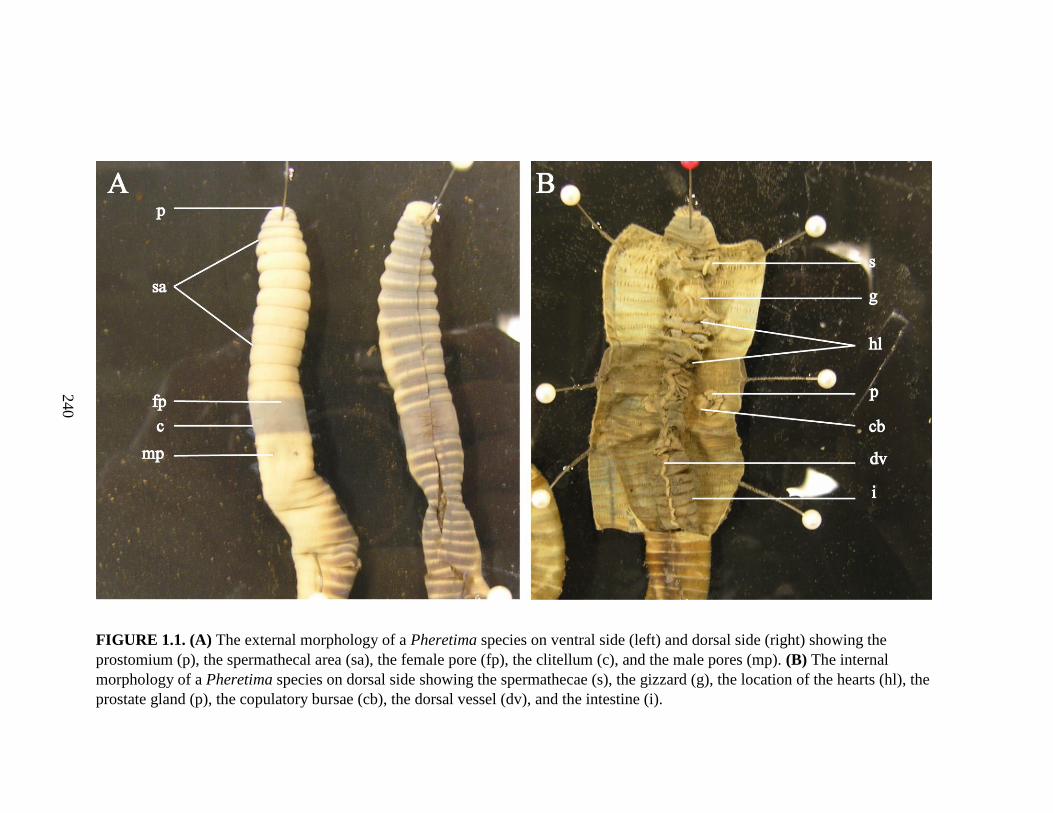
240
240
FIGURE 1.1. (A) The external morphology of a Pheretima species on ventral side (left) and dorsal side (right) showing the
prostomium (p), the spermathecal area (sa), the female pore (fp), the clitellum (c), and the male pores (mp). (B) The internal
morphology of a Pheretima species on dorsal side showing the spermathecae (s), the gizzard (g), the location of the hearts (hl), the
prostate gland (p), the copulatory bursae (cb), the dorsal vessel (dv), and the intestine (i).

241
FIGURE 2.1. (A) Map of the Philippines showing localities where new earthworm
species described in the last decade were collected. (B) Map of Mt. Malindang
showing locations of the barangays where the new Pheretima species reported in this
study were collected (from Aspe & James, 2014).

242
242
FIGURE 2.2. Photographs of living Pheretima worms in dorsal view, showing coloration. (A) P. maculodorsalis;
(B) P. tigris; (C) P. immanis; (D) P. longigula; (E) P. malindangensis. Scale bars: A–D, 5 cm; E, 3 cm (from Aspe & James, 2014).

243
243
FIGURE 2.3. (A) Diagrammatic ventral view of P. maculodorsalis showing the spermathecal pores (sp), clitellum (cl), and male
pores (m). (B, C) Schematic drawings of the internal morphology of Philippine Pheretima species, in dorsal view: (B) P.
maculodorsalis; (C) P. tigris; (D) P. immanis. Abbreviations: s, spermatheca; h, heart; p, prostate gland; cb, copulatory bursa; c,
caecum. Scale bars: 10 mm (from Aspe & James, 2014).

244
244
FIGURE 2.4. Schematic drawings of the internal morphology of Philippine Pheretima species, in dorsal view: (A) P. lago; (B) P.
nunezae; (C) P. boniaoi. Abbreviations: s, spermatheca; h, heart; p, prostate gland; cb, copulatory bursa; c, caecum. Scale bars: 10 mm
(from Aspe & James, 2014).

245
245
FIGURE 2.5. Schematic drawings of the internal morphology of Philippine Pheretima species, in dorsal view: (A) P. malindangensis;
(B) P. misamisensis; (C) P. wati; Abbreviations: s, spermatheca; h, heart; p, prostate gland; cb, copulatory bursa; c, caecum. Scale
bars: 5 mm (from Aspe & James, 2014).

246
246
FIGURE 2.6. Schematic drawings of the internal morphology of Philippine Pheretima species, in dorsal view: (A) P. longiprostata;
(B) P. nolani; (C) P. longigula; Abbreviations: s, spermatheca; h, heart; p, prostate gland; cb, copulatory bursa; c, caecum. Scale bars:
5 mm (from Aspe & James, 2014).

247
247
FIGURE 2.7. (A) Diagrammatic ventral view of polythecal Pheretima adevai, showing the spermathecal pores (sp), clitellum (cl),
and male pores (m). (B–D) Schematic drawings of the internal morphology of Philippine Pheretima species, in dorsal view: (B) P.
adevai; (C) P. lluchi; (D) P. potonganensis. Abbreviations: s, spermatheca; h, heart; p, prostate gland; cb, copulatory bursa; c, caecum.
Scale bars: B–D, 5 mm (from Aspe & James, 2014).

248
248
FIGURE 2.8. (A) Diagrammatic ventral view of monothecal Pheretima vergrandis showing the single spermathecal pore (sp),
clitellum (cl), and single male pore (m). (B–D) Schematic drawings of the internal morphology of Philippine Pheretima species: (B) P.
vergrandis; (C) P. concepcionensis; (D) athecate Pheretima subanensis. (E) Diagrammatic ventral view showing the clitellum (cl) and
a pair of male pores (m), as in P. concepcionensis and P. subanensis. Abbreviations: s, spermatheca; h, heart; p, prostate gland; cb,
copulatory bursa; c, caecum. Scale bars: B–D, 5 mm (from Aspe & James, 2014).
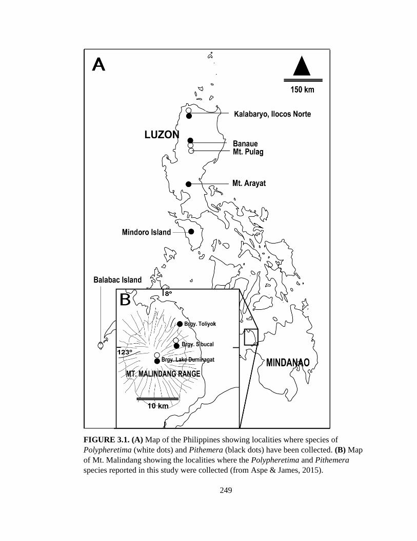
249
FIGURE 3.1. (A) Map of the Philippines showing localities where species of
Polypheretima (white dots) and Pithemera (black dots) have been collected. (B) Map
of Mt. Malindang showing the localities where the Polypheretima and Pithemera
species reported in this study were collected (from Aspe & James, 2015).

250
250
FIGURE 3.2. Schematic drawings of the external anterio-ventral area of (A) Polypheretima mindanaoensis; (B) Pithemera
malindangensis; (C) Pithemera duminagati; and (D) Pithemera donvictorianoi. Abbreviations: fp, female pore; cl, clitellum; mp, male
pore; gm, genital markings. Scale bars: 5 mm (from Aspe & James, 2015).

251
FIGURE 3.3. Schematic drawings of the internal morphology of (A) Polypheretima
mindanaoensis; (B) Pithemera malindangensis; (C) Pithemera duminagati; and (D)
Pithemera donvictorianoi. Abbreviations: s, spermatheca; n, dense tufts of nephridia;
h, heart; p, prostate gland; c, caecum. Scale bars: 5 mm (from Aspe & James, 2015).
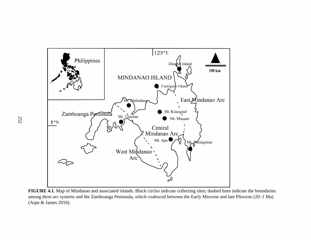
252
252
FIGURE 4.1. Map of Mindanao and associated islands. Black circles indicate collecting sites; dashed lines indicate the boundaries
among three arc systems and the Zamboanga Peninsula, which coalesced between the Early Miocene and late Pliocene (20–1 Ma)
(Aspe & James 2016).

253
FIGURE 4.2. The mountainous landscapes of Mt. Timpoong at > 1300 m asl (top)
and Mt. Apo at > 1500 m asl (bottom), two of the collection sites in Mindanao,
Philippines.
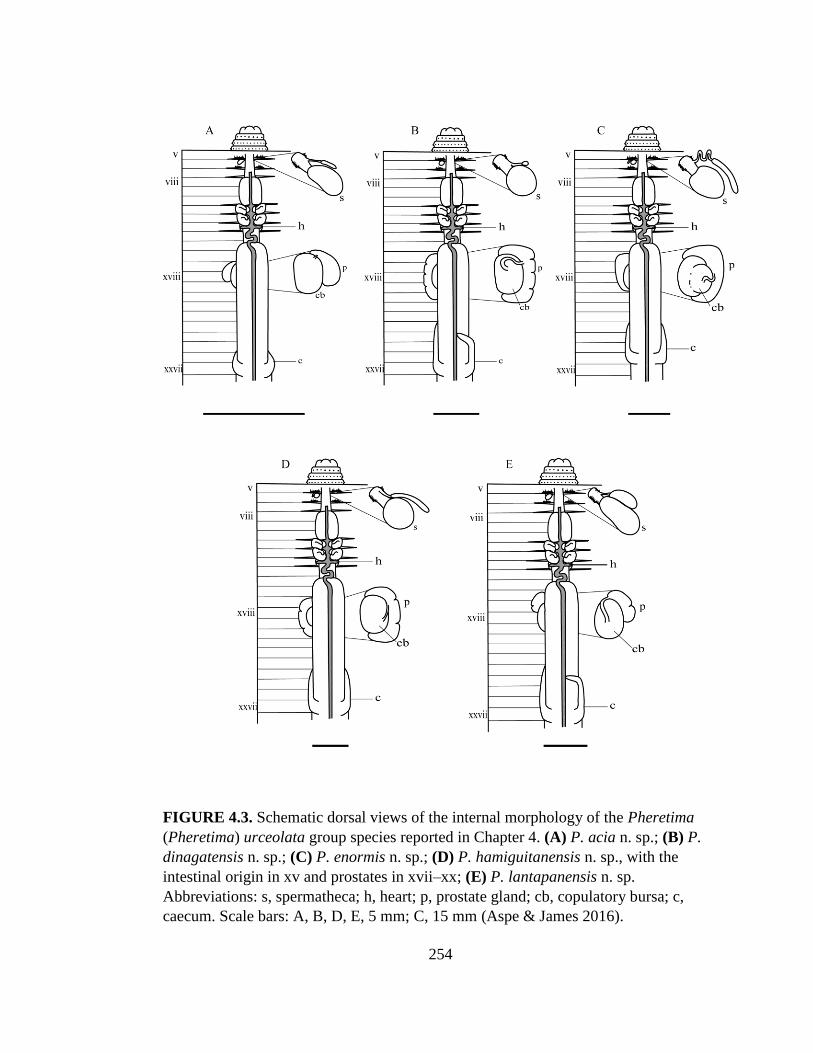
254
FIGURE 4.3. Schematic dorsal views of the internal morphology of the Pheretima
(Pheretima) urceolata group species reported in Chapter 4. (A) P. acia n. sp.; (B) P.
dinagatensis n. sp.; (C) P. enormis n. sp.; (D) P. hamiguitanensis n. sp., with the
intestinal origin in xv and prostates in xvii–xx; (E) P. lantapanensis n. sp.
Abbreviations: s, spermatheca; h, heart; p, prostate gland; cb, copulatory bursa; c,
caecum. Scale bars: A, B, D, E, 5 mm; C, 15 mm (Aspe & James 2016).

255
FIGURE 4.4. Schematic dorsal views of the internal morphology of members of the
Pheretima sangirensis group (A–D) and the Pheretima (Parapheretima) (E, F)
reported in Chapter 4. (A) P. timpoongensis n. sp., with intestinal origin in xv, and
caeca extending from xxvii–xxiv; (B) P. camiguinensis n. sp., with prostates in xvii–
xxi, and caeca extending from xxvii–xxi; (C) P. sibucalensis n. sp.; (D) P. apoensis n.
sp. with spermathecae; (E) P. (Paraph.) pandanensis n. sp.; (F) P. (Paraph.) boaensis
n. sp., with intestinal origin in xiv, prostates in xvii–xix, and caeca extending from
xxvii–xxiii. Abbreviations: s, spermatheca; h, heart; p, prostate gland; cb, copulatory
bursa; c, caecum. Scale bars: A–F, 5 mm (Aspe & James 2016).

256
256
FIGURE 4.5. Schematic dorsal views of the internal morphology of the three Amynthas species reported in Chapter 4. (A) A.
dinagatensis n. sp.; (B) A. cagdianaoensis n. sp.; (C) A. talaandigensis n. sp. Scale bars: A–C: 5 mm. (D) Schematic ventral view of
polythecal A. talaandigensis n. sp., showing the intersegmental spermathecal pores (sp). Abbreviations: s, spermatheca; h, heart; p,
prostate gland; c, caecum; sp, spermathecal pores (Aspe & James 2016).

257
FIGURE 4.6. Schematic views of the internal and external morphology of the
Polypheretima and Pithemera species reported in Chapter 4. (A, B) Polypheretima
bukidnonensis; (A) dorsal view of internal morphology; (B) external ventral view; (C,
D) Polypheretima zamboangensis n. sp.; (C) dorsal view of internal morphology; (D)
external ventral view; (E) Pithemera nolani n. sp., dorsal view of internal morphology,
with prostates in xv–xix. Abbreviations: s, spermatheca; h, heart; p, prostate gland; cb,
copulatory bursa; c, caecum; spbp, spermathecal batteries pores; cl, clitellum, mp,
male pores, gm, genital markings. Scale bars: A–E, 5 mm (Aspe & James 2016).
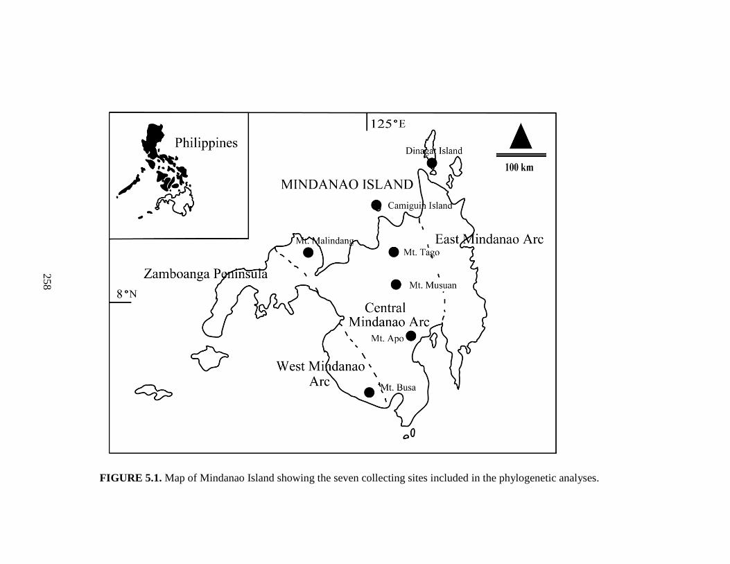
258
258
FIGURE 5.1. Map of Mindanao Island showing the seven collecting sites included in the phylogenetic analyses.

259
FIGURE 5.2. Tree generated from the combined 16S, COI, 28S and H3 genes
showing posterior probability values and bootstrap values. Posterior probability values
lower than 0.8 and bootstrap values lower than 50% indicate weak support and are not
shown.

260
APPENDIX

261
Appendix. Parameters for the concatenated 16S, 28S, COI and H3 genes for
maximum-likelihood (ML) analysis using GTR+G+I model.
Gene Base Frequency Rate Matrix
16S A= 0.345397 A-C= 3.595757
C= 0.193621 A-G= 7.682563
G= 0.190907 A-T= 4.134709
T= 0.270075 C-G= 0.083013
C-T= 14.609200
G-T= 1.000000
28S A= 0.191140 A-C= 1.024950
C= 0.297188 A-G= 0.549549
G= 0.335872 A-T= 0.406212
T= 0.175800 C-G= 0.451803
C-T= 1.887718
G-T= 1.000000
COI-1 A= 0.252159 A-C= 0.000000
C= 0.248523 A-G= 1.567707
G= 0.298522 A-T= 0.000000
T= 0.200796 C-G= 0.000000
C-T= 29.392772
G-T= 1.000000
COI-2 A= 0.124888 A-C= 0.000000
C= 0.265341 A-G= 0.718013
G= 0.173524 A-T= 0.000000
T= 0.436247 C-G= 0.000000
C-T= 0.000000
G-T= 1.000000
COI-3 A= 0.502193 A-C= 68.158957
C= 0.146423 A-G= 670.873380
G= 0.064287 A-T= 0.000000
T= 0.287097 C-G= 69.091061
C-T= 1568.590255
G-T= 1.000000
H3-1 A= 0.277253 A-C= 0.807557
C= 0.312833 A-G= 0.000000
G= 0.318132 A-T= 0.000000
T= 0.091782 C-G= 0.000000
C-T= 1.345784
G-T= 1.000000

262
continued…
H3-2 A= 0.290879 A-C= 0.000000
C= 0.254542 A-G= 0.000000
G= 0.218205 A-T= 0.000000
T= 0.236374 C-G= 0.000000
C-T= 0.000000
G-T= 1.000000
H3-3 A= 0.097697 A-C= 4.846947
C= 0.367040 A-G= 15.202173
G= 0.394050 A-T= 8.519325
T= 0.141212 C-G= 1.906088
C-T= 8.647881
G-T= 1.000000

263
Curriculum Vitae
Nationality : Philippines
Current Address : Rm. 8, 2F, Hoshino Mansion, 3-24, Kita 23 Nishi 8, Kita-ku,
Sapporo City 001-0023, Japan
Name : Nonillon M. Aspe
Date of birth : 1978 March 10
EDUCATIONAL BACKGROUND
Jun. 1991-Mar. 1995 High School
Philippine High School for the Arts
National Arts Center, Mt. Makiling, Los Banos, Laguna,
Philippines
Jun. 1, 1995-Mar. 29, 2000 Bachelor of Science in Marine Biology
Department of Biological Sciences, College of Science
and Mathematics, Mindanao State University-Iligan
Institute of Technology, Iligan City, Philippines
Oct. 22, 2001-Apr. 3, 2006 Master of Science in Biology
Department of Biological Sciences, College of Science
and Mathematics, Mindanao State University-Iligan
Institute of Technology, Iligan City, Philippines
Apr. 1, 2013-Mar. 2016 PhD in Biology
Dept. of Natural History Sciences, Graduate School of
Science, Hokkaido University, Sapporo City, Japan
EMPLOYMENT BACKGROUND
Jun. 2000-Mar. 2001 Instructor
Livingstone Christian Academy, Inc., Cebu, Philippines
Jun. 2002-Oct. 2002 Graduate Teaching Assistant
Department of Biological Sciences, College of Science
and Mathematics, MSU-Iligan Institute of Technology,
Iligan City, Philippines
May 2003-May 2006 Field Biologist
Kansas University-Biodiversity Research Centre
264
“Philippine Terrestrial Annelids and Gastropods
Biodiversity Survey”- A Philippine-wide biodiversity
research based in Kansas University, USA headed by
Dr. Samuel James
May 2006-Feb. 2007 Field Biologist
Haribon Foundation for the Conservation of Natural
Resources, Inc., Philippines, Quezon City, Philippines
Mar. 2007-May 2008 Field Biologist
Kansas University-Biodiversity Research Centre
“Comparative Biogeography and Conservation of
Philippines Vertebrates Project”- A Philippine-wide
biodiversity research based in Kansas University, USA
headed by Dr. Rafe Brown
May 2008-Oct. 2008 Instructor
College of Liberal Arts, St. Michael’s College, Iligan
City, Philippines
Nov. 1, 2008-Mar. 25, 2013 Assistant Professor
Biology Department, College of Natural Sciences and
Mathematics, Mindanao State University, Marawi City,
Philippines
Apr. 1, 2013-Mar.31, 2016 Research Assistant
Department of Natural History Sciences, Graduate
School of Science, Hokkaido University, Sapporo City,
Japan
Sep. 24, 2013-Mar. 31, 2016 Part-time English Tutor
English Department, Hokusei Gakuen University-Junior
College, Sapporo City, Japan
RESEARCH BACKGROUND
2006 During my Master’s program at the Department of Biological Sciences,
College of Science and Mathematics, MSU-Iligan Institute of
Technology, Iligan City, Philippines
Papers:
Aspe NM. 2006. Diversity and Distribution of Earthworms
in Mt. Malindang, Philippines. MS Thesis, School of
Graduate Studies, MSU-Iligan Institute of Technology, Iligan
City, Philippines, 173 pp.

265
Boniao R, Estoista R, de Goede R, Hansel C, Nuñeza O,
Lumactud R, Aspe NM, Poblete M, Roscom B and James
SW. 2006. Soil Ecological Diversity and Relevant
Interrelationships of Critical Resources in Mt. Malindang,
Mis. Occ., Philippines. SEAMEO-SEARCA. Laguna,
Philippines.
Boniao R, Estoista R, Hansel C, de Goede R, Nuñeza O,
Roscom B, James SW, Lumactud R, Poblete M, and Aspe
NM. 2006. Assessment of Soil Properties and Ecological
Diversity in Mt. Malindang Range, Philippines. Society,
Environment and Development: The Mt. Malindang
Experience. SEAMEO-SEARCA. Laguna, Philippines.
Presentations:
Aspe N. Earthworm sampling and field identification -
Capability Building of Research Assistants, Local Partners,
and Researchers on Primary Data Collection for Flora, Fauna
and Soil Ecology in Mt. Malindang. Biodiversity Research
Programme, SEAMEO-SEARCA. Oral presentation in
English. August 2003. Oroquieta City, Philippines
Aspe N. Diversity and distribution of earthworms in Mt.
Malindang”. 6th Annual National Scientific Convention of
the Philippine Society for the Study of Nature. Oral
presentation in English. May 2006. Kapalong, Davao del
Norte, Philippines
2009-2012 When I was working as Assistant Professor at the Department of
Biology, College of natural Sciences and Mathematics, MSU, Marawi
City, Philippines
Papers:
Aspe NM, Nuneza O and Torres MA. 2009. Diversity and
Distribution of Earthworms in Mt. Malindang, Philippines.
Journal of Nature Studies. 8: 59-67.
Boniao R, Aspe NM and Van Ranst E. 2012. Ecological
Properties of the Soil in Mt. Diwata Range, Philippines: A
Physico-chemical and Biological Assessment. LAP Lambert
Academic Publishing. ISBN 978-3-659-22675-5. 73 pp.

266
Presentation:
Aspe NM. Perception of the constituents of Iligan City on
the concept of urban forestry in the City. Science Seminar,
27th CNSM Science Week Celebration “CNSM Call for
Environmental Awareness Curbing Natural Calamities”.
Oral presentation in English. February 2012. Mindanao
State University, Marawi City, Philippines
2014-2016 During my PhD program at the Department of Natural History
Sciences, Graduate School of Science, Hokkaido University, Sapporo
City, Japan
Papers:
Aspe NM and James SW. 2014. New species of Pheretima
(Oligochaeta: Megascolecidae) from the Mt. Malindang
Range, Mindanao Island, Philippines. Zootaxa. 3881: 401-
439. http://dx.doi.org/10.11646/zootaxa.3881.5.1.
Aspe NM and James SW. 2015. New Polypheretima and
Pithemera (Oligochaeta: Megascolecidae) species from the
Mt. Malindang Range, Mindanao Island, Philippines.
Journal of Natural History.
http://dx.doi.org/10.1080/00222933.2015.1021875.
Aspe NM and James SW. 2016. New species of Pheretima,
Amynthas, Polypheretima, and Pithemera (Clitellata:
Megascolecidae) from Mindanao and associated islands,
Philippines. Zoological Studies. 55.
doi:10.6620/ZS.2016.55-08.
Aspe NM. 2016. Taxonomy and phylogeny of pheretimoid
earthworms (Clitellata: Megascolecidae) from Mindanao
and associated islands, Philippines. A PhD Dissertation.
Department of Natural History Sciences, Graduate School
of Science, Hokkaido University, Sapporo City, Japan.
Aspe NM, Kajihara H and James SW. (unpublished) A
molecular phylogenetic study of pheretimoid species
(Megascolecidae) in Mindanao Island, Philippines.
European Journal of Soil Biology.
Aspe NM (unpublished) New species of Pheretima and
Amynthas (Oligochaeta: Megascolecidae) from Mt. Apo,

267
Mindanao Island, Philippines.
Presentations:
Aspe NM, James SW, Dick MH. New species of
earthworms (Oligochaeta: Megascolecidae) from Mt.
Malindang Range, Mindanao Island, Philippines. The 10th
International Symposium on Earthworm Ecology. Oral
presentation in English. June 22-27, 2014. Athens, Georgia,
USA
Aspe NM, Kajihara H, James SW. A molecular
phylogenetic study of pheretimoid species
(Megascolecidae) in Mindanao Island, Philippines. 6th
International Barcode of Life Conference. Oral
presentation in English. Aug. 18-21, 2015. University of
Guelph, Guelph, Ontario, Canada
Aspe NM, Kajihara H, James SW. A taxonomic and
phylogenetic study of earthworms (Clitellata: Annelida)
from Mindanao Island, Philippines. A Research meeting on
taxonomy of Annelida: Previous works and subjects for
further studies in eastern Asia. Oral presentation in English.
Nov. 19-20, 2015. The University of Tokyo, Kashiwa City,
Chiba, Japan
HONORS AND AWARDS
1991-1995 Scholarship Awardee
Philippine High School for the Arts, Mt. Makiling, Los Baños, Laguna,
Philippines
1995-1997 Integrated Performing Arts Guild (IPAG) Scholarship Awardee
MSU-IIT, Iligan City, Philippines
2004-2005 Thesis Grant Awardee
Biodiversity Research Programme, SEARCA, Los Baños, Laguna,
Philippines
Aug. 2005 Best Paper Award (Paper entitled: “Assessment of Soil Properties and
Ecological Diversity in Mt. Malindang Range”)
NOMCARRD Regional Symposium on Research and Development
MOSCAT, Claveria, Misamis Oriental, Philippines
Mar. 2006 Best Thesis Awardee (Thesis entitled: “Diversity and Distribution of
Earthworms in Mt. Malindang, Philippines”)

268
College of Science and Mathematics
MSU-IIT, Iligan City, Philippines
2013-2016 PhD Grant Awardee
Hokkaido University Special Grant Program for International Young
Scientists, Hokkaido University, Sapporo City, Japan
Aug. 2015 Travel Grant Awardee (Title of presentation: “A molecular
phylogenetic study of the pheretimoid species (Megascolecidae) in
Mindanao Island, Philippines”)
2015 Hokkaido University Grant for Attending International
Conference, Hokkaido University, Sapporo City, Japan
Sep. 2015 Grant Awardee
2015 Support Fund for Fostering Researchers at the Faculty of Science
Faculty of Science, Hokkaido University, Sapporo City, Japan
I certify that the above information are true records.
Date:
Name: Nonillon M. Aspe