taxonomic studies on psathyrella sect. spadiceae
TRANSCRIPT

137
CZECH MYCOL. 60(2): 137–171, 2008
Taxonomic studies on Psathyrella sect. Spadiceae
MARTINA VAŠUTOVÁ
Dept. of Biology, Pedagogical Faculty, University of South Bohemia, Jeronýmova 10,CZ-371 15 České Budějovice, Czech Republic
Vašutová M. (2008): Taxonomic studies on Psathyrella sect. Spadiceae. – CzechMycol. 60(2): 137–171.
Descriptions, figures of microscopic characters, data on ecology and distribution of four species ofPsathyrella section Spadiceae known from the Czech Republic and Slovakia are given. These speciesare P. spadicea, P. papyracea, P. pygmaea and P. spintrigeroides. Type material of P. variata,P. imleriana, P. olympiana and P. spintrigeroides has been examined and the insufficiently knowntaxa P. variata, P. imleriana, P. subcernua and P. sarcocephala are discussed. The newly publishedcombination Psathyrella papyracea (Pers.: Fr.) Vašutová is shown to be the correct name for a funguscurrently named P. cernua (Vahl: Fr.) Hirsch. An identification key for Psathyrella species with thick-walled cystidia occurring in Central Europe is presented.
Key words: Psathyrella, Czech Republic, Slovakia, sect. Spadiceae, distribution, ecology.
Vašutová M. (2008): Taxonomická studie křehutek ze skupiny Psathyrella sect.Spadiceae. – Czech Mycol. 60(2): 137–171.
V příspěvku jsou publikovány popisy, nákresy mikroznaků, data o ekologii a rozšíření čtyř druhůkřehutek (Psathyrella) ze sekce Spadiceae známých z České republiky a Slovenska: P. spadicea, P. pa-
pyracea, P. pygmaea a P. spintrigeroides. Zároveň byl studován typový materiál druhů P. variata,P. imleriana, P. olympiana a P. spintrigeroides a diskutovány problematické taxony P. variata,
P. imleriana, P. subcernua a P. sarcocephala. Správným jménem pro druh dosud známý jako P. cernua
(Vahl: Fr.) Hirsch. je Psathyrella papyracea (Pers.: Fr.) Vašutová. Součástí práce je klíč k určováníkřehutek s tlustostěnnými cystidami, které se vyskytují ve střední Evropě.
INTRODUCTION
Psathyrella (Fr.) Quél. (Psathyrellaceae, Agaricales) is a worldwide-distrib-uted genus of about 550 dark-spored saprotrophic taxa (Smith 1972, Kits vanWaveren 1985). Many species have almost identical and variable macroscopiccharacters and so their identification is based on microscopic characters only.Modern infrageneric classifications were published by Smith (1972), Romagnesi(1982), Kits van Waveren (1985), and Singer (1986). Due to the fact that only Kitsvan Waveren published a monograph of the genus in Western Europe, hisinfrageneric classification became the most used there. However, current analy-ses of ITS and part of the LSU area of rDNA (Walther et al. 2005, Larsson and

Örstadius 2008, Padamsee et al. 2008, Vašutová et al. 2008) demonstrate that allthese classifications based on morphological and anatomical characters are moreor less artificial. To prepare a new infrageneric classification respecting the phy-logeny of the genus, it is necessary to analyse more species and to support it byanalyses of other DNA regions.
Section Spadiceae (Morgan) Kits van Wav. emend. is an artificial section(Vašutová et al. 2008), although well usable for identification, characterised as“having pleurocystidia muricate and thick-walled, walls at least 0.5 μm, but almostalways more, up to 2–3.6 μm thick, either everywhere or only locally (apex,ventrical portion or/and in or near pedicel)” (Kits van Waveren 1985). As was sup-posed by Romagnesi (1982) in his classification and then supported by molecularanalysis (Vašutová et al. 2008), thick-walled cystidia with crystals are probablya plesiomorphic character. Other species with slightly thick-walled cystidia (c. 0.5 μmonly) and without crystals were placed by Kits van Waveren into the sectionPennatae Romagn. emend. Kits van Wav. and Hydrophilae (Romagn.) ex Singeremend. Kits van Wav.
In the Czech Republic and Slovakia, section Spadiceae is represented by fourspecies. The occurrence of the fifth species, Psathyrella olympiana A.H. Sm., hasnot been confirmed to date. Based on molecular data (Vašutová et al. 2008), sec-tion Spadiceae is composed of three unrelated, distinct groups of species. Thecore of the section Spadiceae consists of the non-velate species P. spadicea (P.Kumm.) Singer and P. papyracea (Pers.: Fr.) Vašutová (= P. cernua ss. auct.), dis-tinguished by Romagnesi (1982) as subgenus Homophron. This group is a sistergroup of the genus Lacrymaria (Walther et al. 2005, Larsson and Örstadius 2008,Padamsee et al. 2008, Vašutová et al. 2008). The second group consists of thevelate species P. olympiana A.H. Sm. belonging to section Fatuae Romagn. exRomagn. (Romagnesi 1982), and P. pygmaea (Bull.: Fr.) Singer belonging to sec-tion Pygmaeae Romagn. The last group is represented by P. spintrigeroides P.D.Orton – a species the morphological characters of which place it on the border ofsections Spadiceae and Pennatae. However, molecular data (Larsson andÖrstadius 2008, Vašutová et al. 2008) strongly supported relations between P.
spintrigeroides and section Pennatae.The aim of this work is to contribute to the knowledge of species variability in
section Spadiceae by providing descriptions and figures of micromorphologicalcharacters based on Czech and Slovak specimens and to summarise current infor-mation on their ecology and distribution in the Czech Republic and Slovakia. Forthe purpose of practical identification I have added an identification key includingall species with thick-walled cystidia from sections Spadiceae, Pennatae andHydrophilae occurring in Central Europe.
138
CZECH MYCOL. 60(2): 137–171, 2008

METHODS
Descriptions of macromorphological characters are based on fresh basidiomata collected by theauthor in the years 2001–2006 in the Czech Republic and Slovakia, and are deposited in the author’sherbarium (MV) or BRNM. Abbreviations of herbaria follow the Index Herbariorum (Holmgren andHolmgren 2003). Colours were compared with the colour tables by Küppers (1999). Microscopic obser-vations were performed according to Kits van Waveren’s method (Kits van Waveren 1971). At least tenrandomly selected cheilocystidia, pleurocystidia, caulocystidia and basidia and 20 randomly selectedmature spores were measured in each of three representative specimens of each species collected inthe Czech Republic or Slovakia. The size of each microscopic structure is given as the 10– and 90-per-centiles of all measurements, the 5– and 95-percentiles are given in brackets. All specimens listed un-der “Collections studied” were microscopically examined; any deviations were recorded and incorpo-rated into the descriptions of the species. Description terminology is taken from Vellinga (1988) andFouchier (1995). The term “subacute” for the apex of cystidia is used as a term intermediate between“acute” and “obtuse”. Further, type specimens of P. spintrigeroides (K(M) 70569), P. variata (MICH5379), P. imleriana (BR 46447-81, Volders 93222), P. olympiana (MICH 11991), P. kauffmanii (MICH11962), P. indecorosa (MICH 11953), P. avellaneifolia (MICH, Smith 21463) and Psathyrella
artemisiae var. microspora (L 54381) have been examined.The synonymy of names published in Psathyrella is not complete because of the large number of
existing Psathyrella names, which have moreover been mixed with names belonging to the currentgenera Psilocybe, Hypholoma and Stropharia. I have focused mainly on names published in the period1753–1832 and verified only the synonyms of the presented species which are included in monographsof the genus (Kits van Waveren 1985, Enderle 1989, Örstadius 2001).
Notes on ecology and distribution of the species in Europe were taken from publications by mo-nographers of the genus or in cooperation with them to make certain they belonged to the correct spe-cies (Enderle 1987, 1989, 2004; Enderle and Hübner 2005; Kits van Waveren 1985; Örstadius 2001; Legonand Henrici 2005; Tassi 1997; Arnolds et al. 1995). Localisation of the examined specimens is structuredas follows: number of the phytogeographical unit according to the phytogeographical division of theCzech and Slovak Republics (Futák 1966, Skalický 1988, Vašutová 2006 – Figs. 1, 2), name of the local-ity, quadrat of Central European grid mapping (Q).
RESULTS AND DISCUSSION
Identification key for Psathyrella species with thick-walled cystidia
(about 0.5 µm or more)
(Note: it is necessary to observe material in water or 10 % NH4OH, as crystalsare missing in 5 % KOH)
1a Cystidia with distinct crystals at their tops ............................................................................................. 21b Cystidia without distinct crystals at their tops (rarely few scattered small crystals measuring
c. 1 μm are present on a few cystidia only) .............................................................................................. 52a Basidiomata small (pileus up to 2 cm), resembling Coprinus disseminatus, pleurocystidia up to
35 μm long; on decaying stumps of deciduous trees, rarely on soil with wood remnants................................................................................................................................ 3. P. pygmaea (p. 151)
2b Not as above, basidiomata larger, pleurocystidia longer than 35 μm ................................................... 33a Species without veil; often in cavities or at bases of living deciduous trees ....................................... 4
139
VAŠUTOVÁ M.: TAXONOMIC STUDIES ON PSATHYRELLA SECT. SPADICEAE

3b Species with developed veil; on decaying trunks or stumps of deciduous trees. 4. P. olympiana (p. 154)4a Cystidia utriform, crystals forming “caps” on cystidium tops, spores reddish brown, basidiomata
becoming eburneous to sordid white when drying out ................................ 2. P. papyracea (p. 145)4b Cystidia versiform, crystals forming “stars” on cystidium tops, spores very pale, almost colourless,
basidiomata becoming at most pale carneous beige when drying out ........... 1. P. spadicea (p. 141)
N o t e : A species with a pileus distinctively pruinose at margin in a young stage and with a mixtureof lampro– and leptocystidia on the lamellae edge and on their surface is P. variata A.H. Sm. (p. 160).Species with characters between P. cernua and P. spadicea could be P. sarcocephala ss. auct. (p.167).
5a Wall of cystidia thicker in upper part (0.5–2 μm, when rarely 0.5 μm thick only, the very top is al-most thin-walled); on fallen trunks mostly of deciduous trees .......... 5. P. spintrigeroides (p. 156)
5b Wall of cystidia thicker in middle and lower part (0.5–1 μm), wall of upper part sometimes so thinas almost invisible at 200 × magnification ............................................................................................... 6
6a Basidiomata growing on burnt places; spores dark reddish brown almost without germ pore andwith distinct hilar appendix, cystidia mostly with acute apex .......................................... P. pennata
6b Not as above..................................................................................................................................................77a Small basidiomata (conical pileus up to 1.5 cm broad), spores pale brownish yellow without germ
pore; pleurocystidia versiform, often with irregular apex, growing terrestrial in deciduous forests...................................................................................................................... P. umbrina (see note below)
7b Basidiomata medium to large-sized, spores with at least indistinct germ pore; pleurocystidiaversiform or utriform, with regular or irregular apex, growing terrestrially or on wood .................. 8
8a Pleurocystidia mostly versiform to lageniform with regular subacute apex, pleurocystidioidcheilocystidia densely packed, spores slightly amygdaliform with distinct germ pore, dark reddishbrown; growing on wood remnants or fallen trunks of deciduous or coniferous trees.. P. artemisiae
8b Pleurocystidia versiform, some utriform, apex often irregular, sometimes forked, subacute to ob-tuse, pleurocystidioid cheilocystidia not densely packed, spores slightly phaseoliform, germ poredistinct to indistinct, medium brown, growing on fallen trunks, rarely on wood remnants of Fagus
sylvatica or Acer pseudoplatanus ....................... P. rostellata (for description see Vašutová 2008)8c Pleurocystidia utriform, apex almost subcapitate, pleurocystidioid cheilocystidia not densely
packed – sphaeropedunculate cells prevail, germ pore indistinct to distinct, medium brown, grow-ing terrestrially under beech, maple or oak .................................................................... P. kauffmanii
N o t e : P. umbrina Kits van Wav. and P. kauffmanii A.H. Sm. have not been reported from theCzech Republic or Slovakia. P. umbrina is known in Central Europe from one locality in Austria(WU 16345) only. One collection (PRM 897750) similar to P. kauffmanii was found in the ŠumavaMts. but its spores lack a germ pore. Its occurrence in Europe is doubtful. Enderle (1996) describedthe species from Germany but his species has cystidia closer to P. rostellata than to P. kauffmanii.I have studied only herbarium material of these species.
Section Spadiceae (Morgan) Kits van Wav. emend. Kits van Wav.Psilocybe [unranked] Spadiceae Morgan in J. Mycol. 13: 246. 1907. – Drosophila sect. Spadiceae
(Morgan) Romagn. in Bull. Mens. Soc. Linn. Lyon 13: 52. 1944. – Psathyrella sect. Spadiceae (Morgan)Kits van Wav. emend. Kits van Wav. in Persoonia Suppl. Vol. 2: 280. 1985.
Psilocybe [unranked] Spadiceae Morgan consists of 16 species currently belong-ing to the genera Psathyrella, Psilocybe and Panaeolus. Kits van Waveren (1985)placed this section into the genus Psathyrella and reduced the number of species.
140
CZECH MYCOL. 60(2): 137–171, 2008

1. Psathyrella spadicea (P. Kumm.) Singer Figs. 1, 10Agaricus spadiceus Schaeff., Fung. Bavar. Palat. nasc., vol. 4: 27, 1774. (nom. illeg., non Agaricus
spadiceus Scop., Fl. Carniol., 443, 1772); Psilocybe spadicea P. Kumm., Führer Pilzk. 71, 1871;Psathyrella spadicea (P. Kumm.) Singer, Lilloa 22: 468. 1951.
S y n o n y m s : Drosophila spadicea (P. Kumm.) Quél., Enchir. fung.: 116. 1886; Pratella spadicea
(P. Kumm.) J. Schröt. in Cohn, Krypt.-Fl. Schlesien: 568. 1889; Psathyra spadicea (P. Kumm.) Singer,Ann. mycol. 34: 33. 1936. – ?Agaricus curvatus Weinm., Hymen. Gasteromyc. 248. 1836.
M i s a p p l i e d n a m e s : Psathyrella sarcocephala s. J.E. Lange, Fl. agaric. danic. 4: 80, pl. 148F.1939; Crepidotus palmatus s. Bres., Icon. mycol. 16, t. 788, 1930.
“Psathyrella spadicea” auct. non (P. Kumm.) Singer: Bres., Icon. mycol. 18: pl. 859. 1931.Ty p e s p e c i m e n : not preserved. Lectotype (selected by Örstadius 2001): Schaeffer, Fung. Bavar.
Palat. nasc., vol. 1: pl. 60, figs. 4 et 6, 1762. Epitype (selected by Örstadius 2001): Germany, Bavaria,floodplain forests near Leipheim-Riedheim, growing caespitose at the base of a big poplar, 11 Nov.1998, leg. et det. M. Enderle [GB – LÖ 205-98, isoepitype M].
S e l e c t e d i l l u s t r a t i o n s : Kits van Waveren, Persoonia Suppl. Vol. 2: figs. 208–212. 1985;Enderle, Beitr. Kenntn. Pilze Mitteleurop. 5, 1989; Ricken, Die Blätterpilze (Agaricaceae) I: pl. 66, fig. 7.1915; Konrad and Maublanc, Ic. sel. Fung.: pl. 46. 1928, Lange, Fl. agaric. danic.: pl. 148F as P. sarco-
cephala. 1939.S e l e c t e d l i t e r a t u r e : Kits van Waveren, Persoonia Suppl. Vol. 2: 161–164. 1985.
C h a r a c t e r i s t i c s i n b r i e f . Basidiomata medium to large-sized, growingmostly in clusters, without veil, pileus drying out to carneous beige, lamellaebrick-beige to brick-brown. Spores medium– to large-sized, 8–9.5 × 4–5 μm, verypale, slightly phaseoliform, without germ pore. Cystidia broadly versiform, dis-tinctly thick-walled in upper part, with crystals forming “stars”. Growing often atbases or in cavities of living deciduous trees.
D e s c r i p t i o n . Basidiomata caespitose. P i l e u s in young stage paraboloidto convex, some basidiomata with broad umbo, 13–27 mm, at maturity plano-con-vex to plane, rarely broadly conico-paraboloid, 20–52 mm, old pilei plane, oftenwith undulate, even reflexed margin, 72–130 mm. Surface hygrophanous, smooth,in very young stage slightly pruinose in narrow zone along margin (c. 1 mm); inyoung stages deep brown with vinaceous tint (S90Y40M40, S80Y50M60), laterbrown to reddish brown (S70Y50M50), medium brown, slightly irregularly col-oured (S50Y50M50, S50Y40M40), pinkish brown (S50Y50M40), brick-beige(S40Y60M50), brownish pink (S40Y50M40), drying out from the centre, at marginremaining darker, when dry pale carneous beige (S20Y30–40M20, S10Y40M10).Ve i l absent. L a m e l l a e crowded, L = 24–32, l = 3–5, 2–9 mm broad; narrowlyadnate, ventricose, at maturity disrupting transversely, in young stages paleochre-beige with pinkish hue, pale brick-beige (S20Y40M30–40), later pale brick-brown (S40Y70M50); with fimbriate edge, white in young stages, later con-colorous. S t i p e 34–150 × 3–7 mm, cylindrical or tapering to base, often twisted,hollow, creamy, later pale beige with pale pinkish ochre shade; surface fibrillose,at apex pruinose, at base whitish tomentose. F l e s h in pileus 3–7 mm broad,white to pale creamy. S m e l l indistinct. S p o r e p r i n t brick-beige (S40Y60M50).
141
VAŠUTOVÁ M.: TAXONOMIC STUDIES ON PSATHYRELLA SECT. SPADICEAE

S p o r e s 8–9.5 × 4–5 μm, average 8.7 × 4.7 μm, Q = 1.6–2.2(–2.3); slightlyphaseoliform in side view, ellipsoid in front view; without germ pore, with indistinctto small hilar appendix; in water very pale with pink tint, pale beige-ochraceouspink (S20Y20M10, S10Y30M10); in 5 % KOH colourless with slightly reddish shadeor pale beige-ochraceous pink (S20Y20M10); in 10 % NH4OH very pale with pinktint, pale beige-ochraceous pink (S10Y30M10, S40Y20M10). B a s i d i a (21.5–)22–26× (6–)6.5–8 μm, 4-spored, narrowly clavate. P l e u r o c y s t i d i a fairly numerous,(51–)52–64 × 16–21(–22) μm; broadly fusiform with subacute apex, cystidium wallin the middle up to 1 μm thick, in upper part 1–3 μm (thickening starts just belowapex), sometimes entire apex filled with wall structure (up to 15 μm from the top);with crystals forming “stars” at apex, dissolving in KOH; colourless in 10 %NH4OH. E d g e o f l a m e l l a e consisting of: (1) numerous pleurocystidioidcheilocystidia, 45–74 × 14–20 μm, broadly fusiform, with wall up to 1 μm thick inthe middle, 1–3 μm in upper part (thickening starts just below apex), sometimesentire apex filled with wall structure (up to 15 μm from the top); (2) scarceutriform cystidia, 37–55 × 17–20 μm, with wall up to 1 μm thick in middle and up-per parts; (3) scarce (up to 15 %) clavate leptocystidia, 43–57 × 14–17 μm.
142
CZECH MYCOL. 60(2): 137–171, 2008
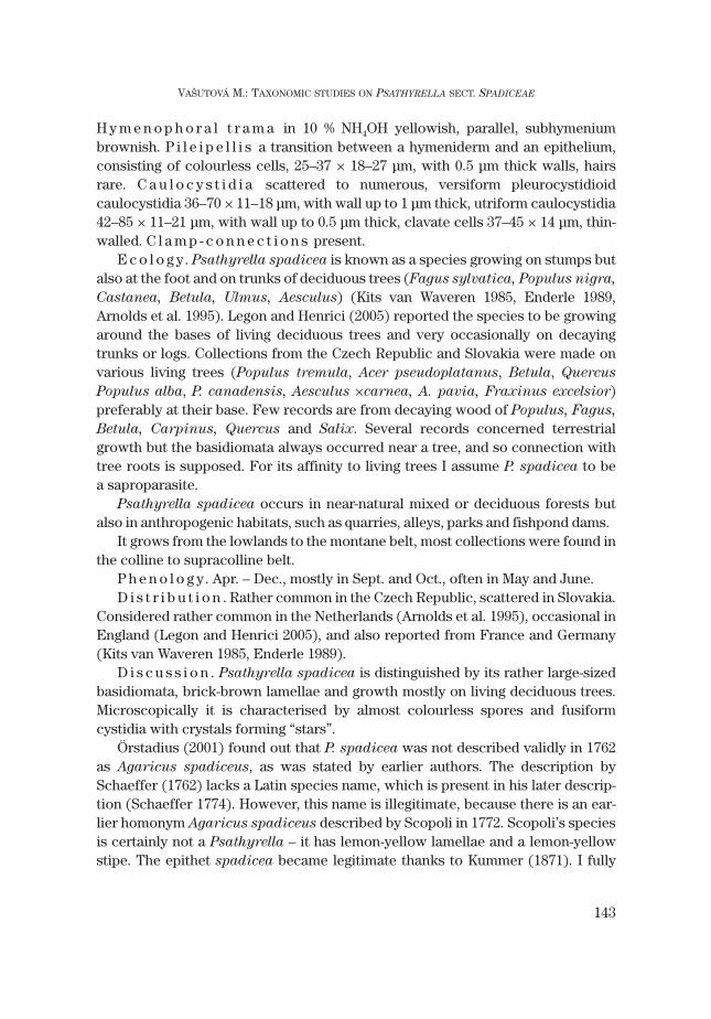
Fig. 1. Psathyrella spadicea (P. Kumm.) Singer (MV06/118): a – spores, b – basidia, c – pleurocystidia,d – cheilocystidia, e – caulocystidia, f – pileipellis. Bars = 10 μm for spores, 50 μm for other microscopicstructures.
H y m e n o p h o r a l t r a m a in 10 % NH4OH yellowish, parallel, subhymeniumbrownish. P i l e i p e l l i s a transition between a hymeniderm and an epithelium,consisting of colourless cells, 25–37 × 18–27 μm, with 0.5 μm thick walls, hairsrare. C a u l o c y s t i d i a scattered to numerous, versiform pleurocystidioidcaulocystidia 36–70 × 11–18 μm, with wall up to 1 μm thick, utriform caulocystidia42–85 × 11–21 μm, with wall up to 0.5 μm thick, clavate cells 37–45 × 14 μm, thin-walled. C l a m p - c o n n e c t i o n s present.
E c o l o g y. Psathyrella spadicea is known as a species growing on stumps butalso at the foot and on trunks of deciduous trees (Fagus sylvatica, Populus nigra,
Castanea, Betula, Ulmus, Aesculus) (Kits van Waveren 1985, Enderle 1989,Arnolds et al. 1995). Legon and Henrici (2005) reported the species to be growingaround the bases of living deciduous trees and very occasionally on decayingtrunks or logs. Collections from the Czech Republic and Slovakia were made onvarious living trees (Populus tremula, Acer pseudoplatanus, Betula, Quercus
Populus alba, P. canadensis, Aesculus ×carnea, A. pavia, Fraxinus excelsior)preferably at their base. Few records are from decaying wood of Populus, Fagus,
Betula, Carpinus, Quercus and Salix. Several records concerned terrestrialgrowth but the basidiomata always occurred near a tree, and so connection withtree roots is supposed. For its affinity to living trees I assume P. spadicea to bea saproparasite.
Psathyrella spadicea occurs in near-natural mixed or deciduous forests butalso in anthropogenic habitats, such as quarries, alleys, parks and fishpond dams.
It grows from the lowlands to the montane belt, most collections were found inthe colline to supracolline belt.
P h e n o l o g y. Apr. – Dec., mostly in Sept. and Oct., often in May and June.D i s t r i b u t i o n . Rather common in the Czech Republic, scattered in Slovakia.
Considered rather common in the Netherlands (Arnolds et al. 1995), occasional inEngland (Legon and Henrici 2005), and also reported from France and Germany(Kits van Waveren 1985, Enderle 1989).
D i s c u s s i o n . Psathyrella spadicea is distinguished by its rather large-sizedbasidiomata, brick-brown lamellae and growth mostly on living deciduous trees.Microscopically it is characterised by almost colourless spores and fusiformcystidia with crystals forming “stars”.
Örstadius (2001) found out that P. spadicea was not described validly in 1762as Agaricus spadiceus, as was stated by earlier authors. The description bySchaeffer (1762) lacks a Latin species name, which is present in his later descrip-tion (Schaeffer 1774). However, this name is illegitimate, because there is an ear-lier homonym Agaricus spadiceus described by Scopoli in 1772. Scopoli’s speciesis certainly not a Psathyrella – it has lemon-yellow lamellae and a lemon-yellowstipe. The epithet spadicea became legitimate thanks to Kummer (1871). I fully
143
VAŠUTOVÁ M.: TAXONOMIC STUDIES ON PSATHYRELLA SECT. SPADICEAE

agree with Enderle (1989) and Örstadius (2001), who regard the holotype(iconotype) to be a mixture of several species.
Smith (1972) distinguishes four species in this group (P. littennii A.H. Sm., P.
sublateritia A.H. Sm., P. conissans (Peck) A.H. Sm. and P. spadicea (Fr.) Singer)based on the colour of the spore deposit, caulocystidium shape and cystidiumcontent, which come very close to the European P. spadicea. I have not studiedhis material. His P. spadicea has a wood-brown spore deposit and is thereforeprobably not identical with the European type.
P. spadicea was often identified as P. sarcocephala by Czech and Slovak my-cologists (see discussion under P. sarcocephala) because they used the work byLange (1939), where they are confused.
Two collections morphologically very similar to P. spadicea were found at thefoot of Picea abies (MV, CB). More specimens are needed to solve their taxo-nomic value.
Collections studied
C z e c h R e p u b l i c : 7d, Prague, Břevnov, Q: 5952a, on base of trunk of Populus sp., 5 Oct. 1941, J.Herink (PRM 735900 as Psilocybe sarcocephala) – 8, Karlštejn, Q: 6051c, 24 June 1943, Mr. Šinták (PRM735906 as Psilocybe sarcocephala). – Prague–Bráník, Q: 5952, on wood of Populus canadensis, 8 Oct.1950, J. Kubička (PRM 735899 as Psilocybe sacrocephala). – Roblín, Q: 6051ab, 26 Sept. 1943, I. Charvát(PRM 735904 as Psilocybe sarcocephala). – 9, Bohnice, Čimický háj forest, Q: 5852, 12 May 1943, J.Malina (PRM 735919). – Ibid., 7 Oct. 1943, J. Malina (PRM 735917 as Psilocybe spadicea). – Prague,Malá Strana, Q: 5952a, on base of Aesculus pavia, 16 Nov. 1963, E. Wichanský (PRM 600818 asPsilocybe spadicea). – Prague, Kinského sady park, Q: 5952a, on base of trunk of living Acer
pseudoplatanus, 30 Oct. 1969, E. Wichanský (PRM 685620). – Ibid., on soil, 27 May 1970, E. Wichanský(PRM 709979). – 10a, Čimice near Prague, Q: 5852d, on (burnt) roots of Betula, 13 May 1948, J. Malina(PRM 735907 as Psilocybe sarcocephala). – 10b, Prague, Hodkovičky, Q: 5952cd, on foot of Betula, 29May 1952, J. Miškovský (PRM 703994 as Psilocybe sarcocephala). – 15c, Stéblová, near Pardubice, Q:5960a, 2 Nov. 1945, K. Kult (PRM 735905 as Psilocybe sarcocephala) – 16, Tulešice, forest with Populus
tremula, Q: 6963c, 18 Juni 1967, L. Čech (BRNM 331956). – 18b, Bzenec-Přívoz, Q: 7069, 15 July 2006, J.Polčák (MV06/146). – 20b, Kobylí, near Hustopeče, Q: 7067c, on ceiling and walls of wine cellar, 13 Oct.1971, E. Hadač (PRM 715642). – Žarošice, Q: 6967d, near stump of Carpinus betulus (?), 16 Sept. 1945,V. Vacek (PRM 735925 as Psilocybe spadicea). – 21b, Tovačov, dam of Donbas pond, Q: 6569, at base ofliving Populus alba, 2 Dec. 2006, M. Vašutová et J. Polčák, (MV06/118). – 35d, Písek, Q: 6650, on roots ofPopulus tremula, 2 May 1974, Kuber (PRM 822335). – 37l, Český Krumlov, Vyšenské kopce national na-ture reserve, Q: 7151, on base of trunk of Populus tremula, 17 July 2004, M. Beran (CB) . – 37p, Klení,on E bank of Velký Klenský rybník pond, Q: 7253, 31 May 2008, T. Papoušek (CB as P. sarcocephala). –39, Turovec, dam of Luční pond, Q: 6654a, on stump of Quercus, 1 June 1989, J. Valter (CB 6577). – 41,
Písek, near a site called “U Vodáka”, on dam of small fishpond, Q: 6650d, at base of Quercus, 31 Aug.1979, A. Štěpánek et J. Kubička (CB 2125). – Čeřenice na Sázavě, settlement called Poříčko, Piceetum,Q: 6155, on decaying trunk of deciduous (?) tree, 29 Oct. 1944, J. Herink (PRM 735903 as Psilocybe
spadicea). – Zbraslav, Q: 6052a (PRM 735901 as Psilocybe sacrocephala). – 42, Tábor, botanical garden,Q: 7554, on wood of Salix, 24 Sept. 1993, M. Beran (CB as P. sarcocephala). – Ibid., on base of trunk ofBetula sp., 12 Oct. 2007, M. Beran et M. Vašutová (MV07/371). – 47, Kamenná Horka, near Krásná Lípa,Q: 5053c, on stump of Populus tremula, 21 Aug. 1961, M. Svrček (PRM 616657 as Psilocybe
sacrocephala). – 50, Turnov, park, Q: 5456b, on decaying trunk of Betula sp., 9 Sept. 1945, J. Herink(PRM 735908 as Psilocybe sarcocephala). – Ibid., on strongly decayed trunk of deciduous tree, 7 July1946, J. Herink (PRM 520223 as Psathyrella sarcocephala). – 51, Kokořínsko, Libovice, Vrátenská hora,
144
CZECH MYCOL. 60(2): 137–171, 2008
mixed forest, under Populus tremula and Acer platanoides, Q: 5553c, 18 July 1998, V. Antonín (BRNM642347). – 61c, Hoděšovice, deciduous forest 2.5 km SE of the village, Q: 5861, 22 July 1984, M.Dobešová (HR, P250/84; P33557). – 64a, Průhonice, Q: 6053a, in cavity of roots of Aesculus carnea, 6Oct. 1989, F. Kotlaba (PRM 867702). – 68, Lelekovice, Mt. Babí lom, Abieto-Piceetum, Q: 6665, 7 Nov.1954, F. Šmarda (BRNM 331950). – Ibid., 21 May 1961, F. Šmarda (BRNM 225197). – Kuřim, Šiberná for-est, Quercetum, Q: 6765a, 17 Juni 1962, Pospíšilová (BRNM 331954). – Ibid., on stump of decaying de-ciduous tree, 17 Juni 1962, F. Šmarda (BRNM 331957). – Ořešín, Q: 6765, 19 Nov. 1961 (BRNM 331959). –Ibid, Kuřimská hora, Q: 6765a, 21 Juni 1952, F. Šmarda (BRNM 331951). – 70, Mokrá u Brna, Mokerskýles, quarry with Carpinetum, Querco-Carpinetum and Piceetum, Q: 6766da, at base of living Populus
tremula, 10 Aug. 2000, A. Vágner (BRNM 665300). – 71a, Javoříčko, Špraněk national nature reserve,Fagetum, Q: 6367a, at base of Acer pseudoplatanus, 19 Oct. 2004, M. Vašutová (MV04/572). – Mladeč,Třesín nature monument, fragment of alluvial forest, Q: 6268c, on base of trunk of Fraxinus excelsior,27 Oct. 2007, M. Beran et M. Vašutová (MV07/510). – 77a, Zdravá Voda near Žarošice, Q: 6967b, on baseof tree, 7 Sept. 1949, V. Vacek (PRM 703995 as Psilocybe spadicea). – 78, Tvarožná Lhota, Čertoryje na-ture reserve, Carpathian meadows, alluvium of the Járkovec stream, Q: 7170, on living Quercus, 16 Aug.2000, A. Vágner (BRNM 665313). – 88, Frymburk, Rašeliniště Bobovec nature monument, Q: 7251c, ontrunk of deciduous tree, 9 Oct. 2004, N. Matočec, A. Mešič et Z. Tkalčec (BRNM 705636). – 89, PohorskáVes, Žofínský prales national nature reserve, Q: 7354a, in frost cavity of living Acer pseudoplatanus, 5Oct. 2004, M. Vašutová (MV04/402).
S l o v a k i a : 6, Bratislava, Q: 7868b, 22 June 1965, I. Fábry (BRA 121). – Ibid., 5 Sept. 1965, J.Vojtašek (BRA 121). – Ibid, on foot of Populus, 16 Nov. 1987, L. Anovčin (BRA 426). – Rača, Q: 7768d, 10Sept. 1965, I. Fábry (BRA 121). – Ibid., 18 Sept. 1966, I. Fábry (BRA 121). – Myslenice, Q: 7769a, 14 Sept.1968, I. Fábry (BRA 121). – Svätý Jur, Šúr national nature reserve, Q: 7769c, 3 Sept. 1968, I. Fábry (BRA121). – 10, Marianka, Svätý vrch, part called Biely kríž, Q: 7768a, terrestrial near Populus tremula (nearQuercus), 25 Apr. 1992, L. Hagara (herb. Hagara). – 31, Nová Sedlica, Stužica national nature reserve,Q: 0169a, on decaying trunk of Fagus, 17 Oct. 1989, J. Kuthan (BRA 407).
2. Psathyrella papyracea (Pers.: Fr.) Vašutová comb. nov. Figs. 2, 9, 11(MycoBank MB512853)
Agaricus papyraceus Pers., Syn. meth. fung. 2: 425. 1801 (basionym); Agaricus papyraceus Pers.:Fr., Syst. mycol. 1: 305. 1821; Psathyrella papyracea (Pers.: Fr.) M.M. Moser in Gams, KleineKryptogamenflora 2: 208. 1953 (combination not valid: basionym not cited).
S y n o n y m s : Prunulus papyraceus (Pers.: Fr.) Gray, Nat. Arr. Brit. Pl.: 631. 1821; Coprinarius
papyraceus (Pers.: Fr.) P. Kumm., Führer Pilzk.: 68. 1871; Psilocybe papyracea (Pers.: Fr.) J. Lange inDansk Bot. Ark. 9(1): 32. 1936. – Agaricus farinulentus Schaeff., Fung. Bavar. Palat. nasc. Vol. 4: 45 (In-dex). 1774; Psilocybe farinulenta (Schaeff.) Sacc., Flora italica cryptogama: 829. 1915. – Psilocybe
cernua var. farinulenta (Schaeff.) Killerm. in Denkschr. Regensburg. Bot. Ges. 6(20): 74. 1936. –Agaricus alneti Schumach., Enum. plant. 2: 280. 1803. – ?Psathyrella ivoeënsis Örstadius, Windahlia16: 155. 1986.
M i s a p p l i e d n a m e s : Agaricus cernuus Vahl: Fr., Syst. mycol. 1: 298. 1821; Psathyra cernua
(Vahl: Fr.) P. Kumm., Führer. Pilzk.: 70. 1871; Psilocybe cernua (Vahl: Fr.) Quél. in Mém. Soc. Émul.Montbéliard, sér. II, 5: 147. 1872; Drosophila cernua (Vahl: Fr.) Quél., Enchir. fung.: 117. 1886; Pratella
cernua (Vahl: Fr.) Kirchner & Eichler in Jahresh. Vereins Vaterl. Naturk. Württemberg 50: 448. 1894;Psathyrella cernua (Vahl: Fr.) Hirsch in Wiss. Z. Friedrich-Schiller-Univ. Jena, Math.-Naturwiss. Reihe33: 815. 1984.
Ty p e s p e c i m e n : Not preserved. Holotype (iconotype): Bolton, Hist. fung. Halifax 1: t. 11. 1788.S e l e c t e d i l l u s t r a t i o n s : Kits van Waveren, Persoonia Suppl. Vol. 2: figs. 203–207. 1985 (as
P. cernua).S e l e c t e d l i t e r a t u r e : Kits van Waveren, Persoonia Suppl. Vol. 2: 160–161. 1985 (as P. cernua).
145
VAŠUTOVÁ M.: TAXONOMIC STUDIES ON PSATHYRELLA SECT. SPADICEAE

C h a r a c t e r i s t i c s i n b r i e f . Basidiomata medium-sized, growing mostly inclusters, without veil, pileus drying out to eburneous white, lamellae creamy toreddish. Spores medium-sized, 7–8 × 4–5 μm, reddish brown, with germ pore.Cystidia utriform, distinctly thick-walled, with crystals forming “caps”. Growingoften at bases or in cavities of living deciduous trees, especially Fagus sylvatica
and Acer pseudoplatanus.D e s c r i p t i o n . Basidiomata caespitose to subcaespitose. P i l e u s in young
stages conico-paraboloid to convex, later conico-convex to convex, somebasidiomata with broad umbo, 6–50 mm, at maturity plano-convex to plane, withoften undulate and reflexed margin, 60–80 mm. Surface hygrophanous, smooth, indry conditions slightly wrinkled in the centre, at the margin sometimes translu-cently striate; in young stages medium brown (S60Y30M30) to reddish brown(S70Y40M40), later reddish beige (S60Y30M40) or greyish beige (S70Y40M20,S60M50C50, S40Y60M40), during drying process discolouring to eburneous to sor-did white with brownish shade, yellowish in the centre (S10Y50M10), greyishcreamy, yellowish creamy to eburneous white. Sometimes pilei eburneous whitein young stages and darkening with age. Ve i l absent. L a m e l l a e crowded tosubcrowded, L = 26–32, l = 3, 1–8 mm broad; narrowly adnate to adnate,
146
CZECH MYCOL. 60(2): 137–171, 2008
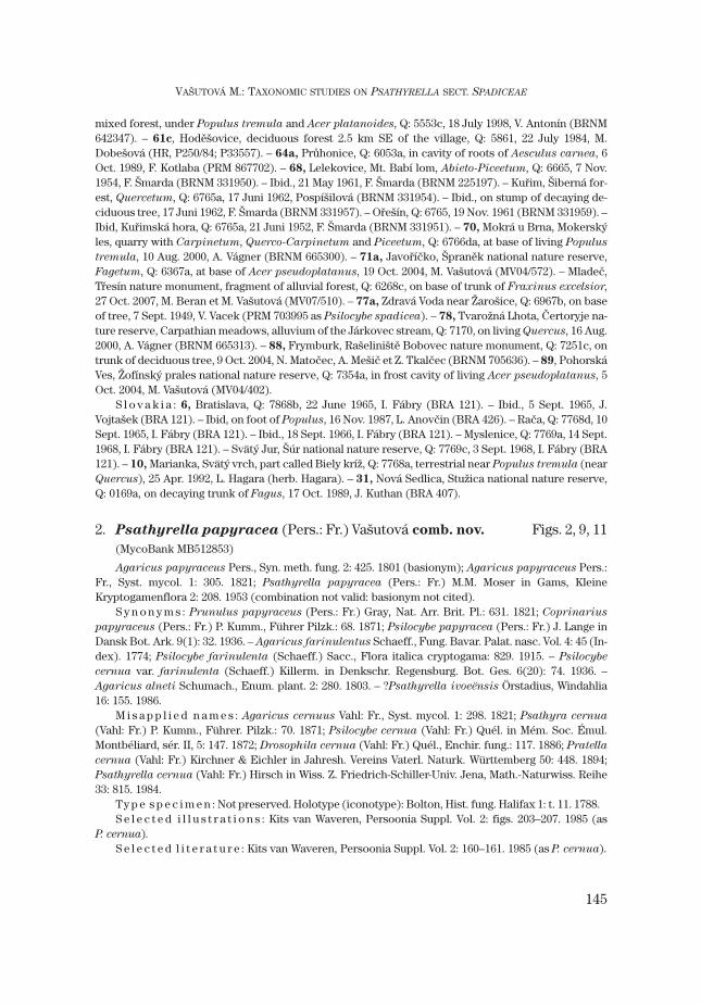
Fig. 2. Psathyrella papyracea (Pers.: Fr.) Vašutová (MV06/117): a – spores, b – basidia, c – pleuro-cystidia, d – cheilocystidia, e – caulocystidia, f – pileipellis. Bars = 10 μm for spores, 50 μm for other mi-croscopic structures.
ventricose; in young stage creamy, creamy with reddish shade, pale brown, laterbrick-brown (S60Y50M40), reddish (S70Y70M50), or dark reddish (S80Y90M70);edge white or concolorous, minutely fimbriate. S t i p e 12–87 × 1.5–12 mm, cylin-drical or tapering to base, sometimes applanate, hollow, white, whitish, creamy oreburneous, at apex pruinose to minutely floccose, surface slightly fibrillose, atbase white tomentose. F l e s h whitish to ochraceous creamy in pileus, creamy instipe. S m e l l indistinct, once a parsley smell was noticed, once a slightlyflowerish smell. S p o r e p r i n t purple-black (S99Y80M50).
S p o r e s 7–8 × 4–5 μm, average 7.7 × 4.3 μm, Q = 1.7–1.9; slightlyamygdaliform in side view (some slightly depressed), ellipsoid in front view; withdistinct to indistinct 1–1.5 μm wide germ pore and small hilar appendix; in waterreddish brown (S60Y60M50, S50Y80M50, S70Y80M50) to dark brown(S80Y60M50–S80Y60M50); in 5 % KOH greyish brown (S60Y30M10, S70Y20M10,S80Y50M30); in 10 % NH4OH reddish brown to dark brown (S70Y60M40,S80Y60M50, S80Y70M50). B a s i d i a (18–)19–25(–26) × (6.5–)7–8 μm, 4-spored,narrowly clavate. P l e u r o c y s t i d i a mostly scarce, sometimes fairly numerousto numerous, (42–)43–53(–55) × (10–)11–15(–16) μm; utriform, with obtuse apex,wall in the middle up to 0.5 μm thick, in upper part mostly thin-walled, exception-ally up to 2 μm thick; with crystals at apex forming “caps”, dissolving in KOH;hyaline. E d g e o f l a m e l l a e consisting of (1) numerous to densely packedpleurocystidioid cheilocystidia, (27–)29–49(–50) × 11–15(–17) μm, utriform, withwall up to 0.5 μm thick in middle and upper parts, at apex with crystals forming“caps” dissolving in KOH; (2) rare to lacking clavate or sphaeropedunculate cells,16–34 × 7–15 μm, thin-walled. H y m e n o p h o r a l t r a m a in 10 % NH4OH yellow-ish to colourless, parallel. Distinct colour differentiation between subhymenium(brown) and hymenium (yellowish). P i l e i p e l l i s a transition between a hymeni-derm and an epithelium, formed of colourless cells, 31–50 × (15.5–)16–28(–31)μm, with 0.5 μm thick walls. Pileocystidia scattered. C a u l o c y s t i d i a rare to nu-merous, consisting of three types: (1) utriform with crystals, 33–66 × 10–17 μm;(2) utriform without crystals, 36–48 × 9–15 μm; (3) clavate to sphaero-pedunculate, 23–37 × 11–19 μm. Walls variably thick between collections, butmostly up to 0.5 μm thick, rarely up to 1 μm. C l a m p - c o n n e c t i o n s present.
D i s t r i b u t i o n . In the Czech Republic and Slovakia rather common in suit-able habitats. Rather rare in the Netherlands (Arnolds et al. 1995), rare in the UK(Legon and Henrici 2005), also reported from France and Germany (Kits vanWaveren 1985, Enderle 1989).
E c o l o g y. P. papyracea grows mostly on bases or in frost cracks of trunks ofliving trees, mainly Fagus sylvatica and Acer pseudoplatanus, less frequently onTilia, Populus, Ulmus glabra, Acer campestre, Aesculus hippocastanus, Alnus
glutinosa and Quercus cerris, but is found also on decaying stumps or fallen de-caying trunks of Fagus, Ulmus glabra, Tilia, Fraxinus excelsior, Aesculus
147
VAŠUTOVÁ M.: TAXONOMIC STUDIES ON PSATHYRELLA SECT. SPADICEAE

hippocastanum and Carpinus betulus. Similarly to P. spadicea, P. papyracea
seems to be a saproparasitic species. In Europe the species is reported eitherfrom stumps or wood of deciduous trees, especially Fagus sylvatica and Populus
(Kits van Waveren 1985, Legon and Henrici 2005) or at the base of living decidu-ous trees, particularly Populus and Fraxinus excelsior (Enderle 1989). One col-lection concerned a group growing from an old house brick discarded in wood-land (Legon and Henrici 2005).
Comparing to P. spadicea, P. papyracea is stronger associated with natural for-ests. It occurs mostly in deciduous and mixed forests; rarely in alleys.
It grows from the lowlands to the montane belt, most collections were found inthe colline and the montane belts.
P h e n o l o g y. Sept.–Dec., mostly in Oct., once collected in Jan.D i s c u s s i o n . P. papyracea is an easily recognisable species with medium-
sized pale-coloured basidiomata, growing mostly on living deciduous trees. Micro-scopically it is characterised by utriform cystidia with crystals forming “caps” ontheir top.
According to Kits van Waveren (1985), spores of P. papyracea (as P. cernua)are 6.5–7 × 4.2–4.5 μm in size, reddish yellow in water, and possess a small (± 1μm) but fairly distinct germ pore. Enderle (1989) reported spores 7–8.5 × 4.2–5.1μm large, dark brown, with an indistinct germ pore. I observed variability in sporesize and distinctiveness of the germ pore between collections. Also the number ofpleurocystidia is very variable (from numerous to even absent). As I did not findany correlation between these characters and moreover, due to the fact thatcarpophores occur late in the year and characters could be influenced by extremeweather conditions, I consider the above-mentioned differences to fall within thespecies’ variability. In my opinion, P. papyracea is a variable species rather thana complex of microspecies.
The pattern in cystidium wall thickness is not in agreement with that observedby Kits van Waveren (1985): “with wall very slightly thickened up to 0.5 μm, rarely1 μm at very apex.” It is necessary to note that due to the presence of crystals atthe cystidium top, it is difficult to measure the cystidium wall thickness there.
Collections studied
C z e c h R e p u b l i c : 8, Srbsko, Q: 6050d, on trunk of living Ulmus glabra, 18 Sept. 1965, M. Svrčeket J. Herink (PRM 610800). – 9, Prague, Bubeneč, Q: 5852, on (burnt) roots of Aesculus
hippocastanum, 8 Nov. 1941, J.A. Herink (PRM 735915 as Psilocybe spadicea). – Ibid.,Dienzenhoferovy sady park, Q: 5852a, on trunk of living Acer campestre, 18 Nov. 1962, E. Wichanský(PRM 568670). – Ibid., on superficial cut of living Acer campestre, 19 Nov. 1963, E. Wichanský (PRM600609). – Ibid., Kinského sady park, Q: 5952a, on soil, around trunk of Tilia, 15 Nov. 1962, E.Wichanský, (PRM 568629, smaller spores than usual). – Ibid., 20 Dec. 1964, E. Wichanský (PRM 735903as Psilocybe cernua). – Ibid, Královská obora park, Q: 5852cd, near cavity of trunk of Tilia, 17 Oct.1941, J.A. Herink (PRM 735918 as Psilocybe cernua). – 14a, Jičín, Q: 5658c, on trunk of Tilia, 14 Nov.1948, A. Příhoda (PRM 609505 as Psathyrella spadicea). – 21a, Náměšť na Hané, Terezské valley, Q:
148
CZECH MYCOL. 60(2): 137–171, 2008

6468a, in frost crack of trunk of living Tilia, 22 Oct. 2003, M. Vašutová (MV03/452). – 21b, Tovačov, damof Donbas pond, Q: 6569, at base of trunk of living Populus alba, 2 Dec. 2006, M. Vašutová et J. Polčák(MV06/117). – 37e, Krajníčko, under Helfenburk ruin, Q: 6850c, on trunk of Acer pseudoplatanus, 27Sept. 1976, J. Kubička (CB 2124). – 39, Příbraz, Fabián nature reserve, Q: 7055b, in cavity of trunk of liv-ing Acer pseudoplatanus, 30 Oct. 2005, M. Beran (CB). – Klikov, Bukové kopce nature reserve, Q:7055d, on base of trunk of living Fagus, 13 Oct. 2007, M. Beran (MV07/362). – 41, Tábor, left bank ofLužnice river, Q: 6553, at base of Tilia platyphyllos, 14 Jan. 2007, M. Beran et M. Vašutová (MV07/004). –Čeřenice na Sázavě, settlement called Poříčko, Q: 6155, on decaying trunk of Carpinus betulus, 29 Oct.1944, J. Herink et J. Kubička (PRM 735922 as Psilocybe spadicea). – 42, Tábor, botanical garden, Q:6554, on base of Populus ×canadensis, 12 Oct. 2007, M. Beran et M. Vašutová (MV07/370). – 50, Turnov,Rývovy sady park, along Jizera river, Q: 5456b, near cavity at base of Ulmus, 14 Nov. 1948, J. Herink(PRM 609504 as Psathyrella spadicea). – 67, Hodonín u Kunštátu, Údolí Hodoňky nature reserve, Q:6564a, on living Alnus glutinosa, 6 Oct. 1999, A. Vágner (BRNM 648528). – 68, Vranov n. Dyjí, decidu-ous forest, Q: 7160b, at base of standing Populus, 19 Oct. 2002, L. Zelený (MV02/530). – 78, Strání, VelkáJavořina national nature reserve, Q: 7172a, on fallen trunk of Fagus sylvatica, 15 Oct. 2004, M.Vašutová, J. Wolfová, K. Bučinová et J.W. Jongepier (MV04/500). – 88a, Železná Ruda, near Čertovojezero lake, on slope of Mt. Jezerní hora, SW of the lake, deciduous forest (Picea, Fagus), Q: 6845a, atbase of living Fagus sylvatica, 16 Oct. 1995, J. Holec (PRM 885578). – 88d, Lenora, Mt. Zátoňská hora,on SW slope, deciduous virgin forest, Q: 7048, terrestrial by trunk of Ulmus glabra, 14 Oct. 1996, J.Holec (PRM 889514). – České Žleby, Mt. Spáleniště, young Fagetum with Sorbus aucuparia, Ulmus
glabra, Acer pseudoplatanus, on SW slope, on trunk of Fraxinus excelsior, 22 Sept. 1998, J. Holec(PRM 897350). – Volary, Mt. Stožec, Medvědice nature reserve, Q: 7148b, on trunk of living Fagus
sylvatica, 15 Oct. 1996, J. Holec (PRM 889545). – Zátoň, Boubínský prales national nature reserve, Q:7048b, on fallen trunk of Fagus sylvatica, 17 Oct. 1979, J. Kubička (PRM 822049 as Psilocybe
sarcocephala). – 89, Pohorská Ves, Žofínský prales national nature reserve, Q: 7354a, at base of stand-ing Fagus sylvatica, 26 Sept. 2003, J. Lederer (BRNM 705614). – Ibid., on base of living trunk of Ulmus
glabra, 30 Oct. 2004, M. Beran (CB). – Ibid., on trunks of living Fagus tree, from base up to a height of1.5 m, 6 Oct. 2005, M. Beran (CB). – Ibid., in cavity of trunk of living Acer pseudoplatanus, 25 Oct. 2005,M. Beran (CB 14532). – Ibid., in cavity of living trunk of Fagus, 25 Oct. 2005, O. Jindřich (CB). – Ibid., incavity of fallen decaying trunk of Fagus, 25 Oct. 2005, M. Beran (CB14532). – Ibid., in cavity of trunk ofliving Acer pseudoplatanus, 25 Oct. 2005, M. Beran (CB). – 91, Čachnov, Q: 6262, in cavity of livingFagus, 21 Oct. 2007, M. Vašutová (MV07/440). – 95a, Orlické Záhoří, Trčkov national nature reserve, Q:5664, in cavity of trunk of Acer pseudoplatanus, 25 Sept. 2007, M. Vašutová (MV07/219) – Ibid., in cavityof Fagus, 25 Sept. 2007, M. Vašutová (MV07/221). – 99a, Horní Lomná, Mionší national nature reserve,Q: 6578a, at base of living Acer pseudoplatanus, 31 Oct. 2002, M. Vašutová et R. Vašut (MV02/582).
S l o v a k i a : 2, Hrachovo, Svetlianská cerina nature reserve, Q: 7585ab, at base of living Quercus
cerris, 24 Oct. 2002, M. Vašutová (MV02/541). – 22, Magurka, Mt. Mestská hora, Q: 8270d, on trunk ofliving Fagus sylvatica, 27 Sept. 1989, J. Holec (PRM 887948 as Psathyrella subcernua). – 28, Žilina, Q:6778, in tree-top of Aesculus hippocastanum, on decaying bark, 30 Oct. 1982, J. Gáper (BRA 334). – 31,Kalná Ráztoka, Havešová nature reserve, Q: 9970, on fallen trunk of Fagus, 4 Oct. 1989, J. Kuthan (BRA342 as P. spadicea). – Nová Sedlica, surrounding of Stužica national nature reserve, Q: 0169, in cavity ofFagus trunk, 17 Oct. 2002, S. Adamčík (MV02/511). – Ibid., at base of Acer pseudoplatanus, 17 Oct.2002, M. Vašutová (MV02/512, MV02/513). – Ibid., Stužica national nature reserve, on slope of Hrúbkyhill, on decaying trunk of Fagus, 28 Sept. 1988, J. Kuthan (BRA 335). – Ibid., on slope of Príkry hill, ondecaying trunk of Fagus, 11 Oct. 1990, J. Kuthan (BRA 367 as P. subcernua). – Ibid., Jarabá skala naturereserve, on hill slope, on fallen trunk of Ulmus glabra, 11 Oct. 1990, J. Kuthan (BRA 367, 90/65, asP. subcernua).
149
VAŠUTOVÁ M.: TAXONOMIC STUDIES ON PSATHYRELLA SECT. SPADICEAE
Nomenclature
Psathyrella papyracea is known as P. cernua in European literature (e. g. Kitsvan Waveren 1985). Nevertheless the name P. cernua cannot be used for this fun-gus as it is based on Agaricus cernuus Vahl which is not P. cernua ss. auct. – seebelow.
Agaricus cernuus VahlAgaricus cernuus Vahl, Fl. Dan. 6(17): 9, 1790; Agaricus cernuus Vahl: Fr., Syst. mycol. 1: 298. 1821.Ty p e s p e c i m e n : not preserved. Holotype (iconotype): Vahl, Fl. Dan. 6(17): pl. 1008, 1790.
Agaricus cernuus Vahl is described as a species growing “in pratis stercoratis”and the picture represents a species with a veil, which is probably P. candolleana.The wrong interpretation came from Fries (1815), whose description, especiallythe substrate “ad truncorum radices” fits well P. cernua, but he referred to Vahland therefore added “in fimo” to the substrate. Vahl’s picture is surely not P.
cernua ss. auct.Another name sanctioned by Fries (1821) which could be a synonym of
Psathyrella cernua ss. auct. is Agaricus papyraceaus Fr.
Agaricus papyraceus Pers.: Fr.Agaricus papyraceus Pers., Syn. meth. fung. 2: 425. 1801; Agaricus papyraceus Pers.: Fr., Syst.
mycol. 1: 305. 1821.Ty p e s p e c i m e n : Not preserved. Holotype (iconotype): Bolton, Hist. fung. Halifax 1: t. 11. 1788.
The name Agaricus papyraceus was proposed by Persoon but its taxonomicconcept was derived from Bolton’s illustration (Bolton 1788), which is very simi-lar to P. cernua ss. auct. Bolton stated that “gills turns quite black in decay”, andFries (1821) even stated “lamellae diffluentes”. This could mean that Bolton’s spe-cies is in fact a Coprinus and the name cannot be used. Therefore the main ques-tion is how to interpret the expression “turn black in decay”. Bolton (1788) pub-lished twelve species probably belonging to the current genera Coprinus s. l.,Panaeolus, Anellaria and Psathyrella. He described their lamellae either as dis-solving or turning black in decay or did not describe them in such a way(Panaeolus, Anellaria). He used the term “dissolving” for short-lived Coprinus
species, the expression “turn black in decay” for the species in question (namedmembraneous agaric), bell agaric (= Coprinus aff. auricomus), domestic agaric(= Coprinus domesticus), striated agaric (Coprinus sp.) and shield or buckeleragaric (mixture of Panaeolus and Coprinus species, possibly Conocybe species).These are, except for Coprinus domesticus, species with non-dissolving lamellae.Unfortunately he did not describe any other current Psathyrella which wouldmake it possible to compare the description of their lamellae with the“membraneous agaric”. Although it cannot be fully excluded that Bolton’s species
150
CZECH MYCOL. 60(2): 137–171, 2008

was a Coprinus, regarding the shape of the pileus it is very unlikely. The fact thatlamellae of old basidiomata of P. cernua are never black but only purplish brownat most, is not so important. At that time Bolton did not use these terms precisely,so I do not consider it as a major discrepancy. Therefore I suggest using the nameAgaricus papyraceus as a basionym for name of the species currently but errone-ously known as P. cernua ss. auct.
3. Psathyrella pygmaea (Bull.: Fr.) Singer Figs. 3, 12Agaricus pygmaeus Bull., Herb. France: pl. 525, fig. 2. 1790; Agaricus pygmaeus Bull.: Fr., Syst.
mycol. 1: 263. 1821; Psathyrella pygmaea (Bull.: Fr.) Singer, Lilloa 22: 467. (‘1949’) 1951.S y n o n y m s : Naucoria pygmaea (Bull.: Fr.) Gillet, Hyménomycètes: 544. 1876; Psathyra
pygmaea (Bull.: Fr.) Quél., C. R. Ass. Franç. Av. Sci. 9: 664. 1881; Drosophila pygmaea (Bull.: Fr.) Quél.,Enchir. fung.: 117. 1886. – Psathyra consimilis Bres. & P. Henn., Verh. Bot. Ver. Prov. Brandenb. 31: 178.1889. – Hypholoma minutellum Höhn., Sitzungsber. Kaiserl. Akad. Wiss., Math.-Naturwiss. Cl., Abt. 1.116: 98.
Ty p e s p e c i m e n : not preserved. Holotype (iconotype): Agaricus pygmaeus Bull., Herb. France:pl. 525 fig. 2. 1790.
S e l e c t e d i l l u s t r a t i o n s : Enderle, Die Pilzflora des Ulmer Raumes: p. 417. 2004; Enderle andHübner, Beitr. Kenntn. Pilze Mitteleurop. 19. 2005; Breitenbach and Kränzlin, Pilze der Schweiz vol. 4:fig. 354. 1995; Kits van Waveren, Persoonia Suppl. Vol. 2: figs. 193–196. 1985; Moser and Jülich,Farbatlas der Basidiomyceten: Psathyrella 9, 1995; J.E. Lange, Fl. agaric. danic.: pl. 151 B – caespitosebasidiomata. 1939.
S e l e c t e d l i t e r a t u r e : Kits van Waveren, Persoonia Suppl. Vol. 2: 156–157. 1985.
C h a r a c t e r i s t i c s i n b r i e f . Basidiomata small-sized, often gregarious, re-minding Coprinus disseminatus. Spores small-sized, 6–7 × 3.5–4 μm, mediumbrown, with distinct germ pore. Cystidia utriform, distinctly thick-walled, withcrystals. Growing often on decaying stumps of deciduous trees.
D e s c r i p t i o n . Basidiomata solitary to caespitose. P i l e u s paraboloid, soonconico-paraboloid, at maturity convex to plano-convex, some basidiomata withsmall umbo, 4–11 mm. Surface hygrophanous, smooth, at margin translucentlystriate; brownish beige (S10Y50M30), medium brown to brownish (S20Y50M20),paler towards margin, later creamy with ochre shade (S10Y50M10). Ve i l a net-work of fibrils on pileus, visible in young stages only, later absent. L a m e l l a ecrowded, L = 14–22, l = 1–3, 1–1.5 mm broad; narrowly adnate to adnate; in youngstage creamy, later pale greyish beige (S20Y40M20), finally brown (S50Y40M30).S t i p e 11–22 × 0.5–2 mm, cylindrical with enlarged base, hollow, white to creamy,pruinose. S m e l l indistinct. S p o r e print not observed.
S p o r e s 6–7 × 3.5–4 μm, average 6.4 × 3.9 μm, Q = 1.5–1.86(–2); slightlyphaseoliform in side view, ellipsoid in front view; with distinct 1.5–2 μm broadgerm pore and small hilar appendix; in water medium brown with reddish shade(S50Y50M40); in 5 % KOH greyish brown (S60Y20M0–30); in 10 % NH4OH reddishbrown (S60Y40M40–S70Y50M40). B a s i d i a (12.5–)13–17 × 6–7 μm, 4-spored, nar-rowly clavate, rarely clavate. P l e u r o c y s t i d i a numerous to fairly numerous,
151
VAŠUTOVÁ M.: TAXONOMIC STUDIES ON PSATHYRELLA SECT. SPADICEAE
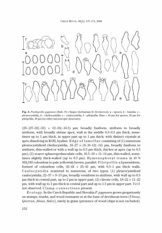
(25–)27–32(–33) × 12–16(–16.5) μm; broadly fusiform, utriform to broadlyutriform, with broadly obtuse apex, wall in the middle 0.3–0.5 μm thick, some-times up to 1 μm thick, in upper part up to 1 μm thick; with distinct crystals atapex dissolving in KOH, hyaline. E d g e o f l a m e l l a e consisting of (1) numerouspleurocystidioid cheilocystidia, 18–27 × (8–)9–12(–14) μm, broadly fusiform toutriform, thin-walled or with a wall up to 0.3 μm thick, thicker at apex (up to 0.5μm); (2) scarce sphaeropedunculate cells, 16.5–19 × 11–14 μm, thin-walled, some-times slightly thick-walled (up to 0.3 μm). H y m e n o p h o r a l t r a m a in 10 %NH4OH colourless to pale yellowish brown, parallel. P i l e i p e l l i s a hymeniderm,formed of colourless cells, 32–44 × 25–41 μm, with 0.5–1 μm thick walls.C a u l o c y s t i d i a scattered to numerous, of two types: (1) pleurocystidioidcaulocystidia, 22–37 × 9–15 μm, broadly versiform to utriform, with wall up to 0.5μm thick in central part, up to 2 μm in upper part; (2) clavate cells, 18–22 × 11–22μm, with wall up to 1 μm thick in central part and up to 1.5 μm in upper part. Ve i lnot observed. C l a m p - c o n n e c t i o n s present.
E c o l o g y. In the Czech Republic and Slovakia P. pygmaea grows gregariouslyon stumps, trunks, and wood remnants or at the base of deciduous trees (Ulmus,Quercus, Alnus, Salix), rarely in grass (presence of wood chips is not excluded).
152
CZECH MYCOL. 60(2): 137–171, 2008
Fig. 3. Psathyrella pygmaea (Bull.: Fr.) Singer (herbarium H. Deckerová): a – spores, b – basidia, c –pleurocystidia, d – cheilocystidia, e – caulocystidia, f – pileipellis. Bars = 10 μm for spores, 50 μm forpileipellis, 30 μm for other microscopic structures.

Kits van Waveren (1985) reported the species from stumps of deciduous species,Enderle (2005) from mossy stumps of various deciduous trees (Alnus, Fraxinus,Populus, Salix, Acer, Ulmus, Fagus), especially on places with a high groundwa-ter level, once at the mossy base of a living deciduous tree, once on a grass-plotwith compost. Legon and Henrici (2005) mention it from decaying wood of a de-ciduous tree, usually on large logs or stumps, occasionally mixed with Coprinus
disseminatus.P. pygmaea occurs in deciduous forests, mostly in alluvial forests but is some-
times found in anthropogenic habitats such as parks or gardens.It is known from the lowlands to the submontane belt, most collections were
found in lowlands and the colline belt.P h e n o l o g y. June–Oct.D i s t r i b u t i o n . Scattered, but probably overlooked. Reported from the
Netherlands as rather common (Arnolds et al. 1995) and occasional but wide-spread in England (Legon and Henrici 2005). Known also from France (Kits vanWaveren 1985), Germany (Enderle 2005) and other European countries (seeEnderle 2005).
D i s c u s s i o n . Although the iconotype of Psathyrella pygmaea is quite sche-matic, it shows the main characters of the species well. The basidiomata grow gre-gariously on wood and have a plano-convex pileus and pale lamellae. AlsoBulliard’s later description agrees with the fungus currently known as P.
pygmaea. A detailed description of the species was given by Kits van Waveren(1985). Czech collections differ in the absence of pink shades in the lamellae,a slightly smaller range of spore size, which are also a little darker, and cystidiahaving thicker walls. The percentage of sphaeropedunculate cells is rather low.There is no visible difference in microscopical characters between collectionsgrowing on wood in alluvial forests and those growing terrestrially in man-madehabitats. Differences in macroscopical characters cannot be evaluated becausemacroscopic descriptions are often absent in herbaria. Also an analysis of ITSrDNA from two specimens of P. pygmaea, each from a different habitat, did notreveal any differences (unpublished data). More data are needed to solve thisquestion.
According to Enderle (Enderle and Hübner 2005), a velum universale ofPsathyrella pygmaea was once observed. It was composed of cylindrical toversiform, yellow, thick-walled elements. I found a similar structure inPsathyrella candolleana (Fr.) Maire. These structures are probably remnants ofthe velum universale and could be used (similarly as in Coprinus s.l.) forinfrageneric classifications. Due to their ephemerality they have not been studiedyet.
153
VAŠUTOVÁ M.: TAXONOMIC STUDIES ON PSATHYRELLA SECT. SPADICEAE

Collections studied
C z e c h R e p u b l i c : 7d, Dolany near Unhošť, Q: 5850d, on decaying trunk of deciduous tree, 19July 1941, J.A. Herink (PRM 735358 as Psathyra consimilis). – 11b, Lysá n. Labem, Stratov, Q: 5855a,on decaying trunk and roots(?) of Ulmus, 17 Oct. 1961, M. Svrček (PRM 616623; Svrček 1960). – 16,
Brno, Arboretum VŠZ, Q: 6765d, on Salix stump, 18 Juni 1993, A. Vágner (BRNM 590179). – Brno-Lesná,Q: 6765d, on stump of Acer pseudoplatanus or Fraxinus, 2 July 1993, A. Vágner (BRNM 590154). –Vranovice nad Svratkou, Plačkův les nature monument, Q: 7065d, on decaying stump of deciduous tree,26 July 2000, A. Vágner (BRNM 664988). – 18a, Lanžhot, Ranšpurk national nature reserve, Q: 6676a, onroot swellings of Quercus robur, 12 Oct. 2001, M. Vašutová (MV01/469). – 20b, Žarošice, Q: 6967d, ontrunk of Alnus glutinosa, 13 Aug. 1946, V. Vacek (PRM 735359 as Psathyra consimilis). – Ibid., ontrunk of Alnus, 19 Aug. 1947, V. Vacek (PRM 735357 as P. consimilis). – 21b, Litovelské Pomoraví Pro-tected Landscape Area, on stump of deciduous tree, L. Kašpárek (MV06/062b). – 37, Frymburk, greenin centre of square, Q: 7351a, terrestrial in grass, 4 Oct. 2004, M. Vašutová et A. Vágner (BRNM705629). – 83, right bank of Odra river, between the villages of Starý Bohumín and Kopytov, Q: 6076c,on decaying wood of deciduous tree, 8 June 2004, H. Deckerová (herbarium H. Deckerová).
S l o v a k i a : 5, Bratislava-Rusovce, Dolná Sihoť, bank of Dunaj river oxbow, Q: 7969a, in grass, 22July 2003, J. Červenka (herbarium J. Červenka).
4. Psathyrella olympiana A.H. Sm. Fig. 4Psathyrella olympiana A.H. Sm., Contr. Univ. Michigan Herb. 5: 36: 1941.Ty p e s p e c i m e n : USA, Olympic National Park, Jackson Guard Station, 6 June 1939 [MICH 11991
(Sm 14118)].S e l e c t e d i l l u s t r a t i o n s : Enderle, Die Pilzflora des Ulmer Raumes: 418. 2004; Ludwig,
Pilzkompendium, t. 379, f. 98.41D. 2007; Tassi, Bull. Soc. Mycol. France 113: t. 329. 1997; Kits vanWaveren, Persoonia Suppl. Vol. 2: figs. 213–217. 1985; Breitenbach and Kränzlin, Pilze der Schweiz 4:fig. 341. 1995.
S e l e c t e d l i t e r a t u r e : Smith, Mem. New York Bot. Gard. 24: 256–257. 1972; Kits van Waveren,Persoonia Suppl. Vol. 2: 167–168. 1985; Derbsch, Zeitsch. f. Pilzkunde 43: 183. 1977.
Although P. olympiana is reported from several countries of Central Europe, it has not been foundin the Czech Republic or Slovakia yet. Therefore the description is only based on my own study of typematerial and Smith’s description of macrocharacters.
C h a r a c t e r i s t i c s i n b r i e f . Basidiomata medium-sized, growing solitaryto gregarious, pileus brown, with distinct veil. Spores large-sized, 9–10.5 × 4.5–5.5μm, reddish brown, with indistinct germ pore. Cystidia mainly broadly versiformwith obtuse apex, thick-walled (up to 2.5 μm), especially in pedicel and upperpart, with crystals. Growing on decaying stumps or trunks of deciduous trees.
D e s c r i p t i o n o f m a c r o s c o p i c c h a r a c t e r s [based on descriptions bySmith (1941, 1972), structure adapted to the pattern used in this paper].Basidiomata solitary to gregarious. P i l e u s obtusely conical to convex, laterplane, 1–4 cm. Surface smooth or slightly rugulose, pileus margin in mature stagetranslucently striate; dark rusty brown, drying out to sordid pinkish buff or dulldirty tan. Ve i l covering the pileus a thin layer of fibrils in young stage, later mar-gin decorated with scattered remains of the thin veil. L a m e l l a e crowded, L =29–35, 0.3 cm broad, broadly adnate to adnate, pallid brownish, later greyishbrown, with white edge. S t i p e 3–5 × 0.2–0.5 cm, equal, hollow, white, upper partfibrillose-squamulose to coarsely pruinose and faintly striate, densely white
154
CZECH MYCOL. 60(2): 137–171, 2008
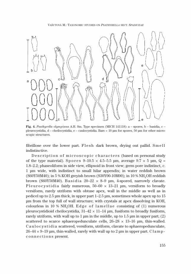
fibrillose over the lower part. F l e s h dark brown, drying out pallid. S m e l lindistinctive.
D e s c r i p t i o n o f m i c r o s c o p i c c h a r a c t e r s (based on personal studyof the type material). S p o r e s 9–10.5 × 4.5–5.5 μm, average 9.7 × 5 μm, Q =1.8–2.2; phaseoliform in side view, ellipsoid in front view; germ pore indistinct, c.1 μm wide, with indistinct to small hilar appendix; in water reddish brown(S60Y50M40); in 5 % KOH greyish brown (S30Y00-10M00); in 10 % NH4OH reddishbrown (S60Y50M40). B a s i d i a 20–22 × 8–9 μm, 4-spored, narrowly clavate.P l e u r o c y s t i d i a fairly numerous, 50–60 × 13–21 μm, versiform to broadlyversiform, rarely utriform with obtuse apex, wall in the middle as well as inpedicel up to 2.5 μm thick, in upper part 1–2.5 μm, sometimes whole apex up to 15μm from the top full of wall structure; with crystals at apex dissolving in KOH,colourless in 10 % NH4OH. E d g e o f l a m e l l a e consisting of (1) numerouspleurocystidioid cheilocystidia, 31–42 × 11–14 μm, fusiform to broadly fusiform,rarely utriform, with wall up to 1 μm in the middle, up to 1.5 μm in upper part; (2)scattered to scarce sphaeropedunculate cells, 26–28 × 13–16 μm, thin-walled.C a u l o c y s t i d i a scattered, versiform, utriform, clavate to sphaeropedunculate,26–44 × 9–19 μm, thin-walled, rarely with wall up to 2 μm in upper part. C l a m p -c o n n e c t i o n s present.
155
VAŠUTOVÁ M.: TAXONOMIC STUDIES ON PSATHYRELLA SECT. SPADICEAE
Fig. 4. Psathyrella olympiana A.H. Sm. Type specimen (MICH 141118): a – spores, b – basidia, c –pleurocystidia, d – cheilocystidia, e – caulocystidia. Bars = 10 μm for spores, 50 μm for other micro-scopic structures.

E c o l o g y. Smith (1941) reported the species as growing scattered to gregari-ous on old wood and debris of alder and cottonwood. In Europe, Derbsch (1977)found it growing on wood remnants, Kits van Waveren (1985) on and againststumps of deciduous trees, Enderle (1987) near and on mossy wood remnants,Legon and Henrici (2005) on buried wood. It occurs in near-natural forests (allu-vial forest – WU 18520, WU 26910; deciduous forest with Quercus, Carpinus andFagus – Derbsch 1977) as well as in man-made ones – “Fichtenparzelle” as re-ported by Enderle (1987).
D i s t r i b u t i o n . Not found in the Czech Republic or Slovakia. Known fromGermany (Derbsch 1977, Enderle 1987), Austria, Hungary, and the UK (Legon andHenrici 2005). Rather rare in the Netherlands (Arnolds et al. 1995), also reportedfrom France (Tassi 1997).
D i s c u s s i o n . According to my observations, the type material has largerspores than described by Smith (1941) and distinctively broader cystidia. Thebroader cystidia were found in their material also by Kits van Waveren (1985) andEnderle (1987). Both emphasised the extremely thick-walled pedicel of thepleurocystidia, which I found in specimen of Enderle too, but in the case of thetype material this character was not so distinct. I have not seen the type materialof P. cloverae A.H. Sm. Therefore I cannot confirm their conspecifity as stated byKits van Waveren (1985).
Collections studied
U S A : type [MICH 11991]. – A u s t r i a : Kärnten, Völkermarkt, Kleindorf II, alluvial forest, 16 Sept.1998, A. Hausknecht (WU 18520). – H u n g a r y : Szabolcs-Szatmár-Bereg megye, Fényi erdö nature re-serve, c. 2 km S of the village of Bátorliget, alluvial forest, wood of a deciduous tree, 27 Oct. 2006, I.Rimóczi (WU 26910). – G e r m a n y : Bavaria, near Unterfahlheim, floodplain forests of Danube, underPicea abies, on and near decaying wood (of Picea or broad-leaved tree?), 17 Aug. 1985, M. Enderle.
5. Psathyrella spintrigeroides P.D. Orton Figs. 5, 6, 13Psathyrella spintrigeroides P.D. Orton, Trans. Brit. Mycol. Soc. 43: 377. 1960.Ty p e s p e c i m e n : UK, England, Surrey, Sheapleas (Mountain Wood): near West Horstley, 5 Nov.
1955, leg. P.D. Orton [K(M) 70569].S e l e c t e d i l l u s t r a t i o n s : Enderle, Die Pilzflora des Ulmer Raumes: 418. 2004.S e l e c t e d l i t e r a t u r e : Derbsch, Zeitsch. f. Pilzkunde 43: 183. 1977; Enderle, Beitr. Kenntn. Pilze
Mitteleurop. 5: 68–70. 1989.
C h a r a c t e r i s t i c s i n b r i e f . Basidiomata medium-sized, growing solitary,pileus brown, with distinct veil. Spores medium-sized, (7–)7.5–8 × 4–4.5 μm, yel-lowish brown to brown, with indistinct germ pore. Cystidia versiform with oftenirregular apex, rather thick-walled (0.5–1.5 μm), especially in upper part, withoutcrystals at the top, if so, then only small and scattered. Growing on decayingstumps or trunks of deciduous trees.
D e s c r i p t i o n (based only on young and mature basidiomata; old basidiomatawere not found). Basidiomata solitary. P i l e u s in young stage paraboloid, 0.8–1.1
156
CZECH MYCOL. 60(2): 137–171, 2008

cm broad, at maturity convex, later broadly convex, 1.3–2.5 cm. Surface smoothor slightly rugulose, pileus margin in mature stage translucently striate; palebrownish (S50Y50M40), medium brownish (S50Y90M50) to dark brown(S90Y90M60), drying out to ochre (S20Y50M20). Ve i l distinct; filamentous,white; entire pileus covered with dispersed fibrils at first, later persisting on widepileus margin as small rather indistinctive squamules. Veil connecting pileus withstipe in young stage, remains as distinct scales on margin at maturity. L a m e l l a esubcrowded, L = 22–32, l = 1–5; 0.2–0.6 cm broad, adnate to broadly adnate, palebrown (S50Y40M20) to brown (S70Y50M30–40), with minutely fimbriate whiteedge. S t i p e 3.2–5 × 0.35–0.4 cm, cylindrical, hollow, white to creamy, pale beigeat base, apex markedly striate from lamella base and with small white squamules,entire stipe fibrillosely scaly to slightly filamentous from veil. F l e s h in pileus0.1–0.25 cm thick, concolorous, pale beige. S m e l l indistinctive.
S p o r e s (7–)7.5–8 × 4–4.5 μm, average 7.8–4.2 μm, Q = (1.6–)1.7–2; some veryslightly phaseoliform in side view, ellipsoid in front view; germ pore indistinct,1–1.5 μm broad; with small hilar appendix; in water yellowish brown (S60Y50M40)to brown (S70Y90M50), in 5 % KOH greyish brown (S80Y40–20M10–20), in 10 %NH4OH brown (S80Y40M40). B a s i d i a (15–)16–20(–22) × (6–)7–9 μm, 4-spored(rarely 2-spored), narrowly clavate. P l e u r o c y s t i d i a n u m e r o u s to very nu-
157
VAŠUTOVÁ M.: TAXONOMIC STUDIES ON PSATHYRELLA SECT. SPADICEAE
Fig. 5. Psathyrella spintrigeroides P.D. Orton (BRNM 705638): a – spores, b – basidia, c –pleurocystidia, d – cheilocystidia, e – caulocystidia, f – pileipellis, h – veil. Bars = 10 μm for spores, 20μm for basidia, 50 μm for other microscopic structures.
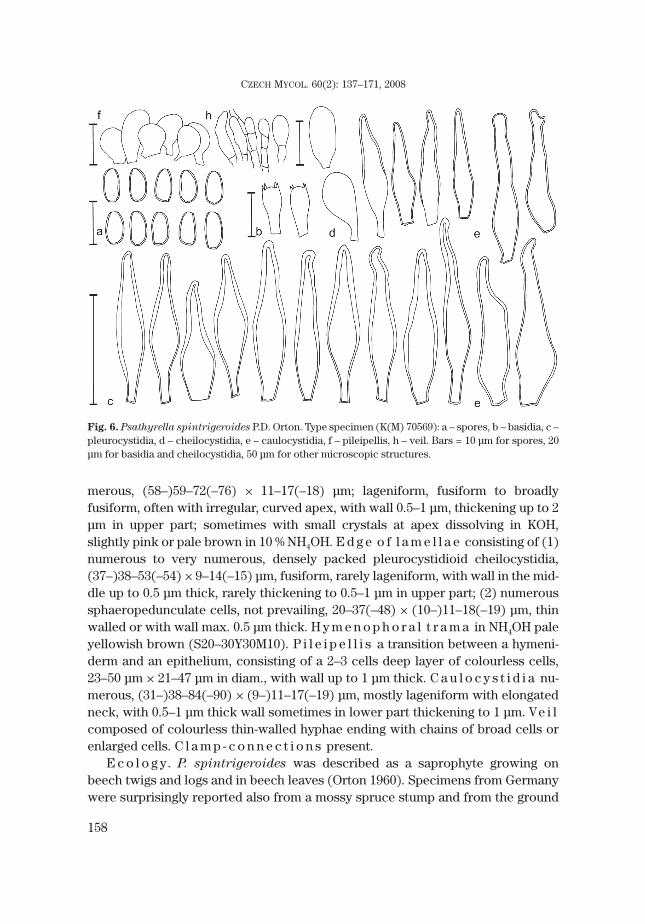
merous, (58–)59–72(–76) × 11–17(–18) μm; lageniform, fusiform to broadlyfusiform, often with irregular, curved apex, with wall 0.5–1 μm, thickening up to 2μm in upper part; sometimes with small crystals at apex dissolving in KOH,slightly pink or pale brown in 10 % NH4OH. E d g e o f l a m e l l a e consisting of (1)numerous to very numerous, densely packed pleurocystidioid cheilocystidia,(37–)38–53(–54) × 9–14(–15) μm, fusiform, rarely lageniform, with wall in the mid-dle up to 0.5 μm thick, rarely thickening to 0.5–1 μm in upper part; (2) numeroussphaeropedunculate cells, not prevailing, 20–37(–48) × (10–)11–18(–19) μm, thinwalled or with wall max. 0.5 μm thick. H y m e n o p h o r a l t r a m a in NH4OH paleyellowish brown (S20–30Y30M10). P i l e i p e l l i s a transition between a hymeni-derm and an epithelium, consisting of a 2–3 cells deep layer of colourless cells,23–50 μm × 21–47 μm in diam., with wall up to 1 μm thick. C a u l o c y s t i d i a nu-merous, (31–)38–84(–90) × (9–)11–17(–19) μm, mostly lageniform with elongatedneck, with 0.5–1 μm thick wall sometimes in lower part thickening to 1 μm. Ve i lcomposed of colourless thin-walled hyphae ending with chains of broad cells orenlarged cells. C l a m p - c o n n e c t i o n s present.
E c o l o g y. P. spintrigeroides was described as a saprophyte growing onbeech twigs and logs and in beech leaves (Orton 1960). Specimens from Germanywere surprisingly reported also from a mossy spruce stump and from the ground
158
CZECH MYCOL. 60(2): 137–171, 2008
Fig. 6. Psathyrella spintrigeroides P.D. Orton. Type specimen (K(M) 70569): a – spores, b – basidia, c –pleurocystidia, d – cheilocystidia, e – caulocystidia, f – pileipellis, h – veil. Bars = 10 μm for spores, 20μm for basidia and cheilocystidia, 50 μm for other microscopic structures.

in a young spruce forest (Enderle 1989); the Hungarian record is from an alluvialforest. Records from the Czech Republic and Slovakia are from fallen mossytrunks (Fagus sylvatica) in natural deciduous forests in the montane belt.
D i s t r i b u t i o n . Rare (Czech Republic: 2 localities, Slovakia: 1 locality), in-cluded in the Red list of fungi of the Czech Republic (Holec and Beran 2006). Re-ported as rare also from the rest of Europe: Germany (3 localities; Enderle 1989,Derbsch 1977), Austria (1 locality – WU 17247), Hungary (1 locality – BRNM),United Kingdom [1 locality – Orton (1960), few other records – herbarium mate-rial not preserved (Legon and Henrici 2005)]. In Sweden rather common(Örstadius, in litt.).
P h e n o l o g y. Sept.–Oct.D i s c u s s i o n . P. spintrigeroides has medium-sized basidiomata and a well-
developed veil. The key character are its cystidia thickened in upper part. Thewall thickness is variable between collections: the type material has rather thick-walled cystidia (1.6 μm on average), the Czech collections 0.8 and 1.8 μm on aver-age, respectively. However, this variability is not correlated with molecular char-acters (unpubl. data) and therefore I consider it of no taxonomical importance.Rarely only slightly thick-walled cystidia are thin-walled at the top.
Although German records do not support the presumption of a strong associa-tion with deciduous natural forests, more data are needed to verify this. Confu-sion is possible with P. rostellata Örstadius and P. artemisiae (Pass.) Konrad etMaubl. The main difference is in pattern of thickness of the cystidium wall. Whilethe cystidium wall in P. spintrigeroides is the thickest on or just below the top, inP. rostellata and P. artemisiae this wall is the thickest in the middle. The Ameri-can species Psathyrella avellaneifolia A.H. Sm. and Psathyrella indecorosa A.H.Sm. are morphologically similar. The first one differs by smaller spores(6.5–)7–7.5(–8) × (3.5–)4(–4.5) μm, the second by a triangular spore shape and oc-currence on alder logs.
Collections studied
C z e c h R e p u b l i c : 88d, České Žleby, Žlebský vrch nature reserve, on fallen trunk of deciduoustree (Fagus sylvatica?), 6 Oct. 2004, M. Vašutová (BRNM 705638). – 89, Pohorská Ves, Žofínský pralesnational nature reserve, on fallen Fagus trunk, 23 Sept. 2003, M. Vašutová (BRNM 705639). – S l o v a k i a :31, Kalná Ráztoka, Havešová nature reserve, Q: 9970b, on stump of Fagus, 5 Oct. 1992, J. Terray (BRA407). – G e r m a n y : Donau-Auwald near Unterfahlheim, in man-made Picea forest, on mossy Picea
stump, 1 Nov. 1987, M. Enderle (herbarium M. Enderle). – H u n g a r y : Szabolcs-Szatmár-Bereg megye,Fényi erdö nature reserve, c. 2 km S of the village of Bátorliget, alluvial forest with Fraxinus
angustifolia and Quercus robur, on decaying trunk of Fraxinus angustifolia, 26 Oct. 2006, V. Antonín(BRNM, 06.174). – A u s t r i a : North Austria, Gföhl, Dobrasperre, on Fagus wood, 13 July 1997, A.Hausknecht and F. Reinwald (WU 17247, as P. artemisiae var. microspora, Hausknecht et al. 2006). –U n i t e d K i n g d o m : type [K(M) 70569].
159
VAŠUTOVÁ M.: TAXONOMIC STUDIES ON PSATHYRELLA SECT. SPADICEAE

Insufficiently known taxa
Psathyrella variata A.H. Sm. Fig. 7Psathyrella variata A.H. Sm., in Mem. New York Bot. Gard. 24: 238, 1972.Ty p e s p e c i m e n : USA, Priest Lake, Bonner Co., Idaho, on cottonwood, 6 Oct. 1964, leg. A.H.
Smith [MICH 5379 (Smith 71092)].S e l e c t e d i l l u s t r a t i o n s : Smith, Mem. New York Bot. Gard. 24: figs. 446–452. 1972; Kits van
Waveren, Persoonia Suppl. Vol. 2: figs. 197–202. 1985; Hausknecht et Krisai-Greilhuber, Fungi nondelineati 2: figs. 8a-g. 1997.
S e l e c t e d l i t e r a t u r e : Smith, Mem. New York Bot. Gard. 24: 238–239. 1972; Hausknecht etKrisai-Greilhuber, Fungi non delineati 2: 26–28. 1997.
D i s c u s s i o n . Psathyrella variata is a species very close to P. spadicea,which was distinguished by Smith according to the presence of a mixture ofleptocystidia and lamprocystidia on one lamella (Smith 1972). My study of thetype material confirms this character in the case of cheilocystidia only, althoughÖrstadius (2001) has seen pleuroleptocystidia as well. This could be due to thefact that I received only a part of the type material from MICH and the charactercould be very variable between single basidiomata, or the type is a mixture of spe-cies. Kits van Waveren (1986) found just a few pleuroleptocystidia in his material,and he reported the presence of hairs at the pileus margin to be a good character.Hausknecht (in Hausknecht et Krisai-Greilhuber 1997) did not observe pleuro-leptocystidia but confirmed cheiloleptocystidia and hairs in his material.
Örstadius (2001) considers P. variata to be a synonym of P. spadicea. Accord-ing to him, the thickness of the cystidium wall is not so taxonomically importantand hairs are found on the pileus of very young species of P. spadicea, too. I haveconfirmed this finding. Moreover, few cheiloleptocystidia are present in P. spadicea
as well (see Kits van Waveren 1986, fig. 210). The ratio of cheiloleptocystidia var-ies between collections; it is even possible to find “transitional forms” between P.
spadicea and P. variata. The conspecificity of the above-mentioned species issupported by current molecular data (Vašutová et al. 2008) but due to the fact thatonly one specimen of P. variata, one of P. spadicea and one transitional form wasused for DNA analysis, it is premature to draw this conclusion yet.
One of my personal collections could be labelled as P. variata. It differs fromthe type by an absence of pleuroleptocystidia. There are also small differences incolour; I did not see such dark chocolate lamellae at maturity as mentioned bySmith (1972) and Kits van Waveren (1985). Macroscopically, the Czech collectionis closest to the Austrian one. Smith described the germ pore as very small, whilethe other authors state it is missing. The collection is described below:
160
CZECH MYCOL. 60(2): 137–171, 2008
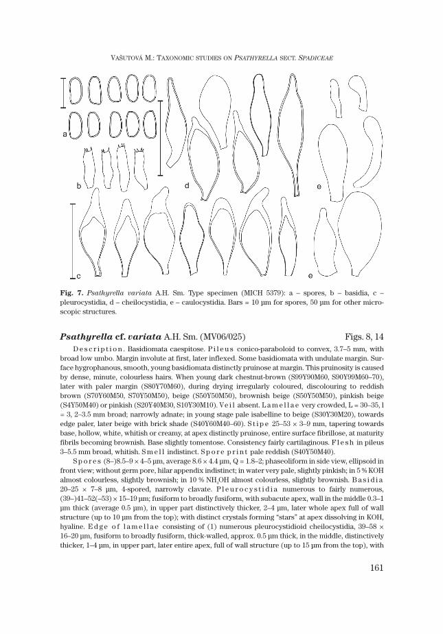
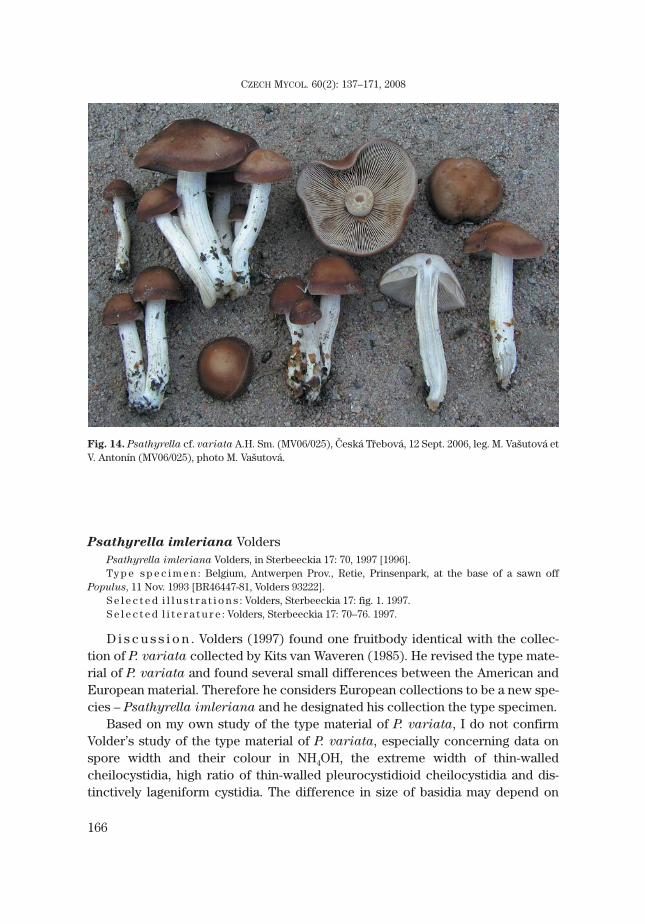
Psathyrella cf. variata A.H. Sm. (MV06/025) Figs. 8, 14D e s c r i p t i o n . Basidiomata caespitose. P i l e u s conico-paraboloid to convex, 3.7–5 mm, with
broad low umbo. Margin involute at first, later inflexed. Some basidiomata with undulate margin. Sur-face hygrophanous, smooth, young basidiomata distinctly pruinose at margin. This pruinosity is causedby dense, minute, colourless hairs. When young dark chestnut-brown (S99Y90M60, S90Y99M60–70),later with paler margin (S80Y70M60), during drying irregularly coloured, discolouring to reddishbrown (S70Y60M50, S70Y50M50), beige (S50Y50M50), brownish beige (S50Y50M50), pinkish beige(S4Y50M40) or pinkish (S20Y40M30, S10Y30M10). Ve i l absent. L a m e l l a e very crowded, L = 30–35, l= 3, 2–3.5 mm broad; narrowly adnate; in young stage pale isabelline to beige (S30Y30M20), towardsedge paler, later beige with brick shade (S40Y60M40–60). S t i p e 25–53 × 3–9 mm, tapering towardsbase, hollow, white, whitish or creamy, at apex distinctly pruinose, entire surface fibrillose, at maturityfibrils becoming brownish. Base slightly tomentose. Consistency fairly cartilaginous. F l e s h in pileus3–5.5 mm broad, whitish. S m e l l indistinct. S p o r e p r i n t pale reddish (S40Y50M40).
S p o r e s (8–)8.5–9 × 4–5 μm, average 8.6 × 4.4 μm, Q = 1.8–2; phaseoliform in side view, ellipsoid infront view; without germ pore, hilar appendix indistinct; in water very pale, slightly pinkish; in 5 % KOHalmost colourless, slightly brownish; in 10 % NH4OH almost colourless, slightly brownish. B a s i d i a20–25 × 7–8 μm, 4-spored, narrowly clavate. P l e u r o c y s t i d i a numerous to fairly numerous,(39–)41–52(–53) × 15–19 μm; fusiform to broadly fusiform, with subacute apex, wall in the middle 0.3–1μm thick (average 0.5 μm), in upper part distinctively thicker, 2–4 μm, later whole apex full of wallstructure (up to 10 μm from the top); with distinct crystals forming “stars” at apex dissolving in KOH,hyaline. E d g e o f l a m e l l a e consisting of (1) numerous pleurocystidioid cheilocystidia, 39–58 ×16–20 μm, fusiform to broadly fusiform, thick-walled, approx. 0.5 μm thick, in the middle, distinctivelythicker, 1–4 μm, in upper part, later entire apex, full of wall structure (up to 15 μm from the top), with
161
VAŠUTOVÁ M.: TAXONOMIC STUDIES ON PSATHYRELLA SECT. SPADICEAE
Fig. 7. Psathyrella variata A.H. Sm. Type specimen (MICH 5379): a – spores, b – basidia, c –pleurocystidia, d – cheilocystidia, e – caulocystidia. Bars = 10 μm for spores, 50 μm for other micro-scopic structures.
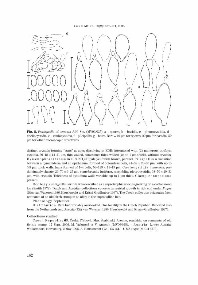
distinct crystals forming “stars” at apex dissolving in KOH; intermixed with (2) numerous utriformcystidia, 30–48 × 14–21 μm, thin-walled, sometimes thick-walled (up to 1 μm thick), without crystals.H y m e n o p h o r a l t r a m a in 10 % NH4OH pale yellowish brown, parallel. P i l e i p e l l i s a transitionbetween a hymeniderm and an epithelium, formed of colourless cells, 41–58 × 23–35 μm, with up to0.5 μm thick walls, hairs formed of 1–4 cells, 53–129 × 13–19 μm. C a u l o c y s t i d i a numerous, pre-dominately clavate, 23–70 × 9–25 μm, some broadly fusiform, resembling pleurocystidia, 38–70 × 10–31μm, with crystals. Thickness of cystidium walls variable; up to 1 μm thick. C l a m p - c o n n e c t i o n spresent.
E c o l o g y. Psathyrella variata was described as a saprotrophic species growing on a cottonwoodlog (Smith 1972). Dutch and Austrian collections concern terrestrial growth in rich soil under Fagus
(Kits van Waveren 1986, Hausknecht and Krisai-Greilhuber 1997). The Czech collection originates fromremnants of an old birch stump in an alley in the supracolline belt.
P h e n o l o g y. September.D i s t r i b u t i o n . Rare but probably overlooked. One locality in the Czech Republic. Reported also
from the Netherlands and Austria (Kits van Waveren 1986, Hausknecht and Krisai–Greilhuber 1997).
Collections studied
C z e c h R e p u b l i c : 63, Česká Třebová, Max Švabinský Avenue, roadside, on remnants of oldBetula stump, 17 Sept. 2006, M. Vašutová et V. Antonín (MV06/025). – A u s t r i a : Lower Austria,Wolkersdorf, Hornsburg, 2 May 1995, A. Hausknecht (WU 13716). – U S A : type [MICH 5379].
162
CZECH MYCOL. 60(2): 137–171, 2008
Fig. 8. Psathyrella cf. variata A.H. Sm. (MV06/025): a – spores, b – basidia, c – pleurocystidia, d –cheilocystidia, e – caulocystidia, f – pileipellis, g – hairs. Bars = 10 μm for spores, 20 μm for basidia, 50μm for other microscopic structures.

163
VAŠUTOVÁ M.: TAXONOMIC STUDIES ON PSATHYRELLA SECT. SPADICEAE
Fig. 9. Psathyrella papyracea (Pers.: Fr.) Vašutová, Žofínský prales, 30 Sept. 2008, photo J. Burel.

164
CZECH MYCOL. 60(2): 137–171, 2008
Fig. 10. Psathyrella spadicea (P. Kumm.)Singer, Troubky, 2 Dec. 2006, leg. M. Vašutováet J. Polčák (MV06/118), photo M. Vašutová.
Fig. 11. Psathyrella papyracea (Pers.: Fr.) Vašutová, Troubky, 2 Dec. 2006, leg. M. Vašutová et J.Polčák (MV06/117), photo M. Vašutová.

165
VAŠUTOVÁ M.: TAXONOMIC STUDIES ON PSATHYRELLA SECT. SPADICEAE
Fig. 12. Psathyrella pygmaea (Bull.: Fr.) Singer, Frymburk, 4 Oct. 2004, leg. M. Vašutová et A. Vágner(BRNM 705629), photo M. Vašutová.
Fig. 13. Psathyrella spintrigeroides P.D. Orton (BRNM 705638), Žofínský prales, 23 Sept. 2003, leg. M.Vašutová (BRNM 705640), photo M. Vašutová.
Psathyrella imleriana VoldersPsathyrella imleriana Volders, in Sterbeeckia 17: 70, 1997 [1996].Ty p e s p e c i m e n : Belgium, Antwerpen Prov., Retie, Prinsenpark, at the base of a sawn off
Populus, 11 Nov. 1993 [BR46447-81, Volders 93222].S e l e c t e d i l l u s t r a t i o n s : Volders, Sterbeeckia 17: fig. 1. 1997.S e l e c t e d l i t e r a t u r e : Volders, Sterbeeckia 17: 70–76. 1997.
D i s c u s s i o n . Volders (1997) found one fruitbody identical with the collec-tion of P. variata collected by Kits van Waveren (1985). He revised the type mate-rial of P. variata and found several small differences between the American andEuropean material. Therefore he considers European collections to be a new spe-cies – Psathyrella imleriana and he designated his collection the type specimen.
Based on my own study of the type material of P. variata, I do not confirmVolder’s study of the type material of P. variata, especially concerning data onspore width and their colour in NH4OH, the extreme width of thin-walledcheilocystidia, high ratio of thin-walled pleurocystidioid cheilocystidia and dis-tinctively lageniform cystidia. The difference in size of basidia may depend on
166
CZECH MYCOL. 60(2): 137–171, 2008
Fig. 14. Psathyrella cf. variata A.H. Sm. (MV06/025), Česká Třebová, 12 Sept. 2006, leg. M. Vašutová etV. Antonín (MV06/025), photo M. Vašutová.

their position on the lamellae – moreover data from collections from Austria andthe Czech Republic are in agreement with Smith’s species. Volder noticed thattype material of P. variata is without crystals but these are often disappearingwith age. Also macroscopic characters (compactness, surface of stem, and cavityor solidity of stem) can be variable and depend on age of basidiomata and growthconditions. The same counts for ecology. My study of the type specimen of P.
imleriana, of which only part of a pileus is preserved now, did not reveal any im-portant differences between this specimen and the type of P. variata. At the mo-ment I consider P. imleriana to be a synonym of P. variata and if the conspecifityof P. spadicea and P. variata is proven, also a synonym of P. spadicea.
Psathyrella sarcocephala (Fr.: Fr.) SingerAgaricus sarcocephalus Fr., Observ. mycol. 1: 51. 1815; Agaricus compactus [unranked] sarco-
cephalus (Fr.: Fr.) Fr., Syst. mycol. 1: 290. 1821; Psathyrella sarcocephala (Fr.: Fr.) Singer ss. Singer nonFr. 1815, Lilloa 22: 468. (‘1949’) 1951.
Ty p e s p e c i m e n : not preserved.M i s a p p l i e d n a m e s : “Agaricus sarcocephalus” auct. non Fr., Epicr. syst. mycol.: 228. 1838;
Monogr. hymenomycet. Suec.: 429. 1857; Hymenomyc. eur.: 297. 1874; Ic. Hymenomyc. 2: pl. 135, fig. 1. 1879.
D i s c u s s i o n . The name Agaricus sarcocephalus is linked to the description byFries (1815), sanctioned by him (1821) at variety level as A. compactus β sarco-
cephalus. This fungus should be a “vera omnimo Pratellae” species with “carnosoflavescente” pileus and “flavescens” stipe. As was already mentioned by Enderle(1989), there is no known similar Psathyrella species to which the description couldfit. Therefore the epithet “sarcocephalus” is not usable for any Psathyrella.
As was already published by Örstadius (2001), Fries wrote in his later publica-tions (1857, 1874) that Agaricus compactus β sarcocephalus is a “forma dubius”and modified his description of Agaricus sarcocephalus. The modified descrip-tion agrees with the figure published in Icones (Fries 1879). This was probably thestarting point for following interpretations of Agaricus sarcocephalus. The pic-ture of Agaricus sarcocephalus (Fries 1879) represents young basidiomata ofa species macroscopically intermediate between Psathyrella papyracea (P.
cernua ss. auct.) and P. spadicea. Noteworthy are small squamules depicted onpilei of Agaricus sarcocephalus by Fries (1879), never mentioned by later au-thors. If this species exists, it cannot be named A. sarcocephalus. A similar funguswas depicted by Cooke (1886, t. 567), Bresadola (1930, t. 855) and Konrad andMaublanc (1929, t. 45). Örstadius (2001) pointed out that the picture of Psilocybe
sarcocephala by Konrad and Maublanc is Psathyrella cernua ss. auct., and a pic-ture of Psilocybe sarcocephala by Bresadola (1930) cannot be interpreted, be-cause of missing crystals on the cystidia. I have studied one specimen morpholog-ically reminding P. sarcocephala sensu Fries 1879 (MV07/138) but molecular dataconfirm its conspecificity with P. spadicea (Vašutová and Urban, unpubl.).
167
VAŠUTOVÁ M.: TAXONOMIC STUDIES ON PSATHYRELLA SECT. SPADICEAE

On another of Cooke’s tables (Cooke 1886, t. 620) other basidiomata named P.
sarcocephala are shown, which look more like P. spadicea but their basidiomataare larger, browner and have darker spores. This concept was followed by Lange(1939, unfortunately he confused the pictures of P. spadicea and P. sarcocephala)and e. g. Michael and Hennig (1967), and Kits van Waveren (1985), who also wrotean extensive discussion on interpretations of the name Psathyrella sarcocephala
by various authors.According to Kits van Waveren (1985) there are only few collections known
from Europe. The first one sensu Fries 1879 comes from a site near Geneva andwas described by Konrad and Maublanc (1929), the second sensu Cooke (1886, t.620) by Monti (1978) from Italy, the third by Weholt from Norway (seen by Kitsvan Waveren 1985). Surprisingly, Legon and Henrici (2005) reported the species aswidespread in the British Isles. I have not revised these specimens from Britainyet. Czech authors identified Psathyrella spadicea as P. sarcocephala sensuCooke (1886, t. 620), because of a wrong interpretation by Lange (1939). Untila well-documented collection is found, I consider this species (in both interpreta-tions), due to inconsistent descriptions based on material not preserved in a her-barium, to be non-existent.
Psathyrella subcernua (Schulzer) SingerAgaricus (Nolanea) subcernuus Schulzer, Verh. Zool.-Bot. Ges. Wien 26: 427. 1877; Psathyrella
subcernua (Schulzer) Singer, Mycologia, 36: 364. 1944.S y n o n y m : Psathyra subcernua (Schulzer) Höhn., Sitzungsber. Kaiserl. Akad. Wiss., Math.-
Naturwiss. Cl., Abt. 1, 66: 99. 1907.Ty p e s p e c i m e n : not preserved.
D i s c u s s i o n . The name Psathyrella subcernua became known among my-cologists thanks to Moser (1953), who included it into his keys. As was correctlystated by Enderle (1989), the name is based on a description which clearly de-scribed a Nolanea species, not a Psathyrella: “Die unregelmässig-kugligen Sporendürften sich bei stärkerer Vergrösserung wohl as fünfeckig erweisen” (Schulzer1877). The wrong interpretation came from Höhnel (1907). According to him,Bresadola thought that Agaricus subcernuus was identical to Clitopilus
connissans Peck. Therefore Höhnel studied specimens of Clitopilus connissans
and decided to place Agaricus subcernuus as an earlier synonym of Clitopilus
connissans into the genus Psathyra.
According to Moser, Psathyrella subcernua should be close to Psathyrella
pygmaea. In his earlier keys (Moser 1953, 1967) he described its spores as “4–7μm, fast rundlich”, later (Moser 1978, 1983) he mentioned a size of 6.5–8 ×3–3.5(–4) μm. I have never come across a species like that. In my opinion, Moser’sdescription of P. subcernua represents old basidiomata of P. pygmaea and there-fore I consider this species to be non-existent.
168
CZECH MYCOL. 60(2): 137–171, 2008
ACKNOWLEDGEMENTS
I wish to thank Jan Holec for supervision and comments on the manuscript, and Zdeněk Pouzarand Anton Hausknecht for fruitful discussion. The curators of the herbaria K, MICH, BR, L, WU, BRA,PRM, BRNM, CB are thanked for arranging loans. Manfred Enderle, Ladislav Hagara, HelenaDeckerová and Ján Červenka for lending their private collections, Jan W. Jongepier for language revi-sion, Jiří Burel for providing his picture of P. papyracea, Ruben Walleyn for sending not easily accessi-ble literature and all my mycological colleagues and friends for their support and assistance in the field.The work was supported by Faculty of Pedagogy UP internal grant no. 11417101. The field excursion toSlovakia was realised thanks to VEGA no. 2/4031/04.
REFERENCES
ARNOLDS E., KUYPER T. W. and NOORDELOOS M. E. (1995): Overzicht van de paddestoelen in Nederland. –871 p. Wijster.
BOLTON J. (1788): An history of fungusses, growing about Halifax. Vol. 1. – 44 p. Halifax.BRESADOLA J. (1930): Iconographia mycologica. Vol. 16. – 50 pl. Mediolani.COOKE M. C. (1886): Illustrations of British fungi. Vol. 4. – London.DERBSCH H. (1977): Seltene Agaricales Arten aus dem Saarland. – Z. Pilzk. 43: 175–186.ENDERLE M. (1987): Bemerkenswerte Agaricales (Psathyrella)-Funde IV. – Beitr. Kenntn. Pilze
Mitteleurop. 3: 241–260.ENDERLE M. (1989): Bemerkenswerte Agaricales (Psathyrella)-Funde VIII. – Beitr. Kenntn. Pilze
Mitteleurop. 5: 55–74.ENDERLE M. (1996): Studien in der Gattung Psathyrella IV. – Beitr. Kenntn. Pilze Mitteleurop. 10: 35–58.ENDERLE M. and HÜBNER H. J. (2005): Studien in der Gattung Psathyrella IX. – Beitr. Kenntn. Pilze
Mitteleurop. 19: 53–65.FOUCHIER F. (1995): Le genre Psathyrella (Fries) Quélet. Flore des espèces européennes et
mediterranées (avec clés dichotomique ďaprès Kits van Waveren). – Monogr. Mycol. 1: 1–97.FRIES E. (1815): Observationes mycologicae. – 230 p. Kjøbenhavn.FRIES E. (1821): Systema mycologicum. Vol. 1. – 520 p. Lund.FRIES E. (1857): Monographia hymenomycetum Sueciae. Vol. 1. – 484 p. Uppsala.FRIES E. (1874): Hymenomycetes europaei. – 755 p. Uppsala.FRIES E. (1879): Icones selectae Hymenomycetum nondum delineatorum. Vol. 2. – pl. 131–140. Stock-
holm.FUTÁK J. (1966): Flóra Slovenska I. – 602 p. Bratislava.HAUSKNECHT A. and KRISAI-GREILHUBER I. (1997): Some rare Agaricales with brown or darker
spores. – Fungi non delineati 2: 1–31.HAUSKNECHT A., PIDLICH-AIGNER H. and FORSTINGER H. (2006): Ergebnisse des Mykologischen Arbeits-
treffens in Langschlag (Waldviertel, Niederösterreich) im September/Oktober 2005. – Österr.Z. Pilzk. 15: 149–179.
HÖHNEL F. X. R. von (1907): Fragmente zur Mykologie III. – Sitzungsber. Kaiserl. Akad. Wiss., Math.-Naturwiss. Cl., Abt. 1. 116: 83–162.
HOLEC J. and BERAN M., eds. (2006): Červený seznam hub (makromycetů) České republiky. [Red list offungi (macromycetes) of the Czech Republic]. – Příroda, Praha 24: 1–282. [in Czech with Englishsummary]
HOLMGREN P. K. and HOLMGREN N. H. (2003): Index Herbariorum: A global directory of public herbariaand associated staff. New York Botanical Garden's Virtual Herbarium. – http://sweetgum.nybg.org/ih.
169
VAŠUTOVÁ M.: TAXONOMIC STUDIES ON PSATHYRELLA SECT. SPADICEAE

KITS VAN WAVEREN E. (1971): Notes on the genus Psathyrella – I. Psathyrella gracilis andP. microrrhiza. – Persoonia 6(2): 249–280.
KITS VAN WAVEREN E. (1985): The Dutch, French and British species of Psathyrella. – Persoonia Suppl.Vol. 2: 1–300.
KONRAD P. and MAUBLANC A. (1929): Icones selectae Fungorum. Vol. 1 – Paris.KUMMER P. (1871): Führer in die Pilzkunde. Der Anleitung zum methodischen, leichten und sichern
Bestimmen der in Deutschland vorkommenden Pilze, mit Ausnahme der Schimmel– und allzuwinzigen Schleim– und Kern-Pilzchen. – 146 p. Zerbst.
KÜPPERS H. (1999): DuMont’s Farbenatlas. – 165 p. Köln.LANGE J. E. (1939): Flora agaricina danica. Vol. 4. – 119 p., 40 pl. Copenhagen.LARSSON E. and ÖRSTADIUS L. (2008) : Fourteen coprophilous species of Psathyrella identified in the
Nordic countries using morphology and nuclear rDNA sequence data. – Mycol. Res. 112(10):1165–1185.
LEGON N. W. and HENRICI A. (2005): Checklist of the British and Irish Basidiomycota. – 517 p. Kew.LUDWIG E. (2007): Pilzkompendium. Vol. 2. Abbildungen. – 209 p. Berlin.MICHAEL E. and HENNIG B. (1967): Handbuch für Pilzfreunde 4. Blätterpilze. – 326 p. Greifswald.MONTI G. (1978): Considerazioni morfologiche e tassonomiche su Psilocybe sarcocephala (Fr.) Gillet,
specie nuova per la Toscana. – Micol. Ital. 7: 45–48.MOSER M. (1953): Die Blätter– und Bauchpilze. – In: Gams H., Kleine Kryptogamenflora von Mittel-
europa, Vol. 2, p. 1–282, Jena.MOSER M. (1967): Die Röhrlinge und Blätterpilze. – In: Gams H., Kleine Kryptogamenflora, Vol. 2b/2, p.
1–443, Stuttgart.MOSER M. (1978): Die Röhrlinge und Blätterpilze. – In: Gams H., Kleine Kryptogamenflora, Vol. 2b/2, p.
1–532, Jena.MOSER M. (1983): Die Röhrlinge und Blätterpilze. – In: Gams H., Kleine Kryptogamenflora, Vol. 2b/2, p.
1–533, Stuttgart.ORTON P. D. (1960): New checklist of British agarics and boleti. Part III. Notes on the genera and spe-
cies in the list. – Trans. Brit. Mycol. Soc. 43: 159–439.ÖRSTADIUS L. (2001): Psathyrella spadicea – taxonomy and nomenclature. – Windahlia 24: 19–24.PADAMSEE M., MATHENY P. B., DENTIGER B. T. M. and MCLAUGHLIN D. J. (2008): The mushroom family
Psathyrellaceae: Evidence for large-scale polyphyly of the genus Psathyrella. – Mol. Phyl. Evol.46(2): 415–429.
ROMAGNESI H. (1982): Études complémentaires de quelques espèces de Psathyrella ss. lato (Droso-
phila Quélet). – Bull. Soc. Mycol. France 98(1): 5–68.SCHAEFFER J. C. (1762): Fungorum qui in Bavaria et Palatinatu circa Ratisbonam nascuntur icones
nativis coloribus expressae. Vol. 1. – Regensburg.SCHAEFFER J. C. (1774): Fungorum qui in Bavaria et Palatinatu circa Ratisbonam nascuntur icones
nativis coloribus expressae. Vol. 4. – Regensburg.SCHULZER VON MÜGGENBURG S. (1877): Mykologische Beiträge. – Verh. Zool.-Bot. Ges. Wien 26:
415–433.SINGER R. (1944): New genera of fungi. – Mycologia 36: 358–368.SINGER R. (1986): The Agaricales in modern taxonomy. Ed. 4. – 981 p. Koenigstein.SKALICKÝ V. (1988): Regionálně fytogeografické členění. – In: Hejný S. and Slavík B., Květena České
republiky, Vol. 1, p. 103–121, Praha.SMITH A. H. (1941): Studies of North American Agarics. – Contr. Univ. Michigan Herb. 5: 1–73.SMITH A. H. (1972): The North American species of Psathyrella. – Mem. New York Bot. Gard. 24: 1–633.SVRČEK M. (1960): Eine mykofloristische Skizze der Umgebung von Karlštejn (Karlstein) im
Mittelböhmen. – Česká Mykol. 14(2): 67–86.TASSI G. (1997): Espèces rares ou interessantes du genre Psathyrella (Fr.) Quél. – Bull. Soc. Mycol.
France 113: 117–124.
170
CZECH MYCOL. 60(2): 137–171, 2008

VAŠUTOVÁ M. (2006): Preliminary checklist of the genus Psathyrella in the Czech Republic andSlovakia. – Czech Mycol. 58(1-2): 1–29.
VAŠUTOVÁ M. (2008): Psathyrella rostellata in the Czech Republic and Slovakia. – Biologia 63(4):487–492.
VAŠUTOVÁ M., ANTONÍN V. and URBAN A. (2008): Phylogenetic studies in Psathyrella focusing on sec-tions Pennatae and Spadiceae – new evidence for the paraphyly of the genus. – Mycol. Res.112(10): 1153–1164.
VELLINGA E. C. (1988): Glossary. – In: Bas C., Kuyper T. W., Noordeloos M. E. and Vellinga E. C. (eds.),Flora Agaricina Neerlandica, Vol. 1, p. 54–64, Leiden.
VOLDERS J. (1997): Psathyrella imleriana spec. nova (Streepsteelfranjehoed). – Sterbeeckia 17: 70–76.WALTHER G., GARNICA S. and WEIß M. (2005): The systematic relevance of conidiogenesis modes in the
gilled Agaricales. – Mycol. Res. 109(5): 525–544.
171
VAŠUTOVÁ M.: TAXONOMIC STUDIES ON PSATHYRELLA SECT. SPADICEAE