taxonomic and ecological notes on some … · didemnum species (ascidiacea, didemnidae) from ... e...
TRANSCRIPT

TAXONOMIC AND ECOLOGICAL NOTES ON SOMEDIDEMNUM SPECIES (ASCIDIACEA, DIDEMNIDAE) FROM
SÃO SEBASTIÃO CHANNEL, SOUTH-EASTERN BRAZIL
ROSANA MOREIRA DA ROCHA1 and FRANÇOISE MONNIOT2lUNICAMP, Instituto de Biologia, Departamento de Zoologia, C. P. 6109
13081-970 Campinas, São Paulo, Brazil
2Muséum National d'Histoire Naturelle, Laboratoire de Biologia des Invertébrés
Marins et Malacologie - 75005 Paris, France
(With 5 figures and 1 plate)
RESUMO
Notas Taxonômicas e Ecológicas sobre Algumas Espécies de Didemnum(Ascidiacea, Didemnidae) do Canal de São Sebastião, Sudeste Brasileiro
Foram estudadas nove espécies coletadas em águas rasas do Canal de São Sebastião (Sudestebrasileiro). Uma das espécies coletadas é endêmica, duas têm uma distribuição geográfica res-trita ao Atlântico ocidental e as espécies restantes podem ser encontradas tanto em águas doAtlântico como do Pacífico. Didemnum ahu, D. granulatum, D. ligulum, D. lutarium e D.perlucidum constituem os primeiros registros para o Brasil.
Palavras-chave:sistemática, Ascidiacea,Didemnum, Sudeste brasileiro.
ABSTRACT
Nine species collected in very shallow waters at the São Sebastião Channel (South-EastemBrazil) were studied. Among the species collected, one is an endemic species, two have a geo-graphical distribution that is restricted to the West Atlantic, and the remaining species arefound in both Atlantic and Pacific waters. Didemnum ahu, D. granulatum, D. ligulum, D. lu-tarium, and D. perlucidum are recorded from Brazil for the first time.
Key words: sistematics,Ascidiacea, Didemnum, South-Eastem Brazil
INTRODUCTION
Ecological studies of the encrusting commu-nity underside intertidal boulders, at the São Se-
Received January 12, 1994
Accepted April5, 1995Distributed November 1, 1995
Correspondence to: R. M. da Rocha
Univ. Federal do Paraná, Depto de Zoologia, C.P, 19020,81531-970 Curitiba, PR, Brazil.
bastião Channel, South-Eastem Brazil, resulted ina regular sampling of ascidians.
Most of the ascidians collected were pre-viously known from Brazil, except for those be-longing to the farnily Didemnidae, which provedto be the most diversified in the intertidal area,
Most species of this farnily belonged to the genusDidemnum.
The nine species referred herein were com-mon components of the communities, five of them
Rev. Brasil. Blol., 55 (4):639-649

640 ROSANA MOREIRA DA ROCHA and FRANÇOISE MONNIOT
are new records for BraziI. Only brief descriptionsare given for D. psammathodes, D. speciosum andD. rodriguesi since they were recently describedin previous papers (Rodrigues and Rocha, 1993;Rocha and Monniot, 1993).
It is very difficult to distinguish between thespecies of Didemnum so it was necessary to com-pare our specimens with the type species. Severaltypes are in the Muséum National d'HistoireNaturelle of Paris, others in the American Mu-seum of Natural History and Amsterdam Museum.Vouchers of the material exarnined have been de-
posited in the Muséum National d'HistoireNaturelle, Paris (MNHN), and in the Museu deZoologia da Universidade de São Paulo, SãoPaulo, Brazil (MZUSP).
STUDY SITE
Boulder fields in the São Sebastião Channelform narrow strips, 3 to 10m wide, between therocky walls of the coast and the sandy substratetoward the sea, in the intertidal zone. A map of theregion was presented elsewhere (Rodrigues andRocha, 1993).
Ascidians are rarely found on small pebblesof 30-50 cm2 in underside surface, but they arevery common underside of boulders over 150 cm2
A
large. These boulders lie on the sandy substrate onthe other layers of boulders.
The boulders are not very exposed to thewaves, but they are subject to displacement threeto five times a year during strong storms, mainlyin the winter. Surface water temperature in the re-gion ranges from 20°C (August and September) to
28°C (February and March).
METHODS
Most of the specimens were collected duringlow tide, by scraping off the colonies from theboulders with a razor blade. They were then fixedin formalin 4%. Only emerged boulders weresampled. Some specimens were collected fromsubmerged artificial substrate hanging between0.5 and 1.0m deep.
The techniques of staining and mounting thematerial on slides were described by Monniot andMonniot (1972).
The figures were prepared under a drawingtube. Small pieces of the tunic were boiled in hy-pocloride in order to obtain spicules, which werephotographed under scanning electronic rnicro-scope.
Didemnum ahu Monniot and Monniot, 1987(Fig. IA, B, C - PI. IA)
EE
Lr\N
o
c
Fig. 1 - Didemnum ahu Monniot and Monniot, 1987: A, thorax; B, abdomen; C, larva.
Rev. Brasil. Biol.. 55 (4):639...(j49

SOME DIDEMNUM SPECIES FROM BRAZIL 641
Didemnum ahu: Monniot and Monniot, 1987: 25,Polynesia.
Localities: Ponta do Baleeiro, Praia Grande - in-tertidal, Praia do Cabelo Gordo de Dentro - shal-low water. Material in MNHN: A2 DID C 183.
The thin colonies (1 mm thick) fonn smallcrusts of 2 cm in diameter at most. They encrustboth natural rocky substrata and submerged metalor ceramic surfaces. They are yellowish or beige.The spicules are not abundant and are restricted tothe outennost layer of the tunic which has a softconsistency. There are two types of spicules (PI.IA): those with a variable number of rays, whichhave conical tapered ends and those that have alarge number of rays which are cylindrical withblunt ends. The size of the spiculesis also variable:from 8 to 42 11mand 64 11min the same colony.
The zooids are easily removed from the tu-nic. They are lesser than 1 mm longoThe oral si-phon is short and the eloacal siphon is usuallysmall in the contracted thorax but it can be ratherwide exposing half of the branchial sacoThere aresix stigmata per half row in the first one, gradingdown to four in the last row (Fig. IA). The tho-racic lateral organs are small, ear-like, placed overthe third transversal vessel or over the fourth row
of stigmata. The retractor musele process is 10ngwhen the thorax is not contracted and is locatedbetween the endostyle and the oesophageal pedi-ele (Fig. IA).
The digestive tube is large, with no distinc-tive feature (Fig. lB). The single testis is sur-rounded by six to eight turns of the spenn duct.The ovary has many oocytes, one of them largerthan the others.
The larvae are small, between 0.3 andOAmm longo They have three adhesive papillaewith long peduneles placed in elose proximitywhich gives the larvae an elongated appearance.In two of the colonies we found one larva withonly two adhesive papillae. The larvae have fourpairs of ampullae. They are not gemmiparous(Fig. 1e).
Didemnum ahu was first found in Po1ynesiaand is recorded in New Caledonia (unpublished).This species is elosely related to D. conchyliatum(Sluiter, 1898) redescribed by F. Monniot, 1983based on the type specimen (ZMA TU 578) andcaribbean samples. D. conchyliatum zooids have alarger oral aperture with pointed lobes, a long tho-
rax, the retractor musele process becoming freefrom the oesophageal pediele very posteriorly; in-testinalloop is wider and the 1arvae contain pig-ment cells in their body walI.
The specimens described here have morecharacters in common with D. ahu from the Pa-cific Ocean than with D. conchyliatum. Becausethere are other Didemnum species common to SãoSebastião Channel and the West Pacific area, theseBrazilian specimens are attributed to Didemnumahu. It is worth mentioning that most of thosespecimens carne from the intertidal zone whereanimaIs are subjected to drying stress. Thus theslightly different morphology can be attributed tothis environmental feature. Deeper collectionswould be necessary to ascertain the identification.
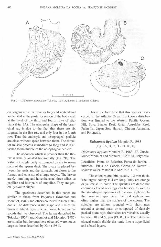
Didemnum granulatum Tokioka, 1954(Fig. 2A, B, C - PI. IR)
Didemnum moseleyi f. granulatum: Tokioka,1954: 244, Japan
Didemnum granulatum: Kott, 1981: 167 (see forsynonymy), Fiji; Kott and Goodbody, 1980: 517,Hong Kong; Monniot and Monniot, 1987: 31,Polynesia; Nishikawa, 1990: 103, Japan.
Localities: Praia Grande, Ponta do Jaroba - interti-daI. Material in MNHN A2 DID C 180.
The colonies are 1-2 mm thick and 10-15em
longo They encrust the lateral surfaces of largeboulders, growing over a1gaeand other encrustinganimals. Their brilliant eo10rs of orange, salmonor brown make them conspieuous. Under magnifi-cation we ean see that the surfaee of the colony isnot smooth but it contains numerous short spieule-filled papillae that make the colonies distinct. Thespicules (PI. IR) are stellate and of variable sizes(11-34 11m).Some of them have thinner rays thanthe others. The spicules are densely distributed inthe test and they fonn a thick basallayer.
The apertures of the oral siphons are stellateand sometimes contracted into the surface of the
colony. The common eloaca are simple holes. Thetunic presents quite extensive thoraeic lacunae,these cavities also extend into the abdominallayerin some eolonies.
The small zooids (lessthan 1mm) areorange.The oral siphon is short and wide. The eloacalaperture is wide, exposing most of the branchialsac (Fig. 2A). It has no languet. The thoraeic lat-
Rev. Brasil. Bio/., 55 (4):639-{j49
642 ROSANA MOREIRA DA ROCHA and FRANÇOISE MONNIOT
A
o.25 mm
B
Fig. 2 - Didemnul1l grallulatul1l Tokioka. 1954: A, thorax; B, abdomen; C, larva.
eral organs are either oval or long and vertical andare located in the posterior region of the body wallat the leveI of the third and fourth rows of stig-mata (Fig. 2A). The triangular shape of the bran-chial sac is due to the fact that there are six
stigmata in the first row and only four in the fourthrow. Thus the endostyle and oesophageal pedicleare close without space between them. The retrac-tor muscle process is medium to long and it is at-tached to the middle of the oesophageal pedicle.
The abdomen which is smaller than the tho-rax is usually located horizontally (Fig. 2B). Thetestis is a single body surrounded by six to sevencoils of the sperm duct. The ovary is placed be-tween the testis and the stomach, but closer to theformer, and consists of a large oocyte. The larvaeare 0.4 mm long and have the usual three adhesivepapillae and four pairs of ampullae. They are gen-erally oval in shape.
The specimens described in this paper aresimilar to those from Polynesia (Monniot andMonniot, 1987) and others collected in New Cale-donia. The difference is the shape and size of thethoracic lateral organs which were larger in thezooids that we observed. The larvae described byTokioka (1954) and Monniot and Monniot (1987)are smaller but the ones here observed were not aslarge as those described by Kott (1981).
Rev. Brasil. Bio/., 55 (4):639--649
This is the fIrst time that this species is re-corded in the Atlantic OceanoIts known distribu-tion was limited to the Westem PacifIc Ocean:
Fiji, Suva Barrier Reef, Great Astrolabe Reef,Palau Is., Japan Sea, Hawaii, Circum Australia,and Polynesia.
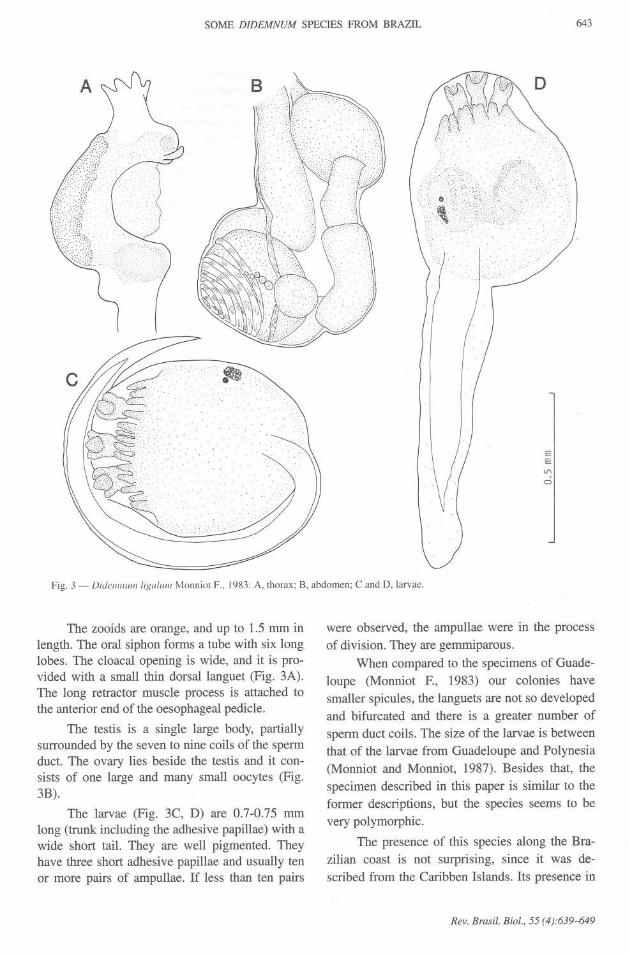
Didemnum ligulum Monniot F., 1983(Fig. 3A, B, C, D - PI. IC, D)
Didemnum ligulum Monniot F., 1983: 27, Guade-loupe; Monniot and Monniot, 1987:34, Polynesia.
Localities: Ponta do Baleeiro, Ponta do Jaroba -intertidal, Praia do Cabelo Gordo de Dentro -shallow water.Material in MZUSP 11.192.
The colonies are thin, usually 1-2 mm thick.The largest colony is 4 cm longoThey are orangeor yellowish in coloroThe spicules are dense butcommon cloacal openings can be seen as well asthe star-shaped apertures of the oral siphons. Inthe preserved specimens, the oral siphons areoften higher than the surface of the colony. Thespicules are almost rounded with short raysaround a central mass or composed of denselypacked blunt rays; their sizes are variable, usuallybetween 10 and 50 J..lm(PI. IC, D). The extensivecloacal canals divide the tunic into a superfIcialand a basallayers.
SOME DIDEMNUM SPECIES FROM BRAZIL 643
EE'"o
Fig. 3 - Didellllllllllligllllllll Monniot F., 1983: A, thorax; B, abdomen; C and D, larvae.
The zooids are orange, and up to 1.5 mm inlength. The oral siphon forms a tube with six longlobes. The c10acalopening is wide, and it is pro-vided with a small thin dorsallanguet (Fig. 3A).The long retractor musc1e process is attached tothe anterior end of the oesophageal pedic1e.
The testis is a single large body, partiallysurrounded by the seven to nine coils of the spermduct. The ovary lies beside the testis and it con-sists of one large and many small oocytes (Fig.3B).
The larvae (Fig. 3C, D) are 0.7-0.75 mmlong (trunk inc1udingthe adhesive papillae) with awide short tail. They are well pigmented. Theyhave three short adhesive papillae and usually tenor more pairs of ampullae. If less than ten pairs
were observed, the ampullae were in the processof division. They are gemmiparous.
When compared to the specimens of Guade-loupe (Monniot F., 1983) our colonies havesmaller spicules, the languets are not so developedand bifurcated and there is a greater number ofsperm duct coils. The size of the larvae is betweenthat of the larvae from Guadeloupe and Polynesia(Monniot and Monniot, 1987). Besides that, thespecimen described in this paper is similar to theformer descriptions, but the species seems to bevery polymorphic.
The presence of this species along the Bra-zilian coast is not surprising, since it was de-scribed from the Caribben Islands. Its presence in
Rev. Brasil. Biol., 55 (4):639-649

644 ROSANA MOREIRA DA ROCHA and FRANÇOISE MONNIOT
the Westem Pacific inclicates that it may also bedistributed world wide in wann shallow waters.
Didemnum lutarium VanName, 1910(Fig. 4A, B, C - PI. IE)
Didemnum lutarium VanName, 1910: 371.
Didemnum candidum lutarium: Van Name, 1921:323; 1945: 86, WestAtlantic (and synonymy).
Localities: Ponta do Baleeiro, Ponta do Jaroba -intertidal. Material in MNHN A2 DID C 181.
This species forros small crusts 2 mm thickunderside the boulders. lt is white, slightly beigeor pink when the zooids are exposed through theoral siphons.
Except for the thin outmost layer of tunicthat has no spicules, the colony is completelyfilled with small rounded spicules, with short andblunt rays (PI. lE). The spicules are slightly morethan 25 11min cliameter.
The zooids are easily removed from the tu-nic. They may be up to 1.3 mm longoThe oral si-phon is curved toward the dorsal side. The eloacalsiphon forros a small rounded aperture at the leveIof the second row of stigmata (Fig. 4A); some-times, when the thorax is very contracted, it forrosa short tube. The thoracic lateral organs are poste-rior to the eloacal siphon (Fig. 4A). There is a
B
strong dorsal musele band along the dorsal side ofthe thorax which continues into the long retractormusele process, that anchors in the tunic when itemerges from the rniddle of the oesophageal pedi-ele. The branchial sac has four rows of stigmatawith up to nine stigmata per half row.
The stomach is round or oval. The posteriorportion of the intestine contains some pyloric ves-ieles (Fig. 4B). Both sexes can be found in thesame individual. The testis is clivided into twolobes which are completely encireled by the six tonine large coils of the sperro duct. The ovary has alarge oocyte but smaller ones can be seen develop-ing at the same time.
The larva (Fig. 4C) is approximately 0.6 mmlong and has three adhesive papillae and four pairsof ampullae. The oozooid has three rows of stig-mata with four to six stigmata per half row. Thetail makes three fourths the coil around the larva.
Van Name (1910), described Didemnum lu-tarium, however, in later works (1921, 1945), hedecided to consider it a sub-species of D. can-didum. This last species is frequently confusedwith many other "white didemnids". The charac-teristics as described by Lafargue (1974) for theneotype of Didemnum candidum Savigny, 1816are somewhat clifferentfrom those of the West At-lantic species. We dissected many colonies and wefound many characters that fully agree with those
0.5 mm
Fig. 4 -Didemnum lutarium Van Name, 1910: A, thorax; B, abdomen; C, larva.
Rev. Brasil. Biol., 55 (4):639-649

SOME DIDEMNUM SPECIES FROM BRAZIL 645
in the original description of Didemnum lutarium,as the co10nythickness and co10r,the strong mus-cular bands in the dorsal side of the thorax, nu-merous stigmata in a row, and two testis vesicles.The only character that did not agree with the de-scription made by Van Name (1945), was the sizeand shape of the spicules. Thus we concluded thatD. lutarium shou1dbe considered a valid species,in which we include the Brazilian specimens.
In the Caribbean area, D. duplicatum Mon-niot, 1983 has also two testis follicles, and rathersmalI cloaca1apertures. But this species differs inseveral characters such as the insertion of the re-
tractor muscle process, the number of stigmata ina row and, above alI, the larval structure.
The distribution of D. lutarium extends from
New England to Brazil along the West AtlanticCoast.
Didemnum perlucidum Monniot F., 1983
Didemnum perlucidum: Monniot F., 1983: 29,Guadeloupe; Monniot et aI., 1985:486, Polynesia;Monniot and Monniot, 1987:40, Polynesia.
Localities:Pontado Baleeiro- intertidal,PraiadoCabelo Gordo de Dentro - shalIow water.Materialin MNHN A2 DID C 184.
The colonies form very thin sheets (1 mm)that grow on multiple substrates such as rock,wood, ropes, PVC tubes, and other ascidians, inshal10wwater. The crusts extend several centime-ters in diameter.
ExternalIy, the colonies resemble marblewith white and gray tones. The gray tones are dueto the presence of an extensive net of cloacal ca-nals where the tunic has only sparse spicules.Spicules are always present, especially in the su-perficiallayers of the tunic, and they can reach 4011mlong, however, most spicu1esreach only 20-3011m.
The internal characters fulIy agree withthose given in the description of the specimensfrom Guadeloupe by F. Monniot (1983). The zo-oids are approximately 1mm in length; the oral si-phon is always short and large; the cloacal siphonis wide exposing almost the entire branchial sac,and has no languet. There are seven to eight stig-mata per half row in the anterior rows. The tho-racic lateral organs are located near the posterioredge of the body walI at the leveI of the third or
fourth row of stigmata. The retractor muscle proc-ess is anterior to the oesophageal pedicle, betweenit and the end portion of the endosty1e.The testisis a single body, surrounded by five to seven coilsof the sperm duct and the ovary is located betweenthe testis and the stomach. The ovary consists ofone large and a number of smaller oocytes. Thelarvae are rounded, with three long adhesive papil-lae and four pairs of ampulIae, and they measure0.4-0.5 mm. The oozooid contains three rows ofstigmata with four to six stigmata per half row.
Didemnum perlucidum is common in NewCaledonia (unpublished data). Present in alIoceans, as a white fouling ascidian, it has prob-ably often been confused with D. candidum.
Didemnum psammathodes (Sluiter, 1895)
Leptoclinum psammathodes: Sluiter, 1895: 171,North Australia.
Didemnum psammathodes: Monniot F., 1983: 31,Guadeloupe; Goodbody, 1984a: 68, West Indies;Rodrigues and Rocha, 1993: 729, South-EasternBrazil.
Localities: Ponta do Baleeiro, Praia Grande - in-tertidal. Material in MNHN A2 DID C 186,MZUSP 11193, 11194, 11195. Type specimenZMA TU 588.
This species is very common in the region,encrusting the lateral surfaces and undersides ofboulders, and vertical walIs, usually shaded sites.The external appearance is exactly the same asthat described for the specimens in other places inthe world. Spicules are sparse and small (5-3011m).The zooids are 0.4 to 0.75 mm in length witha single testis surrounded by six to eight coils ofthe sperm duct. The larvae have three adhesive pa-pillae and four pairs of ampulIae, and they are 0.4to 0.5 mm in trunk length. D. psammathodes hasbeen found in alI warm seas.
Didemnum rodriguesi Rocha and Monniot, 1993
Didemnum rodriguesi: Rocha and Monniot, 1993:261, Brazi1and New Caledonia.Localities: Praia Grande - intertidal. Material inMNHN A2 DID C 187, MZUSP 11121 (Type)11122, 11123.
This species encrusts the lateral surfaces oflarge boulders, growing directly on the rock, al-gae, Bryozoa and other organisms. It forms sheets
Rev. Brasil. Biol.. 55 (4);639-649

646 ROSANA MOREIRA DA ROCHA and FRANÇOISE MONNIOT
several centimeters across and it can have an ir-regular surface due to obstac1esto its growth. Twodistinct external characteristics are the bright or-ange or red color and the microscopic reticulatearrangement of spicules on the colony surface.The larvae also have distinctive features, such asthe three short and wide adhesive papillae andeight pairs of ampullae.
Its distribution is presently restricted to NewCaledonia in the WesternPacific and BraziI.
Didemnum speciosum (Herdman, 1886)
Leptoclinum speciosum: Herdman, 1886: 274.
Didemnum speciosum: Rodrigues and Rocha,1993:930 and synonymy,BraziI.
Localities: Ponta do Baleeiro - intertidal. Materialin MNHN A2 DID C 185.
The colonies are small, thin and white. The
spicules are very abundant and small (15-32 Ilm).The zooids are less than 1 mm in length with widec10acal siphons. The retractor musc1e process isinserted in an anterior position, located betweenthe oesophageal pedic1e and the endostyle. Thetestis is a single body surrounded by six coils ofthe sperm duct. The smalllarvae (0.35 mm) haveno distinctive features; they have three adhesivepapillae and four pairs of ampullae.
The distribution of this species is restrictedto the Brazilian coast.
Didemnum vanderhosti VanName, 1924
(Fig. 5A, B, C - PI. IF)
Didemnum vanderhorsti: Van Name, 1924: 25;1930: 438; 1945: 89; Millar, 1958: 498; 1962: 62;Goodbody, 1984a: 38, 1984b: 65.
Localities: Ilha de São Sebastião, Praia do Araçá,Ponta do Baleeiro, Praia Grande - intertidaI. Ma-terial in MNHN A2 DID C 182, MZUSP 11196,11197.TypeAMNH 714.
This species is very abundant intertidally. Itencrusts the lateral surfaces and undersides ofboulders or vertical rocky walls that are protectedfrom direct sunlight. It forms thin sheets 1-2 mmthick and several centimeters in diameter. Theedges of the colony are sometimes unattached tothe substrate, forrning pendent thick masses. Thecolor varies from dark-purple, dark brown archocolate brown to a marble mixture of these andbeige.
The spicules (10-35 Ilm) are not abundantbut always present. They are rounded with numer-ous short rays (PI. IF).
The zooids are not arranged in systems butthe presence of fecal pellets are evidence that the
B
0.5 mm
Fig. 5 - Didemnum vanderhorsti Van Name, 1924: A, zooid; B, thorax; C, larva.
Rev. Brasil. Biol., 55 (4):639-649

SOME DIDEMNUM SPECIES FROM BRAZIL
canaIs converge to the common c10acafrom alI di-rections. Spicules are absent in the thin layer ofthe transparent test around the common c10acalapertures.
The zooids are rather small and rarely ex-ceed 1 mm in length. The oral siphon is tube-likewith short lobes and the c10acalone is generallynarrow with no languet (Fig. 5A). The large andelliptical thoracic lateral organs are located at theleveI of the third row of stigmata or the thirdtransversal vessel between the edge of the bodywalI and the endostyle (Fig. 5A). The retractormusc1eprocess is long and unattached at the mid-dle of the oesophageal pedic1e.
The digestive tract has no distinct charac-teristics, the stomach is cylindrical. Both sexes arepresent (Fig. 5B). The testis is a single body, par-tially surrounded by nine coils of the sperrn duct.The ovary consists of one large and several smalloocytes located between the testis and the stom-acho
Larvae (Fig. 5e) are 0.5 mm long (inc1udingadhesive papillae) and each has three adhesive pa-pillae and four pairs of ampulIae. Individuals arenot gemmiparous and the oozooid has three rowsof stigmata with five to six stigmata per side. Thisis the first description of D. vanderhorsti larvae.
Except fiom the color of the colony, thisspecies differs in many characters fiom D. ciner-aceum (Sluiter, 1908) redescribed by Monniot F.(1983).
Millar (1958) noted the scarcity of recordsof this species, however, it is very abundant insouth-eastem Brazil. The lack of records of spe-cies found between the São Sebastião Channel and
the Caribbean Sea (Goodbody, 1984a, b) may bedue to the fact that very little colIecting has beendone in this area.
DISCUSSION
Until recently only two species of the genusDidermnum were known in Brazilian waters, D.speciosum and D. vanderhorsti, D. candidum re-ported by Van Name (1945) for São Sebastião Is-land should be reexarnined, since this species wasredescribed by Lafargue (1974) and probably doesnot occur in Brazilian waters. This genus is welIdiversified along alI tropical and subtropicalcoasts in the world and there was no reason to ex-pect that it would not be diversified here. Never-
647
theless, this work should not be considered as a
comprehensive inventory of the species in the SãoSebastião region as it was restricted to intertidal orvery shallow habitats.
Among the nine species colIected, D. spe-ciosum is the only one found to be endemic inBrazil. This species has no distinct features thatwould allow an undoubted identification. It be-
longs to the "white didemnid" group that has al-ways allowed confusion among its species and itmay be found elsewhere with further studies. Twoother species are restricted to the westem Atlantic:D. lutarium and D. vanderhorsti.
Surprisingly, we found many species whosedistributions are much wider and species knownonly fiom the Pacific region. This scattered distri-bution could reflect a bias of sampling due to thesmall number of taxonomic papers on ascidianfauna around the world, so that many speciescould have a wide distribution that is still un-known.
Recent introductions of ascidians have al-
ready been reported in many localities (see Mon-niot et ai., 1985 for a bibliographic review). Thesespecies are generally transported on the hulIs ofships. The newly introduced species usually arerestricted to the port area, and they are rarelyfound among the common species of the region.The São Sebastião Channel is also subjected to theintroduction of new species since it has a largeharbor for oil-tankers that stay in the port for longperiods of time while loading and unloading.Among the species reported in this paper, D. per-lucidum, D. ligulum and D. ahu were found to bevery common on artificial substrata and are con-sidered components of the "fouling community",but they were also present on the natural boulders,which might indicate that they were recently intro-duced.
Acknowledgements - We are indebted to Dr. João E. Lunetta,
director of tbe Centro de Biologia Marinha - Universidade de
São Paulo for providing logistical support during the fieldwork. The first autbor is also tbankful to Drs. F. Monniot and
C. Monniot of tbe Laboratoire de Biologie des Invertébrés Ma-rins et Malacologie of tbe Muséum National d'Histoire
Naturelle for tbe kind award of a training period in ascidian
systematics. This research was supported by grants from CNPqand CAPES to R. M. Rocha.
Rev. Brasil. Biol., 55 (4):639-649
648 ROSANA MOREIRA DA ROCHA and FRANÇOISE MONNIOT
PLATE T
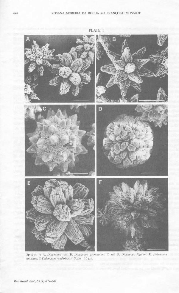
Spicuks in A, lJidellllllllll ulll/: 13, lJidol/lll/lIl grwlI/IllIlIIll; C and D, lJidellllllllll ligullllll; E, lJidelllllulIIlutarium;F, Didemnum vanderhorsti. Scale =tO/lID.
Rev. Brasil. Biol., 55 (4):639-M9

SOME DIDEMNUM SPECIES FROM BRAZIL 649
REFERENCES
GOODBODY, L, 1984a, The ascidian fauna of two contrasting
lagoons in the Netherlands Antilles: Piscadera Baai,
Curaçao and the Lac ofBonaire. Stud. Fauna Curaçao
other Caribb. Islands, 67(202): 21-61.
GOODBODY, L, 1984b, Ascidians from Caribbean shallow
water localities. Stud. Fauna Curaçao other Caribb.
Islands, 67: 62-76.
LAFARGUE, E, 1974, Description d'un néotype de Didem-
num candidum Savigny, 1816 espece type de Mer
Rouge (Ascidie composée). Vie Milieu, /4(2), sér. A:341-356.
KOTI, P., 1981, The ascidians of the reef flats of Fiji. Proc.
Linn. Soe. N. S. w., 105: 147-212.
MILLAR, R. H., 1958, Some Ascidians from Brazil. Ann.
mago nato Hist., 13: 497-514.
MILLAR, R. H., 1962, Some ascidians from the Caribbean.
Stud. Fauna Curaçao other Caribb. Islands, 13: 61-77.
MONNIOT, C. and MONNIOT, E, 1972, Clé mondiale des
gemes d' Ascidies. Arch. Zoo/. Exp. Gen., 113: 311-367.
MONNlOT, C., MONNlOT, E and LABOUTE, P., 1985, As-
cidies du port de Papeete (Polynésie française): rela-tions avec le milieu naturel et apports intercontinentaux
par Ia navigation. Buli. Mus. natn. Hist. nat., ser. 4, A,Paris 7: 481-495.
MONNlOT, C. and MONNlOT, E, 1987, Les ascidies de
Polynésie française. Mém. Mus. natn. Hist. nat., A,Zoo/. 136: 1-155.
MONNIOT, E, 1983, Ascidies littorales de Guadeloupe L
Didemnidae. Buli. Mus. natn. Hist. nat., ser. 4, Paris
5: 5-49.
ROCHA, R. M. and MONNIOT, E, 1993, Didemnum ro-
driguesi sp.nov. - a new didemnid ascidian common toSoutheastem Brazil and New Caledonia. Ann. Inst.
oceanogr., Paris 69: 261-265.
RODRIGUES, S. A. and ROCHA, R. M., 1993, Littoral com-
pound ascidians from São Sebastião, Estado de SãoPaulo, Brazil. Proc. Bio/. Soe. Wash., 106: 728-739.
TOKIOKA, T., 1954, Invertebrate fauna of the intertidal zoneof the Tokara Islands. VII. Ascidians. Pub/. Seto mar.
bio/. Lab. 3: 239-264.
VAN NAME, W. G., 1910, Compound ascidians of the coasts
of New England and neighboring British provinces.Prac. Boston Soe. nato Hist., 34: 339-424.
VAN NAME, W. G., 1921, Ascidians ofthe West Indian regionand South Eastem United States. Buli. Am. Mus. nato
Hist., 44: 283-494.
VAN NAME, W. G., 1945, The North and South American as-cidians. Buli. Am. Mus. nato Hist., 84: 1-476.