tailoring the trajectory of cell rolling with … · 2017-04-10 · tailoring the trajectory of...
TRANSCRIPT

rXXXX American Chemical Society A dx.doi.org/10.1021/la203382k | Langmuir XXXX, XXX, 000–000
ARTICLE
pubs.acs.org/Langmuir
Tailoring the Trajectory of Cell Rolling with Cytotactic SurfacesCollin Edington,†,§Hironobu Murata,§ Richard Koepsel,§,|| Jill Andersen,§ Sungeun Eom,^ Takeo Kanade,^
Anna C. Balazs,‡ German Kolmakov,‡ Carsen Kline,# Daniel McKeel,§ Zvi Liron,3 and Alan J. Russell*,§,||
†Department of Bioengineering, ‡Department of Chemical Engineering, and §McGowan Institute for Regenerative Medicine,University of Pittsburgh, Pittsburgh, Pennsylvania, United States
)Department of Surgery, University of Pittsburgh Medical Center, Pittsburgh, Pennsylvania, United States^Robotics Institute and #Electrical and Computer Engineering, Carnegie Mellon University, Pittsburgh, Pennsylvania, United States3Department of Physical Chemistry, Israel Institute for Biological Research, Ness-Ziona, 74100, Israel
bS Supporting Information
’ INTRODUCTION
The ability to separate specific populations of cells from aheterogeneous sample is a fundamental requirement for studyingcell biology. Medical diagnostics and cell-based therapies alsorely on cell separation technologies to ensure safe and effectivetreatment. Since the 1970s, fluorescent activated cell sorting(FACS) has been the workhorse of cell separation techniquesdue to the high specificity, rapid separation, and wide range ofcommercially available fluorescent markers.1,2 While FACS hasevolved considerably over the past 40 years to increase specificity,efficiency, and speed, it still remains an expensive and often time-consuming process that relies on covalently bound or antibodyconjugated markers to identify specific cell populations. As aresult, the technique is not ideal for sensitive cell populations orapplications where cell phenotype must be preserved. Theadditional cost and complexity of cell labeling also limits theuse of FACS and similar techniques in point-of-care diagnostics.The limitations of label-based cell separation techniques haveinspired the development of numerous label-free methods, manyof which are based onmicrofluidic manipulation of cell solutions.These methods rely primarily on physical properties, such asdielectric charge, cell size, and autofluorescence to sepa-rate different populations.3!9 However, many cell phenotypesare exclusively defined by their expression of specific membrane
proteins, rather than electrical or mechanical properties. Thereexists a need for a device capable of continuous, label-freeseparation of cells based on the expression of surface proteins.
By rationally tailoring the microenvironment of a cell, it maybe possible to manipulate and characterize the cell withoutattaching ligands to its surface. As cells pass over surfaces, thereis an opportunity to deliver signals to the cell from the surfacethat cause the cell to alter its behavior. Surfaces that can deliver astimulus that directs cells toward or away from the surface areknown as cytotactic surfaces. Indeed, in biology, such surfacesalready exist. Following tissue injury, one of the earliest steps ofthe inflammatory response is the recruitment of white blood cells(leukocytes) from nearby vasculature. Leukocyte extravasation isinitiated by the expression of cell adhesion molecules, includingE- and P-selectin, on the surface of activated endothelial cells,which causes free floating leukocytes expressing P-selectin Gly-coprotein Ligand-1 (PSGL-1) to adhere to the vessel walls androll to the site of injury before escaping the blood vessel.10 Selectinmediated rolling has been studied extensively,10!22 and a numberof attempts have been made to purify hematopoietic cells based onthis unique interaction.23!25 Some of these methods are based on
Received: August 29, 2011Revised: October 25, 2011
ABSTRACT: Cell separation technology is a key tool forbiological studies and medical diagnostics that relies primarilyon chemical labeling to identify particular phenotypes.An emergent method of sorting cells based on differentialrolling on chemically patterned substrates holds potentialbenefits over existing technologies, but the underlying mech-anisms being exploited are not well characterized. In order tobetter understand cell rolling on complex surfaces, a micro-fluidic device with chemically patterned stripes of the celladhesion molecule P-selectin was designed. The behavior ofHL-60 cells rolling under flow was analyzed using a high-resolution visual tracking system. This behavior was thencorrelated to a number of established predictive models. Thecombination of computational modeling and widely available fabrication techniques described herein represents a crucial steptoward the successful development of continuous, label-free methods of cell separation based on rolling adhesion.
B dx.doi.org/10.1021/la203382k |Langmuir XXXX, XXX, 000–000
Langmuir ARTICLE
differential rolling adhesion in one dimension or static adhesion,but they are not continuous and require careful collection ofsamples at specified time points.
A recent computational model reveals an improved method ofadhesion-based separation by incorporating arrays of chemically andmechanically patterned surface features.26 The model suggests thatpassive, label-free cell separation could be accomplished in amicrofluidic package by utilizing smart surfaces to identify dif-ferences in cell surface ligands or cell membrane stiffness. The virtualsurface utilizes angled stripes of variable stiffness or adhesiveness. As“cells” roll along the virtual surface under laminar flow, they canobtain a net displacement perpendicular to the directionofflowuponinteracting with a diagonal stripe that has a higher adhesive inter-action or lower modulus than the bulk material. This predicteddisplacement results from a change in shear forces as laminar flowpushes the capsule across each stripe, and is also a function of thecapsule’s stiffness, meaning that two capsules with different com-pliances or adhesive properties will exhibit different amounts ofdisplacement, effectively sorting them.The capsule’s path is altered asit passes from the weakly adhesive bulkmaterial to the “sticky” stripe,and after exposure to multiple stripes the compliant capsules gaindisplacement perpendicular to the direction of flow.
Control of cell motion in two dimensions would allow for thephysical segregation of interacting and noninteracting cells in acontinuous manner, making such a sorting method attractive. Con-sequently, experimentally verifying and understanding this predictionis an important step toward the practical development of an adhesion-based sorting device. Results from experimental manipulations ofleukocytes appear to support the Balazs model, including theobservation of lateral displacement on stripes of P-selectin and theabsence of the effect in the case of rigid microspheres.27,28 Only themacroscopic behavior of cells in these systems has been investigated,and therefore, the data lack the necessary resolution to verify themodel. In order to more fully understand cellular motion acrosspatterned surfaces, we developed a system in which we could observethese interactions on a submicrometer scale. A mechanistic under-standing of this effect would be important in the successful develop-ment of adhesion-based methods of cell sorting.
We designed a microfluidic device to study the directed rollingof cells using a cytotactic surface of covalently immobilized
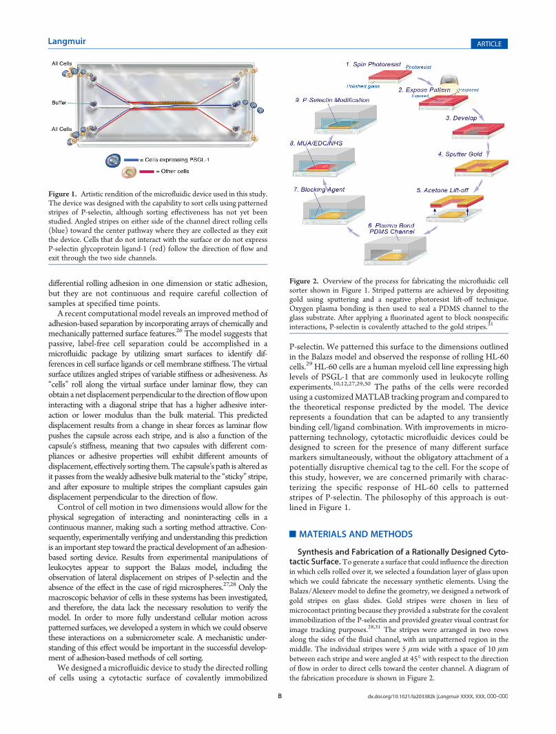
P-selectin. We patterned this surface to the dimensions outlinedin the Balazs model and observed the response of rolling HL-60cells.29 HL-60 cells are a human myeloid cell line expressing highlevels of PSGL-1 that are commonly used in leukocyte rollingexperiments.10,12,27,29,30 The paths of the cells were recordedusing a customizedMATLAB tracking program and compared tothe theoretical response predicted by the model. The devicerepresents a foundation that can be adapted to any transientlybinding cell/ligand combination. With improvements in micro-patterning technology, cytotactic microfluidic devices could bedesigned to screen for the presence of many different surfacemarkers simultaneously, without the obligatory attachment of apotentially disruptive chemical tag to the cell. For the scope ofthis study, however, we are concerned primarily with charac-terizing the specific response of HL-60 cells to patternedstripes of P-selectin. The philosophy of this approach is out-lined in Figure 1.
’MATERIALS AND METHODS
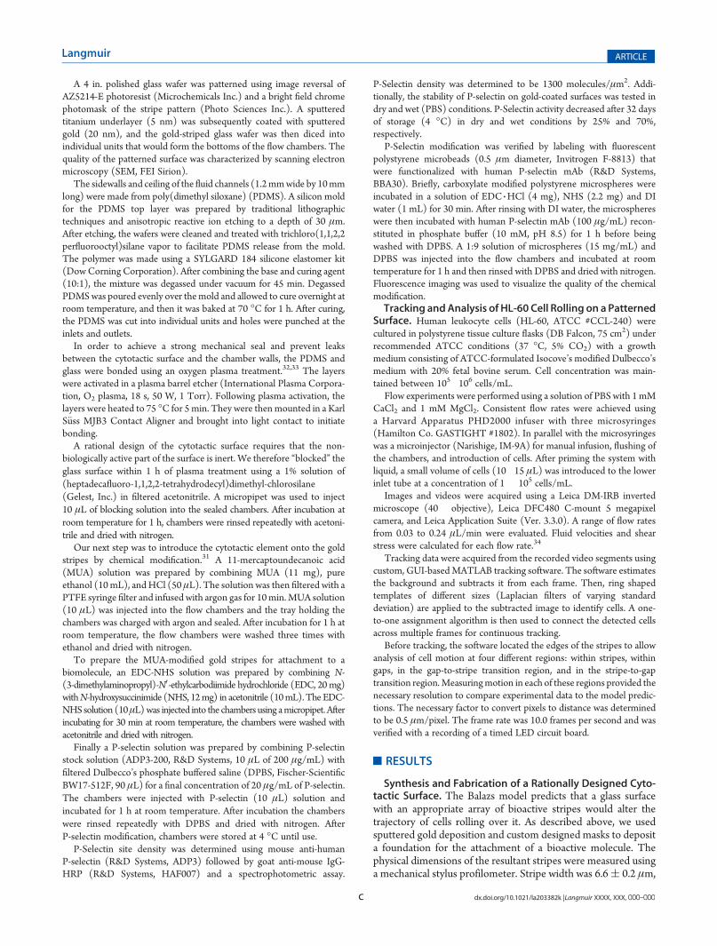
Synthesis and Fabrication of a Rationally Designed Cyto-tactic Surface.To generate a surface that could influence the directionin which cells rolled over it, we selected a foundation layer of glass uponwhich we could fabricate the necessary synthetic elements. Using theBalazs/Alexeev model to define the geometry, we designed a network ofgold stripes on glass slides. Gold stripes were chosen in lieu ofmicrocontact printing because they provided a substrate for the covalentimmobilization of the P-selectin and provided greater visual contrast forimage tracking purposes.28,31 The stripes were arranged in two rowsalong the sides of the fluid channel, with an unpatterned region in themiddle. The individual stripes were 5 μm wide with a space of 10 μmbetween each stripe and were angled at 45! with respect to the directionof flow in order to direct cells toward the center channel. A diagram ofthe fabrication procedure is shown in Figure 2.
Figure 1. Artistic rendition of the microfluidic device used in this study.The device was designed with the capability to sort cells using patternedstripes of P-selectin, although sorting effectiveness has not yet beenstudied. Angled stripes on either side of the channel direct rolling cells(blue) toward the center pathway where they are collected as they exitthe device. Cells that do not interact with the surface or do not expressP-selectin glycoprotein ligand-1 (red) follow the direction of flow andexit through the two side channels.
Figure 2. Overview of the process for fabricating the microfluidic cellsorter shown in Figure 1. Striped patterns are achieved by depositinggold using sputtering and a negative photoresist lift-off technique.Oxygen plasma bonding is then used to seal a PDMS channel to theglass substrate. After applying a fluorinated agent to block nonspecificinteractions, P-selectin is covalently attached to the gold stripes.31
C dx.doi.org/10.1021/la203382k |Langmuir XXXX, XXX, 000–000
Langmuir ARTICLE
A 4 in. polished glass wafer was patterned using image reversal ofAZ5214-E photoresist (Microchemicals Inc.) and a bright field chromephotomask of the stripe pattern (Photo Sciences Inc.). A sputteredtitanium underlayer (5 nm) was subsequently coated with sputteredgold (20 nm), and the gold-striped glass wafer was then diced intoindividual units that would form the bottoms of the flow chambers. Thequality of the patterned surface was characterized by scanning electronmicroscopy (SEM, FEI Sirion).
The sidewalls and ceiling of the fluid channels (1.2mmwide by 10mmlong) were made from poly(dimethyl siloxane) (PDMS). A silicon moldfor the PDMS top layer was prepared by traditional lithographictechniques and anisotropic reactive ion etching to a depth of 30 μm.After etching, the wafers were cleaned and treated with trichloro(1,1,2,2perfluorooctyl)silane vapor to facilitate PDMS release from the mold.The polymer was made using a SYLGARD 184 silicone elastomer kit(DowCorning Corporation). After combining the base and curing agent(10:1), the mixture was degassed under vacuum for 45 min. DegassedPDMSwas poured evenly over themold and allowed to cure overnight atroom temperature, and then it was baked at 70 !C for 1 h. After curing,the PDMS was cut into individual units and holes were punched at theinlets and outlets.
In order to achieve a strong mechanical seal and prevent leaksbetween the cytotactic surface and the chamber walls, the PDMS andglass were bonded using an oxygen plasma treatment.32,33 The layerswere activated in a plasma barrel etcher (International Plasma Corpora-tion, O2 plasma, 18 s, 50 W, 1 Torr). Following plasma activation, thelayers were heated to 75 !C for 5 min. They were thenmounted in a KarlS€uss MJB3 Contact Aligner and brought into light contact to initiatebonding.
A rational design of the cytotactic surface requires that the non-biologically active part of the surface is inert. We therefore “blocked” theglass surface within 1 h of plasma treatment using a 1% solution of(heptadecafluoro-1,1,2,2-tetrahydrodecyl)dimethyl-chlorosilane(Gelest, Inc.) in filtered acetonitrile. A micropipet was used to inject10 μL of blocking solution into the sealed chambers. After incubation atroom temperature for 1 h, chambers were rinsed repeatedly with acetoni-trile and dried with nitrogen.
Our next step was to introduce the cytotactic element onto the goldstripes by chemical modification.31 A 11-mercaptoundecanoic acid(MUA) solution was prepared by combining MUA (11 mg), pureethanol (10mL), andHCl (50 μL). The solution was then filtered with aPTFE syringe filter and infused with argon gas for 10min.MUA solution(10 μL) was injected into the flow chambers and the tray holding thechambers was charged with argon and sealed. After incubation for 1 h atroom temperature, the flow chambers were washed three times withethanol and dried with nitrogen.
To prepare the MUA-modified gold stripes for attachment to abiomolecule, an EDC-NHS solution was prepared by combining N-(3-dimethylaminopropyl)-N0-ethylcarbodiimide hydrochloride (EDC, 20mg)withN-hydroxysuccinimide (NHS, 12mg) in acetonitrile (10mL). TheEDC-NHSsolution (10μL) was injected into the chambers using amicropipet.Afterincubating for 30 min at room temperature, the chambers were washed withacetonitrile and dried with nitrogen.
Finally a P-selectin solution was prepared by combining P-selectinstock solution (ADP3-200, R&D Systems, 10 μL of 200 μg/mL) withfiltered Dulbecco’s phosphate buffered saline (DPBS, Fischer-ScientificBW17-512F, 90 μL) for a final concentration of 20 μg/mL of P-selectin.The chambers were injected with P-selectin (10 μL) solution andincubated for 1 h at room temperature. After incubation the chamberswere rinsed repeatedly with DPBS and dried with nitrogen. AfterP-selectin modification, chambers were stored at 4 !C until use.
P-Selectin site density was determined using mouse anti-humanP-selectin (R&D Systems, ADP3) followed by goat anti-mouse IgG-HRP (R&D Systems, HAF007) and a spectrophotometric assay.
P-Selectin density was determined to be 1300 molecules/μm2. Addi-tionally, the stability of P-selectin on gold-coated surfaces was tested indry and wet (PBS) conditions. P-Selectin activity decreased after 32 daysof storage (4 !C) in dry and wet conditions by 25% and 70%,respectively.
P-Selectin modification was verified by labeling with fluorescentpolystyrene microbeads (0.5 μm diameter, Invitrogen F-8813) thatwere functionalized with human P-selectin mAb (R&D Systems,BBA30). Briefly, carboxylate modified polystyrene microspheres wereincubated in a solution of EDC 3HCl (4 mg), NHS (2.2 mg) and DIwater (1 mL) for 30 min. After rinsing with DI water, the microsphereswere then incubated with human P-selectin mAb (100 μg/mL) recon-stituted in phosphate buffer (10 mM, pH 8.5) for 1 h before beingwashed with DPBS. A 1:9 solution of microspheres (15 mg/mL) andDPBS was injected into the flow chambers and incubated at roomtemperature for 1 h and then rinsed with DPBS and dried with nitrogen.Fluorescence imaging was used to visualize the quality of the chemicalmodification.Tracking andAnalysis of HL-60 Cell Rolling on a Patterned
Surface. Human leukocyte cells (HL-60, ATCC #CCL-240) werecultured in polystyrene tissue culture flasks (DB Falcon, 75 cm2) underrecommended ATCC conditions (37 !C, 5% CO2) with a growthmedium consisting of ATCC-formulated Isocove’s modified Dulbecco’smedium with 20% fetal bovine serum. Cell concentration was main-tained between 105!106 cells/mL.
Flow experiments were performed using a solution of PBS with 1mMCaCl2 and 1 mM MgCl2. Consistent flow rates were achieved usinga Harvard Apparatus PHD2000 infuser with three microsyringes(Hamilton Co. GASTIGHT #1802). In parallel with the microsyringeswas a microinjector (Narishige, IM-9A) for manual infusion, flushing ofthe chambers, and introduction of cells. After priming the system withliquid, a small volume of cells (10!15 μL) was introduced to the lowerinlet tube at a concentration of 1 " 105 cells/mL.
Images and videos were acquired using a Leica DM-IRB invertedmicroscope (40" objective), Leica DFC480 C-mount 5 megapixelcamera, and Leica Application Suite (Ver. 3.3.0). A range of flow ratesfrom 0.03 to 0.24 μL/min were evaluated. Fluid velocities and shearstress were calculated for each flow rate.34
Tracking data were acquired from the recorded video segments usingcustom, GUI-basedMATLAB tracking software. The software estimatesthe background and subtracts it from each frame. Then, ring shapedtemplates of different sizes (Laplacian filters of varying standarddeviation) are applied to the subtracted image to identify cells. A one-to-one assignment algorithm is then used to connect the detected cellsacross multiple frames for continuous tracking.
Before tracking, the software located the edges of the stripes to allowanalysis of cell motion at four different regions: within stripes, withingaps, in the gap-to-stripe transition region, and in the stripe-to-gaptransition region.Measuringmotion in each of these regions provided thenecessary resolution to compare experimental data to the model predic-tions. The necessary factor to convert pixels to distance was determinedto be 0.5 μm/pixel. The frame rate was 10.0 frames per second and wasverified with a recording of a timed LED circuit board.
’RESULTS
Synthesis and Fabrication of a Rationally Designed Cyto-tactic Surface. The Balazs model predicts that a glass surfacewith an appropriate array of bioactive stripes would alter thetrajectory of cells rolling over it. As described above, we usedsputtered gold deposition and custom designed masks to deposita foundation for the attachment of a bioactive molecule. Thephysical dimensions of the resultant stripes were measured usinga mechanical stylus profilometer. Stripe width was 6.6( 0.2 μm,
D dx.doi.org/10.1021/la203382k |Langmuir XXXX, XXX, 000–000
Langmuir ARTICLE
spacing between stripes was 8.0( 0.1 μm, and vertical thicknessof the gold was approximately 29 nm. Scanning electron micro-scopy was used to visualize the profile of the stripes and verify theaccuracy of the gold patterning.The glass surfaces were then bonded to PDMS channels using
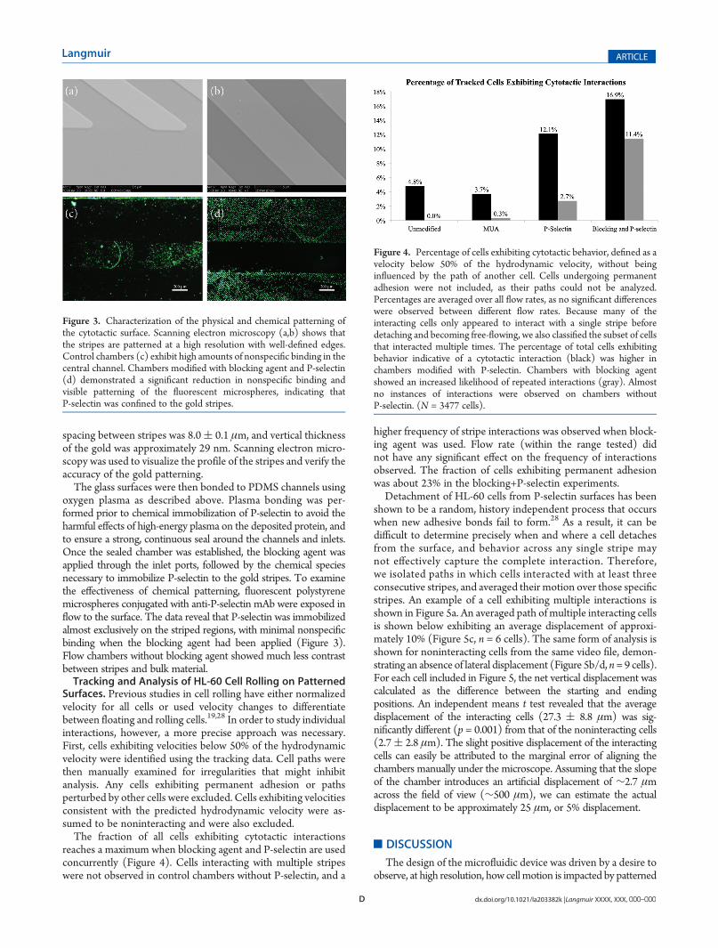
oxygen plasma as described above. Plasma bonding was per-formed prior to chemical immobilization of P-selectin to avoid theharmful effects of high-energy plasma on the deposited protein, andto ensure a strong, continuous seal around the channels and inlets.Once the sealed chamber was established, the blocking agent wasapplied through the inlet ports, followed by the chemical speciesnecessary to immobilize P-selectin to the gold stripes. To examinethe effectiveness of chemical patterning, fluorescent polystyrenemicrospheres conjugated with anti-P-selectin mAb were exposed inflow to the surface. The data reveal that P-selectin was immobilizedalmost exclusively on the striped regions, with minimal nonspecificbinding when the blocking agent had been applied (Figure 3).Flow chambers without blocking agent showed much less contrastbetween stripes and bulk material.Tracking and Analysis of HL-60 Cell Rolling on Patterned
Surfaces. Previous studies in cell rolling have either normalizedvelocity for all cells or used velocity changes to differentiatebetween floating and rolling cells.19,28 In order to study individualinteractions, however, a more precise approach was necessary.First, cells exhibiting velocities below 50% of the hydrodynamicvelocity were identified using the tracking data. Cell paths werethen manually examined for irregularities that might inhibitanalysis. Any cells exhibiting permanent adhesion or pathsperturbed by other cells were excluded. Cells exhibiting velocitiesconsistent with the predicted hydrodynamic velocity were as-sumed to be noninteracting and were also excluded.The fraction of all cells exhibiting cytotactic interactions
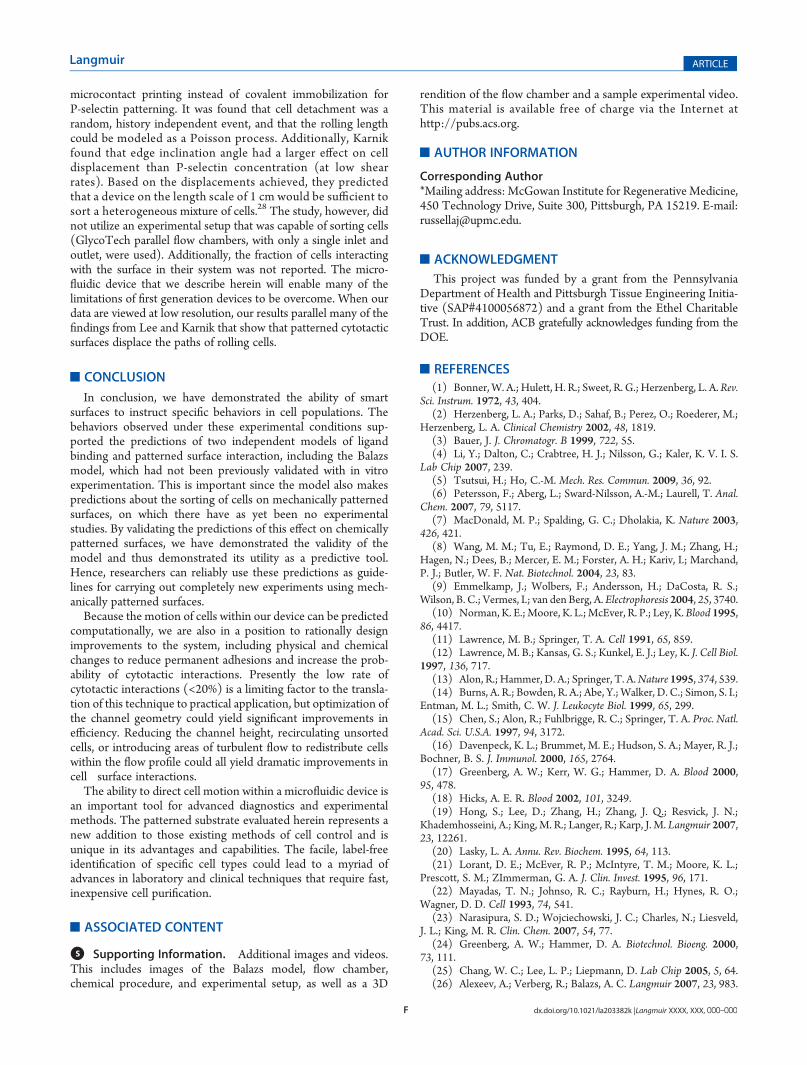
reaches a maximum when blocking agent and P-selectin are usedconcurrently (Figure 4). Cells interacting with multiple stripeswere not observed in control chambers without P-selectin, and a
higher frequency of stripe interactions was observed when block-ing agent was used. Flow rate (within the range tested) didnot have any significant effect on the frequency of interactionsobserved. The fraction of cells exhibiting permanent adhesionwas about 23% in the blocking+P-selectin experiments.Detachment of HL-60 cells from P-selectin surfaces has been
shown to be a random, history independent process that occurswhen new adhesive bonds fail to form.28 As a result, it can bedifficult to determine precisely when and where a cell detachesfrom the surface, and behavior across any single stripe maynot effectively capture the complete interaction. Therefore,we isolated paths in which cells interacted with at least threeconsecutive stripes, and averaged their motion over those specificstripes. An example of a cell exhibiting multiple interactions isshown in Figure 5a. An averaged path of multiple interacting cellsis shown below exhibiting an average displacement of approxi-mately 10% (Figure 5c, n = 6 cells). The same form of analysis isshown for noninteracting cells from the same video file, demon-strating an absence of lateral displacement (Figure 5b/d, n= 9 cells).For each cell included in Figure 5, the net vertical displacement wascalculated as the difference between the starting and endingpositions. An independent means t test revealed that the averagedisplacement of the interacting cells (27.3 ( 8.8 μm) was sig-nificantly different (p = 0.001) from that of the noninteracting cells(2.7( 2.8 μm). The slight positive displacement of the interactingcells can easily be attributed to the marginal error of aligning thechambers manually under the microscope. Assuming that the slopeof the chamber introduces an artificial displacement of ∼2.7 μmacross the field of view (∼500 μm), we can estimate the actualdisplacement to be approximately 25 μm, or 5% displacement.
’DISCUSSION
The design of the microfluidic device was driven by a desire toobserve, at high resolution, how cell motion is impacted by patterned
Figure 3. Characterization of the physical and chemical patterning ofthe cytotactic surface. Scanning electron microscopy (a,b) shows thatthe stripes are patterned at a high resolution with well-defined edges.Control chambers (c) exhibit high amounts of nonspecific binding in thecentral channel. Chambers modified with blocking agent and P-selectin(d) demonstrated a significant reduction in nonspecific binding andvisible patterning of the fluorescent microspheres, indicating thatP-selectin was confined to the gold stripes.
Figure 4. Percentage of cells exhibiting cytotactic behavior, defined as avelocity below 50% of the hydrodynamic velocity, without beinginfluenced by the path of another cell. Cells undergoing permanentadhesion were not included, as their paths could not be analyzed.Percentages are averaged over all flow rates, as no significant differenceswere observed between different flow rates. Because many of theinteracting cells only appeared to interact with a single stripe beforedetaching and becoming free-flowing, we also classified the subset of cellsthat interacted multiple times. The percentage of total cells exhibitingbehavior indicative of a cytotactic interaction (black) was higher inchambers modified with P-selectin. Chambers with blocking agentshowed an increased likelihood of repeated interactions (gray). Almostno instances of interactions were observed on chambers withoutP-selectin. (N = 3477 cells).

E dx.doi.org/10.1021/la203382k |Langmuir XXXX, XXX, 000–000
Langmuir ARTICLE
surfaces. The data clearly demonstrated that not only do the averagepaths of interacting cells differ from those of noninteracting cells,but more importantly cell motion entering, on, and leaving thepatterned stripes bore remarkable similarities to the motionspredicted by the Balazs model. The Balazs model predicts aninitial downwardmotion as the cell enters an adhesive stripe. Thepath of cell #81 (expanded in Figure 5) provides a compellingexample of such behavior in our microfluidic device. Therepetition of this behavior on the same length scale as the stripesfurther verifies the successful patterning of the surface and theinteraction of the cells with that surface.
The Balazsmodel next predicts a change in direction as the cellbecomes localized within the stripe. Because the system wasdesigned at sufficient resolution, we were able to observe thistransition experimentally in almost every case where a cell wasshown to be interacting with the surface. The final element of theBalazs model predicts the path that a cell would take as it exits anadhesive stripe. As shown in Figure 5, the data fit the model.
In addition to demonstrating the existence of a behavioranalogous to the one described by the Balazs model, our observa-tions also support the conclusions of a computational model byChang, in which a state diagram was developed to predict theadhesive interactions between leukocytes and surfaces expressingselectin molecules.35 In the state diagram, the rolling behavior ofcells is divided into five distinct states. Each state is characterizedby a unique profile of displacement versus time and is determinedby the rate of shear, density of ligand!receptor pairs, dissociationrate, and bond interaction length. In addition to “firm adhesion”and “no adhesion”, there exist three unique states of rollingadhesion, termed “fast adhesion”, “transient adhesion”, and“saltation”. Over the range of shear rates tested by Chang(30!400 s!1), cell rolling only occurred within a particularregion of the state diagram, demonstrating that leukocyte rollingrequires a delicate balance between ligand density, shear stress,and dissociation rate.
We were able to identify representative paths for each stateof adhesion, the profiles of which are shown next to theircorresponding graphs from Chang (Figure 6). These graphsrepresent the entire tracked path of a cell, and were not croppedto show only a portion of the path. The existence of all five statesindicates that our experiments successfully captured a wide rangeof potential leukocyte behaviors within our shear stress range(0.29!2.2 dyn/cm2). However, we were also able to observemore than one state of adhesion within each set of experimentalparameters, indicating a considerable variability in the factorsdescribed above. The agreement between our observed data andthe model proposed by Chang support the view that ourcytotactic surface promotes leukocyte rolling.
In 2008, Karnik et al. observed the interaction of rolling HL-60cells with a single patterned edge of P-selectin and a PEG-basedblocking agent.27 They found that rolling HL-60 cells weredisplaced orthogonal to the direction of flow when P-selectinedges were patterned at appropriate angles. A continuation of the2008 study by the Karnik group recently suggested that patternedstripes may provide a label-free method of continuous cellseparation.28 The study was conducted under similar shear ratesand P-selectin concentrations, but with larger stripe and chambergeometry, and at lower angles (e20!). Karnik also used
Figure 6. Plots of displacement versus time for the five states ofadhesion identified by Chang (left) and sample plots of similar behavioridentified in our cell tracking videos (right).35 The five states are noadhesion (a), fast adhesion (b), transient adhesion (c), firm adhesion (d), andsaltation (e). These behaviors represent different dynamic states ofadhesion mediated by the biophysical and kinetic properties of the system.Left figure copyright (2000) National Academy of Sciences, U.S.A.
Figure 5. Behaviors of interacting and noninteracting cells were visiblydifferent. A representative path of an interacting cell (a) and a non-interacting cell (b) are magnified to demonstrate the rolling behaviorand direction of fluid flow (left to right), respectively. By combining thepaths of multiple cells from a single video file, the average path they takeover a single stripe can be plotted. Themean of this path is shown in red,with black lines representing one standard deviation from the mean.Average paths for interacting (c) and noninteracting (d) cells are shown((c) n = 6 cells, 92 data points; (d) n = 9 cells, 96 data points).
F dx.doi.org/10.1021/la203382k |Langmuir XXXX, XXX, 000–000
Langmuir ARTICLE
microcontact printing instead of covalent immobilization forP-selectin patterning. It was found that cell detachment was arandom, history independent event, and that the rolling lengthcould be modeled as a Poisson process. Additionally, Karnikfound that edge inclination angle had a larger effect on celldisplacement than P-selectin concentration (at low shearrates). Based on the displacements achieved, they predictedthat a device on the length scale of 1 cm would be sufficient tosort a heterogeneous mixture of cells.28 The study, however, didnot utilize an experimental setup that was capable of sorting cells(GlycoTech parallel flow chambers, with only a single inlet andoutlet, were used). Additionally, the fraction of cells interactingwith the surface in their system was not reported. The micro-fluidic device that we describe herein will enable many of thelimitations of first generation devices to be overcome. When ourdata are viewed at low resolution, our results parallel many of thefindings from Lee and Karnik that show that patterned cytotacticsurfaces displace the paths of rolling cells.
’CONCLUSION
In conclusion, we have demonstrated the ability of smartsurfaces to instruct specific behaviors in cell populations. Thebehaviors observed under these experimental conditions sup-ported the predictions of two independent models of ligandbinding and patterned surface interaction, including the Balazsmodel, which had not been previously validated with in vitroexperimentation. This is important since the model also makespredictions about the sorting of cells on mechanically patternedsurfaces, on which there have as yet been no experimentalstudies. By validating the predictions of this effect on chemicallypatterned surfaces, we have demonstrated the validity of themodel and thus demonstrated its utility as a predictive tool.Hence, researchers can reliably use these predictions as guide-lines for carrying out completely new experiments using mech-anically patterned surfaces.
Because the motion of cells within our device can be predictedcomputationally, we are also in a position to rationally designimprovements to the system, including physical and chemicalchanges to reduce permanent adhesions and increase the prob-ability of cytotactic interactions. Presently the low rate ofcytotactic interactions (<20%) is a limiting factor to the transla-tion of this technique to practical application, but optimization ofthe channel geometry could yield significant improvements inefficiency. Reducing the channel height, recirculating unsortedcells, or introducing areas of turbulent flow to redistribute cellswithin the flow profile could all yield dramatic improvements incell!surface interactions.
The ability to direct cell motion within a microfluidic device isan important tool for advanced diagnostics and experimentalmethods. The patterned substrate evaluated herein represents anew addition to those existing methods of cell control and isunique in its advantages and capabilities. The facile, label-freeidentification of specific cell types could lead to a myriad ofadvances in laboratory and clinical techniques that require fast,inexpensive cell purification.
’ASSOCIATED CONTENT
bS Supporting Information. Additional images and videos.This includes images of the Balazs model, flow chamber,chemical procedure, and experimental setup, as well as a 3D
rendition of the flow chamber and a sample experimental video.This material is available free of charge via the Internet athttp://pubs.acs.org.
’AUTHOR INFORMATION
Corresponding Author*Mailing address: McGowan Institute for Regenerative Medicine,450 Technology Drive, Suite 300, Pittsburgh, PA 15219. E-mail:[email protected].
’ACKNOWLEDGMENT
This project was funded by a grant from the PennsylvaniaDepartment of Health and Pittsburgh Tissue Engineering Initia-tive (SAP#4100056872) and a grant from the Ethel CharitableTrust. In addition, ACB gratefully acknowledges funding from theDOE.
’REFERENCES(1) Bonner,W. A.; Hulett, H. R.; Sweet, R. G.; Herzenberg, L. A.Rev.
Sci. Instrum. 1972, 43, 404.(2) Herzenberg, L. A.; Parks, D.; Sahaf, B.; Perez, O.; Roederer, M.;
Herzenberg, L. A. Clinical Chemistry 2002, 48, 1819.(3) Bauer, J. J. Chromatogr. B 1999, 722, 55.(4) Li, Y.; Dalton, C.; Crabtree, H. J.; Nilsson, G.; Kaler, K. V. I. S.
Lab Chip 2007, 239.(5) Tsutsui, H.; Ho, C.-M. Mech. Res. Commun. 2009, 36, 92.(6) Petersson, F.; Aberg, L.; Sward-Nilsson, A.-M.; Laurell, T. Anal.
Chem. 2007, 79, 5117.(7) MacDonald, M. P.; Spalding, G. C.; Dholakia, K. Nature 2003,
426, 421.(8) Wang, M. M.; Tu, E.; Raymond, D. E.; Yang, J. M.; Zhang, H.;
Hagen, N.; Dees, B.; Mercer, E. M.; Forster, A. H.; Kariv, I.; Marchand,P. J.; Butler, W. F. Nat. Biotechnol. 2004, 23, 83.
(9) Emmelkamp, J.; Wolbers, F.; Andersson, H.; DaCosta, R. S.;Wilson, B. C.; Vermes, I.; van den Berg, A. Electrophoresis 2004, 25, 3740.
(10) Norman, K. E.;Moore, K. L.;McEver, R. P.; Ley, K.Blood 1995,86, 4417.
(11) Lawrence, M. B.; Springer, T. A. Cell 1991, 65, 859.(12) Lawrence, M. B.; Kansas, G. S.; Kunkel, E. J.; Ley, K. J. Cell Biol.
1997, 136, 717.(13) Alon, R.; Hammer, D. A.; Springer, T. A.Nature 1995, 374, 539.(14) Burns, A. R.; Bowden, R. A.; Abe, Y.;Walker, D. C.; Simon, S. I.;
Entman, M. L.; Smith, C. W. J. Leukocyte Biol. 1999, 65, 299.(15) Chen, S.; Alon, R.; Fuhlbrigge, R. C.; Springer, T. A. Proc. Natl.
Acad. Sci. U.S.A. 1997, 94, 3172.(16) Davenpeck, K. L.; Brummet, M. E.; Hudson, S. A.; Mayer, R. J.;
Bochner, B. S. J. Immunol. 2000, 165, 2764.(17) Greenberg, A. W.; Kerr, W. G.; Hammer, D. A. Blood 2000,
95, 478.(18) Hicks, A. E. R. Blood 2002, 101, 3249.(19) Hong, S.; Lee, D.; Zhang, H.; Zhang, J. Q.; Resvick, J. N.;
Khademhosseini, A.; King, M. R.; Langer, R.; Karp, J. M. Langmuir 2007,23, 12261.
(20) Lasky, L. A. Annu. Rev. Biochem. 1995, 64, 113.(21) Lorant, D. E.; McEver, R. P.; McIntyre, T. M.; Moore, K. L.;
Prescott, S. M.; ZImmerman, G. A. J. Clin. Invest. 1995, 96, 171.(22) Mayadas, T. N.; Johnso, R. C.; Rayburn, H.; Hynes, R. O.;
Wagner, D. D. Cell 1993, 74, 541.(23) Narasipura, S. D.; Wojciechowski, J. C.; Charles, N.; Liesveld,
J. L.; King, M. R. Clin. Chem. 2007, 54, 77.(24) Greenberg, A. W.; Hammer, D. A. Biotechnol. Bioeng. 2000,
73, 111.(25) Chang, W. C.; Lee, L. P.; Liepmann, D. Lab Chip 2005, 5, 64.(26) Alexeev, A.; Verberg, R.; Balazs, A. C. Langmuir 2007, 23, 983.

G dx.doi.org/10.1021/la203382k |Langmuir XXXX, XXX, 000–000
Langmuir ARTICLE
(27) Karnik, R.; Hong, S.; Zhang, H.;Mei, Y.; Anderson, D. G.; Karp,J. M.; Langer, R. Nano Lett. 2008, 8, 1153.(28) Lee, C.-H.; Bose, S.; Van Vliet, K. J.; Karp, J. M.; Karnik, R.
Langmuir 2011, 27, 240.(29) Dong, C.; Lei, X. X. J. Biomech. 2000, 33, 35.(30) Wu, L.; Xiao, B.; Jia, X.; Zhang, Y.; Lu, S.; Chen, J.; Long, M.
J. Biol. Chem. 2007, 282, 9846.(31) Patel, N.; Davies, M. C.; Hartshorne, M.; Heaton, R. J.; Roberts,
C. J.; Tendler, S. J. B.; Williams, P. M. Langmuir 1997, 13, 6485.(32) Bhattacharya, S.; Datta, A.; Berg, J. M.; Gangopadhyay, S.
J. Microelectromech. Syst. 2005, 14, 590.(33) Millare, B.; Thomas, M.; Ferreira, A.; Xu, H.; Holesinger, M.;
Vullev, V. I. Langmuir 2008, 24, 13218.(34) Gaver, D. P.; Kute, S. M. Biophys. J. 1998, 75, 721.(35) Chang, K. C. Proc. Natl. Acad. Sci. U.S.A. 2000, 97, 11262.