t-lymphocyte subsets (cd3 , cd4 , and cd8 ) in systemic
TRANSCRIPT

Journal of Bioscience and Applied Research, 2021, Vol.7, No. 3, P.116 -129 pISSN: 2356-9174, eISSN: 2356-9182 116
Received: July 26, 2021. Accepted: September 18, 2021. Published: September 29, 2021
BioBacta
Journal of Bioscience and Applied Research
www.jbaar.org
T-lymphocyte subsets (CD3+, CD4+, and CD8+) in Systemic Lupus Erythematosus
(SLE): Correlation with Clinical Manifestation
Abeer M. El-Maghraby1, Yasser B.M. Ali2, Eman A. El-maadawy2, Mohamed F. Elshal2, Iman H
Bassyouni3, Islam M El-Garawani1, Roba M. Talaat2
1Zoology Department, Faculty of Science, Menoufia University, Shebin El-Kom 32511, Menoufia, Egypt;
2Molecular Biology Department, Genetic Engineering and Biotechnology Research Institute (GEBRI), University of Sadat City,
[email protected], [email protected],
[email protected], [email protected]
3Rheumatology and Rehabilitation Department, Faculty of Medicine, Cairo University, Cairo,
Corresponding author: Dr. Roba M. Talaat.
Prof. of Molecular Immunology, Molecular Biology Department, Genetic Engineering and Biotechnology Research Institute
(GEBRI), University of Sadat City (USC), Egypt. Fax: +2 048 260 1266/68 Tel. +2 048 260 1264/65
E-mail address: [email protected]
Running Title: T-lymphocyte regulation in SLE patients
DOI: 10.21608/jbaar.2021.198735
Abstract
AIM: Systemic lupus erythematosus (SLE) is a systemic autoimmune disease that has a multifactorial etiology.
T- Lymphocytes are essential in SLE pathogenesis. It plays a crucial role in autoantibody production and the
subsequent immune complex formation, which may induce or directly damage multiple organs. This study was
carried out aiming to quantify certain T lymphocyte subsets (CD3+, CD4+, and CD8+) in SLE patients and to
elucidate if there is a possible influence of disease activity scores and clinical manifestations. Patients and
Methods: This study included 100 SLE patients with various disease activity scores (SLEDAI) and 100 healthy
age and sex-matched controls. The frequency of CD3+, CD4+, and CD8+ was assessed by flow cytometry. Results:
A significant up-regulation in CD3+ (P<0.01), CD8+ (P<0.001) coincides with a significant downregulation in
CD4+cells (P<0.001) were detected in SLE patients compared to controls. A significant up-regulation in CD4+
(P<0.05) was demonstrated in active SLE patients compared with the inactive form of the disease. On the other
hand, no significant change was observed in the frequency of CD3+ and CD8+T cell subsets between active and
inactive patients. Arthritic patients have a significant reduction in CD3+ and CD4+ T cells while those with
Vasculities significantly reduce in CD4+, CD8+ compared with SLE patients without these manifestations.

Journal of Bioscience and Applied Research, 2021, Vol.7, No. 3, P.116 -129 pISSN: 2356-9174, eISSN: 2356-9182 117
Conclusion: The current study results stressed the importance of T cell subsets in SLE disease. They might
participate in disease activity and in controlling several manifestations of lupus. Our findings will help design
more studies on the role of T cell subsets in SLE pathogenesis which may provide new therapeutic targets for SLE.
Keywords: SLE; T-lymphocyte; CD3+, CD4+, CD8+
------------------------------------------------------------------------------------------------------------------------------------------
1. Introduction
Systemic lupus erythematosus (SLE) is
characterized by producing a plethora of autoantibodies
that potentially drive immune-complex-related
inflammation in various tissues and organs (Cheng et
al., 2015; Basta et al., 2020; Justiz Vaillant et al., 2020).
SLE was reported to have a multifactorial etiology.
Several genetic, environmental, and other
immunoregulatory factors are involved in the
pathogenesis of SLE (Mathias and Stohl, 2020; Moore
and Putterman, 2020).
T- and B-lymphocytes are important in SLE
pathogenesis; an increased number of circulating
plasma memory B cell subsets are linked to disease
activity, and B-cell-targeted therapies have shown
some clinical improvement (Cassia et al., 2017).
Moreover, T cells play a crucial role in autoantibody
production and the subsequent immune complex
formation (Koga et al., 2016; Koga et al., 2017).
CD3 are suboptimally synthesized in T cells from
patients with SLE. Moreover, reduction of stability and
increase in degradation of CD3 in lupus T cells are
evident. To replace the deficient CD3 subunits,
FcR receptors are reciprocally activated and expressed
on lupus T cells (Puliaeva et al., 2008).
CD4+ T cells are reported to be essential and
sufficient for lupus induction and driving B cell
production of autoantibodies. Moreover, these cells
might be one of the mechanisms that work to restrict
lupus development (Puliaeva et al., 2008). CD4+ helper
T cells regulate other immune cells' function and play a
key role in self-tolerance, elimination of foreign
microorganisms, development of the adaptive immune
system, and cytokines production that help activate the
immune cells (Kuwabara et al., 2017).
CD8+ in patients with SLE has yielded inconsistent
results. They play a crucial role in recognizing and
removing cells infected by intracellular pathogens and
in antitumor response. The binding of surface receptor
TCR and MHC-I-bound antigen, found on the
professional antigen-presenting cell's (APC) surface,
leads to activation [3]. Since stimulation only through
the TCR receptor is unable to maintain
optimum activation, the second costimulatory signal is
essential for full activation and survival of these cells
(Murphy et al., 2003; Stritesky et al., 2008; Gaffen et
al., 2014; Astry et al., 2015). Other types of Th cells
include Treg cells, which participate in the induction
and maintenance of peripheral tolerance and actively
terminate immune responses (Astry et al., 2015; Koga
et al., 2019).
This study was carried out aiming to quantify CD3+,
CD4+and CD8+ T-cells population in SLE patients and
to elucidate if there is a possible influence on disease
activity scores and clinical manifestations.
2. Materials and methods
2.1. Human subjects:
One hundred SLE patients (13 men and 87 women)
who fulfilled the American College of Rheumatology
(ACR) (Hartman et al., 2012; Aringer et al., 2019)
criteria for the identification of SLE patients were
enrolled in the present study. They all attended the
Rheumatology Department at Kasr El-Aini Hospitals,
Cairo University, Egypt, for months. Patients were
divided according to their disease activity score into
two subgroups; low or inactive (SLEDAI score of <6)

Journal of Bioscience and Applied Research, 2021, Vol.7, No. 3, P.116 -129 pISSN: 2356-9174, eISSN: 2356-9182 118
and high or active (SLEDAI score ≥6) (Gattoet al.,
2020). A physician evaluated patients' disease activity,
clinical features, and medication. Clinical
manifestations and patient data were gathered
retrospectively from hospital records. At the time of
blood sample collection, patients with concomitant
cancers, diabetes, infections, abnormal lipid profile,
and pregnant females were excluded. Demographic and
clinical characteristics were collected through a
structured interview and physical examination. In
parallel, 100 healthy individuals matched for age and
gender with the patients recruited with no history of
autoimmune diseases or treatment with
immunosuppressive agents (15 males and 85 females)
served as a control group.
Ethical approval for this study was granted by the
local ethics committee of the Ministry of Health,
Health, and Human Ethical Clearance Committee
guidelines for Clinical Research. Cairo University's
local Ethics Committee approved the study protocol.
All patients and healthy subjects agreed to be enrolled
in this study, and informed consent was obtained from
all participants.
2.2. Flow cytometric detection of T-lymphocytes
Venous blood (5ml) was collected in EDTA sterile
tubes and centrifuged at 2000 xg for three minutes.
Human peripheral blood mononuclear cells (PBMCs)
were separated from blood by Ficoll-Hypaque
separating media (Biowest SAS, Nuaillé, France). Anti-
CD3+, anti-CD4+, and anti-CD8+ monoclonal
antibodies labeled with PerCP, FITC, and phyco-
erytherin (PE) (BD Bioscience, San Jose, CA),
respectively, were used in lymphocyte staining. A four-
color BD AccuriTM C6 Plus personal flow cytometer
was used to collect data (BD Biosciences, San Jose,
CA).
2.3. Statistical analysis
All statistical analyses were performed using SPSS
21.0 (SPSS, Inc., Chicago, IL). Data were statistically
presented regarding mean ± standard error (SE),
frequencies when appropriate. Univariate analysis and
differences between patient and control groups were
assessed using Student's T-test for statistical analysis.
The correlation between variables was determined
using the Person's correlation test. All values showed
two-sided with a P-value of <0.05 were considered
significant.
3. Results
3.1. Characteristic of SLE patients
Demographic and biochemical characteristics of
SLE patients and healthy controls were summarized in
Table (1). Concerning disease activity, 41 patients were
in the active state while 59 were inactive. The
comprehensive clinical features are presented in Table
(2).
3.2. Flow cytometric detection of T-
lymphocytes
Our results in Figure (1) showed a significant
elevation in percentage of CD3+ (P<0.01) and CD8+
(P<0.001) lymphocytes in SLE patients compared to
healthy controls, with insignificant change in frequency
of CD3+ and CD8+ in disease activity. On the contrary,
a significant reduction was observed in the percentage
of CD4+ T (P<0.001) cells in lupus patients in relation
to normal. On the other side, there was a significant
reduction (P<0.05) in the percentage of CD4+ Th cells
in active vs inactive SLE patients. A negative
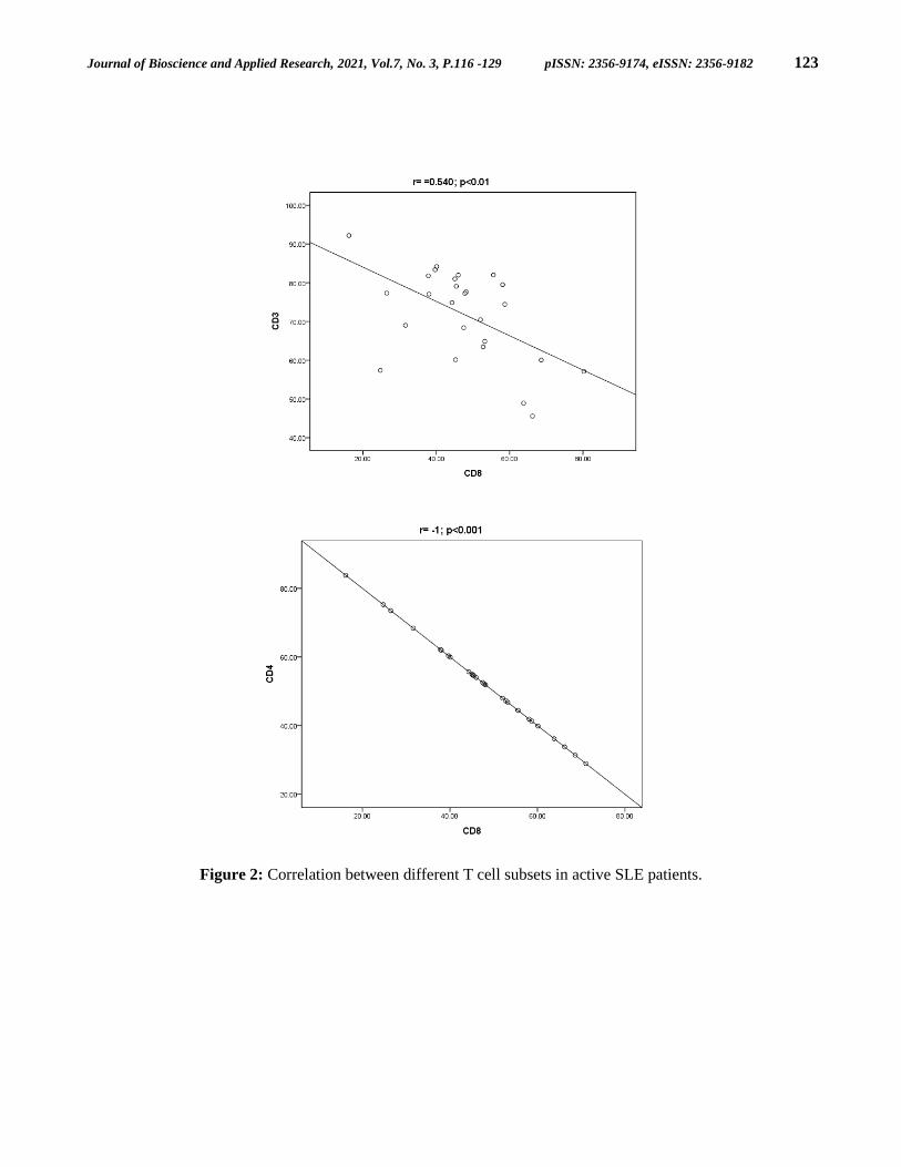
correlation was observed between the frequency of
CD8+ cells and CD3+ (r= -o.541; P<0.01) and CD4+ (r=
-1.00; P<0.001) (Figure 2) in active SLE patients.
3.3. Association between T-lymphocytes and
SLE clinical manifestations
The association between the clinical
manifestations of SLE and detected T- lymphocytes
(CD3+, CD4+, and CD8+) was presented in Table (3). A
significant reduction in CD3+ cells was seen in patients
with Arthritis (P<0.05) and Haemolytic anemia
(P<0.05) compared to patients without these

Journal of Bioscience and Applied Research, 2021, Vol.7, No. 3, P.116 -129 pISSN: 2356-9174, eISSN: 2356-9182 119
manifestations. A significant elevation in CD4+ cells
was observed in patients suffering from Oral Ulcers
(P<0.05). At the same time, there was a significant
reduction (P<0.05) in patients with vasculitis and
Arthritis (P<0.05) compared to patients without those
manifestations. Regarding CD8+cells, there was a
significant diminution (P<0.05) in SLE patients with
vasculitis compared to patients without vasculitis.
Table (1): Demographic and biochemical characteristics of controls and SLE patients.
Parameters
Control
group
(N=100)
SLE
group
(N=100)
P-value
Age (years) 28.22± 7.90 31.8± 1.0 NS
Gender (female/male) 85/15 87/13 NS
ESR (mm/1st hour) 7.2 ± 0.2 56.4 ± 3.4 P<0.01
WBCs (X1000/µl) 8.5 ± 0.14 8.1 ± 0.4 NS
HGB (g/dl) 14.2 ± 0.14 11.0 ± 0.2 P<0.01
Platelets (X1000/µl) 295.5 ± 6.4 257.1 ± 12.3 P<0.01
Serum Creatinine (mg/dl) 0.93 ± 0.23 0.89 ± 0.7 NS
ALT (IU/L) 22.09 ± 0.6 21.2 ± 1.6 NS
AST (IU/L) 24.8 ± 0.6 23.5 ± 1.5 NS
All data are presented as mean ± standard Error (mean ± SE). NS = not significant.
ESR (erythrocyte sedimentation rate); White Blood Cells (WBCs); HGB (Hemoglobin); Alanine
aminotransferase (ALT); Aspartate aminotransferase (AST);

Journal of Bioscience and Applied Research, 2021, Vol.7, No. 3, P.116 -129 pISSN: 2356-9174, eISSN: 2356-9182 120
Table (2): Clinical and laboratory characteristics of SLE patients
Demographic data
Mean ± SD
Laboratory Data Mean ± SD
Age (years) 32.75±10.31 Serum albumin 3.19 ± 0.7
Disease duration (years) 7.97 ±5.73 C3titre 77.4 ± 44.6
Female/Male 87/13 C4 titre 20.6 ± 24.3
SLEDAI 8.37±9.01 Cholesterol 187.5 ± 72.1
ACR criteria of SLE No (%) Triglyceride 162.97 ± 92.9
Malar rash 73 (72.3) HDL 47.2 ± 12.9
Photosensitivity 53 (52.5) LDL 118.3 ± 47.3
Oral Ulcers 63 (62.4) Consumed C3 34 (33.7)
Arthritis 50 (49.5) Consumed C4 20 (19.8)
Osteonecrosis 9 (8.9) Treatment N (%)
Serositis 39 (38.6) Antimalarial drug 89 (94.6)
Glomerulonephritis 62 (61.4) Cyclophosphamide 62 (65.9)
Neuropsychiatric disorders 13 (14.9) Azathioprine 67 (71.2)
PanCytopenia 19 (18.8) Biological 6 (6.3)
Anti-nuclear Ab 71 (70.3)
Anti-dsDNA Ab 71 (70.3)
Hemolytic anemia 16 (15.8)
Leucopenia 34 (33.7)
Neutropenia 13 (12.9)
Lymphopenia 31 (30.7)
All data are presented as mean ± SE. P<0.05(*), P<0.01(**), P<0.001(***)

Journal of Bioscience and Applied Research, 2021, Vol.7, No. 3, P.116 -129 pISSN: 2356-9174, eISSN: 2356-9182 121
Table (3): CD3+, CD4+ and CD8+ level in SLE patients with different clinical manifestations
Clinical parameter CD3+ CD4+ CD8+
Photosensitivity No
Yes
70.81±4.36
71.99±2.95
48.73±2.93
57.20±3.84
51.27±2.93
47.71±3.68
Oral Ulcers No
Yes
69.91±4.84
72.47±2.78
45.32±3.43
59.16±3.64*
54.68±3.43
45.91±3.58
Arthritis No
Yes
75.33±3.30
66.58±3.41*
56.03±4.56
53.31±3.53*
46.36±4.1
52.26±3.55
Serositis No
Yes
71.05±3.35
72.58±3.45
52.66±3.69
59.06±4.90
48.92±3.52
48.39±4.61
Neuropsychiatric disorders No
Yes
71.65±2.40
70.56±2.39
54.87±2.95
54.67±2.82
48.73±2.75
48.61±2.64
PanCytopenia No
Yes
73.80±2.57
63.74±5.31
55.47±3.35
52.54±6.70
47.76±3.21
53.16±4.33
Constitutional symptoms No
Yes
83.35±
71.21±2.45
43.05±9.19
56.23±3.06
56.94±9.19
47.74±2.88
Discoid/subacute cutaneous
lupus
No
Yes
64.47±18.88
72.20±2.33
47.06±13.28
55.44±3.07
52.94±13.28
48.41±2.86
Osteonecrosis No
Yes
71.81±2.70
68.53±6.81
55.53±3.39
48.80±3.07
48.46±3.19
51.19±3.07
Vasculities No
Yes
71.51±2.61
72.79±6.42
54.97±3.29
53.96±0.88*
49.05±3.08
46.04±0.88*
Raynaud's phenomena No
Yes
72.08±2.45
60.13±2.33
54.87±3.06
54.73±3.04
48.86±2.85
45.27±2.72
Alopecia No
Yes
72.75±2.89
69.31±4.56
53.79±3.59
57.67±5.27
48.23±3.12
49.99±5.94
Haemolytic anemia No
Yes
73.89±2.45
61.32±5.85*
54.03±3.22
58.88±7.91
49.07±3.03
46.66±7.29
Leucopenia No
Yes
73.56±2.91
67.19±4.56
55.44±3.85
53.76±4.99
46.60±3.45
53.89±4.89
Neutropenia No
Yes
70.95±2.53
84.18±2.34
54.78±3.17
59.92±3.14
49.09±2.94
40.08±2.84
Lymphopenia No
Yes
73.56±2.91
67.19±4.56
55.44±3.86
53.76±4.99
46.60±3.45
53.89±4.89
All data are presented as mean ± SE. P<0.05(*), P<0.01(**), P<0.001(***)

Journal of Bioscience and Applied Research, 2021, Vol.7, No. 3, P.116 -129 pISSN: 2356-9174, eISSN: 2356-9182 122
Figure 1: Percentage of CD3+, CD4+, and CD8+ in controls and SLE patients (active and inactive) all data are
presented as mean ± SE. "a" significant from the control group, "b" significant from inactive group. P<0.05(*),
P<0.01(**), P<0.001(***).
0
20
40
60
80
100
Control SLE Inactive Active
CD3+
a***
0
20
40
60
80
100
Control SLE Inactive Active
CD4+
a***
b*
0
20
40
60
80
100
Control SLE Inactive Active
CD8+
a***
Journal of Bioscience and Applied Research, 2021, Vol.7, No. 3, P.116 -129 pISSN: 2356-9174, eISSN: 2356-9182 123
Figure 2: Correlation between different T cell subsets in active SLE patients.
Journal of Bioscience and Applied Research, 2021, Vol.7, No. 3, P.116 -129 pISSN: 2356-9174, eISSN: 2356-9182 124
Received: July 26, 2021. Accepted: September 18, 2021. Published: September 29, 2021
4. Discussion
SLE is characterized serologically by B cell
hyperactivity and a panoply of autoantibodies against
nuclear, cytoplasmic, and cell surface antigens. Since
these autoantibodies are mainly of the IgG1 subclass, T
lymphocytes are likely essential in providing help to the
autoantibody-producing B cells (Manjili and Payne,
2016(. Evidence for T cell involvement in disease
pathogenesis is illustrated by the association of SLE
with particular major histocompatibility complex
(MHC) class II alleles and affinity maturation of IgG
autoantibody production (Kailashiyaet al., 2019). It is
thought that T helper (Th) cells drive the production of
pathogenic anti-DNA autoantibodies in SLE, and it has
been shown, in vitro, that some of these are CD47
CD87. These, i.e., double-negative (DN) T cells, have
been shown to express either the αβ T cell receptor
(TCR) or γδ TCR (Abdirama et al., 2021).
Autoreactive CD4+ T cells are implicated in the
pathogenesis of SLE by promoting autoantibody
production by B cells and directly propagating organ
damage in inflamed target organs (Suárez-Fueyoet al.,
2016; Abdirama et al., 2021).
CD8+ cells play a key role in recognizing and
removing cells infected by intracellular pathogens and
in antitumor response. Since stimulation only through
the TCR receptor is unable to maintain optimum
CD8+ activation, the second costimulatory signal is
essential for full activation and survival of these cells
(Jandl and King, 2016). The best-known
costimulatory signal is provided by the interaction of
CD28 molecules presented on the T lymphocyte as well
as the CD86 and CD80 molecules expressed on the
APC's surface. Adequate signal power delivered to
naive T CD8+ results in the proliferation and
differentiation of two cell types. One of these is
cytotoxic T lymphocytes (CTL), which undergo
apoptosis after reaching maturity and fulfilling their
effector function (Yap et al., 2010; Talaat et al., 2015).
The second type is T CD8+ memory cells, both central
and effector. Their continuous presence in the
circulation is essential to control another potential
exposure to the same antigen faster and more
effectively (Suet al., 2012).
Most autoimmune diseases are associated with an
increase in T CD8+CD28− (CD8+CD57+) cells, which
exhibit highly cytotoxic activity and can be related to
more severe disease manifestations (Jin and Dong,
2013; Strzępa and Szczepanik, 2013). Quantitative
changes in the CD8+CD57+ population were observed,
among others, in multiple sclerosis, type 1 diabetes,
Graves' disease, and rheumatoid arthritis. The
decreased number of CD8+CD28− T-cells correlates
with clinical response to abatacept in patients with
rheumatoid arthritis (Shah et al., 2010; Talaat et al.,
2015). Some researchers have reported that
lymphocytes with CD8+CD28− phenotype show
regulatory properties. So far, few studies addressing the
size of the CD8+CD28− subpopulation in patients with
SLE have been conducted. It has been shown that the
number of CD8+CD28− cells might be reduced or
unchanged compared to the control group (Tang et al.,
2019; Yazdani et al., 2020).
5. Conclusion
In conclusion, the current study reported a
significant downregulation in CD4+ cells with a
substantial upregulation of CD3+ and CD8+ frequency
in SLE patients. We stressed the importance of these
subsets in several disease manifestations. Despite the
conflicting reports in T lymphocytes frequencies
between SLE patients and control subjects, the
therapeutic potential of this cell population holds a
great promise for the future employment of effective
treatment modalities using the T cells population.
Further functional research is needed to validate our
findings.

Journal of Bioscience and Applied Research, 2021, Vol.7, No. 3, P.116 -129 pISSN: 2356-9174, eISSN: 2356-9182 125
6. Conflict of interest: The authors declare that
they have no conflict of interest.
7. References
• Abdirama D, Tesch S, Grießbach AS, von
Spee-Mayer C, Humrich JY, Stervbo U, Babel N,
Meisel C, Alexander T, Biesen R, Bacher P, Scheffold
A, Eckardt KU, Hiepe F, Radbruch A, Burmester GR,
Riemekasten G, Enghard P. Nuclear antigen-reactive
CD4+ T cells expand in active systemic lupus
erythematosus, produce effector cytokines, and invade
the kidneys. Kidney Int. 2021 Jan;99(1):238-246. DOI:
10.1016/j.kint.2020.05.051. Epub 2020 Jun 24. PMID:
32592813.
• Aringer M, Costenbader K, Daikh D, Brinks R,
Mosca M, Ramsey-Goldman R, Smolen JS, Wofsy D,
Boumpas DT, Kamen DL, Jayne D, Cervera R,
Costedoat-Chalumeau N, Diamond B, Gladman DD,
Hahn B, Hiepe F, Jacobsen S, Khanna D, Lerstrøm K,
Massarotti E, McCune J, Ruiz-Irastorza G, Sanchez-
Guerrero J, Schneider M, Urowitz M, Bertsias G, Hoyer
BF, Leuchten N, Tani C, Tedeschi SK, Touma Z,
Schmajuk G, Anic B, Assan F, Chan TM, Clarke AE,
Crow MK, Czirják L, Doria A, Graninger W, Halda-
Kiss B, Hasni S, Izmirly PM, Jung M, Kumánovics G,
Mariette X, Padjen I, Pego-Reigosa JM, Romero-Diaz
J, Rúa-Figueroa Fernández Í, Seror R, Stummvoll GH,
Tanaka Y, Tektonidou MG, Vasconcelos C, Vital EM,
Wallace DJ, Yavuz S, Meroni PL, Fritzler MJ, Naden
R, Dörner T, Johnson SR. 2019 European League
Against Rheumatism/American College of
Rheumatology Classification Criteria for Systemic
Lupus Erythematosus. Arthritis Rheumatol. 2019
Sep;71(9):1400-1412. DOI: 10.1002/art.40930. Epub
2019 Aug 6. PMID: 31385462; PMCID: PMC6827566.
• Astry B, Venkatesha SH, Moudgil KD.
Involvement of the IL-23/IL-17 axis and the Th17/Treg
balance in the pathogenesis and control of autoimmune
arthritis. Cytokine. 2015 Jul;74(1):54-61. DOI:
10.1016/j.cyto.2014.11.020. Epub 2015 Jan 13. PMID:
25595306; PMCID: PMC4457562.
• Azab NA, Bassyouni IH, Emad Y, Abd El-
Wahab GA, Hamdy G, Mashahit MA. CD4+CD25+
regulatory T cells (TREG) in systemic lupus
erythematosus (SLE) patients: the possible influence of
treatment with corticosteroids. Clin Immunol. 2008
May;127(2):151-7. DOI: 10.1016/j.clim.2007.12.010.
Epub 2008 Mar 4. PMID: 18299252.
• Basta F, Fasola F, Triantafyllias K, Schwarting
A. Systemic Lupus Erythematosus (SLE) Therapy: The
Old and the New. Rheumatol Ther. 2020 Sep;7(3):433-
446. DOI: 10.1007/s40744-020-00212-9. Epub 2020
Jun 2. PMID: 32488652; PMCID: PMC7410873
• Becker-Merok A, Eilertsen GØ, Nossent JC.
Levels of transforming growth factor-beta are low in
systemic lupus erythematosus patients with active
disease. J Rheumatol. 2010 Oct;37(10):2039-45. DOI:
10.3899/jrheum.100180. Epub 2010 Aug 3. PMID:
20682675.
• Cassia M, Alberici F, Gallieni M, Jayne D.
Lupus nephritis and B-cell targeting therapy. Expert
Rev Clin Immunol. 2017 Oct;13(10):951-962. DOI:
10.1080/1744666X.2017. 1366855. Epub 2017 Aug
18. PMID: 28800401.
• Cepika AM, Soldo Jureša D, MorovićVergles
J, Malenica B, Santak M, Kapitanović S, Mayer M,
Anić B, Sentić M, Gagro A. Decrease in circulating
DNA, IL-10 and BAFF levels in newly-diagnosed SLE
patients after corticosteroid and chloroquine treatment.
Cell Immunol. 2012 Mar-Apr;276(1-2):196-203. DOI:
10.1016/j.cellimm.2012.05.009. Epub 2012 May 29.
PMID: 22703694.
• Chen M, Chen X, Wan Q. Altered frequency of
Th17 and Treg cells in new-onset systemic lupus
erythematosus patients. Eur J Clin Invest.
2018;48(11):e13012.
• Cheng HB, Chen RY, Wu JP, Chen L, Liang
YH, Pan HF, Pan ZF, Zhang QH, Li Q, Du TX, Lv YM,

Journal of Bioscience and Applied Research, 2021, Vol.7, No. 3, P.116 -129 pISSN: 2356-9174, eISSN: 2356-9182 126
Shi JQ. Complement C4 induces regulatory T cells
differentiation through dendritic cell in systemic lupus
erythematosus. Cell Biosci. 2015 Dec 23;5:73. DOI:
10.1186/s13578-015-0052-8. PMID: 26705467;
PMCID: PMC4690337.
• Dai H, He F, Tsokos GC, Kyttaris VC. IL-23
Limits the Production of IL-2 and Promotes
Autoimmunity in Lupus. J Immunol. 2017 Aug
1;199(3):903-910. DOI: 10.4049/jimmunol.1700418.
Epub 2017 Jun 23. PMID: 28646040; PMCID:
PMC5526729.
• Damsker JM, Hansen AM, Caspi RR. Th1 and
Th17 cells: adversaries and collaborators. Ann N Y
Acad Sci. 2010 Jan;1183:211-21. DOI: 10.1111/j.1749-
6632.2009.05133.x. PMID: 20146717; PMCID:
PMC2914500.
• Dienz O, Rincon M. The effects of IL-6 on
CD4 T cell responses. Clin Immunol. 2009
Jan;130(1):27-33. DOI: 10.1016/j.clim.2008.08.018.
Epub 2008 Oct 8. PMID: 18845487; PMCID:
PMC2660866.
• Fattorossi A, Battaglia A, Buzzonetti A,
Ciaraffa F, Scambia G, Evoli A. Circulating and thymic
CD4 CD25 T regulatory cells in myasthenia gravis:
effect of immunosuppressive treatment. Immunology.
2005 Sep;116(1):134-41. DOI: 10.1111/j.1365-
2567.2005.02220.x. PMID: 16108825; PMCID:
PMC1802400.
• Gaffen SL, Jain R, Garg AV, Cua DJ. The IL-
23-IL-17 immune axis: from mechanisms to therapeutic
testing. Nat Rev Immunol. 2014 Sep;14(9):585-600.
DOI: 10.1038/nri3707. PMID: 25145755; PMCID:
PMC4281037.
• Gatto M, Saccon F, Zen M, Regola F, Fredi M,
Andreoli L, Tincani A, Urban ML, Emmi G, Ceccarelli
F, Conti F, Bortoluzzi A, Govoni M, Tani C, Mosca M,
Ubiali T, Gerosa M, Bozzolo E, Canti V, Cardinaletti
P, Gabrielli A, Tanti G, Gremese E, De Marchi G, De
Vita S, Fasano S, Ciccia F, Pazzola G, Salvarani C,
Negrini S, Puppo F, Di Matteo A, De Angelis R,
Orsolini G, Rossini M, Faggioli P, Laria A, Piga M,
Mathieu A, Scarpato S, Rossi FW, de Paulis A,
Brunetta E, Ceribelli A, Selmi C, Prete M, Racanelli V,
Vacca A, Bartoloni E, Gerli R, Larosa M, Iaccarino L,
Doria A. Early Disease and Low Baseline Damage as
Predictors of Response to Belimumab in Patients With
Systemic Lupus Erythematosus in a Real-Life Setting.
Arthritis Rheumatol. 2020 Aug;72(8):1314-1324. DOI:
10.1002/art.41253. Epub 2020 Jun 12. PMID:
32275125.
• Guimarães PM, Scavuzzi BM, Stadtlober NP,
Franchi Santos LFDR, Lozovoy MAB, Iriyoda TMV,
Costa NT, Reiche EMV, Maes M, Dichi I, Simão ANC.
Cytokines in systemic lupus erythematosus: far beyond
Th1/Th2 dualism lupus: cytokine profiles. Immunol
Cell Biol. 2017 Oct;95(9):824-831. DOI:
10.1038/icb.2017.53. Epub 2017 Jun 26. PMID:
28649995.
• Han JW, Zheng HF, Cui Y, Sun LD, Ye DQ,
Hu Z, Xu JH, Cai ZM, Huang W, Zhao GP, Xie HF,
Fang H, Lu QJ, Xu JH, Li XP, Pan YF, Deng DQ, Zeng
FQ, Ye ZZ, Zhang XY, Wang QW, Hao F, Ma L, Zuo
XB, Zhou FS, Du WH, Cheng YL, Yang JQ, Shen SK,
Li J, Sheng YJ, Zuo XX, Zhu WF, Gao F, Zhang PL,
Guo Q, Li B, Gao M, Xiao FL, Quan C, Zhang C,
Zhang Z, Zhu KJ, Li Y, Hu DY, Lu WS, Huang JL, Liu
SX, Li H, Ren YQ, Wang ZX, Yang CJ, Wang PG,
Zhou WM, Lv YM, Zhang AP, Zhang SQ, Lin D, Li Y,
Low HQ, Shen M, Zhai ZF, Wang Y, Zhang FY, Yang
S, Liu JJ, Zhang XJ. Genome-wide association study in
a Chinese Han population identifies nine new
susceptibility loci for systemic lupus erythematosus.
Nat Genet. 2009 Nov;41(11):1234-7. DOI:
10.1038/ng.472. Epub 2009 Oct 18. PMID: 19838193.
• Hartman EAR, van Royen-Kerkhof A, Jacobs
JWG, Welsing PMJ, Fritsch-Stork RDE. Performance
of the 2012 Systemic Lupus International Collaborating
Clinics classification criteria versus the 1997 American

Journal of Bioscience and Applied Research, 2021, Vol.7, No. 3, P.116 -129 pISSN: 2356-9174, eISSN: 2356-9182 127
College of Rheumatology classification criteria in adult
and juvenile systemic lupus erythematosus. A
systematic review and meta-analysis. Autoimmun Rev.
2018 Mar;17(3):316-322. DOI:
10.1016/j.autrev.2018.01.007. Epub 2018 Jan 31.
PMID: 29366725.
• Hayball JD, Lake RA. Altered superantigenic
ligands demonstrate the quantitative nature of T-cell
activation. Immunol Cell Biol. 2000 Dec;78(6):623-32.
DOI: 10.1046/j.1440-1711.2000.00971.x. PMID:
11114973.
• Jandl C, King C. Cytokines in the Germinal
Center Niche. Antibodies (Basel). 2016 Feb 5;5(1):5.
DOI: 10.3390/antib5010005. Erratum in: Antibodies
(Basel). 2016 Apr 27;5(2): PMID: 31557986; PMCID:
PMC6698856.
• Jin W, Dong C. IL-17 cytokines in immunity
and inflammation. Emerg Microbes Infect. 2013
Sep;2(9):e60. DOI: 10.1038/emi.2013.58. Epub 2013
Sep 18. PMID: 26038490; PMCID: PMC3820987.
• Justiz Vaillant AA, Goyal A, Bansal P,
Varacallo M. Systemic Lupus Erythematosus. 2020
Dec 30. In: StatPearls [Internet]. Treasure Island (FL):
StatPearls Publishing; 2021 Jan–. PMID: 30571026.
• Kailashiya V, Singh U, Rana R, Singh NK,
Dash D, Kailashiya J. Regulatory T Cells and Their
Association with Serum Markers and Symptoms in
Systemic Lupus Erythematosus and Rheumatoid
Arthritis. Immunol Invest. 2019 Jan;48(1):64-78. DOI:
10.1080/08820139.2018.1527852. Epub 2018 Oct 16.
PMID: 30325682.
• Karagiannidis C, Akdis M, Holopainen P,
Woolley NJ, Hense G, Rückert B, Mantel PY, Menz G,
Akdis CA, Blaser K, Schmidt-Weber CB.
Glucocorticoids upregulate FOXP3 expression and
regulatory T cells in asthma. J Allergy Clin Immunol.
2004 Dec;114(6):1425-33. DOI:
10.1016/j.jaci.2004.07.014. PMID: 15577848.
• Knochelmann HM, Dwyer CJ, Bailey SR,
Amaya SM, Elston DM, Mazza-McCrann JM, Paulos
CM. When worlds collide: Th17 and Treg cells in
cancer and autoimmunity. Cell Mol Immunol. 2018
May;15(5):458-469. DOI: 10.1038/s41423-018-0004-
4. Epub 2018 Mar 21. PMID: 29563615; PMCID:
PMC6068176.
• Koga T, Ichinose K, Kawakami A, Tsokos GC.
The role of IL-17 in systemic lupus erythematosus and
its potential as a therapeutic target. Expert Rev Clin
Immunol. 2019 Jun;15(6):629-637. DOI:
10.1080/1744666X.2019.1593141. Epub 2019 Mar 25.
PMID: 30874446.
• Koga T, Ichinose K, Tsokos GC. T cells and
IL-17 in lupus nephritis. Clin Immunol. 2017
Dec;185:95-99. DOI: 10.1016/j.clim.2016.04.010.
Epub 2016 Apr 21. PMID: 27109641; PMCID:
PMC5074925.
• Kuwabara T, Ishikawa F, Kondo M, Kakiuchi
T. The Role of IL-17 and Related Cytokines in
Inflammatory Autoimmune Diseases. Mediators
Inflamm. 2017;2017:3908061. DOI:
10.1155/2017/3908061. Epub 2017 Feb 20. PMID:
28316374; PMCID: PMC5337858.
• Li D, Guo B, Wu H, Tan L, Chang C, Lu Q.
Interleukin-17 in systemic lupus erythematosus: a
comprehensive review. Autoimmunity.
2015;48(6):353-361.
• Lourenço EV, La Cava A. Cytokines in
systemic lupus erythematosus. Curr Mol Med. 2009
Apr;9(3):242-54. DOI:

Journal of Bioscience and Applied Research, 2021, Vol.7, No. 3, P.116 -129 pISSN: 2356-9174, eISSN: 2356-9182 128
10.2174/156652409787847263. PMID: 19355907;
PMCID: PMC3589140.
• Manjili MH, Payne KK. Immune Regulatory
Function of Tregs. Immunol Invest. 2016
Nov;45(8):708-711. DOI:
10.1080/08820139.2016.1235394. PMID: 27775448.
• Mathias LM, Stohl W. Systemic lupus
erythematosus (SLE): emerging therapeutic targets.
Expert Opin Ther Targets. 2020 Dec;24(12):1283-
1302. DOI: 10.1080/14728222.2020.1832464. Epub
2020 Dec 1. PMID: 33034541.
• Mok MY, Wu HJ, Lo Y, Lau CS. The relation
of interleukin 17 (IL-17) and IL-23 to Th1/Th2
cytokines and disease activity in systemic lupus
erythematosus. J Rheumatol. 2010 Oct;37(10):2046-
52. DOI: 10.3899/jrheum.100293. Epub 2010 Aug 3.
PMID: 20682672.
• Moore E, Putterman C. Are lupus animal
models useful for understanding and developing new
therapies for human SLE? J Autoimmun. 2020
Aug;112:102490. DOI: 10.1016/j.jaut.2020.102490.
Epub 2020 Jun 11. PMID: 32535128; PMCID:
PMC7384952.
• Murphy CA, Langrish CL, Chen Y,
Blumenschein W, McClanahan T, Kastelein RA,
Sedgwick JD, Cua DJ. Divergent pro- and
antiinflammatory roles for IL-23 and IL-12 in joint
autoimmune inflammation. J Exp Med. 2003 Dec
15;198(12):1951-7. DOI: 10.1084/jem.20030896.
Epub 2003 Dec 8. PMID: 14662908; PMCID:
PMC2194162.
• Okamoto A, Fujio K, Okamura T, Yamamoto
K. Regulatory T-cell-associated cytokines in systemic
lupus erythematosus. J Biomed Biotechnol.
2011;2011:463412. DOI: 10.1155/2011/463412. Epub
2011 Dec 18. PMID: 22219657; PMCID:
PMC3247013.
• Pan L, Lu MP, Wang JH, Xu M, Yang SR.
Immunological pathogenesis and treatment of systemic
lupus erythematosus. World J Pediatr. 2020
Feb;16(1):19-30. DOI: 10.1007/s12519-019-00229-3.
Epub 2019 Feb 22. PMID: 30796732; PMCID:
PMC7040062.
• Puliaeva I, Puliaev R, Via CS. Therapeutic
potential of CD8+ cytotoxic T lymphocytes in SLE.
Autoimmun Rev. 2009 Jan;8(3):219-23. DOI:
10.1016/j.autrev.2008.07.045. Epub 2008 Aug 24.
PMID: 18725326; PMCID: PMC3215296.
• Shah K, Lee WW, Lee SH, Kim SH, Kang SW,
Craft J, Kang I. Dysregulated balance of Th17 and Th1
cells in systemic lupus erythematosus. Arthritis Res
Ther. 2010;12(2): R53. DOI: 10.1186/ar2964. Epub
2010 Mar 24. Erratum in: Arthritis Res Ther.
2010;12(3):402. PMID: 20334681; PMCID:
PMC2888202.
• Singla S, Wenderfer SE, Muscal E, Sagcal-
Gironella ACP, Orange JS, Makedonas G. Changes in
Frequency and Activation Status of Major CD4+ T-Cell
Subsets after Initiation of Immunosuppressive Therapy
in a Patient with New Diagnosis Childhood-Onset
Systemic Lupus Erythematosus. Front Pediatr. 2017
May 15;5:104. DOI: 10.3389/fped.2017.00104. PMID:
28555177; PMCID: PMC5430328.
• Stritesky GL, Yeh N, Kaplan MH. IL-23
promotes maintenance but not commitment to the Th17
lineage. J Immunol. 2008 Nov 1;181(9):5948-55. DOI:
10.4049/jimmunol. 181.9.5948. PMID: 18941183;
PMCID: PMC2678905.
• Strzępa A, Szczepanik M. IL-17-expressing
cells as a potential therapeutic target for treatment of

Journal of Bioscience and Applied Research, 2021, Vol.7, No. 3, P.116 -129 pISSN: 2356-9174, eISSN: 2356-9182 129
immunological disorders. Pharmacol Rep.
2011;63(1):30-44. DOI: 10.1016/s1734-
1140(11)70396-6. PMID: 21441609.
• Su DL, Lu ZM, Shen MN, Li X, Sun LY. Roles
of pro-and anti-inflammatory cytokines in the
pathogenesis of SLE. J Biomed Biotechnol.
2012;2012:347141. DOI: 10.1155/2012/347141. Epub
2012 Feb 15. PMID: 22500087; PMCID:
PMC3303597.
• Su DL, Lu ZM, Shen MN, Li X, Sun LY. Roles
of pro-and anti-inflammatory cytokines in the
pathogenesis of SLE. J Biomed Biotechnol.
2012;2012:347141. DOI: 10.1155/2012/347141. Epub
2012 Feb 15. PMID: 22500087; PMCID:
PMC3303597.
• Suárez A, López P, Gómez J, Gutiérrez C.
Enrichment of CD4+ CD25high T cell population in
patients with systemic lupus erythematosus treated with
glucocorticoids. Ann Rheum Dis. 2006
Nov;65(11):1512-7. DOI: 10.1136/ard.2005.049924.
Epub 2006 Apr 10. PMID: 16606650; PMCID:
PMC1798359.
• Suárez-Fueyo A, Bradley SJ, Tsokos GC. T
cells in Systemic Lupus Erythematosus. Curr Opin
Immunol. 2016 Dec;43:32-38. DOI:
10.1016/j.coi.2016.09.001. Epub 2016 Sep 13. PMID:
27636649; PMCID: PMC5125867.
• Talaat RM, Elmaghraby AM, Barakat SS, El-
Shahat M. Alterations in immune cell subsets and their
cytokine secretion profile in childhood idiopathic
thrombocytopenic purpura (ITP). Clin Exp Immunol.
2014 May;176(2):291-300. DOI: 10.1111/cei.12279.
PMID: 24460857; PMCID: PMC3992042.
• Talaat RM, Mohamed SF, Bassyouni IH,
Raouf AA. Th1/Th2/Th17/Treg cytokine imbalance in
systemic lupus erythematosus (SLE) patients:
Correlation with disease activity. Cytokine. 2015
Apr;72(2):146-53. DOI: 10.1016/j.cyto.2014.12.027.
Epub 2015 Jan 31. PMID: 25647269.
• Tang Y, Tao H, Gong Y, Chen F, Li C, Yang
X. Changes of Serum IL-6, IL-17, and Complements in
Systemic Lupus Erythematosus Patients. J Interferon
Cytokine Res. 2019 Jul;39(7):410-415. DOI:
10.1089/jir.2018.0169. Epub 2019 Jun 7. PMID:
31173544.
• Wang D, Huang S, Yuan X, Liang J, Xu R, Yao
G, Feng X, Sun L. The regulation of the Treg/Th17
balance by mesenchymal stem cells in human systemic
lupus erythematosus. Cell Mol Immunol. 2017
May;14(5):423-431. DOI: 10.1038/cmi.2015.89. Epub
2015 Oct 5. PMID: 26435067; PMCID: PMC5423084.
• Wang X, Wei Y, Xiao H, Liu X, Zhang Y, Han
G, Chen G, Hou C, Ma N, Shen B, Li Y, Egwuagu CE,
Wang R. A novel IL-23p19/Ebi3 (IL-39) cytokine
mediates inflammation in Lupus-like mice. Eur J
Immunol. 2016 Jun;46(6):1343-50. DOI:
10.1002/eji.201546095. Epub 2016 Apr 13. PMID:
27019190.
• Yap DY, Lai KN. Cytokines and their roles in
the pathogenesis of systemic lupus erythematosus: from
basics to recent advances. J Biomed Biotechnol.
2010;2010:365083. DOI: 10.1155/2010/365083. Epub
2010 May 6. PMID: 20467470; PMCID:
PMC2866250.
• Yazdani MR, Aflaki E, Gholijani N.
Inflammatory and T Helper 17/ Regulatory T Cells
Related Cytokines Balance in Cutaneous Lupus
Erythematosus (CLE). Iran J Allergy Asthma Immunol.
2020 Feb 1;19(1):9-17. DOI:
10.18502/ijaai.v19i1.2411. PMID: 32245325.
• Zhang L, Yuan S, Cheng G, Guo B. Type I IFN
promotes IL-10 production from T cells to suppress
Th17 cells and Th17-associated autoimmune
inflammation. PLoS One. 2011;6(12):e28432. DOI:
10.1371/journal.pone.0028432. Epub 2011 Dec 6.
PMID: 22163016; PMCID: PMC3232207.