systematic review and meta-analysis of diagnostic accuracy ... · pdf fileplatinum priority...
TRANSCRIPT

E U R O P E A N U R O L O G Y 6 9 ( 2 0 1 6 ) 6 6 0 – 6 7 3
avai lable at www.sciencedirect .com
journal homepage: www.europeanurology.com
Platinum Priority – Review – Kidney CancerEditorial by Roger Kockelbergh and Leyshon Griffiths on pp. 674–675 of this issue
Systematic Review and Meta-analysis of Diagnostic Accuracy of
Percutaneous Renal Tumour Biopsy
Lorenzo Marconi a, Saeed Dabestani b, Thomas B. Lam c, Fabian Hofmann d, Fiona Stewart c,John Norrie e, Axel Bex f, Karim Bensalah g, Steven E. Canfield h, Milan Hora i, Markus A. Kuczyk j,Axel S. Merseburger k, Peter F.A. Mulders l, Thomas Powles m, Michael Staehler n,Borje Ljungberg o, Alessandro Volpe p,*
a Department of Urology, Coimbra University Hospital, Coimbra, Portugal; b Department of Urology, Skane University Hospital, Malmo, Sweden; c Academic
Urology Unit, University of Aberdeen, Aberdeen, UK; d Department of Urology, Sunderby Hospital, Sunderby, Sweden; e Health Services Research Unit,
University of Aberdeen, UK; f Department of Urology, The Netherlands Cancer Institute, Antoni van Leeuwenhoek Hospital, Amsterdam, The Netherlands;g Department of Urology, University of Rennes, Rennes, France; h Division of Urology, University of Texas Medical School at Houston, Houston, TX, USA;i Department of Urology, Faculty Hospital and Faculty of Medicine in Pilsen, Charles University in Prague, Prague, Czech Republic; j Department of Urology
and Urologic Oncology, Hannover Medical School, Hannover, Germany; k Department of Urology, University Hospital Schleswig-Holstein, Lubeck, Germany;l Department of Urology, Radboud University, Nijmegen, The Netherlands; m Barts Cancer Institute, Queen Mary University of London, St. Bartholomew’s
Hospital, London, UK; n Department of Urology, Ludwig-Maximilians University, Munich, Germany; o Department of Surgical and Perioperative Sciences,
Urology and Andrology, Umea University, Umea, Sweden; p Division of Urology, Maggiore della Carita Hospital, University of Eastern Piedmont, Novara, Italy
Article info
Article history:
Accepted July 31, 2015
Associate Editor:James Catto
Keywords:
Accuracy
Diagnosis
Fine-needle aspiration
Meta-analysis
Needle core biopsy
Renal mass
Renal cell carcinoma
Abstract
Context: The role of percutaneous renal tumour biopsy (RTB) remains controversial dueto uncertainties regarding its diagnostic accuracy and safety.Objective: We performed a systematic review and meta-analysis to determine the safetyand accuracy of percutaneous RTB for the diagnosis of malignancy, histologic tumoursubtype, and grade.Evidence acquisition: Medline, Embase, and Cochrane Library were searched for studiesproviding data on diagnostic accuracy and complications of percutaneous core biopsy(CB) or fine-needle aspiration (FNA) of renal tumours. A meta-analysis was performed toobtain pooled estimates of sensitivity and specificity for diagnosis of malignancy. TheCohen kappa coefficient (k) was estimated for the analysis of histotype/grade concor-dance between diagnosis on RTB and surgical specimen. Risk of bias assessment wasperformed (QUADAS-2).Evidence synthesis: A total of 57 studies recruiting 5228 patients were included. Theoverall median diagnostic rate of RTB was 92%. The sensitivity and specificity ofdiagnostic CBs and FNAs were 99.1% and 99.7%, and 93.2% and 89.8%, respectively. Agood (k = 0.683) and a fair (k = 0.34) agreement were observed between histologicsubtype and Fuhrman grade on RTB and surgical specimen, respectively. A very lowrate of Clavien �2 complications was reported. Study limitations included selection anddifferential-verification bias.Conclusions: RTB is safe and has a high diagnostic yield in experienced centres. Both CB
accanc
and FNA have goodwith better perform
* Corresponding author. DiPiedmont, Corso Mazzini, 1E-mail address: alessandro
http://dx.doi.org/10.1016/j.eururo.2015.07.0720302-2838/# 2015 European Association of Urology. Published by Elsevier
uracy for the diagnosis of malignancy and histologic subtype,e for CB. The accuracy for Fuhrman grade is fair. Overall, the
vision of Urology, Maggiore della Carita Hospital, University of Eastern8, 28100, Novara, Italy. Tel. +39 0321 3733201; Fax: +39 0321 [email protected] (A. Volpe).
B.V. All rights reserved.
quality of the evidence was moderate. Prospective cohort studies recruiting consecutivepatients and using homogeneous reference standards are required.Patient summary: We systematically reviewed the literature to assess the safety anddiagnostic performance of renal tumour biopsy (RTB). The results suggest that RTB hasgood accuracy in diagnosing renal cancer and its subtypes, and it appears to be safe.However, the quality of evidence was moderate, and better quality studies are requiredto provide a more definitive answer.
# 2015 European Association of Urology. Published by Elsevier B.V. All rights reserved.
E U R O P E A N U R O L O G Y 6 9 ( 2 0 1 6 ) 6 6 0 – 6 7 3 661
1. Introduction
The management of renal tumours has evolved, with the
increasing use of nonextirpative therapies for small renal
masses (SRMs) in selected patients and the advent of
effective targeted drugs for metastatic disease [1]. This has
led to an increasing recognition of the importance of
histologic characterisation of renal masses before treatment
to tailor therapy based on tumour histology either in the
localised or metastatic setting [2].
Percutaneous renal tumour biopsy (RTB) has been
criticised due to concerns regarding its safety, diagnostic
accuracy, and ability to distinguish tumour histologic
subtypes and nuclear grade. Although fine-needle aspira-
tion (FNA) and core biopsy (CB) have been used to sample
renal tumours, the best technique has not been clearly
defined [3]. Several recent studies have reported low
complication rates and good diagnostic performance of
RTB, but most studies were limited by small sample sizes,
heterogeneous populations, different biopsy techniques,
and lack of standardised definitions for diagnostic accuracy
[4].
We performed a systematic review of the literature and
meta-analysis to determine the diagnostic performance and
safety of RTB in characterising malignancy, histologic
subtype, and grade of renal tumours.
2. Evidence acquisition
2.1. Search strategy
The review was performed according to Preferred Reporting
Items for Systematic Reviews and Meta-analysis (PRISMA)
[5] and the Cochrane Handbook for Systematic Reviews of
Diagnostic Test Accuracy [6]. Studies on percutaneous RTB
(January 1, 1946, to September 1, 2014) were identified by
highly sensitive searches of electronic databases (Medline,
Medline In-Process, Embase, Cochrane Controlled Trials
Register, and LILACS) and relevant Web sites [7]. The search
was complemented by the reference lists of included
studies and additional reports identified by the European
Association of Urology (EAU) Renal Cell Carcinoma (RCC)
Guideline Panel. No language restrictions were imposed.
Two reviewers (L.M. and S.D.) screened all abstracts and
full-text articles independently. Disagreement was resolved
by a third party (T.L.).
2.2. Selection of studies
Prospective or retrospective cohort studies providing data
on accuracy for malignancy, tumour histotype and grade,
and/or on complications of percutaneous CB or FNA of solid
or cystic renal masses of any size in adult patients were
included. Studies that fulfilled the following criteria were
included for the evaluation of diagnostic accuracy for
malignancy: (1) reference standard for tumour malignancy
represented by pathology on surgical specimen of partial or
radical nephrectomy performed after RTB, or clinical and
radiologic follow-up of at least 12 mo showing presence or
absence of tumour progression and/or onset of tumour-
related symptoms; (2) availability of number of nondiag-
nostic biopsies; and (3) availability of number of diagnostic
biopsies classified as true positives (TPs), false positives
(FPs), false negatives (FNs), and true negatives (TNs) either
as group totals or by case-by-case enumeration of diagno-
ses. Studies that did not provide data on all four elements of
diagnostic accuracy were excluded.
Studies that provided data to assess concordance
between tumour grade and/or histologic subtype between
RTB and surgical pathology were included for the assess-
ment of diagnostic accuracy for histologic subtype and/or
grade.
Studies exclusively reporting complications of RTBs were
also included. Complications were graded according to the
Clavien-Dindo classification [8]. Studies on laparoscopic-
assisted or ex vivo RTBs were excluded.
2.3. Data extraction
A data extraction form was developed a priori to collect
information on study design, patient characteristics (age,
gender, indication for RTB, comorbidities), tumour features
(size, solid or cystic pattern), RTB characteristics (needle
size, image guidance, number of cores, biopsy technique),
reference standard (surgery performed, follow-up length
and protocol), and outcome measures (accuracy and
complications).
2.4. Quality assessment
Risk of bias (QUADAS-2 tool [9]) was assessed for studies
included in the diagnostic accuracy meta-analysis and in
the analysis of accuracy for tumour histotype and grade.

E U R O P E A N U R O L O G Y 6 9 ( 2 0 1 6 ) 6 6 0 – 6 7 3662
2.5. Data analysis and statistical methods
The rate of diagnostic biopsies (diagnostic yield) and
nondiagnostic biopsies was assessed in all studies. When
nondiagnostic cases were not reported by authors, the
following definitions were used to define a nondiagnostic
result: normal renal tissue, extrarenal tissue, blood or necrosis
only, inflammatory or fibrotic tissue only, insufficient material,
or nonadequate tissue.
The accuracy for diagnosing malignancy was assessed on
diagnostic RTBs. For each study, we built a 2� 2 contingency
table consisting of TP, FP, FN, and TN based on concordance
between biopsy result and surgical pathology or clinical/
radiologic follow-up. Meta-analysis was performed where
appropriate on studies demonstrating homogeneity of
population, outcome definition, and methods and timing
of outcome measurement. The joint estimates of sensitivity
and specificity and their 95% confidence intervals (CIs) were
modelled using the metandi command in Stata v.13.1
(StataCorp, College Station, TX, USA). The summary receiver
operating characteristic curve was plotted from this proce-
dure. The xmlelogit default was used, which fits a multilevel
mixed-effects logistic regression using quadrature conver-
gence. The gllamm (generalised linear latent and mixed
model) formulation with spherical quadrature was used in
rare instances with numerical convergence issues. The
pooled estimates for sensitivity and specificity were based
on bivariate analysis. For univariate analysis, forest plots for
both sensitivity and specificity were generated using the
metan command.
Sensitivity analysis was performed for studies with low
risk of selection and flow bias and for studies reporting
exclusively on FNA, CB, SRMs (<4 cm), or cystic masses. The
Egger test was used to identify publication bias [10], and
funnel plots were generated. The influence of potential
publication bias was assessed using the ‘‘trim-and-fill’’
method [11].
Regarding the analysis of complications and accuracy for
histologic subtype and grade, a narrative synthesis was
provided using descriptive statistics. The Cohen kappa
coefficient (k) was calculated to define agreement between
histotype and grade on biopsy and surgical specimen. To
calculate agreement for histotype, a 7 � 7 contingency table
was constructed with the following histologic subtypes: clear
cell RCC, chromophobe RCC, papillary RCC, other malignant
tumour, oncocytoma, angiomyolipoma, and other benign
tumour. Regarding agreement for grade, a 4 � 4 contingency
table was constructed with the four grade categories. The
strength of agreement was considered poor for k < 0.2, fair
for k = 0.21–0.40, moderate for k = 0.41–0.60, good for
k = 0.61–0.80, and very good for k > 0.81.
3. Evidence synthesis
3.1. Quantity of evidence identified
A total of 1500 articles were identified by the literature
search. Of these, 153 articles were selected for full-text
screening and 57 (recruiting 5228 patients) were eligible for
inclusion (Fig. 1). Thirty-three studies recruiting
2867 patients [12–44] were included in the meta-analysis
of diagnostic accuracy for malignancy of FNA and CB.
Nineteen studies were included in the analysis of accuracy
for histotype and grade and 37 studies in the analysis of
complications.
Table 1 lists the main characteristics of the biopsied
tumours in each study and technical details about the RTBs
(needle size and type, image guidance, number of cores).
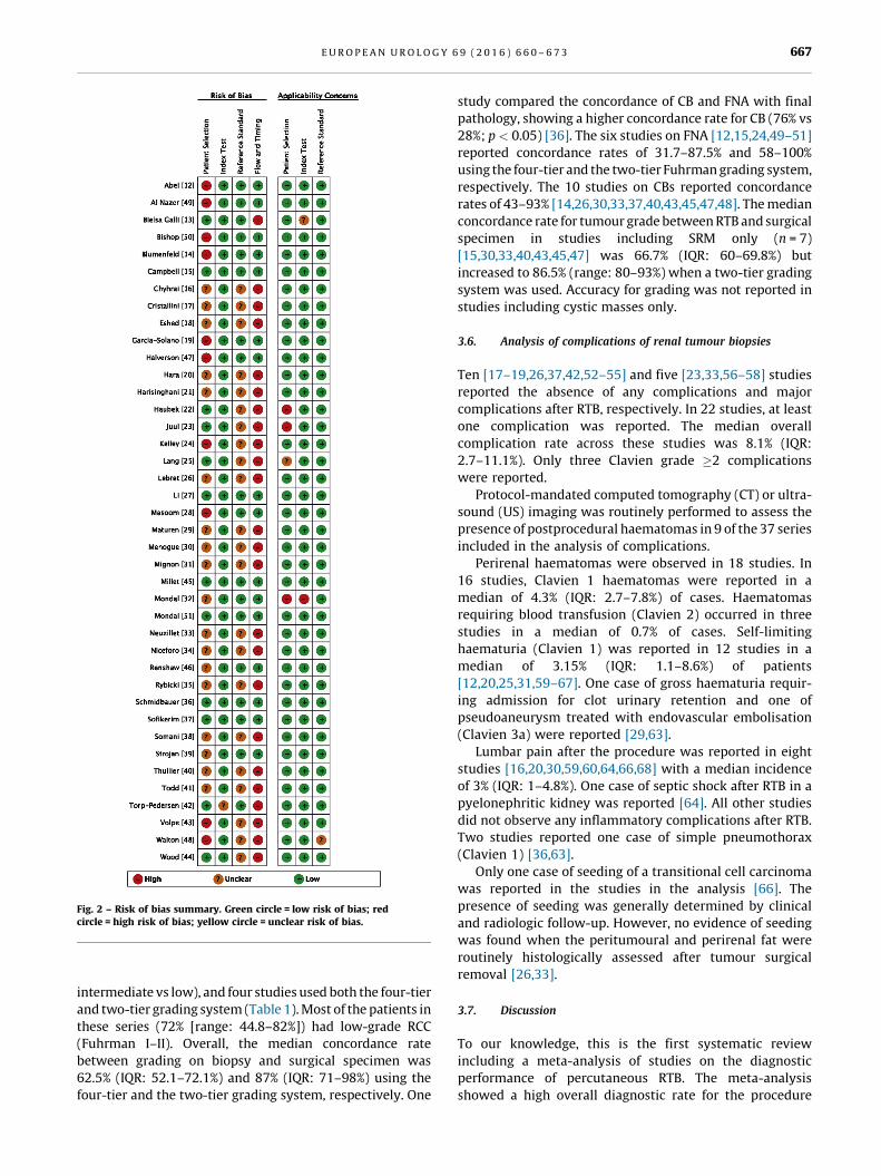
3.2. Risk of bias and quality assessment
Figure 2 shows the risk of bias assessment for studies
included in the analyses of diagnostic accuracy for tumour
malignancy, histotype, and grade. Overall, there was a high
risk of bias across studies. Only five studies included in the
meta-analysis were prospective [15,16,27,36,37]; the
majority were retrospective case series including studies
reporting all RTBs performed at a single institution and
studies reporting only RTBs performed in patients who
ultimately underwent surgery (ie, selection bias). There
was only one prospective not fully paired comparative
study of CB versus FNA [36]. The clinical setting of RTBs
was heterogeneous within studies and across studies.
There were concerns about differential-verification bias
because pathology of the surgical specimen was available
in all patients as a reference standard only in 11 of
33 studies included in the meta-analysis. Of these, only
three high-quality papers reported on consecutive
patients [15,36,37]. In the other 17 studies, pathology
was available in a proportion of cases (median: 56%), and
clinical and radiologic follow-up was used as reference
standard for the remaining patients. Finally, the propor-
tion of patients who had surgical pathology as the
reference standard was not reported in five studies. A
modest evidence of publication bias was found in the FNA
studies (data not shown).
3.3. Meta-analysis of accuracy of renal tumour biopsies for
diagnosis of malignancy
The overall median rate of diagnostic RTBs was 92%
(interquartile range [IQR]: 80.6–96.8%).
Figure 3 shows the forest plot of the 17 studies on CBs
[14,16,18,20,21,26,29,30,33,35–38,40,42–44]. The rate of
nondiagnostic biopsies was 0–22.6%. The estimates for
sensitivity and specificity of diagnostic CBs based on
bivariate analysis were 99.1% (95% CI, 96.4–99.8) and
99.7% (95% CI, 93.7–100), respectively.
Figure 4 shows the forest plot of the 18 studies on FNA
[12,13,15,17,19,22–25,27,28,31,32,34,36,39,41,42]. The
rate of nondiagnostic biopsies was 0–36%. The estimated
sensitivity and specificity of diagnostic FNAs based on
bivariate analysis were 93.2% (95% CI, 83–97.5) and 89.8%
(95% CI, 78.6–95.4), respectively.
The sensitivity analysis for studies reporting RTBs of
SRMs only (n = 7) [15,16,27,30,33,40,43] showed a sensitiv-
ity of 99.7% (95% CI, 81.5–100) and a specificity of 98.2%
(95% CI, 83.3–99.8) (Fig. 5). The sensitivity analysis of

[(Fig._1)TD$FIG]
Fig. 1 – Preferred Reporting Items for Systematic Reviews and Meta-analysis flow diagram: search and study selection process for this review.
E U R O P E A N U R O L O G Y 6 9 ( 2 0 1 6 ) 6 6 0 – 6 7 3 663
studies reporting RTBs of cystic masses only (n = 4)
[13,21,25,41] showed a sensitivity of 83.6% (95% CI, 33.8–
98.1) and a specificity of 98% (95% CI, 80.9–99.8) (Fig. 5). The
estimates of sensitivity and specificity of studies with a low
risk of selection and flow bias were 92.9% (95% CI, 79.6–
97.8) and 84.3% (95% CI, 69.2–92.8).
3.4. Analysis of accuracy of renal tumour biopsies for tumour
histotype
Five studies allowed the analysis of the agreement between
tumour histotype on biopsy and surgical specimen with k
values [14,28,37,45,46]. Median k value was 0.683 (IQR:
0.52–0.95), indicating a good degree of agreement.
Overall, 14 studies reported the concordance of
tumour histotype between RTB and surgical pathology
[12,17,26,28,30,33,36,37,40,43,45–48] (Table 1). The median
concordance rate was 90.3% (IQR: 84–94.4%). One study
compared the concordance for histologic subtype of CB and
FNA with final pathology, showing no significant difference
(91% vs 86%, respectively; p = 0.45) [36]. The median
concordance rate for the diagnosis of histotype in the six
studies including SRMs only was 96% (IQR: 90–100%). Tumour
subtype was not reported in studies including cystic masses
only.
3.5. Analysis of accuracy of renal tumour biopsies for tumour
grade
Seven studies allowed the analysis of the agreement
between tumour grade on biopsy and surgical specimen
with k values [14,15,24,26,37,45,49]. Median k value was
0.34 (0.13–0.52), which indicates a fair degree of agree-
ment. In 17 studies, the authors reported the concordance
rate between grade on RTB and surgical specimen
[12,14,15,24,26,30,33,36,37,40,43,45,47–51]. Ten studies
used the four-tier Fuhrman system, three studies used a
two- or three-tier grading system (high vs low, or high vs

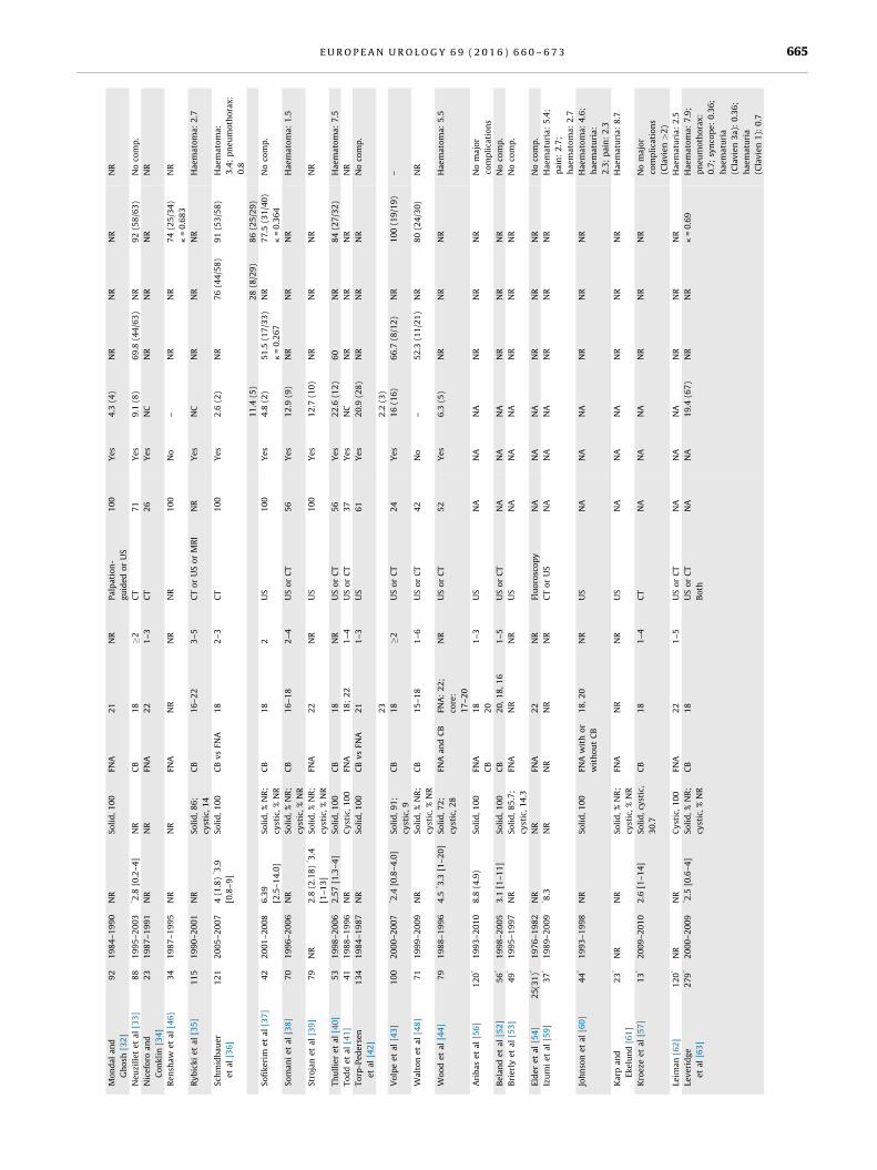
Table 1 – Tumor characteristics, biopsy technical features and diagnostic results of the studies included in the analysis
Study n Time
period
Tumour characteristics Technical characteristics Diagnostic biopsies Nondiagnostic
biopsies, % (n)
Grade concordance Histologic
subtype
concordance,
% (n/n)
Complications,
%Size, mean
(SD) [13_TD$DIFF] *median
[range] (cm)
Solid or
cystic, %
Type, % Needle
size, G
No. of
passes/core
Guidance Pathology
reference
standard, %
Meta-
analysis
Fuhrman
grade,
% (n/n)
High/low
grade,
% (n/n)
Abel et al [12] 166 1991–2007 9.2
[14_TD$DIFF][3.0–32.0[15_TD$DIFF]]
NR FNA or CB 22
20
NR US or CT 100 Yes 5.4 (9) 31.5
(33/104)
67.9
(74/109)
94.4 (75/79) NR
Al Nazer and
Mourad [49]
18 1990–1998 NR NR FNA 20–25 1–4 US or CT 100 No – 77.7 (14/18)
k = 0.65
100 (18/18) NR NR
Bielsa Galli
et al [13]
17 1985–1997 NR Cystic, 100 FNA NR Cyst
aspiration
NR 100 Yes NA NR NR NR NR
Bishop et al [50] 33 1990–2010 NR NR FNA 22 NR US 100 No – NR 85 (28/33) NR NR
Blumenfeld
et al [14]
81 2004–2008 5.3 [16_TD$DIFF][1–17 [15_TD$DIFF]] NR CB 18 2 US or CT 100 Yes 2.5 (2) 43 (29/67)
k = 0.126
– 88 (71/81)
k = 0.679
NR
Campbell
et al [15]
25 1994–1996 [17_TD$DIFF]3.1 [18_TD$DIFF]*2.7
[16_TD$DIFF][1.2–5.0[19_TD$DIFF]]
Solid, 100 FNA 22 2 CT 100 Yes 36 (9) 60 (6/10)
k = 0.375
80 (8/10) NR Haematoma: 40
Chyhrai et al [16] 25 2004–2006 2.5 [16_TD$DIFF][1.5–4.0 [15_TD$DIFF]] Solid, 100 CB 18 3 US 87 Yes 8 (2) NR NR NR Lumbar pain: 4;
haematoma: 4
Cristallini
et al [17]
79 1981–1988 NR NR FNA 21–22 NR US or CT 37.5 Yes 8.9 (7) NR NR NR No comp.
Eshed et al [18] 23 1996–2001 NR NR CB 18 NR CT 82.4 Yes 21.7 (5) NR NR NR No comp.
Garcia-Solano
et al [19]
31 2000–2006 [16_TD$DIFF][2–18 [15_TD$DIFF]] Solid, 100 FNA 25 1–2 US or CT 100 Yes 19.4 (6) NR NR NR No comp.
Halverson
et al [47]
151 1999–2011 2.8 (0.8)
[16_TD$DIFF][1–4[15_TD$DIFF]]
NR CB 18 �2 NR 100 No – 65 NR 94 NR
Hara et al [20] 33 1994–1999 NR Solid, 79;
cystic, 21
CB 18 NR US or CT 45 Yes 0 (0) NR NR NR Lumbar pain:
12.10; haematoma:
6.10; haematuria:
9.10
Harisinghani
et al [21]
28 1997–2000 NR Cystic
Bosniak
III, 100
Both 22 and 18 4–8
4–6
CT 61 Yes 0(0) NR NR NR NR
Haubek et al [22] 169 1982–1988 NR Solid, % NR;
cystic, % NR
FNA 23 NR US NR Yes 4.7 (8) NR NR NR NR
Juul et al [23] 301 1971–1983 NR Solid, 100 FNA 23 4–5 US NR Yes 5.3 (16) NR NR NR No comp.
Kelley et al [24] 43 1989–1993 NR Solid, 100 FNA NR NR US or CT 26 Yes 2.3 (1) NR 58 (7/12)
k = 0.091
NR NR
Lang et al [25] 199 1984–1998 NR Cystic
Bosniak
IIF/III, 100
FNA, 84;
CB, 14
20,21,22
18
NR CT or US 18 Yes 10.1 (20) NR NR NR Haematoma: 1;
haematuria: 8.5
Lebret et al [26] 119 1999–2005 [20_TD$DIFF]3.3 [21_TD$DIFF]*3.0
[16_TD$DIFF][1–10 [19_TD$DIFF]]
Solid, 100 CB 18 1–4 CT 65 Yes 21 (25) 54 (28/52)
k = 0.340
NR 86 No comp.
Li et al [27] 35 NR <4 cm Solid, 100 FNA 22 NR US or CT 100 Yes 0 (0) NR NR NR NR
Masoom et al [28] 31 NR 7.3 [16_TD$DIFF][1.9–14 [15_TD$DIFF]] NR FNA NR NR NR 100 Yes 3.2 (1) NR NR 90 (28/31)
k = 0.890
NR
Maturen et al [29] 152 1999–2005 4.1 [16_TD$DIFF][1–13 [15_TD$DIFF]] Solid, 89;
cystic, 11
CB 18 �4 US or CT NR Yes 3.9 (6) NR NR NR Haematoma:
0.7 (Clavien 1),
0.7 (Clavien 2);
pseudoaneurysm
(Clavien 3a)
Menogue et al [30] 250 1999–2009 [22_TD$DIFF]*2.5 [16_TD$DIFF][0.9–4 [19_TD$DIFF]] Solid, 100 CB 18 [1_TD$DIFF]1–10 [2_TD$DIFF]US or CT 78 Yes 20.8 (52) 69 (50/72) NR 98 (117/120) Lumbar pain:
0.4; haematoma:
0.4 (Clavien 2)
Mignon et al [31] 67 1985–1995 5.2 [16_TD$DIFF][1.5–13 [15_TD$DIFF]] NR FNA 16–21 2 CT NR Yes 28.4 (19) Haematoma:10.40;
haematuria: 3
Millet et al [45] 61 2006–2011 3 [16_TD$DIFF][0.9–4 [15_TD$DIFF]] Solid, 100 CB 17 2–5 CT 100 No NA 75 (46/61)
k = 0.52
93
k = 0.71
100
k = 1
NR
Mondal [51] 24 2005–2009 [16_TD$DIFF][1.5–16[15_TD$DIFF]] NR FNA 22 NR CT 100 No – 87.5 (21/24) NR NR NR
EU
RO
PE
AN
UR
OL
OG
Y6
9(
20
16
)6
60
–6
73
66
4
Mo
nd
al
an
d
Gh
osh
[32
]
92
19
84
–1
99
0N
RS
oli
d,
10
0FN
A2
1N
RP
alp
ati
on
-
gu
ide
do
rU
S
10
0Y
es
4.3
(4)
NR
NR
NR
NR
Ne
uzi
lle
te
ta
l[3
3]
88
19
95
–2
00
3[23_TD$DIFF]* 2
.8[16_TD$DIFF][0
.2–
4[19_TD$DIFF]]
NR
CB
18
�2
CT
71
Ye
s9
.1(8
)6
9.8
(44
/63
)N
R9
2(5
8/6
3)
No
com
p.
Nic
efo
roa
nd
Co
nk
lin
[34
]
23
19
87
–1
99
1N
RN
RFN
A2
21
–3
CT
26
Ye
sN
CN
RN
RN
RN
R
Re
nsh
aw
et
al
[46
]3
41
98
7–
19
95
NR
NR
FNA
NR
NR
NR
10
0N
o–
NR
NR
74
(25
/34
)
k=
0.6
83
NR
Ry
bic
ki
et
al
[35
]1
15
19
90
–2
00
1N
RS
oli
d,
86
;
cyst
ic,
14
CB
16
–2
23
–5
CT
or
US
or
MR
IN
RY
es
NC
NR
NR
NR
Ha
em
ato
ma
:2
.7
Sch
mid
ba
ue
r
et
al
[36
]
12
12
00
5–
20
07
4([3_TD$DIFF]1
.8)[4_TD$DIFF][24_TD$DIFF]* 3
.9[16_TD$DIFF]
[0.8
–9[19_TD$DIFF]]
So
lid
,1
00
CB
vs
FNA
18
2–
3C
T1
00
Ye
s2
.6(2
)N
R7
6(4
4/5
8)
91
(53
/58
)H
ae
ma
tom
a:
3.4
;p
ne
um
oth
ora
x:
0.8
11
.4(5
)2
8(8
/29
)8
6(2
5/2
9)
So
fik
eri
me
ta
l[3
7]
42
20
01
–2
00
86
.39[16_TD$DIFF]
[2.5
–1
4.0[15_TD$DIFF]]
So
lid
,%
NR
;
cyst
ic,
%N
R
CB
18
2U
S1
00
Ye
s4
.8(2
)5
1.5
(17
/33
)
k=
0.2
67
NR
77
.5(3
1/4
0)
k=
0.3
64
No
com
p.
So
ma
ni
et
al
[38
]7
01
99
6–
20
06
NR
So
lid
,%
NR
;
cyst
ic,
%N
R
CB
16
–1
82
–4
US
or
CT
56
Ye
s1
2.9
(9)
NR
NR
NR
Ha
em
ato
ma
:1
.5
Str
oja
ne
ta
l[3
9]
79
NR
2.8
(2.1
8)[4_TD$DIFF][25_TD$DIFF]* 3
.4
[16_TD$DIFF][1–
13[19_TD$DIFF]]
So
lid
,%
NR
;
cyst
ic,
%N
R
FNA
22
NR
US
10
0Y
es
12
.7(1
0)
NR
NR
NR
NR
Th
ull
ier
et
al
[40
]5
31
99
8–
20
06
2.5
7[16_TD$DIFF][1
.3–
4[15_TD$DIFF]]
So
lid
,1
00
CB
18
NR
US
or
CT
56
Ye
s2
2.6
(12
)6
0N
R8
4(2
7/3
2)
Ha
em
ato
ma
:7
.5
To
dd
et
al
[41
]4
11
98
8–
19
96
NR
Cy
stic
,1
00
FNA
18
;2
21
–4
US
or
CT
37
Ye
sN
CN
RN
RN
RN
R
To
rp-P
ed
ers
en
et
al
[42
]
13
41
98
4–
19
87
NR
So
lid
,1
00
CB
vs
FNA
21
1–
3U
S6
1Y
es
20
.9(2
8)
NR
NR
NR
No
com
p.
23
2.2
(3)
Vo
lpe
et
al
[43
]1
00
20
00
–2
00
7[26_TD$DIFF]* 2
.4[16_TD$DIFF][0
.8–
4.0[19_TD$DIFF]]
So
lid
,9
1;
cyst
ic,
9
CB
18
�2
US
or
CT
24
Ye
s1
6(1
6)
66
.7(8
/12
)N
R1
00
(19
/19
)–
Wa
lto
ne
ta
l[4
8]
71
19
99
–2
00
9N
RS
oli
d,
%N
R;
cyst
ic,
%N
R
CB
[5_TD$DIFF]15
–1
8[6_TD$DIFF]1
–6
[2_TD$DIFF]US
or
CT
42
No
–5
2.3
(11
/21
)N
R8
0(2
4/3
0)
NR
Wo
od
et
al
[44
]7
91
98
8–
19
96
[27_TD$DIFF]4.5
[28_TD$DIFF]* 3.3
[16_TD$DIFF][1–
20[19_TD$DIFF]]
So
lid
,7
2;
cyst
ic,
28
FNA
an
dC
BFN
A:
22
;
core
:
17
–2
0
NR
US
or
CT
52
Ye
s6
.3(5
)N
RN
RN
RH
ae
ma
tom
a:
5.5
Ari
ba
se
ta
l[5
6]
12
0*
19
93
–2
01
08
.8(4
.9)
So
lid
,1
00
FNA
CB
18
20
1–
3U
SN
AN
AN
AN
RN
RN
RN
om
ajo
r
com
pli
cati
on
s
Be
lan
de
ta
l[5
2]
56
*1
99
8–
20
05
3.1
[16_TD$DIFF][1–
11[15_TD$DIFF]]
So
lid
,1
00
CB
20
,1
8,
16
1–
5U
So
rC
TN
AN
AN
AN
RN
RN
RN
oco
mp
.
Bri
erl
ye
ta
l[5
3]
49
*1
99
5–
19
97
NR
So
lid
,8
5.7
;
cyst
ic,
14
.3
FNA
NR
NR
US
NA
NA
NA
NR
NR
NR
No
com
p.
Eld
er
et
al
[54
]2
5(3
1)*
19
76
–1
98
2N
RN
RFN
A2
2N
RFl
uo
rosc
op
yN
AN
AN
AN
RN
RN
RN
oco
mp
.
Izu
mi
et
al
[59
]3
7*
19
89
–2
00
9[29_TD$DIFF]* 8
.3[7_TD$DIFF]N
RN
RN
RN
RC
To
rU
SN
AN
AN
AN
RN
RN
RH
ae
ma
turi
a:
5.4
;
pa
in:
2.7
;
ha
em
ato
ma
:2
.7
Joh
nso
ne
ta
l[6
0]
44
*1
99
3–
19
98
NR
So
lid
,1
00
FNA
wit
ho
r
wit
ho
ut
CB
18
,2
0N
RU
SN
AN
AN
AN
RN
RN
RH
ae
ma
tom
a:
4.6
;
ha
em
atu
ria
:
2.3
;p
ain
:2
.3
Ka
rpa
nd
Ek
elu
nd
[61
]
23
*N
RN
RS
oli
d,
%N
R;
cyst
ic,
%N
R
FNA
NR
NR
US
NA
NA
NA
NR
NR
NR
Ha
em
atu
ria
:8
.7
Kro
eze
et
al
[57
]1
3*
20
09
–2
01
02
.6[16_TD$DIFF][1
–1
4[15_TD$DIFF]]
So
lid
,cy
stic
,
30
.7
CB
18
[8_TD$DIFF]1–
4[9_TD$DIFF]C
TN
AN
AN
AN
RN
RN
RN
om
ajo
r
com
pli
cati
on
s
(Cla
vie
n>
2)
Leim
an
[62
]1
20
*N
RN
RC
yst
ic,
10
0FN
A2
21
–5
US
or
CT
NA
NA
NA
NR
NR
NR
Ha
em
atu
ria
:2
.5
Lev
eri
dg
e
et
al
[63
]
27
9*
20
00
–2
00
9[22_TD$DIFF]* 2
.5[16_TD$DIFF][0
.6–
4[19_TD$DIFF]]
So
lid
,%
NR
;
cyst
ic,
%N
R
CB
18
US
or
CT
Bo
th
[10_TD$DIFF]NA
NA
[30_TD$DIFF]19
.4(6
7)
NR
NR
[31_TD$DIFF]k=
0.6
9H
ae
ma
tom
a:
7.9
;
pn
eu
mo
tho
rax
:
0.7
;sy
nco
pe
:0
.36
;
ha
em
atu
ria
(Cla
vie
n3
a):
0.3
6;
ha
em
atu
ria
(Cla
vie
n1
):0
.7
E U R O P E A N U R O L O G Y 6 9 ( 2 0 1 6 ) 6 6 0 – 6 7 3 665

Table 1 (Continued )
Study n Time
period
Tumour characteristics Technical characteristics Diagnostic biopsies Nondiagnostic
biopsies, % (n)
Grade concordance Histologic
subtype
concordance,
% (n/n)
Complications,
%Size, mean
(SD) [13_TD$DIFF] *median
[range] (cm)
Solid or
cystic, %
Type, % Needle
size, G
No. of
passes/core
Guidance Pathology
reference
standard, %
Meta-
analysis
Fuhrman
grade,
% (n/n)
High/low
grade,
% (n/n)
Li et al [58] 90* 2004–2009 <4 Solid, 100 FNA
CB
18 1–3 CT NA NA NA NR NR NR No major
complications
Nadel et al [64] 30* NR 4.6 [16_TD$DIFF][1–11 [15_TD$DIFF]] Solid, % NR;
cystic, % NR
FNA 18,20 1–4 CT or US NA NA NA NR NR NR Haematuria: 3.3;
haematoma
(Clavien 2): 3.3;
pain: 3.3;
infection: 3.3
Park et al [68] 59* 2004–2011 1.9 [16_TD$DIFF][1.1–3.5 [15_TD$DIFF]] Solid, % NR;
cystic, % NR
CB 18 [11_TD$DIFF]1–6 [2_TD$DIFF]US NA NA NA NR NR NR Haematoma:
15.3; pain: 5.1
Pilotti et al [65] 132* 1980–1984 NR Solid, % NR;
cystic, % NR
FNA 22 NR CT
Fluoroscopy
NA NA NA NR NR NR Haematuria: 9.1
Richter et al [55] 517* 1967–1996 NR Cystic, 60;
solid, 40
FNA
CB
18, (20–22) NR Fluoroscopy
US or CT
NA NA NA NR NR NR No comp.
Tikkakoski
et al [66]
180* 1982–1988 4.7 [16_TD$DIFF][1.5–20 [15_TD$DIFF]] Cystic, % NR;
solid, % NR
FNA >20 NR US NA NA NA NR NR NR Seeding: 0.6;
pain: 0.6;
haematuria: 0.6
Veltri et al [67] 145* NR 3.4 [16_TD$DIFF][1–15 [15_TD$DIFF]] Solid, % NR;
cystic, % NR
FNA with or
without CB
18, 21,22 NR US or CT NA NA NA NR NR NR Haematoma:
4; haematuria: 0.7;
AV fistula: 0.7
AV = arteriovenous; CB = core biopsy; CT = computed tomography; FNA = fine-needle aspiration; k = Cohen kappa coefficient; MRI = magnetic resonance imaging; NA = not applicable; No comp. = no complications were
observed; NR = not reported; SD = standard deviation; US = ultrasound.* Study included only in the analysis of complications of RTB[12_TD$DIFF].
EU
RO
PE
AN
UR
OL
OG
Y6
9(
20
16
)6
60
–6
73
66
6
[(Fig._2)TD$FIG]
Fig. 2 – Risk of bias summary. Green circle = low risk of bias; redcircle = high risk of bias; yellow circle = unclear risk of bias.
E U R O P E A N U R O L O G Y 6 9 ( 2 0 1 6 ) 6 6 0 – 6 7 3 667
intermediate vs low), and four studies used both the four-tier
and two-tier grading system (Table 1). Most of the patients in
these series (72% [range: 44.8–82%]) had low-grade RCC
(Fuhrman I–II). Overall, the median concordance rate
between grading on biopsy and surgical specimen was
62.5% (IQR: 52.1–72.1%) and 87% (IQR: 71–98%) using the
four-tier and the two-tier grading system, respectively. One
study compared the concordance of CB and FNA with final
pathology, showing a higher concordance rate for CB (76% vs
28%; p < 0.05) [36]. The six studies on FNA [12,15,24,49–51]
reported concordance rates of 31.7–87.5% and 58–100%
using the four-tier and the two-tier Fuhrman grading system,
respectively. The 10 studies on CBs reported concordance
rates of 43–93% [14,26,30,33,37,40,43,45,47,48]. The median
concordance rate for tumour grade between RTB and surgical
specimen in studies including SRM only (n = 7)
[15,30,33,40,43,45,47] was 66.7% (IQR: 60–69.8%) but
increased to 86.5% (range: 80–93%) when a two-tier grading
system was used. Accuracy for grading was not reported in
studies including cystic masses only.
3.6. Analysis of complications of renal tumour biopsies
Ten [17–19,26,37,42,52–55] and five [23,33,56–58] studies
reported the absence of any complications and major
complications after RTB, respectively. In 22 studies, at least
one complication was reported. The median overall
complication rate across these studies was 8.1% (IQR:
2.7–11.1%). Only three Clavien grade �2 complications
were reported.
Protocol-mandated computed tomography (CT) or ultra-
sound (US) imaging was routinely performed to assess the
presence of postprocedural haematomas in 9 of the 37 series
included in the analysis of complications.
Perirenal haematomas were observed in 18 studies. In
16 studies, Clavien 1 haematomas were reported in a
median of 4.3% (IQR: 2.7–7.8%) of cases. Haematomas
requiring blood transfusion (Clavien 2) occurred in three
studies in a median of 0.7% of cases. Self-limiting
haematuria (Clavien 1) was reported in 12 studies in a
median of 3.15% (IQR: 1.1–8.6%) of patients
[12,20,25,31,59–67]. One case of gross haematuria requir-
ing admission for clot urinary retention and one of
pseudoaneurysm treated with endovascular embolisation
(Clavien 3a) were reported [29,63].
Lumbar pain after the procedure was reported in eight
studies [16,20,30,59,60,64,66,68] with a median incidence
of 3% (IQR: 1–4.8%). One case of septic shock after RTB in a
pyelonephritic kidney was reported [64]. All other studies
did not observe any inflammatory complications after RTB.
Two studies reported one case of simple pneumothorax
(Clavien 1) [36,63].
Only one case of seeding of a transitional cell carcinoma
was reported in the studies in the analysis [66]. The
presence of seeding was generally determined by clinical
and radiologic follow-up. However, no evidence of seeding
was found when the peritumoural and perirenal fat were
routinely histologically assessed after tumour surgical
removal [26,33].
3.7. Discussion
To our knowledge, this is the first systematic review
including a meta-analysis of studies on the diagnostic
performance of percutaneous RTB. The meta-analysis
showed a high overall diagnostic rate for the procedure

[(Fig._3)TD$FIG]
Fig. 3 – Forest plot of estimates of sensitivity and specificity of percutaneous core biopsy for the diagnosis of tumour malignancy (univariate analysis).CI = confidence interval; ES = estimate.
E U R O P E A N U R O L O G Y 6 9 ( 2 0 1 6 ) 6 6 0 – 6 7 3668
(92%), with higher estimates for sensitivity and specificity
for CB compared with FNA. The accuracy of RTB for the
diagnosis of RCC histologic subtype was found to be good. A
fair agreement between tumour grade at biopsy and on the
final specimen was observed.
The use of percutaneous sampling of renal tumours
historically was limited due to concerns about its safety,
diagnostic yield, and accuracy, and for the perceived little
impact of RTBs on clinical management [69].
However, the adoption of modern biopsy techniques and
the growing expertise in performing biopsies, the progres-
sively increased experience of pathologists in interpreting
biopsy specimens, and the increased confidence of urolo-
gists in using biopsy results to support treatment decisions
based on a better knowledge of the natural history of benign
and malignant renal tumours have led to increasing
indications of this procedure for the histologic characteri-
sation of SRMs and metastatic primary renal tumours
[3,70,71]. The current EAU urologic guidelines on RCC
[(Fig._4)TD$FIG]Fig. 4 – Forest plot of estimates of sensitivity and specificity of percutaneous fanalysis).CI = confidence interval; ES = estimate; FNA = fine-needle aspiration.
recommend that RTBs should be performed in patients in
whom active surveillance is pursued and before ablative
therapy and systemic therapy without previous pathology
[1]. However, despite the wider indications and the
encouraging diagnostic performance reported in experi-
enced centres, the use of RTBs still remains limited outside
academic centres and institutions with a special focus on
urologic oncology [72,73].
Improving the quality of the evidence on RTB is crucial to
better define the role of this procedure in the management
of renal tumours. The current evidence base in this field is in
fact limited by several factors. First, most studies on RTBs
are retrospective, have relatively small sample sizes, and
have heterogeneous populations. Second, the assessment of
diagnostic accuracy of RTBs is hampered by the lack of
surgical confirmation of the histology in a variable
proportion of cases, by the use of different follow-up
protocols to monitor the clinical behaviour of tumours that
are not surgically removed after biopsy, by the adoption of
ine-needle aspiration for the diagnosis of tumour malignancy (univariate
[(Fig._5)TD$FIG]
Fig. 5 – Forest plot of estimates of sensitivity and specificity of percutaneous renal tumour biopsy for the diagnosis of tumour malignancy in studiesincluding only small renal masses or cystic renal masses (univariate analysis).CI = confidence interval; ES = estimate.
E U R O P E A N U R O L O G Y 6 9 ( 2 0 1 6 ) 6 6 0 – 6 7 3 669
different definitions for biopsy success, and by the use of
different biopsy techniques and protocols (CB vs FNA, CT vs
US guidance, number and location of biopsies taken).
In the absence of large prospective multicentre studies
using homogeneous biopsy techniques and standardised
protocols for assessment of diagnostic outcomes, the
present paper provides the best available evidence on
diagnostic yield and accuracy of RTBs. The clinical question
we assessed in the review was prioritised by an expert panel
of clinicians (EAU RCC Guideline Panel), and the patient
problem or population, intervention, comparison, and
outcomes (PICO) elements were developed in conjunction
with the panel. The search strategy and entire review
process adhered to PRISMA guidelines and Cochrane review
on diagnostic test accuracy principles. The strict methodo-
logical criteria used also ensured the inclusion in the meta-
analysis of studies with lower risks of bias and a low level of
clinical and methodological heterogeneity.
Our results show that percutaneous sampling of renal
tumours harbours a diagnostic yield of 92% for the diagnosis
of malignancy, indicating that a properly performed RTB can
provide information for treatment decision making in most
cases. However, nondiagnostic biopsies constituted a
variable but non-negligible proportion of cases either for
CB and FNA. This represents a matter of concern for
clinicians; hence surgical exploration or repeat sampling is
recommended when the biopsy of a radiologically suspi-
cious renal mass is nondiagnostic [1]. Repeat biopsies have
been reported to be diagnostic in a high proportion of cases
(83–100%) in several series [38,44,63,74,75].
It should be acknowledged that a relevant proportion of
nondiagnostic RTBs are in fact failed biopsies due to
technical limitations (use of suboptimal technique, needle
type, or image guidance) and/or to the intrinsic challenge of
the procedure (ie, targeting a mass in an organ that moves
with respiration).
The frequent inappropriate allocation of failed biopsies
containing only normal renal parenchyma, blood, or fibrosis
in the category of inaccurate biopsies has led to a potential
underestimation of biopsy accuracy.
A fair evaluation of the diagnostic accuracy of informa-
tive RTBs is crucial for the definition of their utility, safety,
and reliability in clinical practice when histologic informa-
tion is used to make clinical decisions, such as supporting
the choice of observation/active surveillance in the pres-
ence of a benign histology or a low-grade RCC.
In our meta-analysis, CB was shown to have excellent
estimates for sensitivity (99.1%) and specificity (99.7%) in
the assessment of tumour malignancy. Lower estimates for
sensitivity (93.2%) and specificity (89.8%) for the diagnosis
of malignancy were observed for FNA. Based on this data, CB
may therefore be favoured over FNA for percutaneous
sampling of renal tumours. However, because some authors
suggest that the two techniques can provide complemen-
tary results and eventually increase diagnostic rates and
accuracy [76], FNA may be performed in combination with
E U R O P E A N U R O L O G Y 6 9 ( 2 0 1 6 ) 6 6 0 – 6 7 3670
CB in selected cases. A potential advantage of FNA is that it
allows the intraprocedural assessment of the cytologic
specimen, which can potentially confirm the appropriate
location of the guiding cannula and increase the diagnostic
yield of the subsequent CBs.
The subgroup analyses we performed showed excellent
accuracy of RTB for diagnosing malignancy in SRMs,
whereas the estimates for specificity and above all
sensitivity of RTBs of cystic renal masses are significantly
poorer compared with RTBs of solid masses (98% vs 83.6%,
respectively). These findings, together with the potential
risk of spreading of tumour cells resulting from cystic
rupture during biopsy, support the current trend to limit the
indications of RTBs for cystic renal lesions. Percutaneous
sampling may still be indicated for Bosniak grade IV cysts,
where clear enhancing solid nodules are visible within the
lesion [1].
Our analysis also revealed a good degree of concordance
between the diagnosis of RCC subtype on biopsy and on
surgical specimens (90.3% in the overall population,
improving to 96% in the analysis for SRMs; median
k = 0.63). The diagnosis of RCC subtype on RTBs is therefore
reliable in most cases and can be safely used for treatment
decision making. In fact, although an independent prognos-
tic role for RCC histotype has not been clearly established,
single-institution and multicentre series showed signifi-
cantly different oncologic outcomes among RCC histologic
subtypes, with clear cell and papillary type 2 tumours
showing worse outcomes than papillary type 1 and
chromophobe histologies [77,78]. Furthermore, the diagno-
sis of RCC subtype is useful for tailoring the best targeted
therapy in systemic disease.
The assessment of tumour grade on RTBs is challenging
[33,36,43,63]. According to our analysis, the degree of
concordance of tumour grade on RTBs and surgical
specimens is only fair (median k = 0.34). The median
reported concordance rate is 62.5%, improving to 87%
when a simplified two-tier system (Fuhrman I–II = low
grade; Fuhrman III–IV = high grade) is adopted. When only
studies on SRMs are included in the analysis, the
concordance rate using the four-tier system is slightly
improved (66.7%); the concordance is similar using the
two-tier system (86.5%).
Accuracy in the evaluation of tumour grade is important
for clinical decision making but is limited by tumour
heterogeneity and interobserver variability. Intratumoural
grade heterogeneity has been reported in 5–25% of renal
tumours [36,69]. Its potential impact on biopsy accuracy
can be reduced by performing multiple biopsies in different
areas of the tumour. Although the accuracy of RTBs for
tumour grade is not optimal, Jeldres et al observed that
models including other patient and tumour characteristics
cannot reliably predict Fuhrman grade and therefore cannot
substitute percutaneous biopsy for grade assessment [79].
Improving our ability to obtain samples that allow a
reliable and accurate evaluation of tumour grade is clearly
one of the main future goals of clinical research on RTBs. The
classification of grading of renal tumours is evolving, and
the recent International Society of Urological Pathology
2012 Consensus Conference accepted a new grading system
with grades 1–3 of clear cell and papillary RCC based on
nucleolar prominence, and grade 4 defined by the presence
of extreme nuclear pleomorphism or sarcomatoid and/or
rhabdoid differentiation [80]. Future studies will have to
assess the accuracy of RTBs for this newly proposed grading
system.
Finally, the present study indicates that percutaneous
RTB is a safe procedure, with a very limited risk of
significant (Clavien �2) complications. Only one case of
seeding was reported in the eligible studies, also when
peritumoural and perirenal fat were assessed after surgical
removal. Although another case of seeding was recently
published [81], most of the few case reports date back to the
late 1980s to early 1990s when different biopsy techniques
were used [3]. Some authors suggest that the risk of this
worrisome complication is minimal with the use of the
coaxial technique [4], which should always be used in
clinical practice. Other complications of RTB were mainly
lumbar pain and haematomas/haematuria, which mostly
resolved spontaneously without medical intervention.
Although protocol-mandated CT or US imaging was not
performed after biopsy in all included studies, the
incidence of haematomas was relatively low (median:
5%), and blood transfusions were required on average in
only 0.7% of cases.
This study has several limitations. Included studies may
potentially be affected by selection bias and by the use of
different reference standards (differential-verification bias)
including variable clinical follow-up schedules for renal
tumours that were not surgically removed. In spite of our
efforts to standardise outcome definitions and measure-
ments, clinical and methodological heterogeneity was
inevitable, although it was minimised. The threshold for
good quality studies for sensitivity analysis was based on
only two domains of the QUADAS-2 risk of bias assessment
tool (patient selection and flow/timing) because data were
incomplete for all other domains. The duration of follow-up
was also relatively short for most of the studies, and this
confers uncertainty regarding biopsy-negative cases being
TNs.
Nevertheless, this study represents the first meta-
analysis of diagnostic accuracy of RTBs and is based on a
robust methodology, with strict criteria for study selection
that are rigorous, transparent, and reproducible.
Due to the lack of data in the literature, the study does
not provide information on the learning curve needed by
urologists and pathologists to take and interpret biopsy
specimens, and on the optimal number of cores and biopsy
pattern that should be performed to maximise diagnostic
performance. Presently there is agreement that at least two
good quality cores should be obtained in each case, but
increasing the number of cores may increase the diagnostic
yield of the procedure [33].
Further research is also needed to confirm whether
biopsies of the peripheral part of the tumour should be
preferred for larger lesions to avoid central necrosis or if
both central and peripheral biopsies should be performed
for SRMs, as previously suggested [82].

E U R O P E A N U R O L O G Y 6 9 ( 2 0 1 6 ) 6 6 0 – 6 7 3 671
4. Conclusions
RTB has a high diagnostic yield and is associated with a very
low risk of significant complications. Good estimates of
sensitivity and specificity for the diagnosis of malignancy
were found at meta-analysis either for CB and FNA, with
better performance for CB. The accuracy of RTBs for the
diagnosis of RCC subtype was found to be good; accuracy for
nuclear grade was fair and improves using a simplified
system (low vs high grade). However, the quality of the
available evidence is moderate, and well-designed prospec-
tive cohort studies including consecutive patients and using
valid reference standards to assess RTB performance are
required to corroborate these findings and address knowl-
edge gaps.
Author contributions: Alessandro Volpe had full access to all the data in
the study and takes responsibility for the integrity of the data and the
accuracy of the data analysis.
Study concept and design: Volpe, Marconi.
Acquisition of data: Volpe, Marconi, Stewart.
Analysis and interpretation of data: Volpe, Marconi, Norrie, Lam.
Drafting of the manuscript: Volpe, Marconi.
Critical revision of the manuscript for important intellectual content: Volpe,
Marconi, Dabestani, Lam, Hofmann, Stewart, Norrie, Bex, Bensalah,
Canfield, Hora, Kuczyk, Merseburger, Mulders, Powles, Staehler, Ljung-
berg.
Statistical analysis: Norrie, Volpe, Marconi, Lam.
Obtaining funding: None.
Administrative, technical, or material support: None.
Supervision: Volpe, Marconi.
Other (specify): None.
Financial disclosures: Alessandro Volpe certifies that all conflicts of
interest, including specific financial interests and relationships and
affiliations relevant to the subject matter or materials discussed in the
manuscript (eg, employment/affiliation, grants or funding, consultan-
cies, honoraria, stock ownership or options, expert testimony, royalties,
or patents filed, received, or pending), are the following: Thomas B. Lam
is a company consultant for Pfizer, GSK, Astellas, and Ipsen. He receives
company speaker honoraria from Pfizer, GSK, Astellas, and Ipsen. Karim
Bensalah receives grants/research support from Pfizer and honoraria or
consultation fees from Intuitive Surgical. Axel Bex receives company
honoraria from Pfizer, is a trial participant for Pfizer Europe, participates
on advisory boards for GSK and Novartis, is a company consultant for
Pfizer and Novartis, and receives grants/research support from Pfizer.
Steven E. Canfield receives company speaker honoraria from Amgen,
Genomic Health, Algeta, and Bayer. Milan Hora receives company
speaker honoraria from Covidien and Olympus. He participates in trials
for Janssen, receives grants/research support from Ipsen, and receives
company speaker honoraria from Janssen and Astellas. Markus A. Kuczyk
is a shareholder at Bayer Healthcare, Astellas, Storz, Pfizer, Wyeth, and
Novartis. He is a company consultant for Karl Storz, Coloplast, Astra
Zeneca, Astellas, and Storz, and receives company speaker honoraria
from Pfizer, Astellas, Bayer, GSK, Pierre Fabre, and Jansen Cilag & Hexal.
He is a trial participant for the ProtecT Study, Millennium Study C21004,
and Millennium Study C21005 and at Astellas, Ipsen, and Janssen. He
receives grants and research support for Pfizer and is a company
consultant for Hexal AG. Axel S. Merseburger is a company consultant for
Ipsen Pharma, Bayer, Astellas, Janssen Cilag, Novartis, and Pfizer. He
receives company speaker honoraria from Ipsen Pharma, Wyeth,
Astellas, Novartis, Pfizer, and SEP. He participates in trials for Astra
Zeneca, Bayer, Pfizer, TEVA, Novartis, and Astellas. He receives grants/
research support from Wyeth and participates in a company-sponsored
speakers’ bureau for TEVA, Janssen, Pfizer, Astellas, Ferring, and Novartis.
Peter F.A. Mulders receives company speaker honoraria from Astellas,
Pfizer, Novartis, GSK, AstraZeneca, and J&J. He participates in trials for
Astellas, GSK, J&J, and Millennium. He receives grants/research support
from Astellas, Wilex, GSK, and Pfizer. He participates in trials for J&J and
Provenge. He receives grants/research support from Bayer. Thomas
Powles is a company consultant for Novartis, Pfizer, and GSK. He receives
company speaker honoraria from Novartis, Pfizer, and GSK. He
participates in trials for GSK, Pfizer, and BMS. He receives grants/
research support from GSK, Pfizer, and Novartis. He receives company
speaker honoraria and participates in trials for Genentech. Michael
Staehler is a company consultant for Pfizer, Novartis, GSK, Roche, and
Astellas. He receives company speaker honoraria from Pfizer, Novartis,
GSK, Roche, and Astellas. He participates in trials and receives
fellowships and travel grants from Pfizer, Novartis, GSK, and Roche.
He receives grants/research support from Pfizer, Novartis, GSK, Roche,
Aveo, and Bayer. He is a company consultant for Bayer and receives
company speaker honoraria from Bayer and Aveo. He participates in
trials for Bayer, Aveo, Wilex, and Immatics. Borje Ljungberg receives
company speaker honoraria from GSK, Roche, Pfizer, and Novartis. He
participates in trials for GSK, Medivation, Pfizer, and Janssen R&D. He is
on the advisory board at Pfizer and GSK. The other authors have nothing
to disclose.
Funding/Support and role of the sponsor: None.
References
[1] Ljungberg B, Bensalah K, Canfield S, et al. EAU guidelines on renal
cell carcinoma: 2014 update. Eur Urol 2015;67:913–24.
[2] Lane BR, Samplaski MK, Herts BR, Zhou M, Novick AC, Campbell SC.
Renal mass biopsy–a renaissance? J Urol 2008;179:20–7.
[3] Volpe A, Finelli A, Gill IS, et al. Rationale for percutaneous biopsy
and histologic characterisation of renal tumours. Eur Urol 2012;62:
491–504.
[4] Volpe A, Kachura JR, Geddie WR, et al. Techniques, safety and
accuracy of sampling of renal tumors by fine needle aspiration
and core biopsy. J Urol 2007;178:379–86.
[5] Moher D, Liberati A, Tetzlaff J, Altman DG. PRISMA Group. Preferred
reporting items for systematic reviews and meta-analyses: the
PRISMA statement. Open Med 2009;3:e123–30.
[6] The Cochrane Collaboration. Diagnostic Test Accuracy Working
Group: handbook for DTA reviews. Cochrane Collaboration Web
site. http://srdta.cochrane.org/handbook-dta-reviews.
[7] Ljungberg B, Canfield S, Dabestani S, et al. Systematic review method-
ology for the European Association of Urology guidelines for renal cell
carcinoma. European Association of Urology Web site. http://www.
uroweb.org/gls/EU/Systematic_methodology_RCC_2014_update.
pdf2014. Updated 2014.
[8] Dindo D, Demartines N, Clavien PA. Classification of surgical com-
plications: a new proposal with evaluation in a cohort of
6336 patients and results of a survey. Ann Surg 2004;240:
205–13.
[9] Whiting PF, Rutjes AW, Westwood ME, et al. QUADAS-2: a revised
tool for the quality assessment of diagnostic accuracy studies. Ann
Intern Med 2011;155:529–36.
[10] Egger M, Davey Smith G, Schneider M, Minder C. Bias in meta-
analysis detected by a simple, graphical test. BMJ 1997;315:629–34.
[11] Duval S, Tweedie R. Trim and fill: a simple funnel-plot-based
method of testing and adjusting for publication bias in meta-
analysis. Biometrics 2000;56:455–63.
E U R O P E A N U R O L O G Y 6 9 ( 2 0 1 6 ) 6 6 0 – 6 7 3672
[12] Abel EJ, Culp SH, Matin SF, et al. Percutaneous biopsy of primary
tumor in metastatic renal cell carcinoma to predict high risk
pathological features: comparison with nephrectomy assessment.
J Urol 2010;184:1877–81.
[13] Bielsa Gali OT, Angel R, Santamaria R, Garreta J, Gelabert-Mas A. The
preoperative diagnosis of complex renal cystic masses [in Spanish].
Arch Esp Urol 1999;52:19–25.
[14] Blumenfeld AJ, Guru K, Fuchs GJ, Kim HL. Percutaneous biopsy of
renal cell carcinoma underestimates nuclear grade. Urology
2010;76:610–3.
[15] Campbell SC, Novick AC, Herts B, et al. Prospective evaluation of fine
needle aspiration of small, solid renal masses: accuracy and mor-
bidity. Urology 1997;50:25–9.
[16] Chyhrai A, Sanjmyatav J, Gajda M, et al. Multi-colour FISH on
preoperative renal tumour biopsies to confirm the diagnosis of
uncertain renal masses. World J Urol 2010;28:269–74.
[17] Cristallini EG, Paganelli C, Bolis GB. Role of fine-needle aspiration
biopsy in the assessment of renal masses. Diagn Cytopathol
1991;7:32–5.
[18] Eshed I, Elias S, Sidi AA. Diagnostic value of CT-guided biopsy of
indeterminate renal masses. Clin Radiol 2004;59:262–7.
[19] Garcia-Solano J, Acosta-Ortega J, Perez-Guillermo M, Benedicto-
Orovitg JM, Jimenez-Penick FJ. Solid renal masses in adults:
image-guided fine-needle aspiration cytology and imaging techni-
ques–‘‘two heads better than one?’’ Diagn Cytopathol 2008;36:
8–12.
[20] Hara IM, Hara S, Arakawa S, Hanioka K, Kamidono S. Role of
percutaneous image-guided biopsy in the evaluation of renal
masses. Urol Int 2001;67:199–202.
[21] Harisinghani MG, Maher MM, Gervais DA, et al. Incidence of
malignancy in complex cystic renal masses (Bosniak category III):
should imaging-guided biopsy precede surgery? AJR 2003;180:
755–8.
[22] Haubek A, Lundorf E, Lauridsen KN. Diagnostic strategy in renal
mass lesions. Scand J Urol Nephrol Suppl 1991;137:35–9.
[23] Juul N, Torp-Pedersen S, Gronvall S, Holm HH, Koch F, Larsen S.
Ultrasonically guided fine needle aspiration biopsy of renal masses.
J Urol 1985;133:579–81.
[24] Kelley CM, Cohen MB, Raab SS. Utility of fine-needle aspiration
biopsy in solid renal masses. Diagn Cytopathol 1996;14:14–9.
[25] Lang EK, Macchia RJ, Gayle B, et al. CT-guided biopsy of indetermi-
nate renal cystic masses (Bosniak 3 and 2F): accuracy and impact
on clinical management. Eur Radiol 2002;12:2518–24.
[26] Lebret T, Poulain JE, Molinie V, et al. Percutaneous core biopsy for
renal masses: indications, accuracy and results. J Urol 2007;178:
1184–8.
[27] Li G, Cuilleron M, Cottier M, et al. The use of MN/CA9 gene
expression in identifying malignant solid renal tumors. Eur Urol
2006;49:401–5.
[28] Masoom S, Venkataraman G, Jensen J, Flanigan RC, Wojcik EM.
Renal FNA-based typing of renal masses remains a useful adjunc-
tive modality: evaluation of 31 renal masses with correlative
histology. Cytopathology 2009;20:50–5.
[29] Maturen KE, Nghiem HV, Caoili EM, Higgins EG, Wolf Jr JS, Wood Jr
DP. Renal mass core biopsy: accuracy and impact on clinical
management. AJR 2007;188:563–70.
[30] Menogue SR, O’Brien BA, Brown AL, Cohen RJ. Percutaneous core
biopsy of small renal mass lesions: a diagnostic tool to better
stratify patients for surgical intervention. BJU Int 2013;111:
E146–51.
[31] Mignon F, Mesurolle B, Ariche-Cohen M, Vanel D. Value of CT
guided renal biopsies: retrospective review of 67 cases [in French].
J Radiol 2001;82:907–11.
[32] Mondal A, Ghosh E. Fine needle aspiration cytology (FNAC) in the
diagnosis of solid renal masses–a study of 92 cases. Indian J Pathol
Microbiol 1992;35:333–9.
[33] Neuzillet Y, Lechevallier E, Andre M, Daniel L, Coulange C. Accuracy
and clinical role of fine needle percutaneous biopsy with comput-
erized tomography guidance of small (less than 4.0 cm) renal
masses. J Urol 2004;171:1802–5.
[34] Niceforo J, Coughlin BF. Diagnosis of renal cell carcinoma: value of
fine-needle aspiration cytology in patients with metastases or
contraindications to nephrectomy. AJR 1993;161:1303–5.
[35] Rybicki FJ, Shu KM, Cibas ES, Fielding JR, van Sonnenberg E, Silver-
man SG. Percutaneous biopsy of renal masses: sensitivity and
negative predictive value stratified by clinical setting and size of
masses. AJR 2003;180:1281–7.
[36] Schmidbauer J, Remzi M, Memarsadeghi M, et al. Diagnostic accu-
racy of computed tomography-guided percutaneous biopsy of renal
masses. Eur Urol 2008;53:1003–12.
[37] Sofikerim M, Tatlisen A, Canoz O, Tokat F, Demirtas A, Mavili E.
What is the role of percutaneous needle core biopsy in diagnosis of
renal masses? Urology 2010;76:614–8.
[38] Somani BK, Nabi G, Thorpe P. Image-guided biopsy-diagnosed renal
cell carcinoma: critical appraisal of technique and long-term fol-
low-up. Eur Urol 2007;51:1289–97, discussion 1296-7.
[39] Strojan Flezar M, Gutnik H, Jeruc J, Kirbis IS. Typing of renal tumors
by morphological and immunocytochemical evaluation of fine
needle aspirates. Virchows Arch 2011;459:607–14.
[40] Thuillier C, Long JA, Lapouge O, et al. Value of percutaneous biopsy
for solid renal tumours less than 4 cm in diameter based on a series
of 53 cases [in French]. Prog Urol 2008;18:435–9.
[41] Todd TD, Dhurandhar B, Mody D, Ramzy I, Truong LD. Fine-needle
aspiration of cystic lesions of the kidney. Morphologic spectrum
and diagnostic problems in 41 cases. Am J Clin Pathol 1999;111:
317–28.
[42] Torp-Pedersen S, Juul N, Larsen T, Karstrup S, Sehested M, Glenthoj
A. US-guided fine needle biopsy of solid renal masses–comparison
of histology and cytology. Scand J Urol Nephrol Suppl 1991;137:
41–3.
[43] Volpe A, Mattar K, Finelli A, et al. Contemporary results of percuta-
neous biopsy of 100 small renal masses: a single center experience.
J Urol 2008;180:2333–7.
[44] Wood BJ, Khan MA, McGovern F, Harisinghani M, Hahn PF, Mueller
PR. Imaging guided biopsy of renal masses: indications, accuracy
and impact on clinical management. J Urol 1999;161:1470–4.
[45] Millet I, Curros F, Serre I, Taourel P, Thuret R. Can renal biopsy
accurately predict histological subtype and Fuhrman grade of renal
cell carcinoma? J Urol 2012;188:1690–4.
[46] Renshaw AA, Lee KR, Madge R, Granter SR. Accuracy of fine needle
aspiration in distinguishing subtypes of renal cell carcinoma. Acta
Cytol 1997;41:987–94.
[47] Halverson SJ, Kunju LP, Bhalla R, et al. Accuracy of determining
small renal mass management with risk stratified biopsies: confir-
mation by final pathology. J Urol 2013;189:441–6.
[48] Walton TA, Moore D, Mayer N, Rajesh A, Kockelbergh R. Utility of
renal mass biopsy in a UK tertiary referral centre. Br J Med Surg Urol
2012;5:216–23.
[49] Al Nazer M, Mourad WA. Successful grading of renal-cell carcinoma
in fine-needle aspirates. Diagn Cytopathol 2000;22:223–6.
[50] Bishop JA, Hosler GA, Kulesza P, Erozan YS, Ali SZ. Fine-needle
aspiration of renal cell carcinoma: is accurate Fuhrman grading
possible on cytologic material? Diagn Cytopathol 2011;39:168–71.
[51] Mondal SK. Cytohistological study of clear cell carcinoma of kidney
with special reference to nuclear grading and tumor size. Diagn
Cytopathol 2012;40:1083–7.

E U R O P E A N U R O L O G Y 6 9 ( 2 0 1 6 ) 6 6 0 – 6 7 3 673
[52] Beland MD, Mayo-Smith WW, Dupuy DE, Cronan JJ, DeLellis RA.
Diagnostic yield of 58 consecutive imaging-guided biopsies of solid
renal masses: should we biopsy all that are indeterminate? AJR
2007;188:792–7.
[53] Brierly RD, Thomas PJ, Harrison NW, Fletcher MS, Nawrocki JD,
Ashton-Key M. Evaluation of fine-needle aspiration cytology for
renal masses. BJU Int 2000;85:14–8.
[54] Elder DD, Orell SR, Sage MR, Sinclair GR, Marshall VR. The diagnosis
and local staging of renal cancer–an appraisal. Aust N Z J Surg
1984;54:219–21.
[55] Richter F, Kasabian NG, Irwin Jr RJ, Watson RA, Lang EK. Accuracy of
diagnosis by guided biopsy of renal mass lesions classified indeter-
minate by imaging studies. Urology 2000;55:348–52.
[56] Aribas BK, Arda K, Aktas E, et al. Percutaneous US-guided
needle biopsies of solid renal masses. Neoplasma 2011;58:
146–52.
[57] Kroeze SG, Huisman M, Verkooijen HM, van Diest PJ, Ruud Bosch JL,
van den Bosch MA. Real-time 3D fluoroscopy-guided large core
needle biopsy of renal masses: a critical early evaluation according
to the IDEAL recommendations. Cardiovasc Interv Radiol
2012;35:680–5.
[58] Li G, Cuilleron M, Zhao A, et al. Combination of core biopsy and fine-
needle aspiration increases diagnostic rate for small solid renal
tumors. Anticancer Res 2012;32:3463–6.
[59] Izumi K, Narimoto K, Sugimoto K, et al. The role of percutaneous
needle biopsy in differentiation of renal tumors. Japan J Clin Oncol
2010;40:1081–6.
[60] Johnson PT, Nazarian LN, Feld RI, et al. Sonographically guided renal
mass biopsy: indications and efficacy. J Ultrasound Med 2001;20:
749–53, quiz 755.
[61] Karp W, Ekelund L. Ultrasound, angiography and fine needle aspi-
ration biopsy in diagnosis of renal neoplasms. Acta Radiol Diagn
(Stockh) 1979;20:649–59.
[62] Leiman G. Audit of fine needle aspiration cytology of 120 renal
lesions. Cytopathology 1990;1:65–72.
[63] Leveridge MJ, Finelli A, Kachura JR, et al. Outcomes of small renal
mass needle core biopsy, nondiagnostic percutaneous biopsy, and
the role of repeat biopsy. Eur Urol 2011;60:578–84.
[64] Nadel L, Baumgartner BR, Bernardino ME. Percutaneous renal
biopsies: accuracy, safety, and indications. Urol Radiol 1986;8:
67–71.
[65] Pilotti S, Rilke F, Alasio L, Garbagnati F. The role of fine needle
aspiration in the assessment of renal masses. Acta Cytol 1988;32:
1–10.
[66] Tikkakoski T, Paivansalo M, Apaja-Sarkkinen M, Ollikainen A,
Lohela P, Matti K. Ultrasound-guided aspiration cytology of renal
expansions. Rontgenblatter 1990;43:502–6.
[67] Veltri A, Garetto I, Tosetti I, et al. Diagnostic accuracy and clinical
impact of imaging-guided needle biopsy of renal masses. Retro-
spective analysis on 150 cases. Eur Radiol 2011;21:393–401.
[68] Park SY, Park BK, Kim CK, Kwon GY. Ultrasound-guided core biopsy
of small renal masses: diagnostic rate and limitations. J Vasc Interv
Radiol 2013;24:90–6.
[69] Herts BR, Baker ME. The current role of percutaneous biopsy in the
evaluation of renal masses. Semin Urol Oncol 1995;13:254–61.
[70] Campbell SC, Novick AC, Belldegrun A, et al. Guideline for manage-
ment of the clinical T1 renal mass. J Urol 2009;182:1271–9.
[71] Jewett MA, Mattar K, Basiuk J, et al. Active surveillance of small
renal masses: progression patterns of early stage kidney cancer. Eur
Urol 2011;60:39–44.
[72] Breau RH, Crispen PL, Jenkins SM, Blute ML, Leibovich BC. Treat-
ment of patients with small renal masses: a survey of the American
Urological Association. J Urol 2011;185:407–13.
[73] Khan AA, Shergill IS, Quereshi S, Arya M, Vandal MT, Gujral SS.
Percutaneous needle biopsy for indeterminate renal masses: a
national survey of UK consultant urologists. BMC Urol 2007;7:10.
[74] Vasudevan A, Davies RJ, Shannon BA, Cohen RJ. Incidental renal
tumours: the frequency of benign lesions and the role of preopera-
tive core biopsy. BJU Int 2006;97:946–9.
[75] Lechevallier E, Andre M, Barriol D, et al. Fine-needle percutaneous
biopsy of renal masses with helical CT guidance. Radiology
2000;216:506–10.
[76] Parks GE, Perkins LA, Zagoria RJ, Garvin AJ, Sirintrapun SJ, Geisinger
KR. Benefits of a combined approach to sampling of renal neo-
plasms as demonstrated in a series of 351 cases. Am J Surg Pathol
2011;35:827–35.
[77] Patard JJ, Leray E, Rioux-Leclercq N, et al. Prognostic value of
histologic subtypes in renal cell carcinoma: a multicenter experi-
ence. J Clin Oncol 2005;23:2763–71.
[78] Cheville JC, Lohse CM, Zincke H, Weaver AL, Blute ML. Comparisons
of outcome and prognostic features among histologic subtypes of
renal cell carcinoma. Am J Surg Pathol 2003;27:612–24.
[79] Jeldres C, Sun M, Liberman D, et al. Can renal mass biopsy assess-
ment of tumor grade be safely substituted for by a predictive
model? J Urol 2009;182:2585–9.
[80] Delahunt B, Cheville JC, Martignoni G, et al. The International
Society of Urological Pathology (ISUP) grading system for renal
cell carcinoma and other prognostic parameters. Am J Surg Pathol
2013;37:1490–504.
[81] Mullins JK, Rodriguez R. Renal cell carcinoma seeding of a percuta-
neous biopsy tract. Can Urol Assoc J 2013;7:E176–9.
[82] Wunderlich H, Hindermann W, Al Mustafa AM, Reichelt O, Junker K,
Schubert J. The accuracy of 250 fine needle biopsies of renal tumors.
J Urol 2005;174:44–6.