synthetic mast-cell granules as adjuvants to promote and
TRANSCRIPT

ARTICLESPUBLISHED ONLINE: 22 JANUARY 2012 | DOI: 10.1038/NMAT3222
Synthetic mast-cell granules as adjuvants topromote and polarize immunity in lymph nodesAshley L. St. John1*†, Cheryl Y. Chan2, Herman F. Staats1,2,3, Kam W. Leong4
and Soman N. Abraham1,2,5,6
Granules of mast cells (MCs) enhance adaptive immunity when, on activation, they are released as stable particles. Herewe show that submicrometre particles modelled after MC granules augment immunity when used as adjuvants in vaccines.The synthetic particles, which consist of a carbohydrate backbone with encapsulated inflammatory mediators such as tumournecrosis factor, replicate attributes of MCs in vivo including the targeting of draining lymph nodes and the timed release of theencapsulated mediators. When used as an adjuvant during vaccination of mice with haemagglutinin from the influenza virus,the particles enhanced adaptive immune responses and increased survival of mice on lethal challenge. Furthermore, differentialloading of the particles with the cytokine IL-12 directed the character of the response towards Th1 lymphocytes. The syntheticMC adjuvants replicate and enhance the functions of MCs during vaccination, and can be extended to polarize the resultingimmunity.
Adjuvants are added to vaccine formulations to enhance thehost memory response to an antigen when administeredin conjunction with that antigen. There is currently an
urgent need to develop adjuvants that are effective and versatilein modulating immune responses, and that can be tailored toelicit a specific response to counter a unique challenge, such as aninfectious disease or cancer. Under these circumstances, the mosteffective response may be not only maximized but also polarized,defined by a profile of certain cytokines, antibodies and otherhumoural factors. These considerations are hardly addressed bycurrently approved vaccination strategies. Although the adaptiveimmune response that protects an individual from a challengeafter vaccination is initiated and refined in draining lymph nodes(LNs), all currently approved adjuvants are thought to enhanceimmunity through their effects in a distal site where antigen isinjected1. For example, the described mechanisms of the actionsof MF59, CpG or alum are largely confined to the site of vaccineadministration, either by influencing the persistence of antigen orby modulating the function of dendritic cells (DCs; ref. 1). Theactivation of DCs is one of the most important initiating eventsin the adaptive immune response. However, the trafficking ofthese cells to the draining LN, their subsequent interactions withLN-resident lymphocytes and the activation and interactions oflymphocytes themselves are also essential to the amplification ofadaptive immunity and the formation of protective immunologicalmemory2. This makes LN targeting of antigen—or of antigen-activated immune cells—and induction of LN remodelling highlydesirable traits of a candidate adjuvant.
Optimal immunity relies on rapid communication between theperiphery and the draining LN during natural infection, in part
1Department of Immunology, Duke University Medical Center, Durham, North Carolina 27710, USA, 2Department of Pathology, Duke University MedicalCenter, Durham, North Carolina 27710, USA, 3Department of Medicine, Duke University Medical Center, Durham, North Carolina 27710, USA,4Department of Biomedical Engineering, Duke University Medical Center, Durham, North Carolina 27710, USA, 5Department of Molecular Genetics andMicrobiology, Duke University Medical Center, Durham, North Carolina 27710, USA, 6Program in Emerging Infectious Diseases, Duke–National Universityof Singapore, Singapore 169857, Singapore. †Present addresses: Program in Emerging Infectious Diseases, Duke–National University of Singapore,Graduate Medical School, Level 9, Khoo Teck Puat Building, 8 College Road, Singapore 169857, Singapore; Department of Pathology, Duke UniversityMedical Center, Durham, North Carolina 27710, USA. *e-mail: [email protected].
through the actions of MCs, which can influence DCmigration andthe inflammatorymilieu of the draining LN, resulting in heightenedantibody responses3,4. MCs release stable particles in response tovarious stimuli, including pathogens5, and we have shown thatthese particles retain inflammatory mediators and travel with themto the draining LNs (ref. 6). Tumour necrosis factor (TNF), forexample, remains associated with the particles after exocytosis anddrastically reorganizes draining LNs, being responsible for the initialswelling during bacterial infection, where LNs double in size6. Itis likely that the targeting of products by exocytosed granules toLNs also contributes to other processes that are influenced by MCs,including the development of high-affinity antibodies. In support ofthis hypothesis, compounds that have the capacity to activate MCscan act as vaccine adjuvants7. Thus far, efforts to apply adjuvantsto vaccine formulations have focused on enhancing the magnitudeof immune responses. However, the character of the resultingimmune response can also influence the success of a vaccine strategyin preventing illness, which is highly pathogen dependent. MC-promoted responses have been predominantly characterized as Th2responses, defined by efficient antibody production and high levelsof cytokines including IL-4. In contrast, Th1-type responses havebeen shown to be most effective in protection against intracellularpathogens owing to the efficient induction of cytotoxic T cells.These responses are characterized by IL-12-promoted productionof interferon-γ (IFN-γ), particularly by T cells. Although T-cellpolarization is defined by a vast literature, a central reoccurringtheme is that the cytokine profile during activation can determinethe resulting type of functional adaptive immune response.
Cytokines themselves can be used as effective adjuvants,although significant quantities of mediators are required to achieve
250 NATURE MATERIALS | VOL 11 | MARCH 2012 | www.nature.com/naturematerials
© 2012 Macmillan Publishers Limited. All rights reserved

NATURE MATERIALS DOI: 10.1038/NMAT3222 ARTICLES
b
a
c
b
d
MC-derived particles Heparin¬chitosan particles
1 µm
5 µm5 µm500 nm
Acidic pH
Chitosan
Heparin
TNF
¬¬
¬¬
¬¬
¬¬
¬¬
¬¬
¬¬
¬¬
+++++
Figure 1 | Synthetic particles are modelled after MC-derived particles. a, Scanning electron micrograph of an activated rat peritoneal MC, partiallydegranulated. b, Diagram demonstrating the modelling of synthetic particles after MC granules, where chitosan, made positively charged under acidicconditions, is substituted for MC proteases, enabling inflammatory mediators to be entrapped within a similar matrix structure containing heparin.c, Scanning electron micrograph of an individual synthetic particle consisting of heparin and chitosan. d, Comparison of MC-derived particles and syntheticparticles by light microscopy. Images were acquired with×100 magnification.
observable effects8. They are costly to produce and frequently haveadverse side-effects when injected in vivo in soluble form at effectiveconcentrations9,10. Hence, exocytosed MC granules can be thoughtof as acting as physiological drug-delivery devices, ensuring thatminute quantities of pro-inflammatory mediators are efficientlydelivered directly to the draining LNs in a form protected fromdegradation and dilution to promote the adaptive immune process.We therefore proposed to harness and extend the MC strategy tomaximize or polarize immunity through targeted delivery of similarmediator-containing particles.
Particle design as modelled after MC granulesNatural MC-derived particles consist primarily of carbohydrates,specifically heparin, and proteases, and are formed by the processof polyelectrolyte complexation occurring at the cellular level,where submicrometre-sized structures are held together on thebasis of charged interactions11. In vivo, MCs in the process ofgranulating build these structures incrementally, starting with theinitial production of small granule subunits and followed by thefusion of these smaller subunits to build mature granules, eachwithin a single vesicular membrane11. On degranulation, the vesiclefuses with the plasma membrane and the particle that it holdsis released into the extracellular space to act as a slow-releasedepot of inflammatory mediators. In the context of biotechnology,complexes formed similarly (on the basis of charged interactions)and on a larger scale have been used for delivery of cytokinesand nucleic acids in vivo12–14. To most closely approximate theparticles that MCs release during degranulation (Fig. 1a) andreplicate their efficient LN targeting6, we have engineered ourparticles to consist of heparin, but complexed with chitosan, anon-immunogenic carbohydrate derived from crustacean shells
that has significant technical advantages over other positivelycharged compounds. These components are both biocompatibleand biodegradable15,16. We proposed that targeted delivery ofcytokines in heparin–chitosan complexes would recapitulate theadjuvant activity of naturalMCactivation during infection in vivo.
Particles modelled after MC granules were formed by graduallyadding a solution of heparin to a solution of chitosan in the presenceof the cytokine TNF at acidic pH (Fig. 1b). As chitosan becomesprotonated at acidic pH (ref. 17), modulation of the pH caninfluence the extent of complexation, control the size of aggregatesand ensure the stability of particles when the solution is returned toneutral pH. Optimizing this parameter resulted in particles of rel-atively uniform size closely approximating the size of MC-derivedparticles (Fig. 1c,d and ref. 18). Particles were largely between 200and 1,000 nm in diameter (Fig. 2a), and had a slightly positive zetapotential (Fig. 2b). They were stable without crosslinking within therange of physiologic pH, and only began to become disrupted, asdetermined by optical density, in extreme acidic or basic conditions(Fig. 2c). A small peak occurs in this curve around pH 4.5, which isthe optimal pH for particle formation. The particles, therefore, seemto be the correct size to resembleMC-derived particles and be stableunder circumstances approximating physiological conditions.
A major goal for these engineered particles was that theybe able to incorporate cytokines within their structure, similarto MC granules. We expected that, on the basis of chargedinteractions, proteins, including our model cytokine, TNF, wouldbind to heparin in solution, facilitating entrapment within theforming heparin–chitosanmatrix. To validate that TNF is efficientlypackaged within the carbohydrate–particle backbone structure, weused a bioassay to measure TNF functional concentration, relyingon TNF-induced cytotoxicity of the L929 cell line (Fig. 2d). As the
NATURE MATERIALS | VOL 11 | MARCH 2012 | www.nature.com/naturematerials 251
© 2012 Macmillan Publishers Limited. All rights reserved

ARTICLES NATURE MATERIALS DOI: 10.1038/NMAT3222
10
8
6
4
2
01 10 100 1,000 10,000
Particle diameter (nm)
Inte
nsity
(%
)In
tens
ity
100 2000¬100¬200
Zeta potential (mV)
123456
1 ng T
NF
1 ng T
NF (pH 4
.5)
Particle
supern
atant
Theoretic
al enca
psulatio
n
Particu
late T
NF0
50
100
150
200
40
0.005
0.010
0.015
0.020
pH
Abs
orba
nce
at 4
00
nm
5
Incremental release
Calculated
0
6 7 8 9 10
Rele
ase
of fu
nctio
nal
TN
F (n
g)
4
3
2
1
01 2
Time (h)
6 24
Cumulative release
Measured
Rela
tive
func
tiona
l TN
F
a
b
c d
e
∗,∗∗
Figure 2 | Physical characteristics of synthetic particles, and mediatorencapsulation. a,b, The size distribution (a) and zeta potential (b) ofsynthetic particles. c, The stability of particles under varying pH conditionsdetermined by measuring the optical density of the solution. Note the peakthat occurs around pH 4.5, where particles re-aggregate and form.Measurements in a–c were acquired using particles isolated andresuspended in water, and were made at room temperature.d, TNF-encapsulation efficiency was determined using a TNF cytotoxicityassay, based on TNF-induced killing of L929 cells. All values werecompared with the functional activity of 1 ng of TNF, the amount added tothe reaction mixture for encapsulation. Functional assessment of TNFenabled the activity of TNF lost during the protocol step of reaching pH 4.5and returning to neutral pH to be determined. This and the remaining TNFin the reaction supernatant after particle formation and pelleting were usedto calculate the TNF incorporated into the synthetic particles. Thefunctional activity of the particulate fraction was greatly extended over 1 ngof soluble TNF (∗p=0.0147) and over theoretical soluble equivalent(∗∗p=0.005). e, The slow-release kinetics of TNF-loaded syntheticparticles was determined over a 24 h period. At each designated time,particles were pelleted and the supernatant was removed and used toquantify the released TNF for that time interval using an L929 cytotoxicityassay. Graph bars represent the amount of TNF calculated to be releasedduring the given time increment. These values were added to obtain thecumulative activity released during the experimental time course. Errorbars represent s.e.m. in c–e.
particle formation reaction takes place under acidic conditions, wealso investigated whether the pH shift of the solution to 4.5 for theduration of the experiment and back to neutral pH had any impacton TNF potency. Under these conditions, TNF retained ∼80%of its functional activity (Fig. 2d). After precipitation of particles,
the remaining soluble TNF in the supernatant was measured, anddetermined to be only 30% of the initial TNF in the reaction(Fig. 2d). Using this value and the measured loss of activity due tothe pH change of the reaction, we calculated that particles formedunder these conditions entrap approximately 50% of the functionalTNF available in the reaction mixture (Fig. 2d). Measuring theactivity of the particles demonstrated that particulate TNF is muchmore effective in promoting cytotoxicity than soluble TNF. On thebasis of the 50% efficiency in particle loading, this study revealedthat TNF encapsulation in heparin–chitosan particles extended itsfunctional capabilities∼3.6-fold (Fig. 2d).
We presumed that the enhanced activity of particulate TNF oversoluble TNF was due to its protection from degradation withinthe heparin–chitosan complex and its slow release during the 24 hassay. To determine if heparin–chitosan particles slowly releasetheir cargo, we incubated TNF-loaded particles in tissue-cultureconditions approximating physiological pH, salinity and temper-ature, and in the presence of serum proteins. At various times,the particles were pelleted from surrounding medium, which wassaved to measure the released soluble TNF, and then resuspendedin fresh medium. The incremental release of TNF over time wasthen measured by determining the functional TNF in the particlesupernatant at each time (Fig. 2e). The appearance of functionalTNF in themedia supports the idea that these particles slowly releasetheir cargo into soluble formover an extended time course (Fig. 2e).
In vivo LN targeting and particle-induced remodellingTo determine if our particles could replicate the functions andin vivo LN-targeting characteristics of naturalMC-derived particles,we injected these particles into the rear footpads of mice, followedby isolating and examining the draining popliteal LN for their pres-ence. To visualize the particles, during their synthesis we added asmall amount of poly-l-lysine conjugated to the fluorochrome fluo-rescein isothiocyanate (PLL-FITC). The resulting particles were flu-orescent and could be viewed in tissues by confocalmicroscopy aftersectioning (Fig. 3a) and by epifluorescence in whole mount (Sup-plementary Fig. S1). In contrast, soluble PLL-FITC also drained tothe LN; however, the staining was much more diffuse and dim thanthat of the labelled particles (Fig. 3a). Footpad-injected PLL-FITCparticles quickly travel to the LN, where they can be seen in thesubcapsular andmedullary sinuses withinminutes andwith visuallystriking quantities apparentwithin 45min (Fig. 3a). These appearedin a distribution pattern similar to that of peripherally releasedMC-derived particles, which were previously visualized within the LNsinuses6. We anticipate that these particles would also slowly releasetheir cytokine cargo in vivo under physiological conditions becauseour experiments to investigate the kinetics of TNF sustained releasewere carried out under conditions designed to approximate thebiochemical characteristics of the environment in vivo (Fig. 2d).
Furthermore, although some particles could be found insidecells, including DCs (CD11c+), most of the particles did notco-localize with cellular markers CD11c or CD11b (Fig. 3b).Macrophages have been previously reported to line the subcapsularsinuses in the draining LNs (ref. 19) and to acquire soluble antigenfrom their location there20,21, yet images of LNs containing syntheticparticles suggest that DCs are muchmore efficient in taking up par-ticles thanmacrophages (CD11b+CD11c+) within the LN (Fig. 3b).This assumptionwas also supported by flow cytometry data demon-strating that most of the earliest cells to acquire FITC-labelledparticles were DCs. LN-resident DCs also acquired soluble FITC,but more macrophages or monocytes (CD11b+CD11c−) acquiredthis label than when FITC was injected in particulate form (Fig. 3c).As the image in Fig. 3b was acquired 30min after peripheralinjection, there should not be sufficient time forDChoming to LNs,which occurs significantly to the popliteal LN from the footpad onlyabout 3 h after a peripheral insult4. Therefore, it seems that those
252 NATURE MATERIALS | VOL 11 | MARCH 2012 | www.nature.com/naturematerials
© 2012 Macmillan Publishers Limited. All rights reserved

NATURE MATERIALS DOI: 10.1038/NMAT3222 ARTICLES
CD11c CD11b PLL-FITC
B220 Lyve-1 PLL-FITC
a Saline sFITCpFITC
Monocyte DCs
0
1,000
2,000
3,000
4,000
5,000
FIT
C-p
ositi
ve c
ells
per
LN
sFITC pFITC
b cSaline pFITC sFITC
∗∗∗
50 μμm
20 μm
Figure 3 | Synthetic particles flow freely to the draining LN. a, LN sections after injection of particles containing PLL-FITC (pFITC), saline or solublePLL-FITC (sFITC) are presented. These LNs were isolated 45 min post injection, sectioned, and stained for B cells (B220, red) and LN sinuses (Lyve-1, blue).The particles seem predominantly to be localized within the sinuses. b, LNs isolated 30 min after injection of saline, pFITC or sFITC were stained for CD11c(red) and CD11b (blue) to identify populations of phagocytic cells. Most particles are extracellular, although some co-localize with cells. For a,b, imageswere acquired by confocal microscopy. c, Quantitation of FITC-positive cells (monocytes, CD11b+CD11c−; DCs, CD11c+CD11b±) in draining LNs, 1 h afterinjection of pFITC or sFITC. Corresponding representative flow cytometry plots are contained in Supplementary Fig. S2b. Significantly fewer monocyteswere FITC+ after injection of pFITC versus sFITC (∗p=0.044, determined by t-test). No significant difference was observed in the number of FITC+ DCsbetween sFITC- and pFITC-injected animals. For pFITC-injected animals, more DCs were also FITC+ than monocytes (∗∗p=0.0059, determined by t-test).Error bars represent the s.e.m.
particles inside phagocytic cells were taken up within the LN. By24 h, the number of cells containing particulate FITC had increased,yet DCs remained the dominant cell type (Supplementary Fig. S2a).Flow cytometry also revealed that particles were not measurablycontainedwithin non-phagocytic cell types lacking both CD11c andCD11b (Supplementary Fig. S2b). Having established the potentialof our synthetic particles to travel to the LN, we next focused onassessing the functional effects of particulate TNF there.
Particulate TNF in the form of MC granules promotes LNremodelling, including swelling in response to infection6. Thisremodelling involves dynamic re-compartmentalization of cells andthe development of new sub-structures, including germinal centres.Germinal centres contain activated B cells, as well as some DCs andT cells, and are highly consequential to the development of adaptiveimmune responses and to the production of high-affinity antibodiesof multiple subclasses22. Therefore, we examined LNs to determineif particulate TNF in combination with an antigen could inducegerminal centres. Mouse footpads were injected with a vaccine for-mulation containing the soluble experimental antigen haemagglu-tinin (HA) from the influenza virus (Flu), alone or in conjunctionwith particles containing TNF. Responses were also compared withmice administered HA emulsified in the standard vaccine adjuvant,alum, as a positive control. As other groups have reported thatchitosan is able to induce innate immune activation on a cellularlevel23, and that different formulations of chitosan complexes haveadjuvant activity24,25, we included a further control wheremice weregiven HA in combination with empty particles, consisting of onlythe heparin–chitosan core. We then stained single-cell suspensionsfrom the draining LN for the cellular activation marker of germinalcentres, GL7, for quantification by flow cytometry. In this study,alum, but not soluble TNF or empty particles, showed expectedadjuvant activity when compared with antigen alone, indicated by
increased numbers of GL7+ B cells in draining LNs, 10 days aftervaccine administration (Fig. 4a). Similarly, our experimental adju-vant, TNF-loaded nanoparticles, also increased the numbers of ger-minal centre B cells (Fig. 4a). To visually confirm this quantitationof germinal-centre B cells, we stained LN sections for B-cell follicles,and show evidence of structures with germinal centre morphologyat 10 days (Fig. 4b). In B-cell zones, GL7 staining was present, anda characteristic halo of reduced IgD staining on B cells, as occurs onactivated B cells26, was observed (Fig. 4b). We also observed similarresults using a bacterially derived antigen, protective antigen fromBacillus anthracis (data not shown). These findings suggest thatparticulate TNF is sufficient to promote the production of germinalcentres when administered in conjunction with a dose of antigenthat would not otherwise induce their formation.
Particulate cytokines as vaccine adjuvantsOn the basis of the observations that synthetic TNF-loadedparticles show many of the physical characteristics and in vivotrafficking of natural MC-derived particles, and can also promoteLN remodelling, we next investigated if the adjuvant activityof MC granules could be similarly recapitulated. To begin, weexamined antibody production in response to a soluble antigen,HA derived from Flu, injected with particulate TNF or solubleTNF. To compare antibody quality after vaccination with a knownadjuvant, we included a positive control of HA emulsified inalum. Mice were vaccinated, followed by a boost at 14 days, andserum was collected at 21 days to assess the resulting antibodies.When we measured total HA-specific IgG, we observed thatparticulate TNF, but not soluble TNF, showed adjuvant activity,as demonstrated by significantly increased antibody titres overantigen-alone vaccination, which were comparable to those elicitedby the positive control, alum (Fig. 5a). Empty particles also did
NATURE MATERIALS | VOL 11 | MARCH 2012 | www.nature.com/naturematerials 253
© 2012 Macmillan Publishers Limited. All rights reserved

ARTICLES NATURE MATERIALS DOI: 10.1038/NMAT3222
0
20,000
40,000
60,000
80,000
100,000T
otal
GL7
+ C
D19
+ c
ells
in L
Ns
Antigenalone
SolubleTNF
ParticulateTNF
Alum
∗
∗a
b
B220 IgD GL7
B220 IgD GL7
Emptyparticles
50 μm
50 μm
Figure 4 | Particulate TNF as an adjuvant promotes germinal-centreproduction. a, Numbers of activated B cells after immunization werequantified, where LNs were isolated 10 days after injection of mousefootpads with HA alone, or in combination with 1 ng of soluble TNF, or lessthan 1 ng of particulate TNF, or empty particles, or emulsified in alum. LNcells were stained for B cells (CD19) and the activation marker GL7 toquantify the number of germinal-centre B cells. Error bars represent thes.e.m., where N= 3–4 mice. Particulate TNF and alum significantly enhancenumbers of germinal-centre B cells during vaccination; p< 0.05 by analysisof variance. The difference between particulate TNF and alum is notsignificant. b, A draining LN section, isolated 10 days after vaccination with1 µg of HA in combination with less than 1 ng of encapsulated particulateTNF. Sections were stained for B cells (B220, green), IgD (blue) and GL7(red) to reveal germinal-centre activity, including the characteristic halo ofactivated B cells that is revealed owing to their reduced surface expressionof IgD. Individual channel series images are provided. Staining of positiveand negative control groups is included as Supplementary Fig. S3.
not have adjuvant activity when combined with HA (Fig. 5a). This,combined with the observations in Fig. 4, support the idea thatthis low dose of highly purified and heparin-complexed chitosanis not sufficient to promote adjuvant activity. This contradictsthe findings of others where adjuvant activity was observed usingdifferent formulations and/or higher doses of chitosan24,25. Wefound that, for both IgG1 and IgG2a subclasses, particulate TNFproduced significantly increased antibody titres over antigen-alonevaccination (Fig. 5b), whereas alum promoted augmented IgG1
but not IgG2a endpoint titres after vaccination (Fig. 5c). Thisobservation illustrates that particulate TNF may have furtheradvantages over alum, promoting a broader specific antibodyresponse. Individual antibody subclasses have been shown tohave unique activities in vivo and differing effectivenesses againstindividual challenges27; therefore, the ability to promote antibodydiversity is an important attribute of this adjuvant system.
As germinal centres, which we observe are promoted byparticulate TNF (Fig. 4), are key for refining adaptive immunityand thought to be responsible for the generation of high-specificityantibody, we expected to also observe functional improvementsin antibody quality after vaccination using particulate TNF asan adjuvant. To examine antibody avidity, we used a modifiedenzyme-linked immunosorbent assay (ELISA) procedure based onseveral published studies28–31. We found that the avidity of theantigen-specific antibodies that were present was much higher formice given HA and particulate TNF when compared with micegiven HA alone or with alum, whereas the avidities of the latter twocontrols were not dissimilar (Fig. 5d).
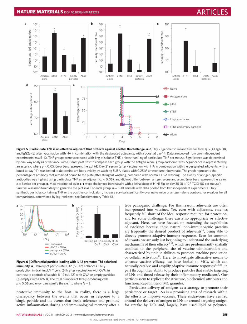
By using HA as our experimental antigen we were able toassess if our adjuvant system conferred any protection to thehost in a lethal challenge model of Flu. For this study, micewere vaccinated as described previously, followed by challengewith a mouse virulent strain of Flu. When mice were monitoreddaily for survival, we observed that vaccination with HA inconjunction with particulate TNF significantly increased survivalof mice after Flu challenge, to levels that did not differ fromalum, which was used as a positive control (Fig. 5e). These datapoint to our particulate cytokine delivery system as an effectiveadjuvant that can be used to protect from an infectious challenge.Cumulatively, these studies demonstrate that this particulate TNFdelivery system promotes both high-magnitude (Fig. 4a–c andhigh-affinity (Fig. 5d) antibodies as part of an adaptive immuneresponse that can be protective, as in the case of a lethal Flu challenge(Fig. 5d), and that this response is possible by potentiating adaptiveresponses within the LNmicroenvironment (Fig. 4).
Differential particle loading to polarize immune responsesAlthough TNF was the target of our initial studies owing to itsincorporation in naturalMC-derived particles, other cytokines havebeen shown to have adjuvant activity8 and could be potentiallytargeted to draining LNs by applying the MC’s strategy. Wesought to address the recognized shortfall of currently approvedvaccine adjuvants in terms of their ability to capitalize on theremarkable plasticity of adaptive responses and to effectively directimmune outcomes differentially depending on the requirements ofa unique challenge. Therefore, we encapsulated IL-12 in heparin–chitosan complexes, because this prominentDC-produced cytokineis important for driving cell-mediated immune responses that areessential to clearmany viral infections32. Soluble IL-12 has also beenpreviously employed to polarize immunity during immunization33.In our studies, injection of particulate IL-12 in conjunction with thesoluble experimental antigen ovalbumin (OVA) greatly increasedthe number of LN T cells producing IFN-γ by 24 h, whereas aneven larger amount of soluble IL-12 or particles alone did not(Fig. 6). This effect was further augmented in combination withparticulate TNF (data not shown). These results demonstrate thatdifferential loading of our synthetic particlesmay be an effectivewayto target minute quantities of cytokines to the draining LN duringvaccination, and that the loaded cytokines need not conform tothe template provided by MC granules. As a result, the cytokinesdelivered in particulate form and the resulting polarization ofimmune responses in the draining LN could be tailored to meet therequirements for protection against an individual challenge.
Much is required of an effective vaccine formulation, whichwould, ideally, consist of a single peptide, capable of conferring
254 NATURE MATERIALS | VOL 11 | MARCH 2012 | www.nature.com/naturematerials
© 2012 Macmillan Publishers Limited. All rights reserved
NATURE MATERIALS DOI: 10.1038/NMAT3222 ARTICLESa
d e
b c∗
∗
Seru
m to
tal I
gG e
ndpo
int t
itre
Seru
m Ig
G1
endp
oint
titr
e
Seru
m Ig
G2a
end
poin
t titr
e
∗
∗
∗
60
70
80
90
sTNFpTNF AlumEmptyparticles
0 1 2 3 4 5 6 7 80
25
50
75
100∗
100
101
102
103
104
105
106
107
108
100 100
101
102
103
104
101
102
103
104
105
106
107
108
Antigenalone
pTNF AlumAntigenalone
sTNFpTNF AlumEmptyparticles
Antigenalone
sTNFpTNF AlumEmptyparticles
Antigenalone
Perc
enta
ge a
ntib
ody
boun
d
Perc
enta
ge s
urvi
val
Days
Naive
Antigen alone
pTNF
sTNF
Empty particles
sTNF and empty particles
Alum
Figure 5 | Particulate TNF is an effective adjuvant that protects against a lethal flu challenge. a–c, Day 21 geometric mean titres for total IgG (a), IgG1 (b)and IgG2a (c) after vaccination with HA in combination with the designated adjuvants, with a boost at day 14. Data are pooled from two independentexperiments; n= 5–10. TNF groups were vaccinated with 1 ng of soluble TNF, or less than 1 ng of particulate TNF per mouse. Significance was determinedby one-way analysis of variance with Dunnet post-test to compare each group with the antigen-alone-group endpoint titres. Significance is represented byan asterisk, where p< 0.05. Error bars represent the s.d. (d) Day 21 serum (after vaccination with HA in combination with the designated adjuvants, with aboost at day 14), was tested to determine antibody avidity by washing ELISA plates with 0.25 M ammonium thiocyanate. The graph represents thepercentage of antibody that remained bound to the plate after stringent washing, compared with normal ELISA washing. The avidity of antigen-specificantibodies was highest using particulate TNF as an adjuvant (p< 0.05), and did not differ between antigen alone and alum. Error bars represent the s.e.m.;n= 5 mice per group. e, Mice vaccinated as in a–c were challenged intranasally with a lethal dose of H1N1 Flu on day 35 (8× 10⁴ TCID-50 per mouse).Survival was monitored daily to generate the plot in e. For each group, n= 5–10 animals with data pooled from two independent experiments. Onlysynthetic particles containing TNF or the positive control, alum, increase survival significantly over naive mice or antigen-alone controls; for p-values for allcomparisons, determined by log-rank test, see Supplementary Table S1.
0
∗
Unstained
a b
1000
5
10
15
20
25
30
101 102 103 104
IFN-γ
Tot
al IF
N-γ
+ c
ells
(×
105 )
pIL-12 + OVAp-empty + OVAsIL-12 + OVA
987654321
Resting pIL-12OVA
p-emptyOVA
sIL-12OVA
Figure 6 | Differential particle loading with IL-12 promotes Th1 polarizedimmunity. a, Delivery of particulate IL-12 (pIL-12) enhances IFN-γproduction in draining LN T cells, 24 h after vaccination with OVA, incontrast to controls of soluble IL-12 (sIL-12) with OVA or empty particles(p-empty) with OVA. b, The total numbers of IFN-γ-producing cells.p< 0.05 and error bars signify the s.e.m., where N= 3.
protective immunity to the host. In reality, there is a largediscrepancy between the events that occur in response to asingle peptide and the events that break tolerance and promoteactive inflammation during and immunological memory after a
true pathogenic challenge. For this reason, adjuvants are oftenincorporated into vaccines. Yet, even with adjuvants, vaccinesfrequently fall short of the ideal response required for protection,and for some challenges there exists no appropriate or effectiveadjuvant. Here, we have focused on extending the capabilitiesof cytokines because these natural non-immunogenic proteinsare frequently the desired product of adjuvants34, being able todirectly promote adaptive immune responses. Even for commonadjuvants, we are only just beginning to understand the underlyingmechanisms of their efficacy1,35, which are predominantly spatiallyconfined to the peripheral site of vaccine administration andcharacterized by unique abilities to promote cytokine productionor cellular activation36. Here, to investigate alternative means toenhance vaccine efficacy, we have looked to MCs, which cannaturally catalyse and amplify adaptive immune responses3,4,7,37, inpart through their ability to produce particles that enable targetingof LNs and timed release by their inflammatory mediators6. Ourparticles seem to replicate the structure, biochemical attributes andfunctional capabilities of MC granules.
Particulate delivery of antigens as a strategy to promote theirpersistence or target LNs is a promising area of research withinthe efforts to improve vaccines. These endeavours have centredaround the delivery of antigen to LNs or around targeting antigenfor uptake by DCs and, largely, have used lipid or polymer-
NATURE MATERIALS | VOL 11 | MARCH 2012 | www.nature.com/naturematerials 255
© 2012 Macmillan Publishers Limited. All rights reserved

ARTICLES NATURE MATERIALS DOI: 10.1038/NMAT3222
based carriers38. Other groups have also employed particles forin vivo delivery of adjuvants39–41. It is our contention, however,that delivery of cytokines to LNs as a mechanism of modulatingthe microenvironment where the immune response is amplifiedand refined may enable the natural processing of peripherallyadministered antigen to proceed under conditions to optimizeand maximize the adaptive immune response. However, as withMC-derived particles, some synthetic particles are able to persistwithin the tissue (data not shown), and these may also promotethe function of encapsulated cytokines at the site of injection.Furthermore, although chitosan has been suggested to promotesome level of innate immune activation23, we find here that particlesmade from this highly purified chitosan do not, alone, haveadjuvant activity, and this property emerges only when the particlesare used as carriers for immuno-modulatory cytokines.
Furthermore, this technology confers a unique plasticity tothe process of rational vaccine design owing to the potential oftargeting precise combinations of cytokines to draining LNs todeliver the optimal combination for a unique challenge. MCs, too,have limited ability to modulate the content of their granules aftera unique challenge, yet MC granules remain largely of the samecomposition5. By capitalizing on the encapsulation technologyMCshave for promoting the persistence of cytokines and targetingthem to the LN, we are able to differentially load these particlesto enhance and also polarize the developing adaptive immuneresponse. Particulate delivery of TNF to the LN during vaccinationhas also given some insight to the role this cytokine plays in the LNduring the development of immunity, because we not only saw thatantibody titres were increased after vaccination, but also observedthat the avidity of these antibodies was greatly enhanced overantigen alone, and even over the standard adjuvant alum. This datasuggests that particulate TNF can play a highly consequential rolein ensuring antibody specificity during the initiation or refinementof adaptive immunity.
MethodsParticle synthesis and cytokine encapsulation. Chitosan (Vanson) was purified todrug grade by dissolving chitosan in 1% acetic acid overnight and filtering throughWhatman paper in a Buchner funnel. The filtered solution was precipitated withNaOH and ultracentrifuged at 6,000 r.p.m. for 20min. The chitosan pellet waswashed with water and frozen at −80 ◦C before lyophilization. The purifiedchitosan used in these studies was 390 kDa with 83.5% deactylation. Syntheticheparin–chitosan particles were generated by gradually combining 1% heparin(Calbiochem) and 1% chitosan (both in dH2O) in a 1:1 ratio at approximately pH4.5. To produce particles, one volume of 1% chitosan was added to five volumes of1% heparin and vortexed for 30 s. This was repeated until a 1:1 ratio of 1% chitosanto 1% heparin was achieved. After 10min at room temperature, the pH was thenadjusted to neutrality to prevent further aggregation. Particles were centrifugedat 14,000× g for 10min at 4 ◦C to form a pellet and washed with water beforeresuspension in PBS for injections or water for visualization on coverslips. To loadparticles with TNF, recombinant TNF (R&D Systems) was vortexed for 10min in1% heparin before the addition of chitosan. Differential loading of particles wasachieved similarly by substituting recombinant IL-12 (R&D Systems). To labelthe particles for fluorescent tracking, 1 µgml−1 of PLL-FITC was added to the 1%heparin before particle synthesis. For encapsulation studies, the amounts of TNFwere measured using a standard L929 cytotoxicity assay. To determine the particlesize distribution and zeta potential, particles were suspended in distilled, deionizedwater and measurements were taken using a Zetasizer Nano instrument (Malvern),according to the manufacturer’s instructions.
Animal sources. Six-week-old female C57BL/6 mice were purchased from theNational Cancer Institute. Sprague-Dawley rats were purchased from Taconic.All animals were housed at the Duke University Vivarium and all experimentalprotocols were approved by the Duke University Institutional Animal Careand Use Committee.
Particle-tracking studies and LN imaging. Synthetic particles containingFITC-PLL were injected in a maximum volume of 20 µl, and draining LNswere subsequently collected at the indicated times. Isolated popliteal LNs wereflash-frozen in OCT (TissueTek), then cryostat-sectioned (10 µm) and fixed onslides with acetone for 15min at 4 ◦C. Before initiating staining protocols, fixed,dried sections were rehydrated in PBS containing 1% BSA. The following primary
antibodies were incubated overnight with tissue sections, as indicated in thefigure legends: anti-B220-biotin, Rat-IgM anti-PNAd, anti-IgD-AlexaFluor647,anti-GL7-FITC, anti-CD11b and anti-CD11c (BD Biosciences). Slides werewashed with PBS before secondary staining with antibodies or probes includingstreptavadin-Alexa546 (Molecular Probes), anti-rat-Cy3, anti-rat-IgM-FITC(Jackson Immunoresearch) and biotinylated anti-hamster (Pharmingen), followedby streptavadin-APC (BD Biosciences). Slides were mounted using ProLongGold Antifade Reagent (Invitrogen). Images were prepared for publicationusing ImageJ software.
Vaccination studies and adjuvant activity evaluation. Mice were vaccinatedwith 1 µg of HA from influenza strain H1N1 A/New Caledonia/20/1999 (ProteinSciences). Antigen was prepared in a solution containing the appropriate adjuvantand injected in a 20 µl volume in a rear footpad. For particulate cytokine adjuvants,particles were produced as outlined above, followed by washing and resuspensionin PBS for injection. For all particle injections a maximum of 10 µg of chitosan and10 µg of heparin (assuming 100% efficiency of particle construction and no loss ofmaterial during washing) were injected per mouse. Assuming approximately 50%efficiency of encapsulation of cytokine (Fig. 1d), 0.5 ng of cytokine would have beeninjected into each mouse; however, this amount is represented as less than 1 ngbecause encapsulation efficiency can vary and because 1 ng per mouse was addedto the reaction mixture during particle formation. Antigen and adjuvant werecombined just before injection to limit antigen encapsulation in the particles. Foralum, an emulsion was produced by mixing HA in 50% PBS, 50% alum (Sigma).Animals received a boost on day 14 and were bled on day 21. Serum was isolatedand stored at−80 ◦C until ELISAs were carried out.
The survival curve was generated by challenging mice vaccinated as describedabove with a dose equivalent to 4× 10² TCID50 of influenza A/PR/8/34(serotype H1N1, Charles River). To infect, mice were briefly anesthetized withisolurane and Flu was instilled into the lungs through the nasal passages in a40 µl volume of PBS. Before inoculation, this suspension of Flu and PBS waspassed through a 0. 22 µm syringe filter. Mice were monitored daily for deathor humane endpoints.
Statistics. For each vaccination study, significance was determined by one-wayanalysis of variance with Dunnet’s post-test to compare treated groups withthe control group. Significance for Kaplan–Meier curves was determined by thelog-rank test. Further comparisons between two groups were carried out usingStudent’s t -tests. Statistics were carried out using Excel and Prism software.
Received 15 July 2011; accepted 30 November 2011;published online 22 January 2012
References1. Lambrecht, B. N., Kool, M., Willart, M. A. & Hammad, H. Mechanism of
action of clinically approved adjuvants. Curr. Opin Immunol. 21, 23–29 (2009).2. Martín-Fontecha, A. et al. Regulation of dendritic cell migration to the draining
lymph node: Impact on T lymphocyte traffic and priming. J. Exp. Med. 198,615–621 (2003).
3. McLachlan, J. B. et al. Mast cell-derived tumor necrosis factor induceshypertrophy of draining lymph nodes during infection. Nature Immunol. 4,1199–1205 (2003).
4. Shelburne, C. P. et al. Mast cells augment adaptive immunity by orchestratingdendritic cell trafficking through infected tissues. Cell Host Microbe. 6,331–342 (2009).
5. Abraham, S. N. & St. John, A. L.Mast cell-orchestrated immunity to pathogens.Nature Rev. Immunol. 10, 440–452 (2010).
6. Kunder, C. A. et al. Mast cell-derived particles deliver peripheral signals toremote lymph nodes. J. Exp. Med. 206, 2455–2467 (2009).
7. McLachlan, J. B. et al. Mast cell activators: A new class of highly effectivevaccine adjuvants. Nature Med. 14, 536–541 (2008).
8. Bradney, C. P., Sempowski, G. D., Liao, H. X., Haynes, B. F. & Staats, H. F.Cytokines as adjuvants for the induction of anti-human immunodeficiencyvirus peptide immunoglobulin G (IgG) and IgA antibodies in serum andmucosal secretions after nasal immunization. J. Virol. 76, 517–524 (2002).
9. Perrillo, R. Benefits and risks of interferon therapy for hepatitis B. Hepatology49, S103–S111 (2009).
10. Anderlini, P., Przepiorka, D., Champlin, R. & Korbling, M. Biologic andclinical effects of granulocyte colony-stimulating factor in normal individuals.Blood 88, 2819–2825 (1996).
11. Stevens, R. L. & Adachi, R. Protease–proteoglycan complexes of mouse andhuman mast cells and importance of their β-tryptase–heparin complexes ininflammation and innate immunity. Immunol. Rev. 217, 155–167 (2007).
12. Roy, K., Mao, H. Q., Huang, S. K. & Leong, K. W. Oral gene delivery withchitosan–DNA nanoparticles generates immunologic protection in a murinemodel of peanut allergy. Nature Med. 5, 387–391 (1999).
13. Kuang, M. et al. Phase II randomized trial of autologous formalin-fixedtumor vaccine for postsurgical recurrence of hepatocellular carcinoma.Clin. Cancer Res. 10, 1574–1579 (2004).
256 NATURE MATERIALS | VOL 11 | MARCH 2012 | www.nature.com/naturematerials
© 2012 Macmillan Publishers Limited. All rights reserved

NATURE MATERIALS DOI: 10.1038/NMAT3222 ARTICLES14. Hanes, J. et al. Controlled local delivery of interleukin-2 by biodegradable
polymers protects animals from experimental brain tumors and liver tumors.Pharm. Res. 18, 899–906 (2001).
15. VandeVord, P. J. et al. Evaluation of the biocompatibility of a chitosan scaffoldin mice. J. Biomed. Mater. Res. 59, 585–590 (2002).
16. Lane, D. A. & Adams, L. Non-anticoagulant uses of heparin. New Engl. J. Med.329, 129–130 (1993).
17. Phaechamud, T., Koizumi, T. & Ritthidej, G. C. Chitosan citrate as film former:Compatibility with water-soluble anionic dyes and drug dissolution fromcoated tablet. Int. J. Pharm. 198, 97–111 (2000).
18. Hammel, I., Lagunoff, D. & Galli, S. J. Regulation of secretory granule sizeby the precise generation and fusion of unit granules. J. Cell Mol. Med. 14,1904–1916 (2010).
19. Ingulli, E., Ulman, D. R., Lucido, M. M. & Jenkins, M. K. In situ analysis revealsphysical interactions between CD11b+ dendritic cells and antigen-specificCD4 T cells after subcutaneous injection of antigen. J. Immunol. 169,2247–2252 (2002).
20. Junt, T. et al. Subcapsular sinusmacrophages in lymphnodes clear lymph-borneviruses and present them to antiviral B cells. Nature 450, 110–114 (2007).
21. Phan, T. G., Grigorova, I., Okada, T. & Cyster, J. G. Subcapsular encounterand complement-dependent transport of immune complexes by lymph nodeB cells. Nature Immunol. 8, 992–1000 (2007).
22. Klein, U. & Dalla-Favera, R. Germinal centres: Role in B-cell physiology andmalignancy. Nature Rev. Immunol. 8, 22–33 (2008).
23. Villiers, C. et al. From secretome analysis to immunology: Chitosan inducesmajor alterations in the activation of dendritic cells via a TLR4-dependentmechanism.Mol. Cell Proteomics 8, 1252–1264 (2009).
24. Sui, Z., Chen, Q., Fang, F., Zheng, M. & Chen, Z. Cross-protection againstinfluenza virus infection by intranasal administration of M1-based vaccinewith chitosan as an adjuvant. Vaccine 28, 7690–7698 (2010).
25. Ghendon, Y., Markushin, S., Akopova, I., Koptiaeva, I. & Krivtsov, G.Chitosan as an adjuvant for poliovaccine. J. Med. Virol. 83, 847–852 (2011).
26. Bhan, A. K., Nadler, L. M., Stashenko, P., McCluskey, R. T. & Schlossman, S. F.Stages of B cell differentiation in human lymphoid tissue. J. Exp. Med. 154,737–749 (1981).
27. Nimmerjahn, F. & Ravetch, J. V. Divergent immunoglobulin g subclass activitythrough selective Fc receptor binding. Science 310, 1510–1512 (2005).
28. Pullen,G. R., Fitzgerald,M. G.&Hosking, C. S. Antibody avidity determinationby ELISA using thiocyanate elution. J. Immunol. Methods 86, 83–87 (1986).
29. Harris, S. L., Tsao, H., Ashton, L., Goldblatt, D. & Fernsten, P. Avidity of theimmunoglobulin G response to aNeisseria meningitidis group C polysaccharideconjugate vaccine as measured by inhibition and chaotropic enzyme-linkedimmunosorbent assays. Clin. Vaccine Immunol. 14, 397–403 (2007).
30. Marcus, H. et al. Contribution of immunological memory to protectiveimmunity conferred by a Bacillus anthracis protective antigen-based vaccine.Infect. Immun. 72, 3471–3477 (2004).
31. McCloskey, N., Turner, M. W. & Goldblatt, T. D. Correlation between theavidity of mouse–human chimeric IgG subclass monoclonal antibodies
measured by solid-phase elution ELISA and biospecific interaction analysis(BIA). J. Immunol. Methods 205, 67–72 (1997).
32. Stetson, D. B. & Medzhitov, R. Type I interferons in host defense. Immunity25, 373–381 (2006).
33. Afonso, L. C. et al. The adjuvant effect of interleukin-12 in a vaccine againstLeishmania major. Science 263, 235–237 (1994).
34. Mosca, F. et al. Molecular and cellular signatures of human vaccine adjuvants.Proc. Natl Acad. Sci. USA 105, 10501–10506 (2008).
35. Datta, S. K. et al. Mucosal adjuvant activity of cholera toxin requires Th17cells and protects against inhalation anthrax. Proc. Natl Acad. Sci. USA 107,10638–10643 (2010).
36. De Gregorio, E., D’Oro, U. & Wack, A. Immunology of TLR-independentvaccine adjuvants. Curr. Opin. Immunol. 21, 339–345 (2009).
37. Galli, S. J., Nakae, S. & Tsai, M. Mast cells in the development of adaptiveimmune responses. Nature Immunol. 6, 135–142 (2005).
38. Liang, M. T., Davies, N. M., Blanchfield, J. T. & Toth, I. Particulate systems asadjuvants and carriers for peptide and protein antigens. Curr. Drug Deliv. 3,379–388 (2006).
39. Kasturi, S. P. et al. Programming the magnitude and persistence of antibodyresponses with innate immunity. Nature 470, 543–547 (2011).
40. Jewell, C. M., Lopez, S. C. & Irvine, D. J. In situ engineering of the lymph nodemicroenvironment via intranodal injection of adjuvant-releasing polymerparticles. Proc. Natl Acad. Sci. USA 108, 15745–15750 (2011).
41. Singh, M. et al. Cationic microparticles are an effective delivery system forimmune stimulatory cpG DNA. Pharm. Res. 18, 1476–1479 (2001).
AcknowledgementsWe thank C. A. Kunder, B. Berwin and A. Alonso for their contributions to producingthe image in Fig. 1a. L. Eibist is thanked for her assistance in acquiring the EM ofsynthetic particles. We also thank A. P. S. Rathore and W. X. G. Ang for their manuscriptreview and discussions. The authors’ work is supported by the US National Institutesof Health grants R01 AI96305, R01 AI35678, R01 DK077159, R01 AI50021, R37DK50814 and R21 AI056101.
Author contributionsExperiments were designed by A.L.S. and S.N.A., with H.F.S. contributing to theexperimental design of vaccination studies and K.W.L. contributing to the experimentaldesign of studies relating to particle synthesis and characterization. Experiments werecarried out by A.L.S. and C.Y.C. Data was analysed by A.L.S. and C.Y.C. with advicefrom S.N.A. and H.F.S. The manuscript was written primarily by A.L.S. All authorscontributed to discussions and manuscript review.
Additional informationThe authors declare no competing financial interests. Supplementary informationaccompanies this paper on www.nature.com/naturematerials. Reprints and permissionsinformation is available online at http://www.nature.com/reprints. Correspondence andrequests for materials should be addressed to A.L.S.
NATURE MATERIALS | VOL 11 | MARCH 2012 | www.nature.com/naturematerials 257
© 2012 Macmillan Publishers Limited. All rights reserved