synthesis of silver nanoparticle synthesis from tea …umpir.ump.edu.my/5903/1/cd7133.pdf ·...
TRANSCRIPT
SYNTHESIS OF SILVER NANOPARTICLE SYNTHESIS FROM TEA LEAF (CAMELLIA
SINENSIS) EXTRACT AND STUDY ON ITS ANTIMICROBIAL PROPERTIES
NURUL AFIQAH BINTI ABDUL RAHIM
Thesis submitted in fulfilment of the requirements
for the award of the degree of
Bachelor of Chemical Engineering in Biotechnology
Faculty of Chemical and Natural Resources Engineering
UNIVERSITI MALAYSIA PAHANG
JUNE 2012

iv
ABSTRACT
Current nanotechnology research uses a lot of chemicals, which are potential threat to both
environment and public health. Therefore the need to find more environmental friendly
procedures for the production of nanoparticles arises. This research is done in with the
purpose of synthesizing silver nanoparticles from tea leaves while studying its
antimicrobial properties on Gram positive and Gram positive bacteria. Tea leaves
(Camellia sinensis) has been used because of its rich source of polyphenolic compounds
used for the reduction and capping of silver nanoparticles, making it a complete green
chemical route. The reduction process of Ag+ to Ag
0 was observed by the change of colour
from clear brown to milky grey. The reaction was followed by the characterization of the
silver nanoparticles using UV-Vis, FTIR, TEM and XRD. An average particle size of 26
nm silver nanoparticles could be obtained using TEM. The antimicrobial activity was tested
using agar well method and it is found that the antimicrobial activity is observed to be
higher against Gram negative bacteria. As a result of the research, it is determined that local
plants such as tea leaves is an alternative to a safer, more eco-friendly alternative of
synthesizing silver nanoparticles.

v
ABSTRAK
Kajian nanoteknologi terkini menggunakan terlalu banyak bahan kima yang menjadi
ancaman terhadap alam sekitar dan kesihatan awam. Oleh itu, timbulnya keperluan untuk
mencari alternatif bagi prosedur menghasilkan nanozarah. Kajian ini dijalankan bertujuan
untuk mengsintesis nanozarah argentum (Ag) dari daun teh serta mengkaji kesan ciri-ciri
antimikrob nanozarah tersebut ke atas bakteria Gram positif dan juga Gram negatif. Daun
teh (Camellia sinensis) telah digunakan berdasarkan sifatnya yang kaya dengan kandungan
polifenol yang boleh menjadi agen pengurangan lantas menjadikan proses ini suatu proses
yang mesra alam. Proses pengurangan dari Ag+ ke Ag
0 telah diperhatikan melalui
perubahan warna dari coklat jernih kepada kelabu keruh. Tindak balas tersebut diikuti
dengan pencirian nanozarah argentum menggunakan UV-Vis, FTIR, TEM dan XRD.
Purata saiz zarah yang telah diukur menggunakan TEM adalah 26 nm. Aktiviti antimikrob
yang telah diperhatikan menerusi kaedah kolam agar menunjukkan bahawa ciri-ciri
antimikrob pada nanozarah argentum bertindak lebih baik ke atas bakteria Gram negatif.
Secara keseluruhannya, telah terbukti bahawa tumbuhan tempatan seperti daun teh boleh
menjadi alternatif kepada kaedah mengsistesis nanozarah argentum yang lebih selamat dan
mesra alam.

vi
TABLE OF CONTENTS
Page
DECLARATION ii
ACKNOWLEDGMENTS iv
ABSTRACT v
ABSTRAK vi
TABLE OF CONTENTS vii
LIST OF FIGURES viii
LIST OF ABBREVIATIONS x
CHAPTER 1 INTRODUCTION
1.1 Background of Research 1
1.2 Problem Statement 4
1.3 Research Objective 4
1.4 Scope of Research 5
1.5 Significance of Research 5
CHAPTER 2 LITERATURE REVIEW
2.1 Silver Nanoparticles 6
2.2 Silver as Antimicrobial Agent 8
2.3 Synthesis of Silver Nanoparticles from Plant 9
CHAPTER 3 METHODOLOGY
3.1 Introduction 12
3.2 Synthesis of Silver Nanoparticles 13

vii
3.3 Washing and Characterization of Silver Nanoparticles 21
3.4 Determination of Antibacterial Properties 16
CHAPTER 4 RESULTS AND DISCUSSION
4.1 Reduction of 19
4.2 Formation of Silver Nanoparticles 22
4.3 Antibacterial Activity of Silver Nanoparticles 26
CHAPTER 5 CONCLUSION
5.1 Conclusion 29
5.2 Recommendation 30
REFERENCES 31
APPENDICES 35

viii
LIST OF FIGURES
FIGURE NO. TITLE PAGE
2.1 Chemical structure of silver 7
2.2 Chemical structure of silver nitrate 10
2.3 Chemical structure of polyphenol 11
3.1 Filtration of tea extract using membrane 14
3.2 Silver nitrate and tea extract mixture stirred
overnight
14
3.3 Cork borer 17
3.4 Laminar flow setup 17
3.5 Application of aseptic technique 18
4.1 (a)
4.1 (b)
Unfiltered tea extract
Filtered tea extract
20
20
4.2 Tea extract reacted with silver nitrate 20
4.3 UV-Vis spectra of Ag (0.1 M) solutions after
reaction with Camellia sinensis extract
21
4.4 Plotted data from UV-Vis spectra 22

ix
4.5 FTIR spectra of tea extract and colloidal solution of
silver nanoparticles after reaction for 24 hours
23
4.6 XRD pattern of the silver nanopowders obtained
from experiment
24
4.7 (a)&(b) TEM images showing silver nanoparticles size
obtained from experiment
25
4.8
4.9
4.10
Silver nanoparticle size obtained in Figure 4.6(b)
Comparison of antimicrobial activity of silver
nanoparticles (4 and 3) on Micrococcus luteus with
blank (1 and 2)
Comparison of antimicrobial activity of silver
nanoparticles (4 and 3) on Escherichia coli with
blank (1 and 2)
25
27
28
34

x
LIST OF ABBREVIATIONS
TEM - Transmission Electron Microscopy
XRD X-ray Diffusion
FTIR - Fourier Traonsform Infra-red Spectroscopy
UV-Vis - Ultra-violet Visible Spectrophotometer

CHAPTER 1
INTRODUCTION
1.1 BACKGROUND OF RESEARCH
Nanoparticles possess a very high surface to volume ratio. This can be utilized in
areas where high surface areas are critical for success. This could for example be in the
catalytic industry; some nanoparticles actually have proven to be good catalysts. Some
nanoparticles also show bactericidal effects and here, a high surface to volume ratio is
important.
In biology and biochemistry, nanoparticles have attracted much attention.
Nanoparticles are often in the range 10-100 nm and this is the size as that of human
proteins. In the production of anti-reflective optical coatings, nanoparticles have also
proven valuable. Using metal oxides to coat polymeric films, anti-reflective surfaces have
been created. Nanoparticle does exhibit many interesting properties, and it is just a matter
of time until more of these properties will be exploited.

2
Recent agriculture and medicine have been applying nanoparticles in multiple ways.
Hence, attempts have been made to synthesize nanoparticles using both chemical and
biological methods. Nanomaterials such as Ag, Au, Pt and Pd have been synthesized by
different methods, including hard template, using bacteria, fungi and plants (Krishnaraj et.
al., 2009). Among the noble metals, silver nanoparticles play a significant role in the field
of biology and medicine due to its attractive physiochemical properties such as increased
optical, electromagnetic and catalytic properties from the bulk materials (Wenseelers et al,
2002; Kelly et al, 2003).
Silver nanoparticles can be synthesized through many different routes. The routes
can be divided into three broad categories, which are physical vapor disposition, ion
implantation and wet chemistry. The silver ion in silver nitrate undergoes reduction to
become silver molecule. The formula is shown in Equation 1.
(1)
Since the first nanoparticles were synthesized, their applications have found their
ways into many different areas of science. It has been applied as a catalyst. In the article
Catalytic Properties of Silver Nanoparticles Supported on Silica Spheres (Jiang et al.,
2004) this catalytic effect has been proven. Another area where silver nanoparticles have
proven to be effective is in controlling and suppressing bacterial growth. Several
applications which use the bactericidal effect of silver nanoparticles have already been
developed.
Various techniques such as chemical and physical mean were developed to prepare
metal nanoparticles such as chemical reduction (Vorobyova et al., 1999), electrochemical

3
reduction (Sandmann et al., 2000), photochemical reduction (Keki et al., 2000), heat
evaporation (Smetana et al., 2005) and so on. However, green process for the synthesis of
silver nanoparticles has been developed and it is an important aspect of current
nanotechnology research. Biosynthesis of nanoparticles has received considerable attention
due to the growing need to develop environmentally benign technologies in material
synthesis. Synthesis of nanoparticles using microorganism or plants can potentially
eliminate this problem by making the nanoparticles more bio-compatible.
The possible mechanism of biosynthesis of nanoparticles by biological system was
reductases and any other equivalent reductants. Polyphenol was reported as one of the
catalysts in the synthesis of nanoparticles. It is known to induce particle formation in
infusions of black decaffeinated tea (Groning et al., 2001). Polyphenol is, in general, a
moderately water-soluble compound with more than 12 phenolic hydroxyl groups and 5-7
aromatic rings per 1000 Da. It is found in virtually all families of plants, and comprising up
to 50% of the dry weight of leaves. It is most commonly found in the leaves of the tea
plant. Therefore, the leaves of Camellia sinensis are chosen to be used as a catalyst in the
synthesis of silver nanoparticles in this experiment.
This research is carried out to synthesis silver nanoparticles from tea leaves extract.
The silver nanoparticles were synthesized by mixing the tea leaves extract with silver
nitrate. The amount of silver nanoparticles is measured by using the Fourier transform
infrared spectroscopy (FTIR), transmission electron microscopy (TEM), ultraviolet-visible
spectrophotometer (UV-Vis) and x-ray diffusion (XRD), and testing of the nanoparticles
for any anti-microbial properties on gram-negative bacteria Escherichia coli and gram-
positive bacteria Micrococcus luteus using agar well method.

4
1.2 PROBLEM STATEMENT
Antibiotic resistance developed by bacteria has been a global concern due to their
capability causing community-acquired infection (Alanis, 2005). Many discussions have
been made on the approaches to deal with antibiotic resistant problem. Beside the
appropriate antibiotic use in the therapeutic and non-therapeutic purpose, searching for safe
and effective antibacterial agents also has been encouraged (Rai et al., 2009). Silver has
been known to possess antimicrobial effects with distinctive properties of conductivity,
stability, and activity (Das et al., 2011). Previous studies have indicated that silver
nanoparticles are effective against a wide spectrum of bacteria, fungi and viruses. In fact,
Quang et al. stated that the antibacterial effect of silver and its compounds have been well
known for a long time as an antibiotic to treat some infectious diseases and burn wounds.
With the emergence and increase of microbial organisms resistant to multiple antibiotics,
and the continuing emphasis on health-care cost, researchers are urged to develop new and
effective antimicrobial reagents free of resistance and cost. Such problems have led to the
resurgence in the use of Ag-based antiseptics that may be linked to broad-spectrum activity
and far lower propensity to induce microbial resistance than antibiotics.
1.3 RESEARCH OBJECTIVE
The purpose of doing this research is to synthesis silver nanoparticles from tea
leaves while studying their antimicrobial properties on Gram positive and Gram negative
bacteria.

5
1.4 SCOPE OF RESEARCH
To achieve the objective of this research, there are a few main scopes that the study
focuses on. Firstly, tea leaves are used in order to synthesize the silver nanoparticles.
Secondly, the characteristic of the silver nanoparticles are determined using FTIR, TEM,
UV-Vis and X-ray diffusion. Thirdly, the antimicrobial properties of silver nanoparticles
synthesized are studied on a gram-negative bacteria Escherichia coli and gram-positive
bacteria Micrococcus luteus.
1.5 SIGNIFICANCE OF RESEARCH
The silver nanoparticles were synthesized using a local plant (tea leaves) because it
has a high content of polyphenol. It is also easier to obtain and more cost effective.
Furthermore, the antimicrobial properties of the silver nanoparticles serve well as an
alternate antiseptic against both gram-negative and gram-positive bacteria.

CHAPTER 2
LITERATURE REVIEW
2.1 SILVER NANOPARTICLES
Silver is a metallic chemical element with the chemical symbol Ag and atomic
number 47. It is a soft, white, lustrous transition metal with the highest electrical
conductivity of any element and the highest thermal conductivity of any metal. It occurs
naturally in its pure, free form (native silver), as an alloy with gold and other metals, and in
minerals such as argentite and chlorargyrite. Most silver is produced as a byproduct of
copper, gold, lead, and zinc refining.
Recently, the development of nanotechnology has resulted in the synthesis of nano
scale silver particles which have particular properties that are superior to the bulk silver
metal. (Quang et al., 2011). Silver nanoparticles are silver particles of between 1 nm and
100 nm in size. It can be synthesized through different methods which included physical
vapor deposition, ion implantation or wet chemistry. Silver nanoparticles have found
applications in catalysis, optics, electronics and other areas due to their unique size-

7
dependent optical, electrical and magnetic properties. Compared to the bulk silver metal,
nano scale silver particles have larger surface areas and unique physical, chemical and
biological properties (Sharma et al., 2009). Moreover, silver nanoparticles can be easily
embedded within porous substrates like mesoporous silica and zeolite (Matsumura, 2003).
Figure 2.1 shows the chemical structure of silver.
Figure 2.1: Chemical structure of silver

8
2.2 SILVER AS ANTIMICROBIAL AGENT
An antimicrobial is a substance that kills or inhibits the growth of microorganisms
such as bacteria, fungi, or protozoans. Antimicrobial agents either kill microbes
(microbiocidal) or prevent the growth of microbes (microbiostatic). Antimicrobials have
long been developed since the observations of Pasteur and Joubert. However, with the
development of antimicrobials, microorganisms have adapted and become resistant to
previous antimicrobial agents. Old antimicrobial technology was mostly based either on
poisons or heavy metals, which may not have killed the microbe completely, allowing the
microbe to survive, change, and become resistant to them. Antimicrobial nanotechnology is
a recent addition to the fight against disease causing organisms, replacing heavy metals and
toxins.
Free silver ion (Ag+) has been well known for its strong toxicity against some
bacteria, viruses, algae and fungi, typical for heavy metals like lead, or mercury, but
without the high toxicity to humans. It is recently shown that silver nanoparticles can be a
promising antimicrobial material. Song et al. reported that silver nanoparticles showed
higher antibacterial ability compared to that of silver compounds like silver nitrate, silver
sulfadiazine, etc. It has been demonstrated that the highly reactive metal oxide
nanoparticles exhibit excellent bactericidal action against Gram positive and Gram negative
bacteria (Klabunde, 2002). Silver nanoparticles also have a high surface area to volume
ratio along with high fraction of surface atoms that elicits elevated antimicrobial activity
compared to the silver metal as a whole (Shahverdi, 2007). Silver supported on various
suitable materials, such as carbon, polyurethane foam, polymers, and sepiolite have also
been effectively used for bactericidal applications (Siva et al., 2004 & Jain et al., 2005).
Various hypotheses have been proposed to explain the mechanism of antimicrobial
activity of silver nanoparticles. It is widely believed that silver nanoparticles are
9
incorporated in the cell membrane, which causes leakage of intracellular substances and
eventually causes cell death (Sondi et al., 2004). Some of the silver nanoparticles also
penetrate into the cells. It is reported that the bactericidal effect of silver nanoparticles
decreases as the size increases and is also affected by the shape of the particles (Pal et al.,
2007). Although most studies have utilized spherical particles, truncated triangular shaped
particles are reported to have greater bactericidal effect compared to that of spherical and
rod shaped particles. The bactericidal efficiency of silver nanoparticles is also reported to
be affected by the type of microorganisms. Kim et al. reported in studies with Gram-
negative, Escherichia coli and Gram-positive, Staphylococcus aureus, that greater
bactericidal efficiency of silver nanoparticle for Escherichia coli is greater and this is
attributed to the difference in cell wall structure between Gram negative and Gram positive
microorganisms.
2.3 SYNTHESIS OF SILVER NANOPARTICLES FROM PLANT
Nanoparticles have been synthesized without aggregation using chemical methods
for its simplicity and the added advantage of high yield for large scale production.
However, hazardous reducing agents used for chemical procedures mounts a bias for an
eco-friendly and feasible approach for the synthesis of nanoparticles. Hence, plants are used
as an alternative trigger for the green synthesis of nanoparticles (Medina-Ramirez et al.
2011). Biological means of synthesizing nanoparticles provides an edge over chemical
means as it is cost effective, does not involve physical barriers with regard to reducing
agents and eliminates the toxic effects of chemicals used for the synthesis (Saxena et al.,
2010 & Song et al., 2009). Figure 2.2 shows the chemical structure of silver nitrate which
can be used to synthesis silver nanoparticles.

10
Figure 2.2: Chemical structure of silver nitrate
Synthesis of nanoparticles using plants can potentially eliminate the problem of the
presence of some toxic chemical species absorbed on the surface by making the
nanoparticle more bio-compatible. Bar et al. stated that the use of plant extract for the
synthesis of nanoparticles could be advantageous over other environmentally benign
biological processes by eliminating the elaborate process of maintaining cell cultures.
Exploration of the plant systems as the potential nanofactories, has increased interest in the
biological synthesis of nanoparticles. Sastry et al. reported the biosynthesis of nanoparticles
using plant leaf extracts and their potential. They studied bioreduction of chloraurate ions
and silver ions by extracts of germanium and neem leaf.
Tea leaf extract (Camellia sinensis) has also been used to synthesize silver
nanoparticles (Vilchis-Nestor et al., 2008). Tea leaf contains polyphenols and terpenoids,
such as β-cariophyllene, linalool, cis-jasmone, α-terpineol, δ-cadiene, indole, geraniol,
among the major bio-components, which have bactericidal and antioxidant activity, and
several other useful properties (Kaufman et al., 1999). Caffeine and theophylline present in
tea extracts were also reported to catalyze the synthesis of nanoparticles (Groning et al.,
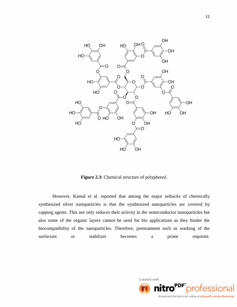
2001). Figure 2.3 shows the chemical structure shows the chemical structure of polyphenol
found in tea.
11
Figure 2.3: Chemical structure of polyphenol.
However, Kamal et al. reported that among the major setbacks of chemically
synthesized silver nanoparticles is that the synthesized nanoparticles are covered by
capping agents. This not only reduces their activity in the semiconductor nanoparticles but
also some of the organic layers cannot be used for bio applications as they hinder the
biocompatibility of the nanoparticles. Therefore, pretreatment such as washing of the
surfactant or stabilizer becomes a prime requisite.

CHAPTER 3
METHODOLOGY
3.1 INTRODUCTION
This chapter includes the methodology applied in order to carry out the experiments
for the synthesis of silver nanoparticles from tea leaves. The experiments will be carried out
in the Chemical Engineering Analytical Laboratory, Faculty of Chemical and Natural
Resources Engineering, Universiti Malaysia Pahang. Before the experiments are carried
out, all the chemicals, raw material and apparatus needed to be set up and ready to run.
The methodology for this research is divided into three main steps; the synthesis of
silver nanoparticles from tea leaf (Camellia sinensis) extract, washing and characterization
of silver nanoparticles, and testing of the nanoparticles for any anti-microbial properties
using agar well method.

13
3.2 SYNTHESIS OF SILVER NANOPARTICLES
3.2.1 Raw Material and Reagents
Silver nitrate (AgNO3, 99.9% pure) was obtained from Sigma-Aldrich, Malaysia.
Commercially available dry tea leaves, Boh Plantations Sdn. Bhd, Malaysia, Was used for
preparation of tea extract and ultra-pure water with a resistivity of 18.2 MΩcm was used as
a medium to dissolve the silver salt and for making the tea extracts.
3.2.2 Procedure
5 g of ground tea leaves were weighed and transferred into a 500ml beaker and was
filled up to the mark with water. The contents of the beaker were thoroughly agitated for 1
hour using a heated stirrer and left to settle. The extract is then filtered through a 0.2 µm
membrane filter and used for further experiments. A dilute solution of silver (0.1 M) was
prepared by dissolving 0.017 g AgNO3 in 1 ml water.
To carry out the reaction, 10 ml of the extract is added with 2 ml of 0.1
M solution of AgNO3. The mixture is gently mixed while being left overnight for the
reaction to occur.

14
Figure 3.1: Filtration of tea extract using membrane
Figure 3.2: Silver nitrate and tea extract mixture stirred overnight

15
3.3 WASHING AND CHARACTERIZATION OF SILVER NANOPARTICLES
3.3.1 Instruments
A desk-top centrifuge is used in the washing process of silver nanoparticles
synthesized from the tea extract. This is to ensure that the polyphenol content is removed
from the nanoparticles and further purify the silver nanoparticles. After the washing
process, the silver nanoparticles are characterized using Fourier transform infrared
spectroscopy (FTIR), transmission electron microscopy (TEM), ultraviolet-visible
spectrophotometer and x-ray diffusion (XRD).
3.3.2 Procedure
12 ml of tea extract was put into a 15 ml centrifuge bottle and centrifuged at 10,000
rpm and 4°C for 10 minutes. After centrifugation, aliquot containing the polyphenols was
separated from the sediment using a dropper. Ultra-pure water was then added to fill up to
12 ml once again. The sample was washed at least three times before it can be tested for
silver nanoparticles characteristics.
The obtained emulsions were characterized using UV-Vis spectrometer. Fourier
transform infra-red spectroscopy was used to study the interactions between the tea extract
and silver nanoparticles for which, a FTIR is used to record the spectra. Silver
nanoparticles were separated from the mother solution by centrifugation at very high
temperature of 20,000 rpm, and the powders thus obtained were placed on silicone (Si)
substrate to obtain the crystal structure using X-ray diffractometer (XRD). The powders
were redispersed in water using a rod ultrasonicator and a drop of the redispersed solution
16
was dropped onto a carbon coated copper grid to carry out transmission electron
microscopy (TEM) studies.
3.4 DETERMINATION OF ANTIBACTERIAL PROPERTIES
The antibacterial activity of silver nanoparticles formed is tested on a gram-negative
strain Escherichia coli and gram-positive strain Micrococcus luteus. The bacteria are used
for inhibitory zone tests to investigate the antibacterial properties of silver nanoparticles
using agar well method. A nutrient broth is used as the growing medium. Bacteria are
grown aerobically in nutrient broth at 37°C for 24 h.
3.4.1 Materials
Escherichia coli and Micrococcus luteus strains were obtained from Chemical
Engineering Laboratory, Faculty of Chemical and Natural Resources Engineering,
Universiti Malaysia Pahang. An agar well method was adopted to assay the nanoparticles
for bactericidal activity against the test strains on Mueller-Hinton agar plates. A cork borer
used to punch the agar wells was from Industial Science and Technology Faculty
laboratory. Sterilized cotton swabs are used to spread the bacterial strain.
3.4.2 Procedure
Using aseptic techniques, petri plates containing 20 ml Mueller-Hinton agar
medium were seeded with 24 hours culture of bacterial strains. A sterilized cork borer was
used to punch through the agar and 20 µl of the silver nanoparticles were added into the