synthesis and characterization of β-co(oh) 2 , cuo and zno...
TRANSCRIPT

Synthesis and Characterization of �-Co(OH)2, CuO and ZnO Nanostructures bySolvothermal Method without Any Additive
Robabeh Mehdizadeh,* Lotf Ali Saghatforoush and Soheila SanatiDepartment of Chemistry, Payam Noor University, 19395-4697, Tehran, I.R. of Iran
(Received: Aug. 2, 2012; Accepted: Oct. 11, 2012; Published Online: Dec. 20, 2012; DOI: 10.1002/jccs.201200419)
�-Co(OH)2, CuO and ZnO nanostructures with plate-like, particle-like and flower-like morphologieswere prepared through the use of simple solvothermal method using of melt salt and 1,10-phenanthrolineas complexing agent and sodium hydroxide. �-Co(OH)2 consisted of a plate-like structure, and the nano-plates size was about 29 nm. The structure was comprised of regular sheets which were assembled to-gether. Furthermore, the as-obtained �-Co(OH)2 nanoplates can be easily converted into Co3O4 nano-plates by calcining in air at 500 °C for 2 h. The results indicate that ZnO powder is of hexagonal wurtzitestructure and well crystallized with high purity. CuO powder is pure monoclinic-structured crystalline.The products were characterized by X-ray diffraction (XRD), scanning electron microscopy (SEM), andFourier transform infrared (FT-IR) spectra. Possible formation mechanism of the nanostructures is pro-posed.
Keywords: Nanostructures; Cobalt hydroxide; ZnO; Solvothermal; CuO.
INTRODUCTION
Inorganic nanostructures (NSs) with well-defined sizes
and complex morphologies have attracted considerable re-
search efforts because of their size, morphology, and nano-
structure dependent properties.1 In particular, hierarchical
NSs of transition metal oxides and hydroxides are a hot
topic due to their attractive structures, large surface-to-vol-
ume ratio, and accessible active interfaces. Among the
transition metal hydroxides, cobalt hydroxides [Co(OH)2]
have versatile applications in catalysis, as electrochemical
supercapacitors, in magnetic recording,2–4 and diverse
other fields. For instance, Co(OH)2 is an important elec-
trode material2,5 and can be used as an effective additive to
improve the electrochemical properties of nickel hydroxide
electrodes.6 Moreover, Co(OH)2 has been investigated as a
precursor for the preparation of cobalt oxide nanomaterials
by a thermal conversion.7,8 Co(OH)2 is polymorphic and
crystallizes in to layered structures with two forms, �- and
�-Co(OH)2.9,10 The hydrotalcite-like �-phase is metastable
and easily transforms into the stable brucite-like �-phase in
strongly alkaline media. The properties of Co(OH)2 materi-
als are closely associated with their microstructures. E.g.
their electrochemical capacitance is significantly influ-
enced by surface area and morphology because double
layer and pseudo capacitances are both interfacial phenom-
ena and pores allow a rapid transfer of electrolytes.11,12
Therefore, much research is devoted to the control of their
microstructure. Divers synthetic methods, such as solution
precipitation,13 precursor conversions,14 and electrode po-
sition,1 have been employed to prepare various NSs of co-
balt hydroxides. Co(OH)2 NSs with different morphologies
including rod,15 needle,16 sheet,17 belt,8 and butterfly-like
shapes15 have been obtained. There are already reports on
three dimensional, hierarchical structures of Co(OH)2, such
as sisal, dandelion, and rose-like shapes,18 and flower-like
hollow core-shell structures.19 Hydrothermal synthesis is
widely employed to prepare various nano-sized inorganic
materials.20 The hydrothermal technique provides a versa-
tile route to control grain size, particle morphology, micro-
structure, and phase composition via adjusting parameters
such as temperature, process duration, and pH value of the
solution.21 The reaction media also play a significant role.22
Compared to the conventional hydrothermal/solvothermal
process, in which usually only a single solvent is used,
mixed solvents allow yet more control via adjusting the
type and ratio of the solvents.23 Synthesis of Co(OH)2 with
abundant morphologies via a facile route still remains a
challenge. Various morphologies of Co(OH)2 with mixed
solvents with different ratios of water to ethanol as media,
are obtained hydrothermally by the aid of dimethylgly-
oxime (dmgH).24 A facile room temperature solution-phase
approach for large-scale synthesis of �-Co(OH)2 nano-
plates reported.25 The synthesis of highly uniform, close to
spherical, coral-like �-Co(OH)2 NSs through a facile, etha-
J. Chin. Chem. Soc. 2013, 60, 339-344 © 2013 The Chemical Society Located in Taipei & Wiley-VCH Verlag GmbH & Co. KGaA, Weinheim 339
JOURNAL OF THE CHINESE
CHEMICAL SOCIETYArticle
* Corresponding author. Tel: +00984612349868; Fax: +00984612349566; E-mail: [email protected]
nol assisted hydrothermal process reported.26
Many studies have shown that the size, shape, and
properties of ZnO nanocrystals depend strongly on the
preparation method and conditions.27 The synthesis, char-
acterization and application of various ZnO nanostructures
including the belts/ ribbons,28 rings,29 tetrapods,30 combs,31
sheets32 and complex structures33 are presently the subject
of intense research. Different synthesis methods have been
devised, including sol–gel technique, microemulsion syn-
thesis, mechanochemical processing, spray pyrolysis and
drying, thermal decomposition of organic precursor, RF
plasma synthesis, supercritical-water processing, self-as-
sembling, hydrothermal processing, vapor transport pro-
cess, sonochemical or microwave-assisted synthesis, direct
precipitation.34 Many methods have been developed to pre-
pare and synthesize of CuO particles with various mor-
phologies. Li et al, synthesized nano-dendrite like CuO via
hydrothermal route.35 CuO nano-shuttle was also prepared
under surfactant assisted conditions using the other same
method.36 CuO nano-rods and nano-ribbons were synthe-
sized by wet chemical methods. In addition, the nanofibers
of CuO were prepared by thermal oxidation on Cu substrate
through importing the polycarbonate membrane template
for initial deposition of Cu nuclei.37 The above mentioned
Methods cannot be departed from complex chemical reac-
tions or processes. Thermal oxidation may assist the pro-
duction of catalysts, semiconductor devices or functional
oxide films under controlled conditions.38 A direct and sim-
ple thermal oxidation method was employed to synthesize
CuO nano-wires and nano-rods. Using this convenient
route, with no catalyst and template assisted, many re-
search teams prepared CuO nano-wires successfully by ox-
idizing copper foils under different conditions such as dif-
ferent annealing temperatures, time or atmosphere.39-42
Metal complexes built from metal ions and polydentate or-
ganic ligands have been grown rapidly in recent years ow-
ing to their potential applications.43 So far, however, the
studies on the syntheses of nano- or microscaled structures
with metal complexes as precursors have been less re-
ported.
In this study, we report the synthesis of plate-like
�-Co(OH)2, ZnO and CuO nanostructures through a facile
solvothermal process. This reliably reproducible method
uses only MCl2 (Co, Zn, Cu), NaOH and 1,10-phenanthro-
line as reactants without templates or further auxiliary re-
agents.
EXPERIMENTAL
Nanostructures synthesis
In a typical experiment, 3 mmol MCl2 (CoCl2.6H2O, ZnCl2,
CuCl2.2H2O) dissolved in 3 mL of distilled water. In the other
beaker, 6 mmol of 1,10-phenanthroline was dissolved in 2 mL of
distilled water and 5 mL of warm ethanol. Then this solution was
added in to the solution of melt chloride under magnetic stirring.
10 mL of NaOH aqueous solution (2M) was added in to the solu-
tion. This alkaline solution was transferred into a Teflon-lined au-
toclave with about 80% capacity. The autoclave was then sealed
and maintained at 160 °C for 24 h. After cooling to room tempera-
ture, the resulting product was centrifuged, washed with distilled
water and absolute ethanol for several times for remove impuri-
ties. Finally, the resulting products was dried at 50 °C. Co3O4
nanoplates were obtained when the as-synthesized Co(OH)2 sam-
ple was directly calcined at 500 °C for 2 h in a muffle furnace.
Materials and physical measurements
All chemical reagents in this experiment were of analytical
grade and used without further purification. Fourier transform in-
frared (FT-IR) spectra were recorded using KBr disks on a
Shimadzu FT-IR model Prestige 21 spectrometer. The morpholo-
gies of products were observed with scanning electron micros-
copy (SEM, Philips XL-30). X-ray powder diffraction (XRD)
measurements were performed using a Philips diffractometer
manufactured by X’pert with monochromatized CuKa radiation.
RESULTS AND DISCUSSION
The morphology, structure and size of the samples are
investigated by Scanning Electron Microscopy (SEM).
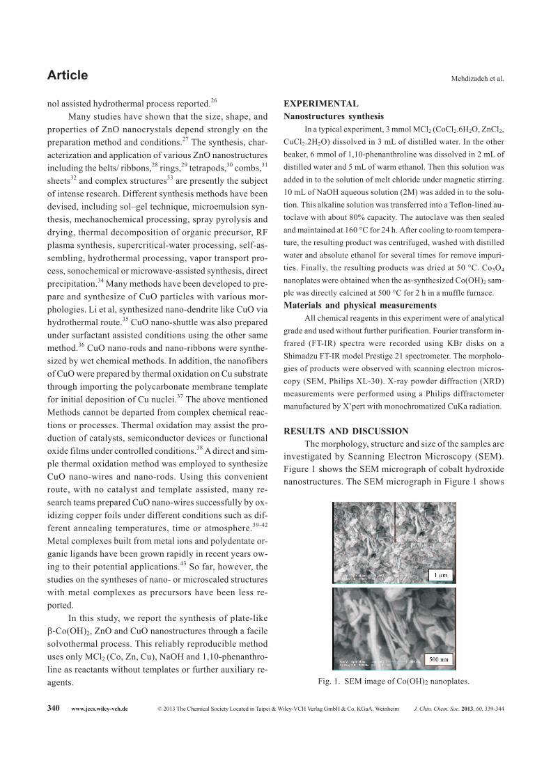
Figure 1 shows the SEM micrograph of cobalt hydroxide
nanostructures. The SEM micrograph in Figure 1 shows
340 www.jccs.wiley-vch.de © 2013 The Chemical Society Located in Taipei & Wiley-VCH Verlag GmbH & Co. KGaA, Weinheim J. Chin. Chem. Soc. 2013, 60, 339-344
Article Mehdizadeh et al.
Fig. 1. SEM image of Co(OH)2 nanoplates.

that the cobalt hydroxide consisted of a plate-like structure,
and the size was in the range between 1 and 2 �m. The
structure was comprised of densely regular sheets which
were assembled together.
Figure 2 shows the SEM micrograph of ZnO nano-
structures. As shown in Fig. 2 the morphology of the ZnO
sample is a flower-like nanomaterial with an average size
of 2 �m in diameter. Nanoflowers size is of 50 nm. In the
case of CuO sample, the CuO crystallites are nanoparticles
with an average size of 53 nm which show in Fig. 3 which
in agreement is by XRD results.
Fig. 4 provides XRD patterns of nanostructures. The
diffraction peaks in the XRD pattern (Fig. 4a) can be
readily indexed to crystalline bulk �-Co(OH)2. The lattice
constants (a = 3.180 Å, c = 4.655 Å) calculated from this
XRD pattern correspond well to the values given in the
standard card (JCPDS 30-0443), which is indexed to the
hexagonal phase of brucite-like �-Co(OH)2.25 Compared
with the standard pattern, the intensity of the (0 0 1) peak is
unusually stronger than others, implying the preferential
orientation of (0 0 1) on the surface. No impurity peaks are
found, suggesting a high purity of the as-synthesized �-
Co(OH)2. The pattern of cobalt hydroxide shows a good
degree of crystallinity. All peaks for sample ZnO (Fig. 4b)
accord with the JCPDS (No. 36-1451) data for ZnO with
hexagonal wurtzite structure. The diffraction peaks in the
XRD pattern can be readily indexed to crystalline bulk
ZnO. The lattice constants (a = 3.24 Å, c = 5.19 Å) calcu-
J. Chin. Chem. Soc. 2013, 60, 339-344 © 2013 The Chemical Society Located in Taipei & Wiley-VCH Verlag GmbH & Co. KGaA, Weinheim www.jccs.wiley-vch.de 341
JOURNAL OF THE CHINESE
Synthesis and Characterization of �-Co(OH)2, CuO and ZnO CHEMICAL SOCIETY
Fig. 2. SEM image of ZnO nanoflowers.
Fig. 3. SEM image of CuO nanoparticles.
Fig. 4. XRD pattern of nanostructures, a) Co(OH)2, b)ZnO, c) CuO.

lated from this XRD pattern correspond well to the values
given in the standard card. No impurity peaks are found,
suggesting a high purity of the as-synthesized ZnO. While
the peaks for sample CuO (Fig. 4c) can be readily ascribed
to the monoclinic CuO form. The observed indexed peaks
in this XRD pattern are fully matched with the correspond-
ing pure monoclinic-structured crystalline CuO (a = 4.68
Å, b = 3.42 Å, c = 5.12 Å, JCPDS card No. 05-0661). Esti-
mated from the Scherrer formula, D = 0.891�/�cos�,
where, D is the average grain size, � is the X-ray wave-
length (0.15405 nm), and � and � are the diffraction angle
and full-width at half maximum of an observed peak, re-
spectively. The average size of the ZnO nanoflowers and
CuO nanoparticles is calculated were about 37 nm for ZnO
by using the strongest peak (1 0 1) at 2� = 36.20, 32 nm for
CuO by using the strongest peak (0 0 2) at 2� = 35.50 and
25.2 nm for Co(OH)2 by using the strongest peak (0 0 1) at
2� = 19.20.
The nanostructures composition can be confirmed via
FT-IR spectroscopy as depicted in Fig. 5a,b,c. A narrow
band (Fig. 5a) is located at 3630 cm-1, which corresponds to
the � O–H stretching of the OH groups in the brucite-like
structure. A broad band at about 3447 cm-1 is characteristic
of the stretching vibration of interlayer water molecules
and of hydroxyl groups hydrogen-bonded to H2O.26 The
bands at 1655 and 1457 cm-1 corresponds to the bending
mode of water molecules. The peak in the region of 486
cm-1 can be assigned to Co–O stretching vibration.26 In the
FT–IR spectrum of the nanostructures (Fig. 5b,c), a strong
band around 445 and 522 cm-1 was observed, which is re-
lated to the Zn–O and Cu–O stretching vibration, respec-
tively. The broad peaks at ca. 3438 and 3425 cm-1 in (Fig.
5b,c) are due to adsorbed water on the external surface of
the samples during handling to record the spectra.
M(phen)2 complex is formed firstly by the reaction of
M2+ with phen during the precursor preparation, and it then
releases M2+ slowly during solvothermal processing. For
Co2+, Co(OH)2 precipitate is produced gradually from the
reaction of Co2+ with OH-. The plate-like nanostructures of
Co(OH)2 are expected to be promoted by the guide of
Co(phen)2 complex. The process of decomposition was a
controlling step, because complex didn’t decompose as
quickly as other Co-containing inorganic salts, which may
provide enough time and opportunity for the growth of
Co(OH)2 nanomaterial. Thus, �-Co(OH)2 nuclei slowly
grew along reaction, resulting in the plate-like structure of
the sample.
For Zn2+ and Cu2+, under the condition of heavy alka-
line solution, [Zn(OH)4]2- or [Cu(OH)4]
2- ions were first
formed. Then [Zn(OH)4]2- or [Cu(OH)4]
2- ions were dehy-
drated under hydrothermal conditions and they in situ gen-
erated a bit of ZnO or CuO nuclei which acted as the seeds
for the growth of melt oxide. The surface of ZnO or CuO
nuclei is either positively charged or negatively charged. In
either case the surface will selectively adsorb ions of oppo-
site charges (OH� or Cu2+) on it, and the new surface cov-
ered with ions will in turn adsorb ions with opposite charges
to cover the surface next.20,21 In the heavy alkaline syn-
thetic system, more OH� may neutralize positive charges
342 www.jccs.wiley-vch.de © 2013 The Chemical Society Located in Taipei & Wiley-VCH Verlag GmbH & Co. KGaA, Weinheim J. Chin. Chem. Soc. 2013, 60, 339-344
Article Mehdizadeh et al.
Fig. 5. FT-IR spectra of nanostructures a) Co(OH)2, b)ZnO, c) CuO.

on the surface of melt oxide, preventing them from possible
crystallite aggregation. Thus, ZnO or CuO nuclei slowly
grew and resulting in the flower-like structure of the sam-
ple ZnO and nanoparticle structure for CuO.44
The as-prepared �-Co(OH)2 nanoplates acting as a
precursor is converted into cobalt oxide through dehydra-
tion in our experiment. This reaction can be schematized as
follows:
�-Co(OH)2dehydration� ��� Co3O4 + H2O
The product of the thermal decomposition from the
Co(OH)2 precursor was studied by FT-IR, XRD and SEM.
The FT-IR spectra of the Co3O4 show at Fig. 6a. The FT-IR
spectra of the Co3O4 show absorption peak at 3430 cm-1 are
assigned to the stretching vibration of hydroxyl group.
Peaks around 663 and 567 cm-1 are ascribed to the Co–O
stretching mode.
Figure 6b shows the XRD patterns of the dehydration
product. The diffraction peaks were indexed to the phase of
crystalline cubic structured cobalt oxide with the lattice
constant a = 8.08 which are consistent with the values in the
standard card (JCPDS card No. 42-1467). No peaks from
impurities are observed in this pattern. Figure 7 shows the
SEM micrograph of Co3O4 after calcination, which is com-
posed of nanoplates. Size of nanoplates was in about 35 nm
in diameter.
CONCLUSIONS
In summary, �-Co(OH)2 nanoplates, ZnO nanoflow-
ers and CuO nanoparticles were successfully synthesized
in one step via a template and surfactant free solvothermal
route. �-Co(OH)2 consisted of a plate-like structure, and
the nanoplates size was about 29 nm. Furthermore, the as-
obtained �-Co(OH)2 nanoplates can be easily converted
into Co3O4 nanoplates by calcining in air. The results indi-
cate that ZnO powder is of hexagonal wurtzite structure
and well crystallized with high purity. CuO powder is pure
monoclinic-structured crystalline. Chemical composition,
morphology, and size of the nanostructures have been sys-
tematically characterized using XRD, SEM and FTIR.
Thermal decomposition was employed to produce Co3O4
nanoplatelets of Co(OH)2 precursor. The present method is
simple and low-cost which makes it feasible for scale-up
production nanostructures.
ACKNOWLEDGEMENTS
We are grateful to Payame Noor University for finan-
cial support of this work.
REFERENCES
1. Lou, X. W.; Deng, D.; Lee, J. Y.; Feng, J.; Archer, L. A. Adv.
Mater. 2008, 20, 258.
J. Chin. Chem. Soc. 2013, 60, 339-344 © 2013 The Chemical Society Located in Taipei & Wiley-VCH Verlag GmbH & Co. KGaA, Weinheim www.jccs.wiley-vch.de 343
JOURNAL OF THE CHINESE
Synthesis and Characterization of �-Co(OH)2, CuO and ZnO CHEMICAL SOCIETY
Fig. 6. a) FT-IR spectra, b) XRD pattern of Co3O4
nanoplates after calcination of Co(OH)2 precur-sor.
Fig. 7. SEM image of Co3O4 nanoplates after calcina-tion of Co(OH)2 precursor.

2. Gupta, V.; Kusahara, T.; Toyama, H.; Gupta, S.; Miura, N.
Electrochem. Commun. 2007, 9, 2315.
3. Chen, H. M.; Zhao, Y. Q.; Yang, M. Q.; He, J. H.; Chu, P. K.;
Zhang, J.; Wu, S. H. Anal. Chim. Acta 2010, 659, 266.
4. Qiao, R.; Zhang, X. L.; Qiu, R.; Kim, J. C.; Kang, Y. S.
Chem. Eur. J. 2009, 15, 1886.
5. Cao, L.; Xu, F.; Liang, Y. Y.; Li, H. L. Adv. Mater. 2004, 16,
1853.
6. Li, W. Y.; Zhang, S. Y.; Chen, J. J. Phys. Chem. B 2005, 109,
14025.
7. Li, Y. G.; Tan, B.; Wu, Y. Y. J. Am. Chem. Soc. 2006, 128,
14258.
8. Yang, J. H.; Hyodo, H.; Kimura, K.; Sasaki, T. Nanotechnol-
ogy 2010, 21, 045605.
9. Bish, D. L.; Livingstore, A. Miner. Mag. 1981, 44, 339.
10. Oliva, P.; Leonardi, J.; Laurent, J. F.; Delmas, C.;
Braconnier, J. J.; Figlarz, M.; Fievet, F. J. Power Sources
1982, 8, 229.
11. Zhou, H.; Li, D.; Hibino, M.; Honma, I. Angew. Chem. Int.
Ed. 2005, 44, 797.
12. Hosono, E.; Fujihara, S.; Honma, I.; Ichihara, M.; Zhou, H.
J. Power Sources 2006, 158, 779.
13. Liang, Y. Y.; Cao, L.; Kong, L. B.; Li, H. L. J. Power Sources
2004, 136, 197.
14. Barde, F.; Palacin, M. R.; Beaudoin, B.; Delahaye-Vidal, A.;
Tarascon, J. M. Chem. Mater. 2004, 16, 299.
15. Sampanthar, J. T.; Zeng, H. C. J. Am. Chem. Soc. 2002, 124,
6668.
16. Zhang, L. Q.; Dutta, A. K.; Jarero, G.; Stroeve, P. Langmuir
2000, 16, 7095.
17. Hosono, E.; Fujihara, S.; Honma, I. J. Mater. Chem. 2005,
15, 1938.
18. Li, B.; Xie, Y.; Wu, C.; Li, Z.; Zhang, J. Mater. Chem. Phys.
2006, 99, 479.
19. Yang, L. X.; Zhu, Y. J.; Li, L.; Zhang, L.; Tong, H.; Wang, W.
W.; Cheng, G. F.; Zhu, J. F. Eur. J. Inorg. Chem. 2006, 23,
4787.
20. Wang, X.; Zhuang, J.; Peng, Q.; Li, Y. D. Nature 2005, 437,
121.
21. Tang, S. C.; Tang, Y. F.; Vongehr, S.; Zhao, X. N.; Meng, X.
K. Appl. Surf. Sci. 2009, 255, 6011.
22. Pang, H.; Gao, F.; Lu, Q. Y. Cryst. Eng. Commun. 2010, 12,
406.
23. Yao, W. T.; Yu, S. H. Adv. Funct. Mater. 2008, 18, 3357.
24. Wang, B.; Lin, H.; Yin, Z. Mater. Lett. 2011, 65, 41.
25. Wang, W. Z.; Zhou, Q.; Wang, L.; Yang, T.; Zhang, G. J.
Cryst. Growth 2010, 312, 3485.
26. Tang, S.; Vongehr, S.; Wang, Y.; Chen, L.; Meng, X. J. Solid.
State. Chem. 2010, 183, 2166.
27. Zhan, P. J. Alloy. Compd. 2009, 478, 823.
28. a) Pan, Z. W.; Dai, Z. R.; Wang, Z. L. Science 2001, 291,
1947. b) Yang, Z.; Liu, Q. H. Physica E 2008, 40, 531.
29. Kong, X. Y.; Ding, Y.; Yang, R.; Wang, Z. L. Science 2004,
303, 1348.
30. a) Yan, H. Q.; He, R. R.; Pham, J.; Yang, P. D. Adv. Mater.
2003, 15, 402, b) Wu, R.; Jiang, Y. X.; Cong, S. H.; Kong, J.
Y.; Xie, C. S. Mater. Lett. 2004, 58, 3792.
31. Wang, Z. L.; Kong, X. Y.; Zuo, J. M. Phys. Rev. Lett. 2003,
91, 185502.
32. Hu, J. Q.; Bando, Y.; Zhan, J. H.; Li, Y. B.; Sekiguchi, T.
Appl. Phys. Lett. 2003, 83, 4414.
33. Gao, P. J. Phys. Chem. B 2002, 106, 12653.
34. Salavati-Niasari, M.; Davar, F.; Mazaheri, M. Mater. Lett.
2008, 62, 1890.
35. Liu, Z.; Bando, Y. Adv. Mater. 2003, 15, 303.
36. Wang, W.; Liu, Z.; Liu, Y.; Xu, C.; Zheng, C.; Wang, G. Appl.
Phys. A 2003, 76, 417.
37. Wen, X.; Zhang, W.; Yang, S.; Dai, Z. R.; Wang, Z. L. Nano
Lett. 2002, 2, 1397.
38. Chang, Y.; Zeng, H. C. Cryst. Growth Des. 2004, 4, 397.
39. Yue, G. H.; Yan, P. X.; Yan, D.; Liu, J. Z.; Qu, D. M.; Yang,
Q.; Fan, X. Y. J. Cryst. Growth. 2006, 293, 428.
40. Hu, J. Q.; Bando, Y.; Zhan, J. H.; Li, Y. B.; Sekiguchi, T.
Appl. Phys. Lett. 2003, 83, 4414.
41. Wang, J. J.; Zhu, M. Y.; Outlaw, R. A.; Zhao, X.; Manos, D.
M.; Holloway, B. C.; Mammana, V. P. Appl. Phys. Lett. 2004,
85, 1265.
42. Ghijsen, J.; Tjeng, L. H.; van Elp, J.; Eskes, H.; Westerink,
J.; Sawatzky, G. A.; Czyzyk, M. T. Phys. Rev. B 1988, 38,
11322.
43. Mu, Y.; Yang, J.; Han, S.; Hou, H.; Fan, Y. Mater. Lett. 2010,
64, 1287.
44. Mehdizadeh, R.; Saghatforoush, L. A.; Sanati, S. Super-
lattices Microstruct. 2012, 52, 92.
344 www.jccs.wiley-vch.de © 2013 The Chemical Society Located in Taipei & Wiley-VCH Verlag GmbH & Co. KGaA, Weinheim J. Chin. Chem. Soc. 2013, 60, 339-344
Article Mehdizadeh et al.