synip arrests snare-dependent membrane fusion as a ... · the t-snare complex and preventing the...
TRANSCRIPT
A novel inhibitory mechanism in vesicle fusion regulation
1
Synip arrests SNARE-dependent membrane fusion as a selective t-SNARE-binding inhibitor
Haijia Yu1, Shailendra S. Rathore1, and Jingshi Shen1
1 From the Department of Molecular, Cellular and Developmental Biology
University of Colorado at Boulder
Boulder, CO 80309, USA
* Running title: A novel inhibitory mechanism in vesicle fusion regulation
To whom correspondence should be addressed: Jingshi Shen, Department of Molecular, Cellular and Developmental Biology, University of Colorado at Boulder, Boulder, CO 80309, USA. Phone: 303-492-6166; Fax: 303-492-7744; E-mail: [email protected]
Keywords: membrane fusion; membrane trafficking; membrane proteins; membrane reconstitution
_____________________________________________________________________________________
Background: Synip is a SNARE-binding regulatory factor whose molecular mechanism remains unclear.
Results: Synip acts as a selective t-SNARE-binding inhibitor that arrests membrane fusion by preventing the initiation of ternary SNARE complex assembly.
Conclusion: Synip function likely represents a novel regulatory mechanism of vesicle fusion.
Significance: Studies of vesicle fusion regulation provide key insights into the mechanisms of vesicle transport.
SUMMARY
The vesicle fusion reaction in regulated exocytosis requires the concerted action of the SNARE core fusion engine and a group of SNARE-binding regulatory factors. The regulatory mechanisms of vesicle fusion remain poorly understood in most exocytic pathways. Here, we reconstituted the SNARE-dependent vesicle fusion reaction of GLUT4 exocytosis in vitro using purified components. Using this defined fusion system, we discovered that the regulatory factor synip binds to GLUT4 exocytic SNAREs and inhibits the docking, lipid mixing, and content mixing of the fusion reaction. Synip arrests fusion by binding the t-SNARE complex and preventing the initiation of ternary SNARE complex assembly. While
synip also interacts with the syntaxin-4 monomer, it does not inhibit the pairing of syntaxin-4 with SNAP-23. Interestingly, synip selectively arrests the fusion reactions reconstituted with its cognate SNAREs, suggesting that the defined system recapitulates the biological functions of the vesicle fusion proteins. We further showed that the inhibitory function of synip is dominant over the stimulatory activity of Sec1/Munc18 proteins. Importantly, the inhibitory function of synip is distinct from how other fusion inhibitors arrest SNARE-dependent membrane fusion, and therefore likely represents a novel regulatory mechanism of vesicle fusion.
________________________________________
Regulated exocytosis is the basis of a wide range of fundamental biological processes including neurotransmitter release, hormone secretion, and inside-outside distributions of surface transporters and receptors (1,2). One prominent example of regulated exocytosis is the insulin-controlled trafficking of the glucose transporter GLUT4, which plays a central role in maintaining blood glucose homeostasis (3-5). GLUT4 is normally sequestered in intracellular vesicles in adipocytes and skeletal muscles. In response to elevated levels of blood glucose, insulin binds to cell surface receptors and activates a complex signaling cascade; ultimately leading to the exocytosis of GLUT4-containing vesicles. Once
http://www.jbc.org/cgi/doi/10.1074/jbc.M113.465450The latest version is at JBC Papers in Press. Published on May 12, 2013 as Manuscript M113.465450
Copyright 2013 by The American Society for Biochemistry and Molecular Biology, Inc.
by guest on March 15, 2020
http://ww
w.jbc.org/
Dow
nloaded from

A novel inhibitory mechanism in vesicle fusion regulation
2
on the cell surface, GLUT4 facilitates the uptake of excess blood glucose into the cell for disposal (6-12).
GLUT4 exocytosis is mediated by the fusion of GLUT4-containing exocytic vesicles with the plasma membrane (13). The core engine of intracellular membrane fusion is the soluble N-ethylmaleimide-sensitive factor attachment protein receptors (SNAREs) (1,14-16). SNAREs are membrane-associated proteins localized to both the vesicle (v-SNAREs, or R-SNAREs) and the target membrane (t-SNAREs, or Q-SNAREs) (17-23). Membrane fusion is initiated when the v-SNARE pairs with the t-SNAREs to form a four-helix trans-SNARE complex. N- to C-terminal zippering of the trans-SNARE complex brings the two membranes into close proximity to fuse (24-27). In GLUT4 exocytosis, syntaxin-4 and SNAP-23 constitute the t-SNAREs while VAMP2 serves as the primary v-SNARE (28-31).
In addition to SNAREs, regulated exocytosis also requires a group of regulatory factors that are superimposed upon the SNAREs to achieve the spatial and temporal regulation of vesicle fusion (7,9,13). One of the SNARE regulators in GLUT4 exocytosis is synip, a soluble factor expressed in insulin-responsive tissues (32,33). It has been suggested that synip binds to the syntaxin-4 monomer and negatively regulates GLUT4 exocytosis (33). The molecular mechanism of synip in membrane fusion, however, remains unclear due to the complexity of the cellular environment.
Here, we reconstituted the SNARE-dependent vesicle fusion reaction of GLUT4 exocytosis in vitro using purified components. Using this defined fusion system, we demonstrated that the regulatory factor synip binds to GLUT4 exocytic SNAREs and arrests fusion at an intermediate stage. Synip arrests fusion by binding the t-SNARE complex and preventing the initiation of ternary SNARE complex assembly. We showed that synip inhibits the docking, lipid mixing, and content mixing of the SNARE-dependent fusion reaction. While synip also interacts with the syntaxin-4 monomer, it does not inhibit the pairing of syntaxin-4 with SNAP-23. Interestingly, synip selectively arrests the fusion reactions reconstituted with its cognate SNAREs, suggesting that the defined system recapitulates
the physiological function of synip in exocytosis. We further showed that synip inhibit the fusion reaction in the presence of the Sec1/Munc18 (SM) protein, a positive regulator of GLUT4 exocytosis. Hence, the inhibitory function of synip is dominant over the stimulatory activity of the SM protein. The inhibitory function of synip is distinct from how other fusion inhibitors arrest SNARE-dependent membrane fusion, and therefore represents a novel regulatory mechanism of vesicle fusion.
EXPERIMENTAL PROCEDURES
Protein expression and purification
Recombinant t- and v-SNARE proteins were expressed in E. coli and purified by affinity chromatography. GLUT4 exocytic t-SNAREs, comprised of the untagged syntaxin-4 and the His6-tagged SNAP-23, were expressed using the same procedure as previously described for synaptic t-SNAREs (34,35). The v-SNARE proteins were expressed in a similar way as VAMP2 (36), and had no extra residues left after the tags were proteolytically removed. Lysosomal and yeast exocytic SNAREs were purified as previously described (34-37). SNAREs were stored in a buffer containing 25 mM HEPES (pH 7.4), 400 mM KCl, 1% n-octyl-β-D-glucoside (OG), 10% glycerol and 0.5 mM Tris(2-carboxyethyl)phosphine (TCEP).
Genes encoding mouse synip (Open Biosystems) was subcloned into a pET28a-based SUMO vector, and expressed in a similar way as Munc18-1 (34,36). After proteolysis, no tag remained on the recombinant protein. Recombinant untagged Munc18c protein was produced in Sf9 insect cells using baculovirus infection (31). The insect cells were lysed in a lysis buffer (25 mM HEPES [pH 7.4], 400 mM KCl, 10% glycerol, 20 mM imidazole, 1% Triton and 1 mM DTT, 2 mM b-mercaptoethanol and EDTA-free protease inhibitor cocktail). The cell extract was centrifuged at 18,500 rpm for 30 min at 4 oC. Munc18c protein in the cell extract was purified by nickel affinity chromatography. The His6 tag was removed from Munc18c by TEV protease and the protein was subsequently dialyzed overnight against a storage buffer (25 mM HEPES [pH 7.4], 150 mM KCl, 10% glycerol and 0.5 mM TCEP). Mutant SNAREs and
by guest on March 15, 2020
http://ww
w.jbc.org/
Dow
nloaded from

A novel inhibitory mechanism in vesicle fusion regulation
3
regulators were generated by site-directed mutagenesis, and purified similarly to wild-type (WT) proteins.
Reconstitution of proteoliposomes and membrane nanodiscs
All lipids were obtained from Avanti Polar Lipids Inc. For t-SNARE reconstitution, 1-palmitoyl-2-oleoyl-sn-glycero-3-phosphocholine (POPC), 1-palmitoyl-2-oleoyl-sn-glycero-3-phosphoethanolamine (POPE), 1-palmitoyl-2-oleoyl-sn-glycero-3-phosphoserine (POPS) and cholesterol were mixed in a molar ratio of 60:20:10:10. For v-SNARE reconstitution, POPC, POPE, POPS, cholesterol, (N-(7-nitro-2,1,3-benzoxadiazole-4-yl)-1,2-dipalmitoyl phosphatidylethanolamine (NBD-DPPE) and N-(Lissamine rhodamine B sulfonyl)-1,2-dipalmitoyl phosphatidylethanolamine (rhodamine-DPPE) were mixed at a molar ratio of 60:17:10:10:1.5:1.5. SNARE proteoliposomes were prepared by detergent dilution and isolated on a Nycodenz density gradient flotation (36,38). Detergents were removed by overnight dialysis of the samples in Novagen dialysis tubes against the reconstitution buffer (25 mM HEPES [pH 7.4], 100 mM KCl, 10% glycerol and 1 mM DTT). To prepare calcein-containing liposomes, the t-SNARE liposomes were reconstituted in the presence of 50 mM calcein. Free calcein was removed by overnight dialysis, followed by liposome flotation on a nycodenz gradient.
The v-SNARE membrane nanodiscs were prepared as described (39,40). Briefly, lipid mixtures (of the same composition as unlabeled t-SNARE liposomes) were dried and then resuspended in a reconstitution buffer (20 mM Tris-HCl, pH 7.4, 100 mM NaCl, and 1% OG), together with His6-MSP and VAMP2. The molar ratio of the molecules was MSP: VAMP2: lipid=2:6:120. SM-2 Bio-Beads (Bio-Rad) were subsequently added to remove the detergent. After overnight incubation, the v-SNARE nanodiscs were purified using nickel affinity chromatography, and dialyzed overnight in a Novagen dialysis tube. Each v-SNARE nanodisc contained 7-8 copies of VAMP2.
Liposome lipid- and content-mixing assays
A standard lipid mixing reaction contained 45 μl of unlabeled t-SNARE liposomes and 5 μl of v-SNARE liposomes labeled with NBD and rhodamine, and was conducted in a 96-well Nunc plate at 37 oC. Prior to fusion, NBD emission from the v-SNARE liposomes was quenched by neighboring rhodamine molecules through FRET. After fusion, the NBD dyes were diluted, resulting in the dequenching of their fluorescence. Increase in NBD-fluorescence at 538 nm (excitation 460 nm) was measured every two minutes in a BioTek Synergy HT microplate reader. At the end of the reaction, 10 μl of 10% CHAPSO was added to the liposomes. Fusion data were presented as the percentage of maximum fluorescence change. The maximum fusion rate within the first 10 min of the reaction was used to represent the initial rate of a fusion reaction. Full accounting of statistical significance was included for each figure based on at least three independent experiments. For content mixing assays, calcein-containing t-SNARE liposomes were directed to fuse with v-SNARE membrane nanodiscs. The fusion of liposomes with nanodiscs released the self-quenched calcein dye, leading to the massive dilution and dequenching of calcein. The increase of calcein fluorescence at 515 nm (excitation 495 nm) was measured every two minutes.
Liposome co-flotation assay measuring SNARE-regulator interactions
Association of soluble factors with liposomes was examined using a liposome co-flotation assay (34). A soluble factor was incubated with liposomes at 4 oC with gentle agitation. After 1 hour, an equal volume of 80% Nycodenz (w/v) in reconstitution buffer was added and transferred to 5 mm by 41 mm centrifuge tubes. The liposomes were overlaid with 200 μl each of 35% and 30% Nycodenz, and then with 20 μl reconstitution buffer on the top. The gradients were centrifuged for 4 hours at 52,000 rpm in a Beckman SW55 rotor. Samples were collected from the 0/30% Nycodenz interface (2 x 20 μl) and analyzed by SDS-PAGE.
Dynamic light scattering (DLS)
DLS was performed on a Wyatt/ProteinSolutions DynaPro 99-D instrument using a five-second acquisition time at 25 oC. SNARE liposomes were
by guest on March 15, 2020
http://ww
w.jbc.org/
Dow
nloaded from

A novel inhibitory mechanism in vesicle fusion regulation
4
diluted to 10 μM of final lipid concentration, and centrifuged at 12,000 rpm for 10 minutes before DLS measurements. The sizes (in diameters) and size distributions of the liposomes were calculated using the Dynamics V6 software.
Liposome docking assay
The t-SNARE liposomes were prepared in a similar way as in the liposome fusion assay except that 2% biotin-conjugated DOPE lipid was included. The biotin-labeled t-SNARE liposomes were incubated with avidin-conjugated agarose beads at room temperature for 1 hour. The bead-bound t-SNARE liposomes were then used to pull down rhodamine-labeled v-SNARE liposomes. The rhodamine-labeled v-SNARE liposomes were identical to the v-SNARE liposomes used in lipid mixing assays. The pull-down reactions were performed in the liposome reconstitution buffer at 4 oC in the presence or absence of 5 M synip. After washing three times with the reconstitution buffer, CHAPSO was added to the final concentration of 1% to solubilize the bead-bound liposomes. The avidin beads were removed by centrifugation at 4,000 rpm for two minutes. Rhodamine fluorescence in the supernatant was measured in a BioTek microplate reader. In the negative control reaction, 20 M VAMP2 CD was added to prevent the assembly of the ternary SNARE complex.
RESULTS
Synip inhibits the assembly of the ternary SNARE complex, but not the formation of the t-SNARE complex
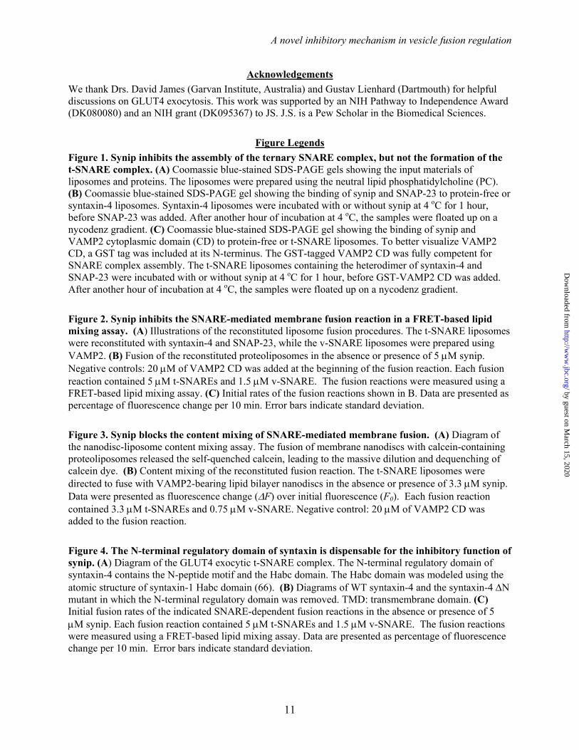
The formation of the binary t-SNARE complex is a key regulatory step in exocytic vesicle fusion (15,16). In solution, synip binds to syntaxin-4 monomer and appears to inhibit the SNARE assembly (32,33). However, it is unclear how synip regulates SNARE assembly and membrane fusion in the membrane environment. In a liposome co-flotation assay, we observed that synip bound to proteoliposomes reconstituted with syntaxin-4 monomer (Fig. 1A-B). Synip did not bind to protein-free liposomes (Fig. 1B), indicating that the synip-syntaxin-4 interaction was specific. When added as a soluble protein, SNAP-23 readily assembled with syntaxin-4 to
form the binary t-SNARE complex on the membrane (Fig. 1B). We found that synip binding did not prevent the pairing of syntaxin-4 with SNAP-23 to form the binary t-SNARE complex (Fig. 1B). These data demonstrate that the synip-associated syntaxin-4 monomer is fully competent for t-SNARE complex assembly (33).
After t-SNARE complex formation, the v-SNARE zippers with the t-SNAREs to form the ternary SNARE complex that pulls two membranes into close proximity to fuse (41). Next we examined whether synip regulates the assembly of the ternary SNARE complex. We prepared proteoliposomes using the pre-formed t-SNARE complex of syntaxin-4 and SNAP-23 (Fig. 1A). Addition of VAMP2 cytoplasmic domain (CD) to the t-SNARE liposomes resulted in the formation of the ternary SNARE complex on the membrane (Fig. 1C). Interestingly, synip bound to the t-SNAREs and strongly inhibited its pairing with VAMP2 CD (Fig. 1C). Together, these results indicate that synip binds to the t-SNAREs and inhibits the assembly of the ternary SNARE complex on the membrane bilayer.
Synip arrests the SNARE-mediated membrane fusion reaction
Next we examined how synip regulates the dynamic SNARE-mediated membrane fusion reaction. GLUT4 exocytic SNAREs were reconstituted into a defined fusion system, in which the v- and t-SNAREs were anchored in separate populations of lipid bilayers (Figs. 2A and S1). In this defined fusion system, SNAREs and regulators can be added or altered individually in the absence of other potentially confounding factors.
The fusion of v- and t-SNARE liposomes was first monitored using a fluorescence resonance energy transfer (FRET)-based lipid mixing assay (42). GLUT4 exocytic SNAREs alone drove an efficient level of lipid mixing (Fig. 2B-C). In the presence of synip, the SNARE-mediated membrane fusion was reduced to a background level similar to the negative control reaction in which the dominant negative inhibitor VAMP2 CD was added (Fig. 2B-C). We also examined the content mixing of the fusion reaction using a nanodisc-liposome fusion assay. VAMP2 was reconstituted into nanodiscs, small synthetic
by guest on March 15, 2020
http://ww
w.jbc.org/
Dow
nloaded from

A novel inhibitory mechanism in vesicle fusion regulation
5
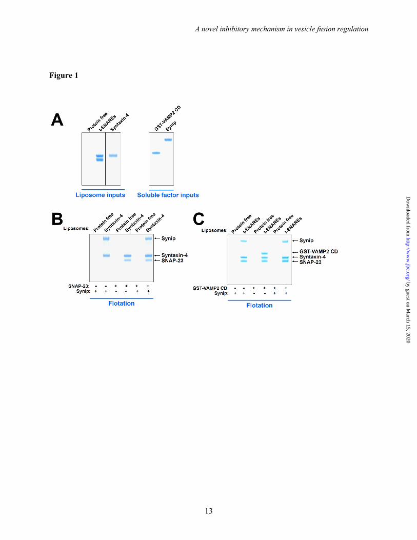
lipoprotein complexes that harbor a small piece of circular membrane bilayer wrapped by two molecules of membrane scaffold protein (MSP) (40). The soluble dye calcein (50 mM) was encapsulated in the t-SNARE liposomes. At that concentration, the fluorescent emission of calcein was inhibited by self-quenching. The fusion of VAMP2-bearing nanodiscs with calcein-containing t-SNARE liposomes led to the release and massive dequenching of the calcein dye (Fig. 3A). Using this liposome-nanodisc fusion assay, we observed that GLUT4 exocytic SNAREs drove an efficient level of content mixing (Fig. 3B). This SNARE-mediated content mixing was strongly inhibited by synip (Fig. 3B). Therefore, synip can arrest both the lipid and content mixing of SNARE-mediated membrane fusion. Since the lipid- and content-mixing experiments yielded similar results, lipid-mixing assays were used in the rest of this study.
Next we sought to further dissect how synip arrests SNARE-dependent membrane fusion. We used a liposome docking assay to examine how synip regulates the docking of v- and t-SNARE liposomes. The t-SNARE liposomes were immobilized on avidin agarose beads, and were used to pull down rhodamine-labeled v-SNARE liposomes (Fig. S2A). We found that GLUT4 exocytic v- and t-SNAREs promoted the docking of the liposomes (Fig. S2B). The SNARE-dependent liposome docking was strongly inhibited when synip was added (Fig. S2B). The ability of synip to block liposome docking suggests that the v- and t-SNAREs remained unpaired in the presence of synip, in agreement with liposome co-flotation findings (Fig. 1). Thus, synip arrests membrane fusion by blocking the initiation of ternary SNARE complex assembly. Together, these results demonstrate that synip functions as a t-SNARE-binding inhibitor that arrests SNARE-mediated membrane fusion at an intermediate state.
The N-terminal regulatory domain of syntaxin-4 is dispensable for the inhibitory function of synip
The zippering of the ternary trans-SNARE complex is mediated by the SNARE motifs (core domains) of the v- and t-SNAREs (18,43). Besides the universal SNARE motif, the syntaxin subunit
also possesses an N-terminal regulatory domain comprised of an N-peptide motif and a Habc domain (Fig. 4A-B). The N-terminal regulatory domain of syntaxin plays critical roles in multiple SNARE-regulator interactions (44,45). To determine its role in the inhibitory function of synip, the N-terminal regulatory domain was removed from syntaxin-4 (Fig. 4B). When reconstituted into proteoliposomes, the syntaxin-4 N mutant behaved similarly to WT syntaxin-4 in driving membrane fusion (Fig. 4C). The syntaxin-4 N-containing fusion reaction was still strongly inhibited by synip, with the inhibitory efficiencies comparable to those in WT fusion reactions (Fig. 4C). Thus, the N-terminal regulatory domain of syntaxin is dispensable for the inhibitory function of synip.
Synip selectively inhibits its cognate SNAREs
Next we examined the intrinsic specificity of synip in regulating the SNARE-mediated fusion. The GLUT4 exocytic SNAREs in the reconstituted fusion system were substituted with SNARE isoforms involved in other fusion pathways including mammalian lysosomal fusion (syntaxin-7, syntaxin-8, Vti1b, and VAMP8), and yeast exocytosis (Sso1p, Sec9p, and Snc2p) (Fig. 5A). While the v- and t-SNAREs of these fusion pathways can cross-pair to drive membrane fusion, they exhibit little sequence similarities (34,41,46,47). Strikingly, synip failed to inhibit the fusion reactions driven by lysosomal SNAREs (labeled “lysosomal fusion t + v”), or yeast exocytic SNAREs (labeled “yeast exocytosis t + v”) (Fig. 5A-B). Thus, the inhibitory function of synip is specific to GLUT4 exocytic SNAREs.
Next we fused the liposomes containing VAMP2, the GLUT4 exocytic v-SNARE, with the liposomes bearing the lysosomal t-SNAREs (syntaxin-7, syntaxin-8, and Vti1b), or yeast exocytic t-SNAREs (Sso1p and Sec9p). Again, neither of these fusion reactions (labeled “lysosomal fusion t” or “yeast exocytosis t”) was blocked by synip (Fig. 5A-B). We also fused GLUT4 t-SNARE liposomes with v-SNARE liposomes reconstituted with either the lysosomal v-SNARE VAMP8 or the yeast exocytic v-SNARE Snc2p (Fig. 5A). Interestingly, these fusion reactions (labeled “lysosomal fusion v” and “yeast exocytosis v”) were strongly inhibited by
by guest on March 15, 2020
http://ww
w.jbc.org/
Dow
nloaded from

A novel inhibitory mechanism in vesicle fusion regulation
6
synip (Fig. 5B). Therefore, the specificity of synip is determined by the t-SNAREs, but not the v-SNARE, in agreement with the interaction of synip with the t-SNARE complex in the liposome co-flotation assays (Fig. 1C). In the cell, the interactions between SNAREs and regulatory factors are exclusively specific to ensure the accuracy of vesicle transport (41). The stringent specificity of synip observed in this study supports that our reconstituted system has recapitulated the biological function of synip in exocytosis.
The inhibitory function of synip is dominant over the stimulatory activity of Munc18c
In addition to SNAREs, intracellular membrane fusion also requires the conserved Sec1/Munc18 (SM) family proteins, which promote membrane fusion through binding to their cognate SNAREs (34,48-50). SM proteins exhibit similar loss-of-function phenotypes as SNAREs (i.e., abrogation of fusion), and are involved in every intracellular vesicle fusion pathway (51-53). In GLUT4 exocytosis, the cognate SM protein is Munc18c (also known as Munc18-3) (54-56). We expressed and purified recombinant Munc18c protein from Sf9 insect cells using baculovirus. When added to the reconstituted SNARE-mediated fusion reaction (Fig. 6A), Munc18c strongly accelerated the fusion kinetics (Fig. 6B). The stimulation of fusion by Munc18c was abrogated when synip was added to the SNAREs (Fig. 6B), indicating that synip can arrest the fusion reaction in the presence of Munc18c. Therefore, the inhibitory function of synip is dominant over the stimulatory activity of Munc18c in vesicle fusion.
DISCUSSION
In regulated exocytosis, the SNARE-dependent membrane fusion reaction is controlled by a group of SNARE-binding regulatory factors. While the regulatory mechanisms of synaptic neurotransmitter release have been extensively studied, our knowledge about other exocytic pathways such as GLUT4 exocytosis remains primitive. Although conceptually similar to synaptic neurotransmitter release, the GLUT4 pathway is distinct in fundamental ways: 1) the kinetics of the fusion reaction is markedly slower (minutes vs. sub-millisecond); 2) specialized
fusion regulators are involved; and 3) the fusion reaction of GLUT4 exocytosis is coupled to insulin signaling (13,57). Thus, the regulatory mechanisms of GLUT4 vesicle fusion cannot be directly derived from the available knowledge of synaptic release.
While the physiological and medical importance of the GLUT4 exocytic pathway is well established, the underlying molecular mechanisms remain largely unknown. It is challenging to delineate complex membrane trafficking systems that involve the dynamic assembly of multiple layers of functional units at membrane-cytosol interfaces. We sought to address the question by reconstituting GLUT4 vesicle fusion in a defined system using purified components. Regulatory factors can be individually added or perturbed without the complications of other molecules naturally present in the cell, allowing their kinetic effects on fusion to be causally established.
Using the defined system, we demonstrated that the fusion regulator synip arrests the SNARE-dependent fusion reaction at an intermediate stage. Synip arrests membrane fusion by binding to the t-SNAREs and preventing the initiation of ternary SNARE complex assembly. We showed that synip inhibits the docking, lipid mixing, and content mixing of the SNARE-dependent fusion reaction. Notably, although originally isolated as a syntaxin-binding protein (33), synip does not affect the formation of syntaxin-4 with SNAP-23 into the t-SNARE complex on the membrane bilayer.
SM proteins are universal fusion regulators that promote intracellular vesicle fusion by binding to their cognate SNAREs (34,48-50). To arrest exocytosis, a fusion inhibitor is expected to block SNARE-mediated fusion in the presence of the SM protein. Indeed, we observed that the inhibitory function of synip is dominant over the stimulatory activity of Munc18c, the cognate SM protein of GLUT4 exocytosis.
In the highly synchronized synaptic release, fusion-competent vesicles are immobilized on the plasma membrane through the docking and priming processes (57). GLUT4-containing vesicles, however, are mobile and cycle continuously beneath the cell surface. Upon insulin stimulation, GLUT4 vesicles begin to dock
by guest on March 15, 2020
http://ww
w.jbc.org/
Dow
nloaded from

A novel inhibitory mechanism in vesicle fusion regulation
7
and fuse with the plasma membrane (58-60). These observations are consistent with our biochemical findings that synip arrests the fusion at the t-SNARE stage. With the t-SNARE complex blocked by synip, the vesicle-rooted v-SNARE cannot pair with the t-SNAREs to form the trans-SNARE complex, allowing the vesicles to be fully mobile.
It remains to be determined whether synip serves as a reversible fusion inhibitor. It has been proposed that synip is dissociated from SNAREs when its serine 99 residue is phosphorylated by Akt2, but the physiological role of this phosphorylation is being debated (32,61). Nevertheless, it is conceivable that an insulin-induced phosphorylation may destabilize the t-SNARE-synip interaction to permit the entry of the v-SNARE. Alternatively, synip may form an irreversible inhibitor on GLUT4 exocytic t-SNAREs to help demarcate vesicle fusion sites on the plasma membrane, thereby allowing for spatial regulation of GLUT4 vesicle fusion.
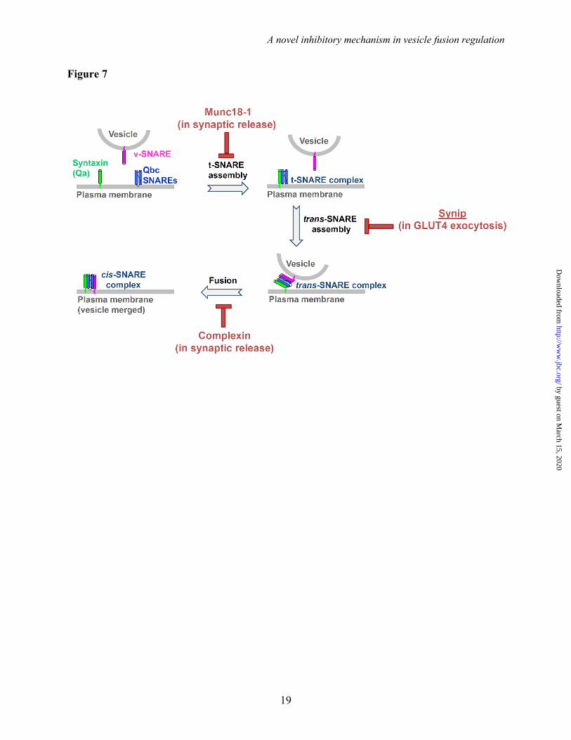
Importantly, the inhibitory function of synip is distinct from how other fusion inhibitors arrest membrane fusion. For example, the synaptic factor Munc18-1 binds to syntaxin monomer and locks the latter in a “closed” configuration incompatible with t-SNARE complex assembly (44,45). The small soluble protein complexin, on the other hand, negatively regulates synaptic vesicle fusion by arresting SNAREs at a partially-zippered trans-SNARE
configuration (62,63). In the presence of complexin, the v- and t-SNAREs can initiate pairing but the trans-SNARE complex cannot complete zippering (Fig. 7) (62,64). Therefore, SNARE-dependent membrane fusion can be arrested at each stage of the fusion pathway (Fig. 7). This versatility of fusion regulation likely allows the SNARE-mediated fusion reaction to adjust according to specific demands of a physiological response.
In summary, our studies revealed the molecular mechanism by which synip regulates the SNARE-dependent GLUT4 vesicle fusion. The inhibitory function of synip likely represents a novel regulatory mechanism of vesicle fusion. Genetic studies of GLUT4 fusion regulators such as synip often reached inconsistent and sometimes even contradictory conclusions (32,33,61,65), reminiscent of the studies of synaptic fusion regulation (15,16). In regulated exocytic pathways, multiple fusion regulators usually operate at similar or overlapping steps of the fusion reaction such that deletion of one factor might lead to unpredictable outcomes. In addition, loss of one fusion regulator might be compensated by another protein present in the cell, further complicating the analysis (57). Now, with the intrinsic regulatory mechanism of synip established, more precisely targeted in vivo experiments can be designed to delineate how it acts in concert with SNAREs and other fusion regulators to mediate GLUT4 exocytosis.
by guest on March 15, 2020
http://ww
w.jbc.org/
Dow
nloaded from
A novel inhibitory mechanism in vesicle fusion regulation
8
References
1. Sudhof, T. C., and Rothman, J. E. (2009) Science 323, 474-477 2. Schekman, R., and Novick, P. (2004) Cell 116, S13-15, 11 p following S19 3. Birnbaum, M. J. (1989) Cell 57, 305-315 4. James, D. E., Strube, M., and Mueckler, M. (1989) Nature 338, 83-87 5. Charron, M. J., Brosius, F. C., 3rd, Alper, S. L., and Lodish, H. F. (1989) Proc Natl Acad Sci U S
A 86, 2535-2539 6. Huang, S., and Czech, M. P. (2007) Cell Metab 5, 237-252 7. Jewell, J. L., Oh, E., and Thurmond, D. C. (2010) Am J Physiol Regul Integr Comp Physiol 298,
R517-531 8. Bryant, N. J., Govers, R., and James, D. E. (2002) Nat Rev Mol Cell Biol 3, 267-277 9. Watson, R. T., and Pessin, J. E. (2006) Trends Biochem Sci 31, 215-222 10. Vassilopoulos, S., Esk, C., Hoshino, S., Funke, B. H., Chen, C. Y., Plocik, A. M., Wright, W. E.,
Kucherlapati, R., and Brodsky, F. M. (2009) Science 324, 1192-1196 11. Blot, V., and McGraw, T. E. (2008) Mol Biol Cell 19, 3477-3487 12. Lavan, B. E., and Lienhard, G. E. (1994) Biochem Soc Trans 22, 676-680 13. Rowland, A. F., Fazakerley, D. J., and James, D. E. (2011) Traffic 12, 672-681 14. Wickner, W., and Schekman, R. (2008) Nat Struct Mol Biol 15, 658-664 15. Rizo, J., and Sudhof, T. C. (2012) Annu Rev Cell Dev Biol 28, 279-308 16. Jahn, R., and Fasshauer, D. (2012) Nature 490, 201-207 17. Weber, T., Zemelman, B. V., McNew, J. A., Westermann, B., Gmachl, M., Parlati, F., Sollner, T.
H., and Rothman, J. E. (1998) Cell 92, 759-772 18. Sutton, R. B., Fasshauer, D., Jahn, R., and Brunger, A. T. (1998) Nature 395, 347-353 19. Schwartz, M. L., and Merz, A. J. (2009) J Cell Biol 185, 535-549 20. Burgoyne, R. D., and Morgan, A. (2007) Curr Biol 17, R255-258 21. Kramer, L., and Ungermann, C. (2011) Molecular biology of the cell 22, 2601-2611 22. Martens, S., and McMahon, H. T. (2008) Nat Rev Mol Cell Biol 9, 543-556 23. Ohya, T., Miaczynska, M., Coskun, U., Lommer, B., Runge, A., Drechsel, D., Kalaidzidis, Y.,
and Zerial, M. (2009) Nature 459, 1091-1097 24. Melia, T. J., Weber, T., McNew, J. A., Fisher, L. E., Johnston, R. J., Parlati, F., Mahal, L. K.,
Sollner, T. H., and Rothman, J. E. (2002) J Cell Biol 158, 929-940 25. Pobbati, A. V., Stein, A., and Fasshauer, D. (2006) Science 313, 673-676 26. Sollner, T., Whiteheart, S. W., Brunner, M., Erdjument-Bromage, H., Geromanos, S., Tempst, P.,
and Rothman, J. E. (1993) Nature 362, 318-324 27. Gao, Y., Zorman, S., Gundersen, G., Xi, Z., Ma, L., Sirinakis, G., Rothman, J. E., and Zhang, Y.
(2012) Science 337, 1340-1343 28. Brandie, F. M., Aran, V., Verma, A., McNew, J. A., Bryant, N. J., and Gould, G. W. (2008) PLoS
One 3, e4074 29. D'Andrea-Merrins, M., Chang, L., Lam, A. D., Ernst, S. A., and Stuenkel, E. L. (2007) J Biol
Chem 282, 16553-16566 30. Vicogne, J., Vollenweider, D., Smith, J. R., Huang, P., Frohman, M. A., and Pessin, J. E. (2006)
Proc Natl Acad Sci U S A 103, 14761-14766 31. Latham, C. F., Lopez, J. A., Hu, S. H., Gee, C. L., Westbury, E., Blair, D. H., Armishaw, C. J.,
Alewood, P. F., Bryant, N. J., James, D. E., and Martin, J. L. (2006) Traffic 7, 1408-1419 32. Yamada, E., Okada, S., Saito, T., Ohshima, K., Sato, M., Tsuchiya, T., Uehara, Y., Shimizu, H.,
and Mori, M. (2005) J Cell Biol 168, 921-928 33. Min, J., Okada, S., Kanzaki, M., Elmendorf, J. S., Coker, K. J., Ceresa, B. P., Syu, L. J., Noda,
Y., Saltiel, A. R., and Pessin, J. E. (1999) Mol Cell 3, 751-760 34. Shen, J., Tareste, D. C., Paumet, F., Rothman, J. E., and Melia, T. J. (2007) Cell 128, 183-195
by guest on March 15, 2020
http://ww
w.jbc.org/
Dow
nloaded from
A novel inhibitory mechanism in vesicle fusion regulation
9
35. Scott, B. L., Van Komen, J. S., Liu, S., Weber, T., Melia, T. J., and McNew, J. A. (2003) Methods Enzymol 372, 274-300
36. Shen, J., Rathore, S., Khandan, L., and Rothman, J. E. (2010) J Cell Biology 190, 55-63 37. Rathore, S. S., Ghosh, N., Ouyang, Y., and Shen, J. (2011) Molecular biology of the cell 22,
2612-2619 38. Yu, H., Rathore, S. S., Davis, E. M., Ouyang, Y., and Shen, J. (2013) Mol Biol Cell 39. Shi, L., Shen, Q. T., Kiel, A., Wang, J., Wang, H. W., Melia, T. J., Rothman, J. E., and Pincet, F.
(2012) Science 335, 1355-1359 40. Ritchie, T. K., Grinkova, Y. V., Bayburt, T. H., Denisov, I. G., Zolnerciks, J. K., Atkins, W. M.,
and Sligar, S. G. (2009) Methods Enzymol 464, 211-231 41. Jahn, R., and Scheller, R. H. (2006) Nat Rev Mol Cell Biol 7, 631-643 42. Rathore, S. S., Bend, E. G., Yu, H., Hammarlund, M., Jorgensen, E. M., and Shen, J. (2010) Proc
Natl Acad Sci U S A 107, 22399-22406 43. Stein, A., Weber, G., Wahl, M. C., and Jahn, R. (2009) Nature 460, 525-528 44. Burkhardt, P., Hattendorf, D. A., Weis, W. I., and Fasshauer, D. (2008) EMBO J 27, 923-933 45. Misura, K. M., Scheller, R. H., and Weis, W. I. (2000) Nature 404, 355-362 46. Brandhorst, D., Zwilling, D., Rizzoli, S. O., Lippert, U., Lang, T., and Jahn, R. (2006) Proc Natl
Acad Sci U S A 47. Wang, C. C., Ng, C. P., Lu, L., Atlashkin, V., Zhang, W., Seet, L. F., and Hong, W. (2004) Dev
Cell 7, 359-371 48. Novick, P., and Schekman, R. (1979) Proc Natl Acad Sci U S A 76, 1858-1862 49. Hata, Y., Slaughter, C. A., and Sudhof, T. C. (1993) Nature 366, 347-351 50. Dulubova, I., Khvotchev, M., Liu, S., Huryeva, I., Sudhof, T. C., and Rizo, J. (2007) Proc Natl
Acad Sci U S A 104, 2697-2702 51. Carr, C. M., and Rizo, J. (2010) Curr Opin Cell Biol 22, 488-495 52. Burgoyne, R. D., Barclay, J. W., Ciufo, L. F., Graham, M. E., Handley, M. T., and Morgan, A.
(2009) Ann N Y Acad Sci 1152, 76-86 53. Toonen, R. F., and Verhage, M. (2007) Trends Neurosci 30, 564-572 54. Hu, S. H., Latham, C. F., Gee, C. L., James, D. E., and Martin, J. L. (2007) Proc Natl Acad Sci U
S A 104, 8773-8778 55. Tellam, J. T., McIntosh, S., and James, D. E. (1995) J Biol Chem 270, 5857-5863 56. Jewell, J. L., Oh, E., Ramalingam, L., Kalwat, M. A., Tagliabracci, V. S., Tackett, L., Elmendorf,
J. S., and Thurmond, D. C. (2011) J Cell Biol 193, 185-199 57. Sudhof, T. C. (2004) Annu Rev Neurosci 27, 509-547 58. Lizunov, V., Stenkula, K., Lisinski, I., Gavrilova, O., Yver, D. R., Chadt, A., Al-Hasani, H.,
Zimmerberg, J., and Cushman, S. W. (2012) American journal of physiology. Endocrinology and metabolism
59. Bai, L., Wang, Y., Fan, J., Chen, Y., Ji, W., Qu, A., Xu, P., James, D. E., and Xu, T. (2007) Cell Metab 5, 47-57
60. Xu, Y., Rubin, B. R., Orme, C. M., Karpikov, A., Yu, C., Bogan, J. S., and Toomre, D. K. (2011) J Cell Biol
61. Sano, H., Kane, S., Sano, E., and Lienhard, G. E. (2005) Biochem Biophys Res Commun 332, 880-884
62. Giraudo, C. G., Eng, W. S., Melia, T. J., and Rothman, J. E. (2006) Science 313, 676-680 63. Tang, J., Maximov, A., Shin, O. H., Dai, H., Rizo, J., and Sudhof, T. C. (2006) Cell 126, 1175-
1187 64. Kummel, D., Krishnakumar, S. S., Radoff, D. T., Li, F., Giraudo, C. G., Pincet, F., Rothman, J.
E., and Reinisch, K. M. (2011) Nature structural & molecular biology 18, 927-933 65. Saito, T., Okada, S., Yamada, E., Ohshima, K., Shimizu, H., Shimomura, K., Sato, M., Pessin, J.
E., and Mori, M. (2003) J Biol Chem 278, 36718-36725
by guest on March 15, 2020
http://ww
w.jbc.org/
Dow
nloaded from
A novel inhibitory mechanism in vesicle fusion regulation
10
66. Fernandez, I., Ubach, J., Dulubova, I., Zhang, X., Sudhof, T. C., and Rizo, J. (1998) Cell 94, 841-849
by guest on March 15, 2020
http://ww
w.jbc.org/
Dow
nloaded from
A novel inhibitory mechanism in vesicle fusion regulation
11
Acknowledgements
We thank Drs. David James (Garvan Institute, Australia) and Gustav Lienhard (Dartmouth) for helpful discussions on GLUT4 exocytosis. This work was supported by an NIH Pathway to Independence Award (DK080080) and an NIH grant (DK095367) to JS. J.S. is a Pew Scholar in the Biomedical Sciences.
Figure Legends
Figure 1. Synip inhibits the assembly of the ternary SNARE complex, but not the formation of the t-SNARE complex. (A) Coomassie blue-stained SDS-PAGE gels showing the input materials of liposomes and proteins. The liposomes were prepared using the neutral lipid phosphatidylcholine (PC). (B) Coomassie blue-stained SDS-PAGE gel showing the binding of synip and SNAP-23 to protein-free or syntaxin-4 liposomes. Syntaxin-4 liposomes were incubated with or without synip at 4 oC for 1 hour, before SNAP-23 was added. After another hour of incubation at 4 oC, the samples were floated up on a nycodenz gradient. (C) Coomassie blue-stained SDS-PAGE gel showing the binding of synip and VAMP2 cytoplasmic domain (CD) to protein-free or t-SNARE liposomes. To better visualize VAMP2 CD, a GST tag was included at its N-terminus. The GST-tagged VAMP2 CD was fully competent for SNARE complex assembly. The t-SNARE liposomes containing the heterodimer of syntaxin-4 and SNAP-23 were incubated with or without synip at 4 oC for 1 hour, before GST-VAMP2 CD was added. After another hour of incubation at 4 oC, the samples were floated up on a nycodenz gradient.
Figure 2. Synip inhibits the SNARE-mediated membrane fusion reaction in a FRET-based lipid mixing assay. (A) Illustrations of the reconstituted liposome fusion procedures. The t-SNARE liposomes were reconstituted with syntaxin-4 and SNAP-23, while the v-SNARE liposomes were prepared using VAMP2. (B) Fusion of the reconstituted proteoliposomes in the absence or presence of 5 M synip. Negative controls: 20 M of VAMP2 CD was added at the beginning of the fusion reaction. Each fusion reaction contained 5 M t-SNAREs and 1.5 M v-SNARE. The fusion reactions were measured using a FRET-based lipid mixing assay. (C) Initial rates of the fusion reactions shown in B. Data are presented as percentage of fluorescence change per 10 min. Error bars indicate standard deviation.
Figure 3. Synip blocks the content mixing of SNARE-mediated membrane fusion. (A) Diagram of the nanodisc-liposome content mixing assay. The fusion of membrane nanodiscs with calcein-containing proteoliposomes released the self-quenched calcein, leading to the massive dilution and dequenching of calcein dye. (B) Content mixing of the reconstituted fusion reaction. The t-SNARE liposomes were directed to fuse with VAMP2-bearing lipid bilayer nanodiscs in the absence or presence of 3.3 M synip. Data were presented as fluorescence change (F) over initial fluorescence (F0). Each fusion reaction contained 3.3 M t-SNAREs and 0.75 M v-SNARE. Negative control: 20 M of VAMP2 CD was added to the fusion reaction.
Figure 4. The N-terminal regulatory domain of syntaxin is dispensable for the inhibitory function of synip. (A) Diagram of the GLUT4 exocytic t-SNARE complex. The N-terminal regulatory domain of syntaxin-4 contains the N-peptide motif and the Habc domain. The Habc domain was modeled using the atomic structure of syntaxin-1 Habc domain (66). (B) Diagrams of WT syntaxin-4 and the syntaxin-4 N mutant in which the N-terminal regulatory domain was removed. TMD: transmembrane domain. (C) Initial fusion rates of the indicated SNARE-dependent fusion reactions in the absence or presence of 5 M synip. Each fusion reaction contained 5 M t-SNAREs and 1.5 M v-SNARE. The fusion reactions were measured using a FRET-based lipid mixing assay. Data are presented as percentage of fluorescence change per 10 min. Error bars indicate standard deviation.
by guest on March 15, 2020
http://ww
w.jbc.org/
Dow
nloaded from

A novel inhibitory mechanism in vesicle fusion regulation
12
Figure 5. The specificity of synip in regulating the SNARE-mediated fusion reaction. (A) Illustrations of the liposome fusion pairs. The proteoliposomes were reconstituted with SNAREs isoforms involved mammalian GLUT4 exocytosis, mammalian lysosomal fusion or yeast exocytosis. (B) Initial fusion rates of the indicated SNARE-dependent fusion reactions in the absence or presence of 5 M synip. Each fusion reaction contained 5 M t-SNAREs and 1.5 M v-SNARE. The fusion reactions were measured using a FRET-based lipid mixing assay. The fusion reaction labeled “No Regulator” represents the control SNARE-mediated fusion reaction in the absence of regulatory factors. Data are presented as percentage of fluorescence change per 10 min. Error bars indicate standard deviation.
Figure 6. The inhibitory function of synip is dominant over the stimulatory activity of Munc18c in fusion. (A) Diagram illustrating the experimental procedures for the reconstituted fusion reactions. (B) Initial fusion rates of the indicated SNARE-mediated fusion reactions showing the inhibitory activity of synip in the presence or absence of Munc18c. Each fusion reaction contained 5 M t-SNAREs and 1.5 M v-SNARE. The final concentration of each SNARE regulator was at 5 M. The fusion reactions were measured using a FRET-based lipid mixing assay. Data are presented as percentage of fluorescence change per 10 min. Error bars indicate standard deviation.
Figure 7. Model showing fusion inhibitors in SNARE-mediated membrane fusion. In synaptic neurotransmitter release, the fusion regulator Munc18-1 binds to the syntaxin monomer and blocks the formation of the t-SNARE complex on the plasma membrane. Complexin, on the other hand, recognizes the partially-zippered trans-SNARE complex and arrests fusion at a late step of the fusion pathway. Our research established that synip negatively regulates the vesicle fusion reaction of GLUT4 exocytosis by binding the t-SNARE complex and preventing the initiation of trans-SNARE assembly. The regulatory mechanism of synip is distinct from how Munc18-1 and complexin arrest vesicle fusion. It should be noted that, in addition to their inhibitory functions, Munc18-1 and complexin also positively regulate membrane fusion.
by guest on March 15, 2020
http://ww
w.jbc.org/
Dow
nloaded from
A novel inhibitory mechanism in vesicle fusion regulation
13
Figure 1
by guest on March 15, 2020
http://ww
w.jbc.org/
Dow
nloaded from

A novel inhibitory mechanism in vesicle fusion regulation
14
Figure 2
by guest on March 15, 2020
http://ww
w.jbc.org/
Dow
nloaded from
A novel inhibitory mechanism in vesicle fusion regulation
15
Figure 3
by guest on March 15, 2020
http://ww
w.jbc.org/
Dow
nloaded from

A novel inhibitory mechanism in vesicle fusion regulation
16
Figure 4
by guest on March 15, 2020
http://ww
w.jbc.org/
Dow
nloaded from

A novel inhibitory mechanism in vesicle fusion regulation
17
Figure 5
by guest on March 15, 2020
http://ww
w.jbc.org/
Dow
nloaded from

A novel inhibitory mechanism in vesicle fusion regulation
18
Figure 6
by guest on March 15, 2020
http://ww
w.jbc.org/
Dow
nloaded from
A novel inhibitory mechanism in vesicle fusion regulation
19
Figure 7
by guest on March 15, 2020
http://ww
w.jbc.org/
Dow
nloaded from

Haijia Yu, Shailendra S. Rathore and Jingshi Sheninhibitor
Synip arrests SNARE-dependent membrane fusion as a selective t-SNARE-binding
published online May 12, 2013J. Biol. Chem.
10.1074/jbc.M113.465450Access the most updated version of this article at doi:
Alerts:
When a correction for this article is posted•
When this article is cited•
to choose from all of JBC's e-mail alertsClick here
Supplemental material:
http://www.jbc.org/content/suppl/2013/05/12/M113.465450.DC1
by guest on March 15, 2020
http://ww
w.jbc.org/
Dow
nloaded from