syncoilin modulates peripherin filament networks and is
TRANSCRIPT

Research Article 2543
IntroductionSyncoilin is an atypical type III intermediate filament (IF) proteinthat lacks the key features of IFs, including the L1 linker domainand a periodic distribution of charged residues across the roddomain (Kemp et al., 2008). Syncoilin has never been observedunder normal conditions to form filament structures either alone orwith other IF proteins, but thick syncoilin filament-like structuresare known to form in some myopathies where syncoilinupregulation occurs (Brown et al., 2005).
Syncoilin is highly expressed in skeletal and cardiac musclewhere it interacts with a-dystrobrevin, a component of thedystrophin-associated protein complex, and desmin, the major IFprotein expressed in mature myocytes (Newey et al., 2001; Poonet al., 2002). Knockout animal models for both a-dystrobrevin anddesmin have a mild muscular dystrophy (Grady et al., 1999; Li etal., 1996). Myofibre regeneration in a-dystrobrevin (Dtna)-nullmice results in the upregulation of syncoilin, but syncoilinexpression is reduced and completely lost from the neuromuscular-myotendinous junctions in desmin (Des)-null mice (McCullagh etal., 2007). Given the alterations to syncoilin expression in thesemodels of muscle myopathies, mice null for syncoilin (Sync–/–)show surprisingly few signs of disease. Sync–/– mice have anunremarkable muscle phenotype with the exception of a reductionin maximal-force capacity in EDL muscle (McCullagh et al., 2008;Zhang et al., 2008) and an increase in serum levels of creatinekinase after a period of enforced endurance running (McCullagh etal., 2008).
Two new isoforms for syncoilin (Sync2 and Sync3) have beenidentified in addition to the original and dominant isoform in
muscle (Sync1). These isoforms are spliced from a single gene anddiffer only in their C-terminal ends. Syncoilin isoforms aredifferentially upregulated during atrophy and regeneration,suggesting distinct but possibly significant functions for syncoilin(Kemp et al., 2008).
In addition to their expression in muscle, IFs of the type III, IVand V families are also expressed in neuronal tissues (Jing et al.,2007; Lariviere and Julien, 2004). Most notably, similarly tosyncoilin, peripherin is a type III IF protein with several knownisoforms. Per58 is generated by all nine exons of the human andmouse gene and forms filaments; the Per45 isoform does not formfilaments and is generated using a downstream in-frame initiationcodon. The absence of Per45 or disruption of Per45 to Per58 ratioshas been shown to disrupt the normal peripherin filamentousstructures (McLean et al., 2008). Two peripherin isoforms areassociated with amyotrophic lateral sclerosis (ALS): aggregate-inducing Per28 is upregulated in patients with ALS and is associatedwith round inclusions in disease pathology (Xiao et al., 2008);Per61 is neurotoxic and has been observed in ALS mouse modelsand human patients, but not in controls (Robertson et al., 2003).
In this study, we show that syncoilin is not limited to muscle andis expressed in the peripheral and central nervous systems.Antibodies generated specifically against Sync1 and Sync2demonstrate that Sync2 is dominant in the peripheral nervoussystem and spinal cord whereas Sync1 is dominant in the brain. Todetermine the function of syncoilin, we show that syncoilin co-localises and interacts with peripherin and also functions tomodulate peripherin filament network assembly in vitro. A neuronalassessment of Sync–/– mice revealed reactions that were similar to
Syncoilin modulates peripherin filament networks andis necessary for large-calibre motor neuronsW. Thomas Clarke1,*, Ben Edwards1,*, Karl J. A. McCullagh1,‡, Matthew W. Kemp1,§, Catherine Moorwood1,¶,Diane L. Sherman2, Matthew Burgess1,** and Kay E. Davies1,‡‡
1MRC Functional Genomics Unit, Department of Physiology, Anatomy and Genetics, University of Oxford, South Parks Road, Oxford, OX1 3PT, UK2Centre for Neuroscience Research, The University of Edinburgh, Summerhall, Edinburgh, EH9 1QH, UK*These authors contributed equally to this work‡Present address: Regenerative Medicine Institute, National Centre for Biomedical Engineering Science, National University of Ireland, Galway, Ireland§Present address: School of Women’s and Infants’ Health, Faculty of Medicine, Dentistry and Health Sciences, The University of Western Australia, Western Australia, WA 6009¶Present address: Department of Physiology and Pennsylvania Muscle Institute, University of Pennsylvania School of Medicine, Philadelphia, PA 19104, USA**Present address: Botnar Research Centre, Institute of Musculoskeletal Sciences, Nuffield Department of Orthopaedics, Rheumatology and Musculoskeletal Sciences,University of Oxford, Oxford OX3 7LD, UK‡‡Author for correspondence ([email protected])
Accepted 20 April 2010Journal of Cell Science 123, 2543-2552 © 2010. Published by The Company of Biologists Ltddoi:10.1242/jcs.059113
SummarySyncoilin is an atypical type III intermediate filament (IF) protein, which is expressed in muscle and is associated with the dystrophin-associated protein complex. Here, we show that syncoilin is expressed in both the central and peripheral nervous systems. IsoformSync1 is dominant in the brain, but isoform Sync2 is dominant in the spinal cord and sciatic nerve. Peripherin is a type III IF proteinthat has been shown to colocalise and interact with syncoilin. Our analyses suggest that syncoilin might function to modulate formationof peripherin filament networks through binding to peripherin isoforms. Peripherin is associated with the disease amyotrophic lateralsclerosis (ALS), thus establishing a link between syncoilin and ALS. A neuronal analysis of the syncoilin-null mouse (Sync–/–) revealeda reduced ability in accelerating treadmill and rotarod tests. This phenotype might be attributable to the impaired function of extensordigitorum longus muscle and type IIb fibres caused by a shift from large- to small-calibre motor axons in the ventral root.
Key words: Syncoilin, Peripherin, Intermediate filaments, Amyotrophic lateral sclerosis, Neurons, Ventral root, Axon calibre
Jour
nal o
f Cel
l Sci
ence

those of Sync+/+ mice in a sensory neuron test, but significantlyworse than wild-type mice in motor neuron tests. An analysis ofspinal cord and dorsal and ventral roots revealed a significantreduction in the number of large-calibre motor axons from the L4-5 region of ventral root motor neurons. The results of this studyidentify syncoilin as a neuronal IF protein, describe a function forsyncoilin, detail a significant neuronal phenotype in the Sync–/–
mouse and provide a link between syncoilin, peripherin and ALS.
ResultsExpression of syncoilin in neuronal tissueImmunoblots, Q-PCR and immunohistochemistry were performedto determine whether syncoilin is expressed in neurons. Syncoilinwas detectable by immunoblot in brain, spinal cord and sciaticnerve using a pan-syncoilin antibody against an N-terminal epitope(anti-syncoilin N-term) (Fig. 1A). Within neurons, spinal cord hadthe highest relative expression of Sync1 and Sync2, isoforms thatare indistinguishable by immunoblot. Detection of Sync3 wasvisible only in the spinal cord and brain, which are both componentsof the central nervous system. Antibodies were generated thatspecifically recognise either Sync1 or Sync2; an antibody againstSync3 was not generated. An immunoblot identically loaded to theone probed for pan-syncoilin was probed for Sync1 and thenstripped and re-probed for Sync2. Sync1 was predominantlyexpressed in the brain, with lower levels in the spinal cord. Sync2expression was seen in the spinal cord and sciatic nerve. Q-PCRanalysis of Sync1 and Sync2 mRNA transcript levels reflected therelative protein expression of the two syncoilin isoforms (Fig. 1B).mRNA levels were normalised against GAPDH, and Sync2 mRNAvalues were calculated relative to Sync1 mRNA. In the brain, therewas high expression of Sync1 protein and mRNA. Sync2 mRNAlevels were 56% lower than levels of Sync1 mRNA, and no Sync2protein was detected by immunoblot. Spinal cord is the only tissueto express both Sync1 and Sync2 protein. Higher Sync2 proteinexpression was confirmed, with sixfold more Sync2 mRNA thanSync1 mRNA. The sciatic nerve exclusively expresses Sync2protein and had marginally more Sync2 mRNA than Sync1 mRNA.Sync3 transcripts were detectable by Q-PCR at very low levelsthat were not of sufficient amplitude to allow for meaningfulcalculations.
Immunohistochemistry showed syncoilin expression in thesciatic nerve, dorsal root ganglion (DRG) and spinal cord (Fig.1C). The transverse image of sciatic nerve showed that syncoilinwas contained within the axon and not in the surroundingSchwann cells. In the longitudinal image of a sciatic nerve single-teased-fibre preparation, syncoilin localised along the length ofthe axon and had a punctate appearance around the node ofRanvier in what are probably microvilli. Syncoilin expressionwas seen in sensory axons and neuron cell bodies of the DRG aswell as motor axons and neuron cell bodies of the ventral hornregion of the spinal cord.
Co-localisation and interaction of syncoilin and peripherinImmunohistochemistry analysis was performed in the DRG,spinal cord and sciatic nerve to show that syncoilin co-localisedwith peripherin (Fig. 2). Extensive but variable overlap ofsyncoilin and peripherin protein expression exists in neuron cellbodies of the DRG, because expression of one protein in a DRGcell body does not necessarily coincide with strong expressionof the other protein. Within the spinal cord, syncoilin wasexpressed almost exclusively in neuronal cell bodies expressingperipherin, but peripherin was more widely distributed in areasof the spinal cord that did not appear to contain syncoilin. Thesciatic nerve, which contains a mixed population of sensory andmotor axons, revealed the greatest amount of syncoilin andperipherin co-localisation. This longitudinal view of sciatic nervedemonstrates that syncoilin co-localises with peripherin to thecentre of the axon.
Blot-overlay experiments allow for an assessment of syncoilinand peripherin isoform binding. Recombinant syncoilin isoformsSync1, Sync2 and Sync3, syncoilin domains N-term (residues 1-157; identical in all three isoforms) and rod (residues 158-452;identical in Sync1 and Sync2), and experimental controls maltose-binding protein tag (MBP) and Per58 were used to create twoidentically loaded protein blots (Fig. 3A). These blots were overlaidwith equal amounts of recombinant Per45 or recombinant Per58and then probed for peripherin under identical conditions. Per45had strong binding affinity for Sync1, Sync2 and the rod domainwith a lesser affinity for Sync3 and N-term syncoilin. Per58 hadless binding affinity than Per45 for Sync1, Sync2 and the rod
2544 Journal of Cell Science 123 (15)
Fig. 1. Syncoilin isoforms are differentially expressed in thenervous system. (A)Immunoblots of mouse brain, spinal cord(SC) and sciatic nerve (SN). The top blot probed using a pan-syncoilin antibody (anti-syncoilin N-term) cannot distinguishbetween Sync1 and Sync2. Below, an identically loaded blot wasprobed with an antibody specific to Sync1, then stripped andreprobed with an antibody specific to Sync2. An antibody blockperformed with the Sync1 antibody shows specificity to theisoform-specific peptide antigen and is representative of the blankblot also observed for Sync2. (B)Q-PCR of Sync1 and Sync2mRNA transcript levels in brain, spinal cord and sciatic nerve(n3). Sync2 mRNA is measured relative to Sync1 mRNA, which isset to 1. (C)Immunohistological analysis of syncoilin (green)expression in transverse (top) and longitudinal (bottom) sciaticnerve, dorsal root ganglion (DRG) and motor neurons of the ventralhorn spinal cord. Syncoilin is costained with myelin (red) in thetransverse sciatic nerve. The arrowhead in longitudinal sciatic nerveindicates the node of Ranvier. In DRG and spinal cord, arrowheadsindicate axons and arrows neuronal cell bodies. Scale bars: 10m.
Jour
nal o
f Cel
l Sci
ence

domain. Per58 also had only weak affinity for Sync3, and noapparent affinity for syncoilin N-term. Neither Per45 nor Per58interacted with the isolated MBP, confirming the fidelity of theinteraction.
Co-immunoprecipitation was used to further demonstratesyncoilin and peripherin binding. Protein lysate from mouse spinalcord was immunoprecipitated with peripherin and syncoilinantibodies (Fig. 3B). Desmin antibody was used as a negativecontrol because desmin is an IF that is not expressed in neurons.One immunoblot probed for syncoilin showed that syncoilin wasimmunoprecipitated by both peripherin and syncoilin antibodies.The no-antibody and desmin-negative controls were negative forperipherin, demonstrating specificity. The second immunoblotprobed for peripherin showed that both Per58 and Per45 co-immunoprecipitated with syncoilin. The no-antibody negativecontrol showed that only very low levels of Per45 and Per58 werenon-specifically pulled down by the beads; the desmin-negativecontrol lane showed secondary antibody recognition of the mouseheavy chain IgG.
The role of syncoilin in formation of peripherin filamentnetworksGiven that syncoilin and peripherin co-localised and interacted,experiments were performed to determine whether a functionalrelationship exists between the proteins. A subclone of the humanadrenal cortex carcinoma-derived cell line known as SW13vim(–) lacks all known classes of cytoplasmic IF proteins (Hedberg andChen, 1986). SW13vim(–) cells transfected with peripherin (bothPer45 and Per58) (Fig. 4A), Per45 (Fig. 4C) and Per58 (Fig. 4E)confirm previous reports of formation of peripherin filamentnetworks (McLean et al., 2008). The peripherin filament networkof each transfection was blindly graded as ‘full’, ‘partial’ or ‘none’using parameters of filament length, thickness and arborisation.Transfection of peripherin (Prph), the gene that encodes Per58 aswell as Per45 via an in-frame downstream initiation codon,typically resulted in a fully formed filament network. When Per45was transfected alone it formed no filament network, but insteadresulted in dense and diffuse cytoplasmic immunostaining. Per58transfected alone formed a filament network consisting of shorter
2545Syncoilin expression and function in neurons
Fig. 2. Syncoilin partially co-localises with peripherin.Syncoilin (green) is costained with peripherin (red) in DRG,motor neurons of the ventral horn of the spinal cord and sciaticnerve. Arrow indicates a spinal-cord cell body. Scale bars: 50m(DRG and spinal cord), 20m (sciatic nerve).
Fig. 3. Syncoilin and peripherin interact. (A)Identicallyloaded immunoblots of recombinant syncoilin isoforms,syncoilin domains and experimental controls were overlaidwith equal amounts of either recombinant Per45 orrecombinant Per58 and probed under identical conditionsfor peripherin. A Ponceau-S-stained blot shows equalprotein loading and maltose-binding protein and per58serve as negative and positive controls, respectively.(B)Co-immunoprecipitations from mouse spinal cord usingrabbit anti-peripherin, anti-syncoilin N-term and anti-desmin antibodies. Immunoblots were probed with anti-syncoilin N-term and mouse anti-peripherin. The no-antibody and desmin co-immunoprecipitations served asnegative controls.
Jour
nal o
f Cel
l Sci
ence

and less-organised filaments than the network in Prph-transfectedcells.
As Sync2 is the dominant syncoilin isoform in spinal cord andsciatic nerve, Sync2 was co-transfected with peripherin (Fig. 4B),Per45 (Fig. 4D) and Per58 (Fig. 4F). Co-transfections of Sync1with the different peripherin constructs had very similar results(not shown). Peripherin staining in the syncoilin co-transfectedcells is noticeably different from staining in cells singly transfectedwith peripherin. Approximately 78% of Prph-transfected cells werecategorised as having a full filament network. When Sync2 wasco-transfected with peripherin, only 34% of cells had a full filamentnetwork, whereas 59% formed a partial filament network and 8%of cells formed no filament network. The difference betweenperipherin filament networks with and without transfected Sync2is statistically significant (P<0.05) by ANOVA analysis followedby t-test Bonferroni post-hoc correction. To demonstrate specificityof syncoilin in the modulation of peripherin filament networks, theneuronal IF protein neurofilament-L (NF-L) was co-transfectedinto SW13 vim(–) cells. Full peripherin filament networks wereformed in the presence of other IF proteins. The interaction ofsyncoilin and peripherin was also studied in the neuronal N2a cellline (supplementary material Fig. S1). Endogenously expressed
peripherin formed an IF network whereas endogenous syncoilinexpression was diffuse. The overexpression of transfected syncoilinalmost completely disrupted the endogenous peripherin network.
Fig. 4D shows Per45 expression when co-transfected withsyncoilin to be punctate and diffuse throughout the cytoplasm,which was almost identical to the expression in cells singlytransfected with Per45 in Fig. 4C. The Per58 and syncoilin co-transfected cell in Fig. 4F resembles the partial filament networkof Per58 transfected alone in Fig. 4E. Cells co-transfected withSync2 and Per45 or Per58 were analysed for co-localisation (Fig.4H, images in supplementary material Fig. S2). Sync2 co-localisedwith both isoforms, because 53.9% of Sync2 staining overlappedwith Per45 and 21.3% of Sync2 staining overlapped with Per58.The greater co-localisation of Sync2 with Per45 was statisticallysignificant (P<0.001).
The solubility of peripherin in transfected SW13vim(–) cells wasanalysed by immunoblot to further understand how the addition ofsyncoilin affects peripherin filament network formation. Inperipherin-transfected cells without syncoilin (Fig. 5A), both Per45and Per58 were found in the insoluble fraction whereas Per58 wasalso found in the soluble fraction. These results are similar topreviously reported data (McLean et al., 2008). The addition of
2546 Journal of Cell Science 123 (15)
Fig. 4. Syncoilin modulates peripherin filament networkformation. (A-G)SW13vim(–) cells were singly transfectedwith peripherin (A), Per45 (C) and Per58 (E). SW13vim(–)cells were co-transfected with Sync2 and peripherin (B),Sync2 and Per45 (D), Sync2 and Per58 (F) and peripherinand NF-L (G). Cells were stained for peripherin. Scale bars:10m. To the right of the representative transfection imagesis a graph showing the percentage of cells in the threeperipherin filament network classifications. 60 cells werecounted in each of three separate transfection experiments.The preferential co-localisation of syncoilin with Per45 overPer58 (H) was statistically significant (**P<0.001) whenanalysed using Student’s t-test.
Jour
nal o
f Cel
l Sci
ence

Sync2 resulted in the solubilisation of some Per45 (Fig. 5B). In cellstransfected with only Per58 or Per45, the addition of Sync2 made nodifference. Per58 was always found in the insoluble fraction andPer45 was equally split between insoluble and soluble fractions.
Neuronal analysis of Sync–/– miceSince syncoilin expression affects formation of the peripherinfilament network and peripherin abnormalities are linked toneuronal disease, a number of experiments were performed toidentify a neurological phenotype in the Sync–/– mouse.
Immunoblots of Sync+/+ and Sync–/– spinal cord and sciatic nerveprobed for Per45, Per58 and NF-L demonstrated that the loss ofsyncoilin has an effect on the expression of Per58 protein but noeffect on the expression of Per45 or NF-L protein (Fig. 6A). Fourindependent repetitions analysed by ImageJ and normalised to -actin loading controls revealed no difference in Per45 in eithertissue or Per58 in sciatic nerve, but did show Per58 levels in spinalcord to be 40% lower in Sync–/– mice (P<0.05) (Fig. 6B). SincePer45 results from an in-frame downstream start site, individualmRNA isoform analysis could not be achieved. Prph (includesPer45 and Per58) mRNA levels in sciatic nerve were unchangedbut in spinal cord were 50% lower in Sync–/– mice (P<0.001) (Fig.6C).
A standard hotplate test was performed to assess whether Sync+/+
and Sync–/– mice reacted differently to a mild thermal stimulus.The mice were timed from the moment their paws touched theplate until they responded with a hindpaw lick, hindpaw flick orjump. The absence of syncoilin appeared to have no effect on amouse’s pain reflex in response to a thermal stimulus (Fig. 6D).Dorsal roots from the L4-5 region exclusively contain sensoryneurons. The loss of syncoilin had no effect on myelinated sensoryaxon body area (Fig. 6E). The histological appearance of the L4-5dorsal root showed no overt difference between mice (Fig. 6F).
Several standard motor tests were performed to determinewhether Sync–/– mice had a motor neuron phenotype. Anaccelerating rotarod tests both motor coordination and balance.Sync+/+ mice were able to stay on the rotarod for 266 secondswhereas Sync–/– mice averaged only 212 seconds (Fig. 7A). The20% decline in time spent on the rotarod was found to bestatistically significant (P<0.001). Given that the acceleratingrotarod tests both motor coordination and balance, two experiments
2547Syncoilin expression and function in neurons
Fig. 5. Syncoilin disrupts the insolubility of Per45 in peripherin-transfected cells. Immunoblots of SW13vim(–) cells transfected with (A)Peripherin, Per45 or Per58 and (B) Sync2 with peripherin, Per45 or Per58.Cell lysates were fractionated, and the insoluble (I) and soluble (S) fractionswere run separately. Ponceau loading controls are shown to verify equalloading. The blot in B was stripped and re-probed for syncoilin to verify thecells were co-transfected.
Fig. 6. Per58 is reduced in the Sync–/– spinal cord. (A)Immunoblot of Sync+/+ and Sync–/– mouse spinal cord (SC) and sciatic nerve (SN) probed for Per45,Per58, neurofilament-L (NF-L) and -actin loading control. (B)Graphical representation of Per58 protein expression in spinal cord and sciatic nerve of Sync+/+ andSync–/– mice (n4). Sync–/– protein levels are measured relative to Sync+/+ protein, which is set to 1. The reduction of Per58 protein in Sync–/– spinal cord was foundto be statistically significant (*P<0.05) when analysed using Student’s t-test. No difference was found in Per58 protein levels in sciatic nerve or in Per45 and NF-Lprotein levels. (C)Q-PCR of Prph mRNA levels in Sync+/+ and Sync–/– mice in spinal cord and sciatic nerve (n3). Sync–/– mRNA is measured relative to Sync+/+
mRNA, which is set to 1. The reduction of peripherin mRNA in Sync–/– spinal cord was found to be statistically significant (**P<0.001; Student’s t-test). (D)Therewas no difference between Sync+/+ and Sync–/– mice in reaction time to a mild thermal stimulus (n45). (E)There was no difference in body area of sensory neuronsfrom the dorsal root (n5). (F)Representative images of Toluidine-Blue-stained L4-5 dorsal roots.
Jour
nal o
f Cel
l Sci
ence
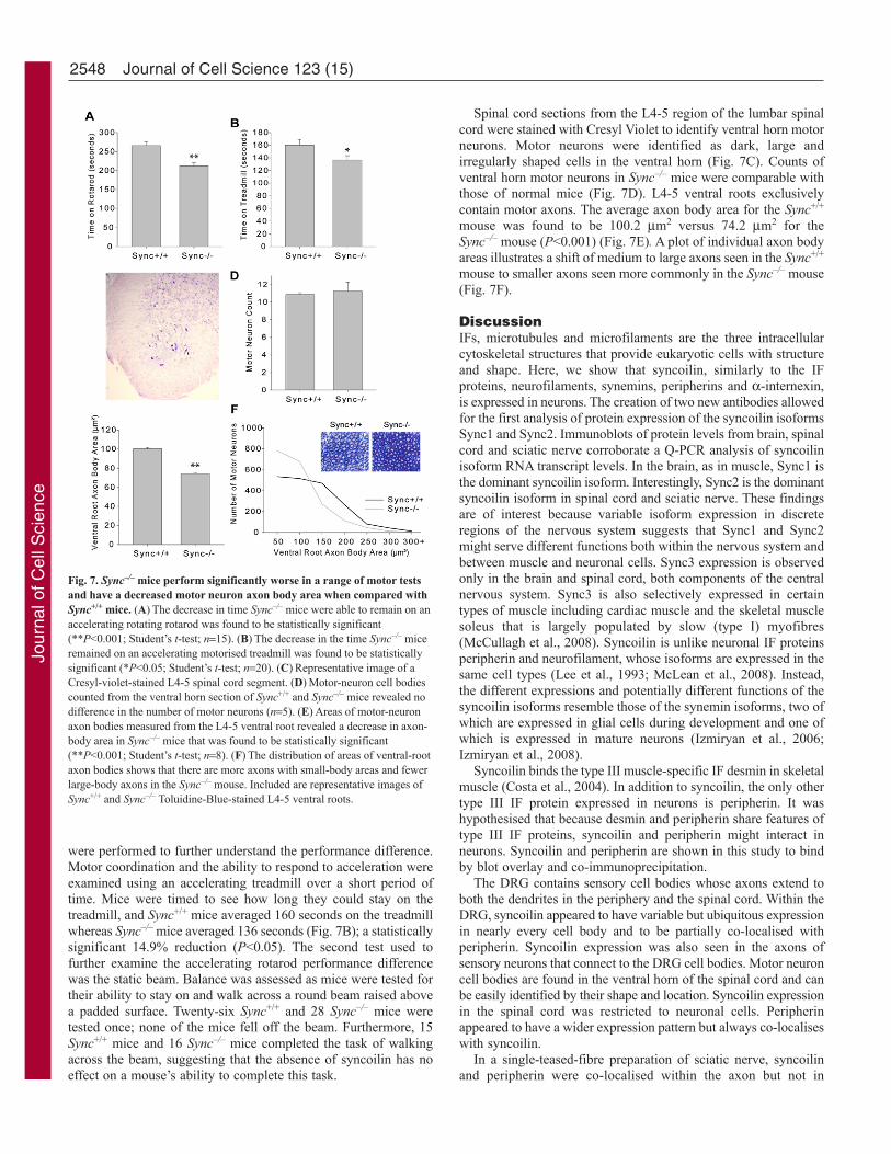
were performed to further understand the performance difference.Motor coordination and the ability to respond to acceleration wereexamined using an accelerating treadmill over a short period oftime. Mice were timed to see how long they could stay on thetreadmill, and Sync+/+ mice averaged 160 seconds on the treadmillwhereas Sync–/– mice averaged 136 seconds (Fig. 7B); a statisticallysignificant 14.9% reduction (P<0.05). The second test used tofurther examine the accelerating rotarod performance differencewas the static beam. Balance was assessed as mice were tested fortheir ability to stay on and walk across a round beam raised abovea padded surface. Twenty-six Sync+/+ and 28 Sync–/– mice weretested once; none of the mice fell off the beam. Furthermore, 15Sync+/+ mice and 16 Sync–/– mice completed the task of walkingacross the beam, suggesting that the absence of syncoilin has noeffect on a mouse’s ability to complete this task.
Spinal cord sections from the L4-5 region of the lumbar spinalcord were stained with Cresyl Violet to identify ventral horn motorneurons. Motor neurons were identified as dark, large andirregularly shaped cells in the ventral horn (Fig. 7C). Counts ofventral horn motor neurons in Sync–/– mice were comparable withthose of normal mice (Fig. 7D). L4-5 ventral roots exclusivelycontain motor axons. The average axon body area for the Sync+/+
mouse was found to be 100.2 m2 versus 74.2 m2 for theSync–/– mouse (P<0.001) (Fig. 7E). A plot of individual axon bodyareas illustrates a shift of medium to large axons seen in the Sync+/+
mouse to smaller axons seen more commonly in the Sync–/– mouse(Fig. 7F).
DiscussionIFs, microtubules and microfilaments are the three intracellularcytoskeletal structures that provide eukaryotic cells with structureand shape. Here, we show that syncoilin, similarly to the IFproteins, neurofilaments, synemins, peripherins and a-internexin,is expressed in neurons. The creation of two new antibodies allowedfor the first analysis of protein expression of the syncoilin isoformsSync1 and Sync2. Immunoblots of protein levels from brain, spinalcord and sciatic nerve corroborate a Q-PCR analysis of syncoilinisoform RNA transcript levels. In the brain, as in muscle, Sync1 isthe dominant syncoilin isoform. Interestingly, Sync2 is the dominantsyncoilin isoform in spinal cord and sciatic nerve. These findingsare of interest because variable isoform expression in discreteregions of the nervous system suggests that Sync1 and Sync2might serve different functions both within the nervous system andbetween muscle and neuronal cells. Sync3 expression is observedonly in the brain and spinal cord, both components of the centralnervous system. Sync3 is also selectively expressed in certaintypes of muscle including cardiac muscle and the skeletal musclesoleus that is largely populated by slow (type I) myofibres(McCullagh et al., 2008). Syncoilin is unlike neuronal IF proteinsperipherin and neurofilament, whose isoforms are expressed in thesame cell types (Lee et al., 1993; McLean et al., 2008). Instead,the different expressions and potentially different functions of thesyncoilin isoforms resemble those of the synemin isoforms, two ofwhich are expressed in glial cells during development and one ofwhich is expressed in mature neurons (Izmiryan et al., 2006;Izmiryan et al., 2008).
Syncoilin binds the type III muscle-specific IF desmin in skeletalmuscle (Costa et al., 2004). In addition to syncoilin, the only othertype III IF protein expressed in neurons is peripherin. It washypothesised that because desmin and peripherin share features oftype III IF proteins, syncoilin and peripherin might interact inneurons. Syncoilin and peripherin are shown in this study to bindby blot overlay and co-immunoprecipitation.
The DRG contains sensory cell bodies whose axons extend toboth the dendrites in the periphery and the spinal cord. Within theDRG, syncoilin appeared to have variable but ubiquitous expressionin nearly every cell body and to be partially co-localised withperipherin. Syncoilin expression was also seen in the axons ofsensory neurons that connect to the DRG cell bodies. Motor neuroncell bodies are found in the ventral horn of the spinal cord and canbe easily identified by their shape and location. Syncoilin expressionin the spinal cord was restricted to neuronal cells. Peripherinappeared to have a wider expression pattern but always co-localiseswith syncoilin.
In a single-teased-fibre preparation of sciatic nerve, syncoilinand peripherin were co-localised within the axon but not in
2548 Journal of Cell Science 123 (15)
Fig. 7. Sync–/– mice perform significantly worse in a range of motor testsand have a decreased motor neuron axon body area when compared withSync+/+ mice. (A)The decrease in time Sync–/– mice were able to remain on anaccelerating rotating rotarod was found to be statistically significant(**P<0.001; Student’s t-test; n15). (B)The decrease in the time Sync–/– miceremained on an accelerating motorised treadmill was found to be statisticallysignificant (*P<0.05; Student’s t-test; n20). (C)Representative image of aCresyl-violet-stained L4-5 spinal cord segment. (D)Motor-neuron cell bodiescounted from the ventral horn section of Sync+/+ and Sync–/– mice revealed nodifference in the number of motor neurons (n5). (E)Areas of motor-neuronaxon bodies measured from the L4-5 ventral root revealed a decrease in axon-body area in Sync–/– mice that was found to be statistically significant(**P<0.001; Student’s t-test; n8). (F)The distribution of areas of ventral-rootaxon bodies shows that there are more axons with small-body areas and fewerlarge-body axons in the Sync–/– mouse. Included are representative images ofSync+/+ and Sync–/– Toluidine-Blue-stained L4-5 ventral roots.
Jour
nal o
f Cel
l Sci
ence

surrounding Schwann cells. Syncoilin expression was punctate inthe vicinity of the nodes of Ranvier in what are likely to bemicrovilli, because the staining extends beyond axonal syncoilinlabelling. The nodes of Ranvier are gaps between myelin sheathsthat contain protein complexes including voltage-gated Na+
channels, Na+/K+ ATPases, and Na+/Ca2+ exchangers that assist inthe propagation of action potentials along myelinated nerve fibres(Susuki and Rasband, 2008). In muscle, syncoilin expression isenriched at the neuromuscular junction (Newey et al., 2001), andmany morphological and molecular similarities exist between nodesof Ranvier and neuromuscular junctions (Caldwell, 2000; Ellisman,1979). Several proteins including N-cadherin (Cifuentes-Diaz etal., 1994), N-CAM and cytotactin (Rieger et al., 1986) are expressedor enriched at both the neuromuscular junction and node of Ranvierand function in cell-cell adhesion, neurite outgrowth and synapticplasticity. It is unknown why the syncoilin expression patternchanges at the node of Ranvier, but it is possible that syncoilin isinvolved with one of these functions or interacts with any of theprotein complexes at the node.
Upon transfection in SW13vim(–) cells lacking an IF network,peripherin (Per45+Per58) forms a full IF network. Alone, Per58 iscapable of forming a partially disrupted network, but Per45 aloneforms no network. Syncoilin transfected into SW13vim(–) cellswas incapable of forming an IF network. The addition of Sync2 toperipherin resulted in a partially disrupted peripherin network thatresembled the Per58 partially disrupted filament network. Thissuggests that the addition of Sync2 to a peripherin-transfected cellhas the same effect as the removal of Per45 from a peripherin-transfected cell. In a solubility assay of transfected SW13vim(–)cell protein lysates, filament network-forming peripherin (Per45 +Per58) was found largely in the insoluble fraction although aportion of Per58 was found in the soluble fraction. This is puzzlingbecause Per58 alone is entirely insoluble. The presence of Per45 istherefore able to solubilise a portion of the Per58 isoform from theinsoluble filament structures, possibly reflecting the role of Per45in regulating peripherin filament networks. More importantly, theaddition of Sync2 to a peripherin-transfected cell resulted in therelocation of some Per45 from the insoluble to the soluble fraction.The Sync2-dependent redistribution of some Per45 from theinsoluble to the soluble fraction is a likely explanation for thepartial disruption of peripherin filament networks. It is possiblethat Sync2 disrupts the peripherin filament network by sequesteringPer45 away from peripherin. This hypothesis is further supportedby the fact that the addition of Sync2 to Per58 in transfected cellsappeared to have no affect on the solubility of Per58 and no furtherimpact on the already partially disrupted Per58 network.Furthermore, Sync2 exhibited preferential co-localisation withPer45 over Per58 in transfected cells. Since Per45 is necessary forfull formation of filament networks, it is possible that Sync2 actsto modulate peripherin filament network formation. Syncoilin couldactively sequester and release Per45, thereby controlling the growthand maintenance of the peripherin IF network in response to furthercellular stimuli.
Several neuronal IF proteins are associated with human disease.Peripherin and the neurofilaments are often found in inclusionbodies in motor neurons of patients with the disease ALS (Al-Chalabi et al., 1999; Figlewicz et al., 1994; Mitchell and Borasio,2007; Xiao et al., 2006; Kemp and Davies, 2007). It was firstdetermined whether the loss of syncoilin had an effect on any otherneuronal IF proteins. NF-L is essential for the assembly ofneurofilaments and serves as an indicator of normal neuronal
structures (Zhu et al., 1997). NF-L and peripherin protein levelswere analysed by immunoblot. NF-L was unchanged in spinal cordand sciatic nerve in the Sync–/– mouse, suggesting that syncoilin isnot essential for neurofilament expression and stability. Alsounchanged were Per45 protein levels in spinal cord and sciaticnerve as well as Per58 protein and Prph (Per45+Per58) mRNAlevels in sciatic nerve. However, Per58 protein and Prph mRNAlevels were significantly reduced in the spinal cord. If syncoilinacts to modulate peripherin filament network assembly, it is possiblethat peripherin filament networks, which consist primarily of Per58,are unstable and then degraded in the absence of syncoilin.However, it remains unknown why Per58 levels are affected onlyin the spinal cord or why Per45 is not reduced in the spinal cordwith Per58. This could be due to the differences between spinalcord and sciatic nerve syncoilin isoform expression, neuronstructure or neuron function.
Given the range of phenotypes from IF transgenic and knockoutmice and the minimal muscle phenotype in the Sync–/– mouse, theSync–/– mouse was examined in a wide range of sensory and motorneuron tests. The hotplate test is an established method for testingthe response to a thermal stimulus (Gu et al., 2002). The fact thatthere was no difference in hotplate-response time between Sync+/+
and Sync–/– mice suggests that syncoilin is not essential fornociception – the neural processing in the central and peripheralnervous system of painful stimuli. An analysis of sensory neuronsfrom the dorsal root also revealed no difference between Sync+/+
and Sync–/– axon-body area. This is interesting because the loss ofperipherin results in a substantial reduction in the number of L5unmyelinated sensory fibres (Lariviere et al., 2002). Unfortunately,owing to Home Office regulations, no further relevant sensoryneuron tests could be performed.
We also performed several motor-neuron-specific tests. Mostof these tests could also assess muscle function. However,voluntary and extended forced running tests and extensivehistological analyses revealed minimal muscular differencesbetween Sync+/+ and Sync–/– mice (McCullagh et al., 2008).Therefore, it is possible that any motor phenotype discovered inthe Sync–/– mouse is the result of a neuronal change. Anaccelerating rotarod requires motor coordination and balance(Dunham and Miya, 1957). This test demonstrated the mostsignificant difference between Sync+/+ and Sync–/– mice, confirmeda neuromuscular deficiency in the Sync–/– mouse and introducedpossible defects including balance and coordination. Theaccelerating treadmill test is designed to examine the ability of amouse in a rapidly accelerating environment akin to a sprint. Thistreadmill test differs from the rotarod because it does not requirethe same degree of balance or coordination needed to stay on theapparatus. A previous study of forced treadmill running at constantspeeds was designed to run the mice to exhaustion but revealedno difference between Sync+/+ and Sync–/– mice (McCullagh et al.,2008). Sync–/– mice performed significantly worse than Sync+/+
mice in the accelerating treadmill test. EDL is a fast glycolytictissue containing approximately 77% type IIb fibres that areprimarily used for activities such as sprinting (Nemeth and Pette,1980). An analysis of EDL muscle in the Sync–/– mouse found a27.9% reduction in force generation (McCullagh et al., 2008). Itis probable that the defect in EDL muscle contributes to thereduced ability of Sync–/– mice in these tests. To specificallyexamine balance, a factor in the accelerating-rotarod test, astandard static-beam test was used (Le Marec et al., 1997). Theability of Sync–/– mice to stay on the beam, turn and to walk to the
2549Syncoilin expression and function in neurons
Jour
nal o
f Cel
l Sci
ence

end of the beam at the same frequency of Sync+/+ mice suggeststhat syncoilin is not essential for proper balance or coordination.
Taken together, the range of behavioural tests suggests aneuromuscular deficiency in Sync–/– mice. Two histologicalexaminations of the quantity and quality of motor neurons wereperformed in an effort to find a cause for the neuromuscularphenotype. An examination of motor neurons in the ventral hornof the lumbar spinal cord in Sync+/+ and Sync–/– mice revealed thatthe loss of syncoilin does not affect the quantity of motor neurons.Motor neurons from the L4-5 region are known to innervatemuscles of the lower limb such as EDL (Tyc and Vrbova, 2007).Ventral roots from the L4-5 region were used to exclusivelyexamine the quality of motor neurons. No obvious histologicaldifference were seen, but motor neuron axon body area wasmeasured and found to be significantly smaller in the Sync–/–
mouse, which is possibly an indication of reduced neuronaltransmission. It is also possible that syncoilin functionsindependently or via some other unknown mechanism to regulatethe diameter of large-calibre axons (Hirokawa, 1982). AlthoughSync–/– mice have no overt phenotype throughout their lifetimes,moderate neuronal deficiencies caused by the absence of syncoilinmight contribute to a reduced neuromuscular ability. In theaccelerating-rotarod and accelerating-treadmill tests, Sync–/– werealways able to complete the tasks; instead, the difference betweenSync+/+ and Sync–/– mice was how well the task was completed.This distinction fits well with the observed histological phenotypeof motor neurons in Sync–/– mice. The absence of syncoilin doesnot result in the loss of motor neurons. Instead, it is hypothesisedthat the loss of syncoilin results in a shift of large-calibre to small-calibre axons with a reduced ability to transmit neurological signalsto muscles. We hypothesise that the reduction in large-calibremotor neurons in the L4-5 region inhibits full EDL function duringaccelerating tasks such as the rotarod and treadmill, and results inthe reduction in force generation previously described (McCullaghet al., 2008; Zhang et al., 2008). This suggests that EDL musclehas undergone an inherent change in its muscle contractilecharacteristics owing to a chronic lack of proper neuraltransmission. The specific impairment of EDL function can beexplained by the Henneman size principle, which states large-calibre motor neurons preferentially innervate type IIb fibres thatare abundant in EDL muscle (Henneman, 1957).
In conclusion, we found that the IF protein syncoilin is expressedin neurons and that it interacts with peripherin isoforms. The lossof syncoilin results in a significant neuronal phenotype, and weshow that syncoilin might mediate assembly of peripherin filamentnetworks by interacting with Per45. Previous studies haveimplicated syncoilin in muscle disease. The findings of this studyraise the potential for the involvement of syncoilin in diseasesassociated with neuronal IFs. In particular, the association ofperipherin with ALS and the ability of syncoilin to alter assemblyof peripherin filament networks introduces the possibility of theinvolvement of syncoilin in this disease.
Materials and MethodsAnimals and antibodiesSyncoilin null (Sync–/–) and wild type (Sync+/+) mice were generated and bred in-house as described previously (McCullagh et al., 2008). All experimental proceduresinvolving the use of animals were performed in accordance with guidelines approvedby the Home Office. All experiments were performed with 6- to 8-week-old mice.Antibodies used include rabbit anti-Sync-N-term (Newey et al., 2001), mouse anti-desmin (Abcam 8592), mouse anti-peripherin (Chemicon MAB5380), rabbit anti-peripherin (Chemicon MAB 1530), chicken anti-peripherin (Chemicon AB9282),rabbit anti-NF-L (Abcam 9035), mouse anti-myelin basic protein (Dytrych et al.,
1998), goat anti-a-actinin (Santa Cruz N-19) and Alexa Fluor 594- and AlexaFluor 488-conjugated IgG secondary antibodies (Invitrogen). Newly describedrabbit polyclonal antibodies anti-Sync1 and anti-Sync2 were generated byEurogentec from the Keyhole limpet haemocyanin-coupled peptide antigensH2N-CTSQAGGVEAQSPGTV-COOH and H2N-CSPETRKHLLKDH-COOH,respectively.
Histology and immunofluorescenceMice were perfused with 2% paraformaldehyde in a phosphate buffer (25 mMNaH2P04, 8 mM Na2HPO4). Tissues were dissected, post-fixed in 4%paraformaldehyde and cryoprotected in 30% sucrose solution. For motor neuroncounts of the spinal cord ventral horn, 10 m sections were stained with 0.5% CresylViolet (Sigma), washed in a H2O and alcohol series, cleaned with HistochoiceClearing Agent (Sigma) and mounted with Histomount (RA Lamb). Dorsal andventral roots were dissected from the L4-L5 region of the spinal cord in perfusedmice and stored in 70% ethanol until embedding, sectioning and staining were doneby Mohan Masih (DPAG, University of Oxford, Oxford, UK). Dorsal-ventral rootswere embedded in Araldite resin, sectioned at a thickness of 1 m and stained witha solution of 1% Toluidine Blue and 1% borax.
Tissue for immunofluorescence was perfused, fixed and cryoprotected as above.Tissue was embedded in OCT (BDH), sectioned on a cryostat to a thickness of 10m. Teased fibre preparations from fixed isolated sciatic nerve were perfused andcryoprotected as described above and teased using fine instruments in PBS on 3-aminopropyltriethoxysilane (TESPA)-coated slides. All immunofluorescence sampleswere blocked for 1 hour in 10% fetal calf serum in PBS. Primary antibodies asdescribed above were diluted in blocking solution and applied to fixed sections for1 hour. Sections were rinsed in PBS and incubated with secondary antibodiesdescribed above in blocking solution for 1 hour. Finally, sections were rinsedthoroughly and mounted in Vectashield aqueous medium with DAPI (NationalDiagnostic).
All microscope work was performed at room temperature. Co-localisation imageswere taken using a 510 Meta confocal laser-scanning microscope and analysedusing LSM Image 4.0 software. All parameters [(63�/1.4 oil DIC objective), zoom(2), averaging (4)] were kept equal and images were adjusted so no saturationoccurred and background was slightly above zero. Whole cells were selected asregions of interest (ROIs) and threshold was set from the ROIs. Other imaging wasperformed with an AxioPlan microscope, AxioCam HRc and Axiovision LE 4.6software. Plan-neofluar 10�/0.3, 20�/0.5, 40�/0.75 objective lenses were used inboth microscopes (all imaging products from Carl Zeiss). Axon calibre was measuredblindly using Axiovision by outlining individual axons. Classification of peripherinfilament networks was determined using three parameters: length, thickness offilaments and arborisation. Full networks contained long, continuous filamentbundles with extensive arborisation and minimal punctate spots. Partial networkscontained shorter filaments or thick bundles with little arborisation and significantpunctate staining. Cells classified as having no filament network contained noevidence of filament formation and only punctate expression. Counts were performedblindly on 60 cells of each transfection during three different experiments for a totalof 180 cells.
Protein and RNA preparationTissue from Sync+/+ and Sync–/– mice was homogenised in an appropriate volume ofcold Newcastle buffer (75 mM Tris-HCl, pH 6.8, 3.8% SDS, 4 M urea and 20%glycerol) with protease inhibitor cocktail (Sigma). Protein quantification wasperformed with the Bio-Rad DC Protein assay according to the manufacturer’sprotocol. For total RNA extraction, dissected tissue was snap frozen and milled inliquid nitrogen immediately before using Trizol (Invitrogen) according to themanufacturer’s instructions. Total RNA (300 ng) was reverse transcribed into cDNAusing random decameric primers and SuperScript II Reverse Transcriptase(Invitrogen). Generated cDNA was used as a template to perform PCR analysis. Thesolubility assay protocol was adapted from published techniques (McLean et al.,2008). Briefly, cells were harvested in low-salt lysate buffer [20 mM Tris-HCl, pH7.5, 150 mM NaCl, 1 mM EDTA, 1% (v/v) Triton X-100 and protease inhibitors (allfrom Sigma)], briefly homogenised and incubated on ice for 30 minutes. The lysateswere centrifuged at 16,000 g for 30 minutes at 4°C to separate soluble and insolublefractions. The insoluble fraction was solubilised in 2% (w/v) SDS in lysate bufferand made up to a volume equal to the soluble fraction. Samples were concentratedequally in a Centriplus filter device (Millipore) according to the manufacturer’sinstructions. Equal volumes of each fraction were run on a gel and immunoblottedas described below.
Immunoblotting80 g total protein from tissue lysates was separated on 8% SDS polyacrylamidegels (Bio-Rad) and electrophoretically transferred to nitrocellulose membranes. Afterblocking membranes for 1 hour in TBS-T [Tris-buffered saline containing 0.1%(v/v) Tween 20] and 5% nonfat dry milk, blots were incubated overnight withprimary antibody in blocking buffer and washed three times in TBS-T beforeincubation for 1 hour with horseradish-peroxidase-conjugated IgG secondary antibody(GE) in blocking buffer. Membranes were washed and developed using ECL detectionreagents (GE) according to the manufacturer’s instructions. Blots were stripped at
2550 Journal of Cell Science 123 (15)
Jour
nal o
f Cel
l Sci
ence

50°C for 20 minutes in stripping buffer (100 mM 2 mercaptoethanol, 2% SDS,62.5 mM Tris-HCl) before re-probing with different antibodies. Exposed film wasscanned with a HP Scanjet 5400c. Protein expression was measured from immunoblotintensity using ImageJ (Abramoff et al., 2004).
Blot-overlay analysisUsing standard cloning techniques and endonuclease (5�, EcoRI; 3�, HindIII)sequence-tagged primers, Sync1-Sync3, Per45 and Per58 were subcloned frompcDNA3 into pMAL (New England Biolabs) using standard methodologies. SyncoilinN-term (residues 1-157) and rod domains (residues 158-452) were subcloned intopMAL using the following primers: N-termF, TTT GAA TTC ATG GCC AGC CCGGAA CCC CT; N-termR, TTT AAG CTT AGA GGG GAT CCT CCT CGG TGT;RodF, TTT GAA TTC AGC GTG GAG GAC CTG GAG CG; RodR, TTT AAGCTT ATG CAT CAG CCT GTT CCA GAC. Expression and purification of maltose-binding protein (MPB)-tagged proteins were performed using the pMAL proteinfusion and purification system, in accordance with the manufacturer’s instructions(New England Biolabs). 10 g MBP-tagged recombinant proteins were separated ongels and transferred to nitrocellulose membranes as described above. Membraneswere blocked for 1 hour and incubated with 5 ml MBP-tagged Per45 or Per58 (3g/ml) in blocking buffer for 2 hours. Membranes were washed, probed with mouseanti-peripherin antibody and then treated as described above.
Co-immunoprecipitationSpinal-cord tissue was crushed in liquid nitrogen and then added toimmunoprecipitation lysis buffer with CHAPS, which partially solubilises syncoilinand peripherin (50 mM Tris-HCl, pH 7.5, 150 mM NaCl, 1% w/v CHAPS, 1% v/vprotease inhibitor cocktail). Samples were homogenised, lysed on ice, centrifugedat 13,000 g for 10 minutes at 4°C and supernatants removed. Lysate supernatantswere pre-cleared by rotating with 20 l Protein-G-Sepharose beads for 2 hours at4°C. Pre-cleared beads were removed, and 5 g antibody was added to samplesand incubated rotating overnight at 4°C. 30 l of pre-equilibrated Protein-G-Sepharose beads were added to each sample and rotated at 4°C for 2 hours. Beadswere washed three times with 1 ml lysis buffer without CHAPS and proteins wereeluted by boiling in 1� NuPAGE LDS sample buffer (Invitrogen) with -mercaptoethanol for 2 minutes. Supernatants were used for immunoblotting asdescribed above.
Quantitative PCRQ-PCR analysis of Sync1, Sync2 and peripherin was performed on an ABI Prism7000 using 2� power SYBR master mix (ABI) in a total volume of 20 l. Measuredfluorescence was normalised against GAPDH. Syncoilin primers were designed totake advantage of unique exon boundaries created by the syncoilin splice isoforms.Primers used were: Sync1F, CAAAAACGCGATGAAGAGGT, Sync1R, GGGTA -GCATAGCCTTATATGTGGA; Sync2F, CAAAAACGCGATGAAGAGGT, Sync2R,TCTAAACAGTCCTTATATGTGGAAAGC; PeripherinF, AGCTACTGGAAGG -GGAGGAG, PeripherinR, CGGGTCTCAATTGTCCTGAT; GAPDH-F, ACTCC -ACTCACGGCAAATTC, GAPDH-R, TCTCCATGGTGGTGAAGACA. Three age-and sex-matched adult animals from each genotype were used for each transcriptanalysis and all reactions were performed in triplicate. Data was exported usingSDS1.2.3 from ABI. Reaction efficiency was calculated for each primer pair usingaveraged kinetic data obtained from at least 50 individual reactions (Tichopad et al.,2003). Data were processed using a method described by Funke-Kaiser and co-workers (Schefe et al., 2006).
Cell CultureSW-13vim(–) cells were seeded on uncoated coverslips in 12-well plates. After 24hours, they were transfected with 0.5 g DNA per well using FuGENE (Roche) ata ratio of 1 g DNA to 3 l FuGENE following the manufacturer’s protocol. Cellswere cultured for 48 hours before being fixed and permeabilised for 20 minutes incold methanol at –20°C and immunostained as described above. Syncoilin isoformconstructs were used as previously described (Kemp et al., 2008). Peripherinexpression vectors were kind gifts from Jean-Pierre Julien (Université Laval, Quebec,Canada) and Janice Robertson (University of Toronto, Canada). The Per45 andPer58 constructs were generated as described (McLean et al., 2008). NF-L constructwas created using primers NF-L F, GCACCGGCCGCCACCATGAGT, NF-L R,GTTGGGAATAGGGCTCAATG. PCR products were cloned into pCDNA3 with 5�EcoRI and 3�NotI adapters. All transfection data expressed as mean ± s.e.m., multiplestatistical comparison between groups was performed by ANOVA test, withBonferroni’s t-test post-hoc correction for a better evaluation of intra-group andinter-group variability and avoiding false positives (De Luca et al., 2003; De Lucaet al., 2005).The source of all cDNA used in this paper was mouse. N2a cells werecultured in MEM + Earle’s, 1% non-essential amino acids (NEAA), 10% foetal calfserum (FCS), 2 mM glutamine, 100 U/ml penicillin and 0.1 mg/ml streptomycin andwere passaged approximately 1:5 twice a week.
Behavioural testingThe rotarod device consists of a mechanically controlled grooved plastic beam 5 cmin diameter (Ugo Basile, Italy). Mice were placed on the beam rotating initially at aspeed of 5 rotations per minute. Rod speed was gradually accelerated to a maximum
of 40 rotations per minute over 5 minutes. Fifteen 2-month-old mice of eachgenotype were tested for their ability to stay on the rod and were tested four timeseach on separate days.
Mice were placed on a 55°C hotplate surrounded by a clear acrylic cage. Micewere timed from the moment they were put on the hotplate until the observation ofa physical response identified by hindpaw lick, hindpaw flick or jump. Forty-five 2-month-old mice of each genotype were allowed to stay on the hotplate for amaximum of 30 seconds and were tested only once.
Mice were placed on a motorised treadmill (Exer6-M, Columbus Instruments)initially at a speed of 5 m/minute. The treadmill speed was gradually accelerated toa maximum of 30 m/minute over 5 minutes. Twenty-two-month-old mice of eachgenotype were tested for their ability to stay on the treadmill. Mice were tested onlyonce.
A wooden dowel 28 mm diameter and 60 cm in length was clamped to a bench60 cm above a padded surface. Mice were placed on the protruding end of the rodfacing away from the bench. The time taken to reach the end of the beam attachedto the bench or fall from the rod was recorded up to a maximum of 3 minutes.Twenty-six Sync+/+ and 28 Sync–/– 2-month-old mice were tested only once.
This work was funded by the Medical Research Council, MuscularDystrophy Association USA and Association Française Contre lesMyopathies. W.T.C. was funded by a Marshall Scholarship. The authorsare grateful to Mohan Masih, Dirk Baumer, Simon D’Alton and PeterOliver (all Department of Physiology, Anatomy and Genetics,University of Oxford) for technical assistance, University of OxfordBiomedical Services staff for animal husbandry and Jean-Pierre Julien(Université Laval, Quebec, Canada) and Janice Robertson (Universityof Toronto, Canada) for kindly providing peripherin expression vectors.Deposited in PMC for release after 6 months. This article is freelyaccessible online from the date of publication.
Supplementary material available online athttp://jcs.biologists.org/cgi/content/full/123/15/2543/DC1
ReferencesAbramoff, M. D., Magelhaes, P. J. and Ram, S. J. (2004). Image processing with
ImageJ. Biophotonics International 11, 36-42.Al-Chalabi, A., Andersen, P. M., Nilsson, P., Chioza, B., Andersson, J. L., Russ, C.,
Shaw, C. E., Powell, J. F. and Leigh, P. N. (1999). Deletions of the heavy neurofilamentsubunit tail in amyotrophic lateral sclerosis. Hum. Mol. Genet. 8, 157-164.
Brown, S. C., Torelli, S., Ugo, I., De Biasia, F., Howman, E. V., Poon, E., Britton, J.,Davies, K. E. and Muntoni, F. (2005). Syncoilin upregulation in muscle of patientswith neuromuscular disease. Muscle Nerve 32, 715-725.
Caldwell, J. H. (2000). Clustering of sodium channels at the neuromuscular junction.Microsc. Res. Tech. 49, 84-89.
Cifuentes-Diaz, C., Nicolet, M., Goudou, D., Rieger, F. and Mege, R. M. (1994). N-cadherin expression in developing, adult and denervated chicken neuromuscular system:accumulations at both the neuromuscular junction and the node of ranvier. Development120, 1-11.
Costa, M. L., Escaleira, R., Cataldo, A., Oliveira, F. and Mermelstein, C. S. (2004).Desmin: molecular interactions and putative functions of the muscle intermediatefilament protein. Braz. J. Med. Biol. Res. 37, 1819-1830.
De Luca, A., Pierno, S., Liantonio, A., Cetrone, M., Camerino, C., Fraysse, B.,Mirabella, M., Servidei, S., Ruegg, U. T. and Conte Camerino, D. (2003). Enhanceddystrophic progression in mdx mice by exercise and beneficial effects of taurine andinsulin-like growth factor-1. J. Pharmacol. Exp. Ther. 304, 453-463.
De Luca, A., Nico, B., Liantonio, A., Didonna, M. P., Fraysse, B., Pierno, S., Burdi,R., Mangieri, D., Rolland, J. F., Camerino, C. et al. (2005). A multidisciplinaryevaluation of the effectiveness of cyclosporine a in dystrophic mdx mice. Am. J. Pathol.166, 477-489.
Dunham, N. W. and Miya, T. S. (1957). A note on a simple apparatus for detectingneurological deficit in rats and mice. J. Am. Pharm. Assoc. Am. Pharm. Assoc. (Baltim)46, 208-209.
Dytrych, L., Sherman, D. L., Gillespie, C. S. and Brophy, P. J. (1998). Two PDZdomain proteins encoded by the murine periaxin gene are the result of alternative intronretention and are differentially targeted in schwann cells. J. Biol. Chem. 273, 5794-5800.
Ellisman, M. H. (1979). Molecular specializations of the axon membrane at nodes ofranvier are not dependent upon myelination. J. Neurocytol. 8, 719-735.
Figlewicz, D. A., Krizus, A., Martinoli, M. G., Meininger, V., Dib, M., Rouleau, G. A.and Julien, J. P. (1994). Variants of the heavy neurofilament subunit are associatedwith the development of amyotrophic lateral sclerosis. Hum. Mol. Genet. 3, 1757-1761.
Grady, R. M., Grange, R. W., Lau, K. S., Maimone, M. M., Nichol, M. C., Stull, J. T.and Sanes, J. R. (1999). Role for alpha-dystrobrevin in the pathogenesis of dystrophin-dependent muscular dystrophies. Nat. Cell Biol. 1, 215-220.
Gu, Y., McIlwain, K. L., Weeber, E. J., Yamagata, T., Xu, B., Antalffy, B. A., Reyes,C., Yuva-Paylor, L., Armstrong, D., Zoghbi, H. et al. (2002). Impaired conditionedfear and enhanced long-term potentiation in Fmr2 knock-out mice. J. Neurosci. 22,2753-2763.
2551Syncoilin expression and function in neurons
Jour
nal o
f Cel
l Sci
ence

Hedberg, K. K. and Chen, L. B. (1986). Absence of intermediate filaments in a humanadrenal cortex carcinoma-derived cell line. Exp. Cell Res. 163, 509-517.
Henneman, E. (1957). Relation between size of neurons and their susceptibility todischarge. Science 126, 1345-1347.
Hirokawa, N. (1982). Cross-linker system between neurofilaments, microtubules, andmembranous organelles in frog axons revealed by the quick-freeze, deep-etching method.J. Cell Biol. 94, 129-142.
Izmiryan, A., Cheraud, Y., Khanamiryan, L., Leterrier, J. F., Federici, T., Peltekian,E., Moura-Neto, V., Paulin, D., Li, Z. and Xue, Z. G. (2006). Different expression ofsynemin isoforms in glia and neurons during nervous system development. Glia 54,204-213.
Izmiryan, A., Franco, C. A., Paulin, D., Li, Z. and Xue, Z. (2008). Synemin isoformsduring mouse development: multiplicity of partners in vascular and neuronal systems.Exp. Cell Res. 315, 769-783.
Jing, R., Wilhelmsson, U., Goodwill, W., Li, L., Pan, Y., Pekny, M. and Skalli, O.(2007). Synemin is expressed in reactive astrocytes in neurotrauma and interactsdifferentially with vimentin and GFAP intermediate filament networks. J. Cell. Sci. 120,1267-1277.
Kemp, M. W. and Davies, K. E. (2007). The role of intermediate filament proteins in thedevelopment of neurological disease. Crit. Rev. Neurobiol. 19, 1-27.
Kemp, M. W., Edwards, B., Burgess, M., Clarke, W. T., Nicholson, G., Parry, D. A.and Davies, K. E. (2008). Syncoilin isoform organization and differential expressionin murine striated muscle. J. Struct. Biol. 165, 196-203.
Lariviere, R. C. and Julien, J. P. (2004). Functions of intermediate filaments in neuronaldevelopment and disease. J. Neurobiol. 58, 131-148.
Lariviere, R. C., Nguyen, M. D., Ribeiro-da-Silva, A. and Julien, J. P. (2002). Reducednumber of unmyelinated sensory axons in peripherin null mice. J. Neurochem. 81, 525-532.
Le Marec, N., Caston, J. and Lalonde, R. (1997). Impaired motor skills on static andmobile beams in lurcher mutant mice. Exp. Brain Res. 116, 131-138.
Lee, M. K., Xu, Z., Wong, P. C. and Cleveland, D. W. (1993). Neurofilaments areobligate heteropolymers in vivo. J. Cell Biol. 122, 1337-1350.
Li, Z., Colucci-Guyon, E., Pincon-Raymond, M., Mericskay, M., Pournin, S., Paulin,D. and Babinet, C. (1996). Cardiovascular lesions and skeletal myopathy in micelacking desmin. Dev. Biol. 175, 362-366.
McCullagh, K. J., Edwards, B., Poon, E., Lovering, R. M., Paulin, D. and Davies, K.E. (2007). Intermediate filament-like protein syncoilin in normal and myopathic striatedmuscle. Neuromuscul. Disord. 17, 970-979.
McCullagh, K. J., Edwards, B., Kemp, M. W., Giles, L. C., Burgess, M. and Davies,K. E. (2008). Analysis of skeletal muscle function in the C57BL6/SV129 syncoilinknockout mouse. Mamm. Genome 19, 339-351.
McLean, J., Xiao, S., Miyazaki, K. and Robertson, J. (2008). A novel peripherinisoform generated by alternative translation is required for normal filament networkformation. J. Neurochem. 104, 1663-1673.
Mitchell, J. D. and Borasio, G. D. (2007). Amyotrophic lateral sclerosis. Lancet 369,2031-2041.
Nemeth, P. M. and Pette, D. (1980). The interrelationship of two systems of fiberclassification in rat EDL muscle. J. Histochem. Cytochem. 28, 193.
Newey, S. E., Howman, E. V., Ponting, C. P., Benson, M. A., Nawrotzki, R., Loh, N.Y., Davies, K. E. and Blake, D. J. (2001). Syncoilin, a novel member of the intermediatefilament superfamily that interacts with alpha-dystrobrevin in skeletal muscle. J. Biol.Chem. 276, 6645-6655.
Poon, E., Howman, E. V., Newey, S. E. and Davies, K. E. (2002). Association ofsyncoilin and desmin: Linking intermediate filament proteins to the dystrophin-associatedprotein complex. J. Biol. Chem. 277, 3433-3439.
Rieger, F., Daniloff, J. K., Pincon-Raymond, M., Crossin, K. L., Grumet, M. andEdelman, G. M. (1986). Neuronal cell adhesion molecules and cytotactin are colocalizedat the node of ranvier. J. Cell Biol. 103, 379-391.
Robertson, J., Doroudchi, M. M., Nguyen, M. D., Durham, H. D., Strong, M. J., Shaw,G., Julien, J. P. and Mushynski, W. E. (2003). A neurotoxic peripherin splice variantin a mouse model of ALS. J. Cell Biol. 160, 939-949.
Schefe, J. H., Lehmann, K. E., Buschmann, I. R., Unger, T. and Funke-Kaiser, H.(2006). Quantitative real-time RT-PCR data analysis: current concepts and the novel“gene expression’s CT difference” formula. J. Mol. Med. 84, 901-910.
Susuki, K. and Rasband, M. N. (2008). Molecular mechanisms of node of ranvierformation. Curr. Opin. Cell Biol. 20, 616-623.
Tichopad, A., Pfaffl, M. W. and Didier, A. (2003). Tissue-specific expression pattern ofbovine prion gene: quantification using real-time RT-PCR. Mol. Cell. Probes 17, 5-10.
Tyc, F. and Vrbova, G. (2007). Modification of motoneuron size after partial denervationin neonatal rats. Arch. Ital. Biol. 145, 337-344.
Xiao, S., McLean, J. and Robertson, J. (2006). Neuronal intermediate filaments andALS: A new look at an old question. Biochim. Biophys. Acta 1762, 1001-1012.
Xiao, S., Tjostheim, S., Sanelli, T., McLean, J. R., Horne, P., Fan, Y., Ravits, J.,Strong, M. J. and Robertson, J. (2008). An aggregate-inducing peripherin isoformgenerated through intron retention is upregulated in amyotrophic lateral sclerosis andassociated with disease pathology. J. Neurosci. 28, 1833-1840.
Zhang, J., Bang, M. L., Gokhin, D. S., Lu, Y., Cui, L., Li, X., Gu, Y., Dalton, N. D.,Scimia, M. C., Peterson, K. L. et al. (2008). Syncoilin is required for generatingmaximum isometric stress in skeletal muscle but dispensable for muscle cytoarchitecture.Am. J. Physiol. Cell. Physiol. 294, C1175-C1182.
Zhu, Q., Couillard-Despres, S. and Julien, J. P. (1997). Delayed maturation of regeneratingmyelinated axons in mice lacking neurofilaments. Exp. Neurol. 148, 299-316.
2552 Journal of Cell Science 123 (15)
Jour
nal o
f Cel
l Sci
ence