sustained cgrp1 receptor stimulation modulates development of ec coupling by camp/pka signalling...
TRANSCRIPT

J Physiol 584.1 (2007) pp 47–57 47
Sustained CGRP1 receptor stimulation modulatesdevelopment of EC coupling by cAMP/PKA signallingpathway in mouse skeletal myotubes
Guillermo Avila, Citlalli I. Aguilar and Roberto Ramos-Mondragon
Departamento de Bioquımica, Cinvestav-IPN, AP 14-740, Mexico, DF 07000, Mexico
We investigated modulation of excitation–contraction (EC) coupling by calcitonin gene-related
peptide (CGRP), which is released by motorneurons during neuromuscular transmission. Mouse
skeletal myotubes were cultured either under control conditions or in the presence of 100 nM
CGRP (∼4–72 h). T- and L-type Ca2+ currents, immobilization resistant charge movement,
and intracellular Ca2+ transients were characterized in whole-cell patch-clamp experiments.
CGRP treatment increased the amplitude of voltage-gated Ca2+ release ((ΔF/F)max) ∼75–350%
and moderately increased both maximal L-current conductance (Gmax) and charge movement
(Qmax). In contrast, CGRP treatment did not affect their corresponding voltage dependence of
activation (V 1/2 and k) or T-current density. CGRP treatment enhanced voltage-gated Ca2+
release in ∼4 h, whereas the effect on L-channel magnitude took longer to develop (∼24 h),
suggesting that short-term potentiation of EC coupling may lead to subsequent long-term
up-regulation of DHPR expression. CGRP treatment also drastically increased caffeine-induced
Ca2+ release in ∼4 h (∼400%). Thus, short-term potentiation of EC coupling is due to an
increase in sarcoplasmic reticulum Ca2+ content. Both application of a phosphodiesterase
inhibitor (papaverine) and a membrane-permeant cAMP analogue (Db-cAMP) produced a
similar potentiation of EC coupling. Conversely, this potentiation was prevented by pretreatment
with either CGRP1 receptor antagonist (CGRP8-37) or a PKA inhibitor (H-89). Thus, CGRP acts
through CGRP1 receptors and the cAMP/PKA signalling pathway to enhance voltage-gated
Ca2+ release. Effects of CGRP on both EC coupling and L-channels were attenuated at later times
during myotube differentiation. Therefore, we conclude that CGRP accelerates maturation of
EC coupling.
(Received 30 May 2007; accepted after revision 25 July 2007; first published online 26 July 2007)
Corresponding author G. Avila: Departamento de Bioquımica, Cinvestav-IPN, AP 14–740, Mexico, DF 07000, Mexico.
Email: [email protected]
In skeletal muscle, excitation–contraction (EC) couplingis under the control of voltage sensors of the transversetubules (Schneider & Chandler, 1973). The molecularidentity of the voltage sensor is the α1-subunit of thevoltage-dependent L-type Ca2+ channels (L-channels),also known as the dihydropyridine receptor or DHPR(Rios & Brum, 1987; Tanabe et al. 1988). The DHPRs arebelieved to be physically bound to ryanodine receptors orRyR1s (Marty et al. 1994), which are intracellular Ca2+
release channels located at the sarcoplasmic reticulum(SR). In response to sarcolemmal depolarization, DHPRsactivate nearby RyR1s, producing a massive release of Ca2+
from the SR and a brief increase in intracellular Ca2+
concentration (termed a Ca2+ transient). This increasein intracellular Ca2+ subsequently activates proteins ofthe contractile machinery, ultimately resulting in muscle
contraction (for reviews see Melzer et al. 1995; Dirksen,2002).
Calcitonin gene-related peptide (CGRP) is a37-amino-acid neuropeptide that is synthesized andreleased by motor neurons at the neuromuscular junctionduring skeletal muscle development (Matteoli et al. 1990).CGRP binds to at least two classes of membrane receptors(CGRP1 and CGRP2), which are positively coupled toGs proteins and activation of adenylate cyclase (Juanedaet al. 2000; Hay et al. 2003). The 30 amino acid variantof CGRP, CGRP8-37, is commonly used as a selectiveantagonist of the CGRP1 receptor (Chiba et al. 1989).CGRP released from motor neuron terminals followingelectrical stimulation (Uchida et al. 1990; Sakaguchi et al.1991; Sala et al. 1995) binds to membrane receptorsof skeletal muscle (Fernandez et al. 2003; Rossi et al.
C© 2007 The Authors. Journal compilation C© 2007 The Physiological Society DOI: 10.1113/jphysiol.2007.137687

48 G. Avila and others J Physiol 584.1
2003). Activation of muscle CGRP receptors increasescontraction force and the activity of the Na+/K+ pumpvia increasing levels of cyclic-adenosine monophosphate(cAMP) generation and subsequent protein kinase A(PKA) activation (Takami et al. 1985; Takami et al. 1986;Uchida et al. 1990; Andersen & Clausen, 1993).
In developing skeletal muscle (i.e. myotubes and/orcultured embryonic/neonate muscle fibres), CGRPincreases the rate of acetylcholine receptor (AchR)desensitization (Mulle et al. 1988), potentiates AChRchannel activity (Lu et al. 1993), increases the numberof AChRs (Fontaine et al. 1986; New & Mudge, 1986),and decreases acetylcholinesterase expression (Boudreau-Lariviere & Jasmin, 1999; Rossi et al. 2003). Nevertheless,the role of CGRP in modulating skeletal muscle ECcoupling remains unexplored, even though this processis critical in determining force generation.
Skeletal myotubes represent a widely used experimentalmodel to investigate the molecular and cellularmechanisms of EC coupling. Myotubes are small andelectrically compact, which is required to ensure adequatevoltage clamp in whole-cell patch-clamp experiments(Beam & Franzini-Armstrong, 1997).
In the present study, we demonstrate that CGRPmodulates development of EC coupling in skeletalmyotubes. We determined the involvement of thefollowing downstream molecules in the CGRP response:CGRP1 receptors, cAMP and PKA. Our results indicatethat CGRP exposure activates a CGRP receptor–cAMP–PKA pathway that accelerates maturation of the ECcoupling ‘machinery’ by enhancing SR Ca2+ content,voltage-gated SR Ca2+ release, and the density ofsarcolemmal DHPRs.
Methods
Primary cultures of myotubes
Primary cultured myotubes were obtained as describedin Mejia-Luna & Avila (2004). Animal manipulationswere performed according to the Mexican Official NormNOM-062-ZOO-199 and the Guide for the Care andUse of Laboratory Animals as adopted of the NationalInstitutes of Health (USA). Briefly, newborn micewere decapitated and skeletal muscle myoblasts wereisolated from forelimb and hindlimb muscles. Muscle wasremoved, digested with trypsin (at 37◦C, 45 min) andthen mechanically dispersed. The resulting preparationwas filtered and preplated to decrease fibroblast content.Semipurified myoblasts were then plated on sterile glasscoverslides (8000 cells cm−2) placed in 35 mm Petri dishes.Myoblasts were allowed to proliferate for 24 h in platingmedium, which consisted of Dulbecco’s modified Eagle’smedium (DMEM) supplemented with 10% horse serum(HS), 100 U ml−1 penicillin, 100 μg ml−1 streptomycin
and 4 mm l-glutamine. Experiments were performed incultures 3–6 days after exchanging plating medium withfusion medium, which was similar to the plating mediumbut only contained 2% HS (as opposed to 10%).
Exposure of myotubes to CGRP, caffeineand other compounds
Myotubes were initially grown for 3 days in fusionmedium. Afterwards cells either remained in the standardfusion medium (control myotubes) or were cultured infusion medium supplemented with either 100 nm CGRP(α-CGRP; rat, synthetic) or other compounds (Db-cAMP,H-89, CGRP8-37) as indicated. Fusion medium in allexperiments was subsequently replenished every 24 h.Control traces in all figures represent myotubes culturedin standard fusion medium for the same additional periodof time as that used for treated myotubes (i.e. time-matched controls). Except for caffeine, myotubeswere not exposed to CGRP or other compoundsduring patch-clamp experiments, which were performed∼15–90 min following removal of medium.
Caffeine (30 mm) was locally applied under whole-cellpatch-clamp conditions by gravity using a custom-madefast perfusion system. Applications were performed ∼20 safter brief depolarizing steps delivered from a holdingpotential of −80 mV (30 ms to +70 mV). Caffeinewas dissolved in a rodent Ringer solution consistingof (mm): 145 NaCl, 5 KCl, 2 CaCl2, 1 MgCl2, and10 4-(2-hydroxyethyl)-1-piperazineethanesulphonic acid(Hepes); pH 7.4 with NaOH.
Measurements of ionic and gating currents
Ionic and gating currents were measured using thewhole-cell mode of the patch-clamp technique, aspreviously described (Mejia-Luna & Avila, 2004). Briefly,a coverslide of myotubes was withdrawn from a Petri dishcontaining fusion medium and transferred to a recordingchamber filled with extracellular recording solution(see Recording solutions). Myotubes were observedthrough an IX71 inverted microscope (Olympus AmericaInc., Melville, NY, USA) and patch-clamp experimentswere conducted within the next ∼15–90 min using anAxopatch 200B amplifier, a Digidata 1322A digitizer (AxonInstruments Inc., Union City, CA, USA), and pCLAMP9.2 software (Axon Instruments) installed on a personalcomputer (1.6 GHz CPU, Compaq/Hewlett-Packard).Sampling frequencies were 5 kHz and 50 kHz, for ionicand gating currents, respectively. Whole-cell patch clampelectrodes exhibited electrical resistances of ∼2.0 M�
when filled with the internal solution (see Recordingsolutions). The patch electrodes were fabricated fromborosilicate glass capillaries using a two-stage verticalpuller (L/M-3P-A, List-Electronik, Darmstadt/Elberstadt,
C© 2007 The Authors. Journal compilation C© 2007 The Physiological Society

J Physiol 584.1 CGRP modulates development of EC coupling via cAMP/PKA signalling 49
Germany) and a MF-830 microforge (Narishige Inter-national USA, Inc., Long-Island, NY, USA). Total cellmembrane capacitance (Cm) and series resistance (Rs)were estimated, analogically cancelled, and the remaininglinear components digitally removed using a p/N(n = −3) online subtraction protocol. If necessary, Rs wascompensated (∼40–85%) to ensure a time constant forcharging the membrane capacitance (τ ) of 100–300 μs.Investigated myotubes typically exhibited Cm values of∼70–300 pF. Ca2+ currents were elicited by 200 ms pulsesof variable amplitude, in the absence (T- and L-type Ca2+
currents) or the presence (L-type Ca2+ current) of a 1 sprepulse to −30 mV (followed by 50 ms at −50 mV). Theholding potential (HP) was −80 mV. All current signalswere normalized by Cm. In some cases outward gatingcurrents and inward tail currents are truncated for clarity.Peak L-type Ca2+ current density at the end of each 200 mspulse was plotted as a function of membrane potential andfitted according to:
I = Gmax(Vm − Vrev)/(1 + exp{[V1/2,G − Vm]/kG}) (1)
Where Gmax is the maximal L-channels conductance, V m
is the test potential, V rev is the extrapolated reversalpotential, V 1/2,G represents the voltage for half-maximalconductance activation, and kG is a slope factor.
Immobilization-resistant intramembrane chargemovement was measured as described by Avila et al.(2001). Test pulses were applied following the prepulseprotocol described above in the presence of extracellularCd2+ (0.5 mm) and La3+ (0.2 mm). The amount ofcharge movement was estimated by integrating outwardnonlinear capacitive currents after onset of the test pulse(QON). Immobilization-resistant charge movement (QON)was plotted as a function of membrane potential andfitted according to the following equation:
QON = Qmax/(1 + exp[(V1/2,Q − Vm)/kQ]) (2)
where Qmax, V m, V 1/2,Q and kQ have their usual meaningswith regard to charge movement.
Measurements of Ca2+ transients
Intracellular Ca2+ transients were measured as previouslydescribed (Avila et al. 2001). Briefly, myotubes weretransferred from the incubator to an extracellularrecording solution (see Recording solutions) and subjectedto whole-cell voltage-clamp experiments, as describedabove. In this case, the internal recording solutionwas supplemented with 0.2 mm of the Ca2+ sensitive(free-acid) dye fluo-4 (K5fluo-4; see Recording solutions).Emitted fluo-4 fluorescence was acquired from a smallrectangular area of the investigated myotube, which didnot include the patch pipette. Extracellular fluorescence(due to dye leak from the pipette prior to seal formation)
was eliminated by perfusing excess of external solution.Fluo-4 was excited using a 100 W mercury arc lamp.The excitation cube (U-MNIBA, Olympus America Inc.)consisted of the following elements: dichroic mirror(505 nm), excitation filter (470–490 nm bandwidth), andemission filter (515–550 nm bandwidth). Except forexperiments where myotubes were exposed to caffeine,depolarizing pulses (of 30 ms) were applied followingthe prepulse protocol used to inactivate T-type Ca2+
channels (see previous section). A computer-controlledshutter (Vincent Associates, Rochester, NY, USA) was usedto block illumination during interpulse intervals. Thefluorescence signal was acquired using a photomultiplierdetection system (Photon Technology International, Inc.;Birmingham, NJ, USA) working in the analog mode,digitized, and stored for subsequent offline analysis.Sampling frequencies were 2 kHz and 10 kHz, for caffeine-and voltage-gated Ca2+ transients, respectively. Relativechanges in intracellular Ca2+ are expressed as fluorescencechange (�F) divided by basal fluorescence (F) observedjust before stimulation (�F/F). The amplitude of voltage-gated Ca2+ transients measured at the end of test pulseswas plotted as a function of membrane potential and fittedaccording to:
�F/F = (�F/F)max/1 + exp[(V1/2,F − Vm)/kF])
(3)
where (�F/F)max, V 1/2,F, V m, and kF have their usualmeanings with regard to Ca2+ transients. The peak of thefirst derivative of �F/F was used as an indirect measure ofthe maximal rate of SR Ca2+ release (as described in Avila& Dirksen, 2005).
Recording solutions
All ionic and gating current measurements wereperformed in the presence of the following externalrecording solution (mm): 145 tetraethylamonium-Cl(TEA-Cl), 10 CaCl2, 0.003 tetrodotoxin (TTX) and 10Hepes. Immobilization-resistant intramembrane chargemovement was measured following block of Ca2+ currentsby supplementing the external solution with 0.5 mm CdCl2
and 0.2 mm LaCl2. Ionic and gating current measurementswere recorded using the following internal solution(mm): 135 caesium aspartate, 10 caesium ethylene glycoltetraacetic acid (Cs2EGTA), 5 MgCl2 and 10 Hepes. Intra-cellular Ca2+ transients were measured using an internalsolution that consisted of (mm): 145 caesium aspartate,10 CsCl, 0.1 Cs2EGTA, 1.2 MgCl2, 5 MgATP, 0.2 fluo-4pentapotassium (K5Fluo-4), and 10 Hepes. The pH ofall solutions was adjusted to 7.4. All measurements werecarried out at room temperature (22–24◦C).
C© 2007 The Authors. Journal compilation C© 2007 The Physiological Society
50 G. Avila and others J Physiol 584.1
Statistical analysis
All results are expressed as means ± s.e.m. and wereanalysed using Microsoft Excel, pCLAMP 9.2 (AxonInstruments), and SigmaStat 3.5 (Systat Software Inc.,San Jose, CA, USA) software. Significant differenceswere determined at the P < 0.05 level unless otherwisespecified. One-way analysis of variance (ANOVA) was usedwhen comparisons involved more than two experimentalconditions (Fig. 7). Two-way ANOVA was used to examinethe effects of CGRP as a function of time in culture (Figs 2and 4). Pairs of means were compared post hoc, using theHolm–Sidak method. Other statistical comparisons wereperformed using Student’s t test for unpaired data.
Results
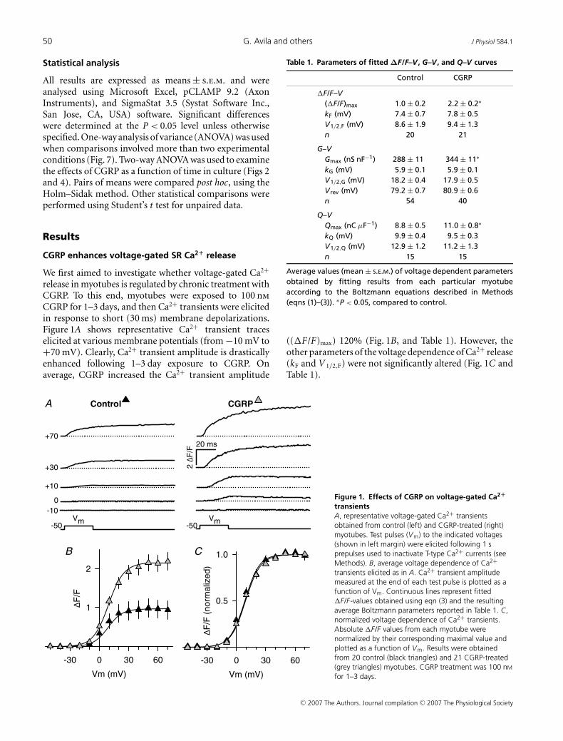
CGRP enhances voltage-gated SR Ca2+ release
We first aimed to investigate whether voltage-gated Ca2+
release in myotubes is regulated by chronic treatment withCGRP. To this end, myotubes were exposed to 100 nm
CGRP for 1–3 days, and then Ca2+ transients were elicitedin response to short (30 ms) membrane depolarizations.Figure 1A shows representative Ca2+ transient traceselicited at various membrane potentials (from −10 mV to+70 mV). Clearly, Ca2+ transient amplitude is drasticallyenhanced following 1–3 day exposure to CGRP. Onaverage, CGRP increased the Ca2+ transient amplitude
Vm (mV)
-30 0 30 60
F/F
1
2
Vm (mV)
-30 0 30 60
F/F
(n
orm
aliz
ed
)
0.5
1.0
-50Vm
-50Vm
+70
+30
+10
-10
0
2F
/F
A
B C
Control CGRP
20 ms
Figure 1. Effects of CGRP on voltage-gated Ca2+
transientsA, representative voltage-gated Ca2+ transientsobtained from control (left) and CGRP-treated (right)myotubes. Test pulses (Vm) to the indicated voltages(shown in left margin) were elicited following 1 sprepulses used to inactivate T-type Ca2+ currents (seeMethods). B, average voltage dependence of Ca2+transients elicited as in A. Ca2+ transient amplitudemeasured at the end of each test pulse is plotted as afunction of Vm. Continuous lines represent fitted�F/F-values obtained using eqn (3) and the resultingaverage Boltzmann parameters reported in Table 1. C,normalized voltage dependence of Ca2+ transients.Absolute �F/F values from each myotube werenormalized by their corresponding maximal value andplotted as a function of Vm. Results were obtainedfrom 20 control (black triangles) and 21 CGRP-treated(grey triangles) myotubes. CGRP treatment was 100 nM
for 1–3 days.
Table 1. Parameters of fitted ΔF/F–V , G–V , and Q–V curves
Control CGRP
�F/F–V(�F/F)max 1.0 ± 0.2 2.2 ± 0.2∗
kF (mV) 7.4 ± 0.7 7.8 ± 0.5V1/2,F (mV) 8.6 ± 1.9 9.4 ± 1.3n 20 21
G–VGmax (nS nF−1) 288 ± 11 344 ± 11∗
kG (mV) 5.9 ± 0.1 5.9 ± 0.1V1/2,G (mV) 18.2 ± 0.4 17.9 ± 0.5V rev (mV) 79.2 ± 0.7 80.9 ± 0.6n 54 40
Q–VQmax (nC μF−1) 8.8 ± 0.5 11.0 ± 0.8∗
kQ (mV) 9.9 ± 0.4 9.5 ± 0.3V1/2,Q (mV) 12.9 ± 1.2 11.2 ± 1.3n 15 15
Average values (mean ± S.E.M.) of voltage dependent parametersobtained by fitting results from each particular myotubeaccording to the Boltzmann equations described in Methods(eqns (1)–(3)). ∗P < 0.05, compared to control.
((�F/F)max) 120% (Fig. 1B, and Table 1). However, theother parameters of the voltage dependence of Ca2+ release(kF and V 1/2,F) were not significantly altered (Fig. 1C andTable 1).
C© 2007 The Authors. Journal compilation C© 2007 The Physiological Society

J Physiol 584.1 CGRP modulates development of EC coupling via cAMP/PKA signalling 51
The increase in the amplitude of the Ca2+ transientcould result from an increase in the rate of voltage-gatedSR Ca2+ release. Thus, we calculated the peak of thefirst derivative of �F/F (i.e. dF/dt), which represents areasonable approximation of the maximum rate of SRCa2+ release (Avila & Dirksen, 2005). CGRP was foundto significantly increase the maximum dF/dt by ∼90%,from 97 ± 17 to 184 ± 25 �F/F s−1 (P < 0.01, same cellsas in Fig. 1).
To investigate the onset of Ca2+ transient potentiationby CGRP, we determined (�F/F)max in myotubestreated from ∼1 h to up to ∼79 h compared to thatof time-matched controls. The experimental resultswere subsequently pooled as follows: ∼1–7 h (4 h),∼24–31 h (1 day), ∼48–55 h (2 days), and ∼72–79 h(3 days). Figure 2A shows representative Ca2+ transientsrecorded from each time point (time in days is indicatedin italic numbers). The corresponding average valuesof (�F/F)max are shown in Fig. 2B. Under controlconditions, these values grow steadily between 0 and3 days (from ∼0.7 �F/F , to up to ∼1.7 �F/F). Incontrast, CGRP-treated myotubes exhibit a markedincrease in voltage-gated Ca2+ release even as early as4 h post-exposure, a similar magnitude as that observedfor control myotubes after 3 days in control medium.However, voltage-gated Ca2+ release does not significantlyincrease further even following 3 days of CGRP exposure.These data suggest that CGRP accelerates the developmentof voltage-gated SR Ca2+ release in culture.
CGRP selectively increases L-channel expression
The results shown in Figs 1 and 2 prompted us toinvestigate whether CGRP enhancement of voltage-gated
Time after CGRP treatment (days)
0 1 2 3
F/F
) max
1
2
3
∗ ∗ ∗
A BControl CGRP
-50 -50+70+70
0.15
1
0.15
1
22
3 3
25 ms
2F
/F
Figure 2. Time course of CGRP effect on voltage-gated Ca2+ transientsA, representative voltage-gated Ca2+ transients obtained from myotubes cultured for 0–3 days either under controlconditions (left) or in the presence of 100 nM CGRP (right). Ca2+ transient amplitude was estimated as describedin Fig. 1. The approximated time (in days) following initiation of treatment is indicated with italicized numbers.B, average amplitude of voltage-gated Ca2+ transients ((�F/F)max) obtained at different times after CGRP treatment(100 nM). Results were obtained from 6 to 12 myotubes for each condition. (�F/F)max was estimated by eitherfitting experimental data to eqn (3) or averaging peak �F/F values obtained at saturating voltages (+30 mV to+70 mV). Results from two-way ANOVA and post hoc Holms–Sidak test indicated significant differences betweencontrol and CGRP-treated groups (∗P ≤ 0.05). Specifically, the P values between CGRP-treated and time-matchedcontrols obtained for 0.15, 1, 2, and 3 day treatments were 0.021, 0.008, 0.050, and 0.086, respectively.
SR Ca2+ release might involve a possible effect onvoltage-gated Ca2+ channels. Figure 3 shows L-type(Fig. 3B and D) and total Ca2+ currents (i.e. T-typeplus L-type, Fig. 3A and C) in control and CGRP-treatedmyotubes (1–3 days). As can be seen from both therepresentative currents (Fig. 3A and B) and average I–Vcurves (Fig. 3C and D), CGRP significantly increased theL-type Ca2+ current density by ∼25% (P < 0.0005 at+30 mV), while not significantly affecting T-type Ca2+
current density (P = 0.2 at −30 mV). The increase inL-type Ca2+ current density was entirely explained by aselective effect on the maximal L-channels conductance(Gmax), the only parameter of the Boltzamann fit (eqn (1))that was significantly altered (see Table 1).
We next set out to investigate the time course for theobserved effect on Gmax in a similar way as shown for Ca2+
transients in Fig. 2. An important difference, however,was that the shortest treatment we investigated for theL-type Ca2+ current was 0.5 days (∼8–15 h treatments),as opposed to 0.15 days for Ca2+ release (Fig. 2). Thus, wecompared L-type Ca2+ current density in time-matchedcontrol and CGRP-treated myotubes (Fig. 4). The resultsindicate that L-current density was significantly increased1–2 days following CGRP treatment, but not following 0.5or 3 days. Thus, similar to that observed for voltage-gatedCa2+ release, the CGRP-induced increase in L-currentdensity is also diminished after several days in culture.However, more importantly, the increase of L-type Ca2+
current density occurred only after a significant delay(∼1 day) compared to that observed for potentiation ofvoltage-gated Ca2+ release (∼4 h).
We next investigated whether the CGRP-inducedenhancement in L-current density results from anincrease in the number of voltage sensors in the
C© 2007 The Authors. Journal compilation C© 2007 The Physiological Society

52 G. Avila and others J Physiol 584.1
sarcolemma. Specifically, we compared the amountof immobilization-resistant intramembrane chargemovement between control and CGRP-treated (1–3 days)myotubes. Charge movement magnitude was estimatedby integrating the nonlinear capacitive (gating) currentselicited at the onset of membrane depolarization (QON).Figure 5A shows representative gating currents obtainedfrom control (left) and CGRP-treated (right) myotubes.Gating currents were significantly larger in CGRP-treatedmyotubes. In fact, CGRP increased the maximalmagnitude of QON (Qmax; Fig. 5B) without significantlyaltering the steepness (kQ) or voltage distribution (V 1/2,Q)of the charge movement–voltage relationship (Fig. 5C).Thus, the observed increase in QON (∼25%) is sufficient
Vm (mV)
-30 0 30 60
L-c
ure
nt
de
nsi
ty (
pA
/pF
)
-15
-7
Vm (mV)
-30 0 30 60
Cure
nt
densi
ty(p
A/p
F)
-15
-7
A B
1s prepulse
C D
1s prepulse
Control
CGRP
-80 -50
Vm Vm
1s prepulse
50 ms
10 p
A/p
F
Figure 3. CGRP selectively increases L-type Ca2+ current densityA and B, representative traces of total (A) and L-type (B) Ca2+ currentsobtained from control (top) and CGRP-treated (bottom) myotubes.Total (T-type and L-type) Ca2+ currents were first elicited from theholding potential (−80 mV). Subsequently, a family of L-type Ca2+currents were elicited following a 1 s prepulse to −30 mV to inactivateT-type Ca2+ channels. Ca2+ current traces are shown for the followingmembrane potentials (mV): −20, −10, 0, +10, +20 and +30. C andD, average current–voltage relationships (I–V curves) obtained for total(C) and L-type (D) Ca2+ currents. Results were obtained from 31 (C)and 54 (D) control myotubes (black triangles), and 29 (C) and 40 (D)CGRP-treated myotubes (1–3 days, 100 nM; grey triangles). Thecontinuous lines in D represent fits to the data using eqn (1) andaverage values of the parameters of these fits are shown in Table 1(G–V data). A spline curve was used to generate the smooth linesthrough the data in C.
to account for a similar 25% increase in Gmax observed inFigs 3 and 4.
CGRP increases SR Ca2+ content
While an increase in charge movement is sufficient toexplain a parallel increase in L-type Ca2+ current density(∼1.25-fold increase in both measurements), the slowertime course of this effect makes it unlikely to account for aneven larger effect on voltage-gated Ca2+ release (∼2.2-foldincrease in only 4 h). Thus, we speculated that potentiationof Ca2+ release may involve an increase in luminal SRCa2+ content. To test this possibility, we compared thecaffeine-releasable Ca2+-releasable pool in control and 4 hCGRP-treated myotubes (i.e. prior to a change in L-typeCa2+ current). Specifically, we measured the amplitude ofCa2+ transients induced by caffeine (30 mm), a widely usedryanodine receptor agonist, under identical conditionsas those used to assess voltage-gated Ca2+ release(i.e. measurements of both voltage- and caffeine-inducedCa2+ transients on the same myotube, Fig. 6). In both,control and CGRP-treated myotubes, caffeine applicationproduced a relatively fast increase in intracellular Ca2+
concentration whose maximal amplitude was higher thanthe peak Ca2+ transient elicited by voltage (Fig. 6).
In this set of experiments we used higher concentrationsof CGRP (100–300 nm, as opposed to 100 nm), andvoltage-gated Ca2+ release was stimulated to an even largerdegree (3.5-fold; Fig. 6, Voltage). The larger effect onvoltage-gated Ca2+ release in these experiments could beexplained by the use of a higher CGRP concentration.However, a somewhat smaller effect on voltage-inducedrelease (∼2.3-fold) was observed using 100–300 nm
CGRP in a separate set of experiments (Fig. 7, culture3). This apparent contradiction may deserve furtherinvestigation. In any event, 100–300 nm CGRP-treatedmyotubes exhibited a remarkable increase (4.0-fold) in thepeak amplitude of caffeine-induced Ca2+ release (Fig. 6B,Caffeine). Moreover, the ratios of Ca2+ release inducedby caffeine and voltage (i.e. caffeine-to-voltage ratio)were not statistically different for control (1.1 ± 0.2) andCGRP-treated (1.3 ± 0.2) myotubes. These results indicatethat CGRP drastically enhances the magnitude of thecaffeine-releasable Ca2+-releasable pool, an effect that issufficient to explain the corresponding potentiation ofvoltage-gated SR Ca2+ release.
Potentiation of Ca2+ release by CGRP is mediated byCGRP1 receptor signalling through cAMP/PKA
CGRP binds to at least two different membrane receptors,CGRP1 and CGRP2, and blockade of CGRP effects byCGRP8-37 defines a CGRP1 receptor mediated response(Chiba et al. 1989; reviewed by Juaneda et al. 2000). Thus,
C© 2007 The Authors. Journal compilation C© 2007 The Physiological Society

J Physiol 584.1 CGRP modulates development of EC coupling via cAMP/PKA signalling 53
Time after CGRP treatment (days)
0 1 2 3
L-c
urr
ent
density
(pA
/pF
)
-17
-14
-11
A BControl CGRP
-50 -50+30+30
0
1
0.5
1
2 2
3 3
50 ms9 p
A/p
F ∗
∗
Figure 4. Time course of CGRP-mediated increase in L-type Ca2+ current densityA, representative L-type Ca2+ currents from myotubes cultured for 0–3 days in either the absence (left) or thepresence of 100 nM CGRP (right). The time after initiation of CGRP treatment (in days) is shown in italicized numbers.Current traces were elicited using 200 ms depolarizing pulses to +30 mV that were preceded by a 1 s prepulse toinactivate T-type Ca2+ channels. B, time course of average peak L-type Ca2+ current density obtained from control(black triangles) and CGRP-treated (grey triangles) myotubes. Data represent means ± S.E.M. from 12–19 myotubesfor each condition. Two-way ANOVA indicates significant differences between control and CGRP-treated (100 nM)groups (P < 0.002). The P-values obtained from post hoc Holms–Sidak tests between time-matched control andCGRP-treated for 0.5, 1, 2 and 3 days of treatment were 0.700, 0.049, 0.036 and 0.069, respectively. ∗P < 0.05.
we investigated the possibility that CGRP potentiationof voltage-gated SR Ca2+ release might be mediated bysignalling through a CGRP1 receptor. We found thatCGRP8-37 blocks CGRP stimulation of voltage-gated SRCa2+ release (Fig. 7, Culture 1). These results indicatethat CGRP augments voltage-gated Ca2+ release by actingthrough CGRP1 receptors.
CGRP1 receptors couple via Gs proteins to stimulateadenylate cyclase leading to the generation of cAMPand activation of PKA (Juaneda et al. 2000; Hay et al.2003). Thus we next investigated the role of the cAMP/PKA signalling pathway in the CGRP enhancement ofvoltage-gated SR Ca2+ release. To this end, we preincubatedmyotubes with 10 μm H-89, a specific inhibitor of PKA,for ∼60 min prior to CGRP exposure. Interestingly, thestimulatory effect of CGRP was completely inhibited byH-89 pretreatment (Fig. 7, Culture 2), similarly to thatobserved for CGRP8-37. Thus, CGRP requires PKA topotentiate voltage-gated SR Ca2+ release.
The next step was to investigate a possible contributionof cAMP. To achieve this goal, we used two differentpharmacological approaches. We first investigated whetherthe effect of CGRP could be mimicked by Db-cAMP, amembrane-permeant cAMP analogue. As can be observedin Fig. 7 (Culture 3), Db-cAMP exposure also increasedthe amplitude of voltage-gated Ca2+ release, albeit to asomewhat lesser degree than CGRP. We also preincubatedmyotubes with papaverine, a nonselective inhibitor ofphosphodiesterases, which promotes cAMP accumulationby inhibiting its degradation. Papaverine treatmentproduced a 2.6-fold increase in the amplitude ofvoltage-gated Ca2+ release (Fig. 7, PAP), similar to theeffect seen with CGRP. Representative voltage-gated Ca2+
transients elicited at +70 mV are shown in Fig. 7B. Othermyotubes were treated with papaverine plus Db-cAMP
Vm (mV)
-30 0 30 60
QO
N (
nC
/µF
)
3
7
11
Vm (mV)
-30 0 30 60
QO
N (
norm
aliz
ed)
0.5
1.0
-50Vm -50
Vm
+30
+20
+10
0
-40
2.5
pA
/pF
10 ms
A
B C
Control CGRP
Figure 5. CGRP increases immobilization-resistant chargemovementA, representative non-capacitative gating currents (asymmetric chargemovements) obtained from control (left) and CGRP-treated (right)myotubes elicited at different membrane potentials (left margin).B, average voltage dependence of asymmetric charge movementestimated from integrating the non-capacitative current during theonset of each test depolarization (QON) plotted as a function of Vm.The continuous lines through the data were generated by fitting eachdata set with eqn (2). Average values of the fitted parameters areshown in Table 1 (Q–V data). C, normalized voltage dependence ofcharge movement. Absolute values of QON obtained from eachmyotube were normalized by their corresponding maximum values(Qmax), averaged, and plotted as a function of Vm. Experimentalresults were obtained from 15 control (black triangles) and 15CGRP-treated (1–3 days, 100 nM; grey triangles) myotubes.
C© 2007 The Authors. Journal compilation C© 2007 The Physiological Society

54 G. Avila and others J Physiol 584.1
(Fig. 7, Culture 4), in order to determine whether thesetwo agents exert an additive effect on release. However,combined treatment with papaverine and Db-cAMP failedto further increase voltage-gated Ca2+ release. These resultsindicate that cAMP stimulates voltage-gated SR Ca2+
release. Taken together, results presented in Fig. 7 indicatethat potentiation of SR Ca2+ release by CGRP is mediatedby activation of the CGRP1 receptor, production cAMP,and subsequent stimulation of PKA.
Discussion
This study represents to our knowledge the firstdemonstration that CGRP regulates the developmentof the EC coupling machinery in skeletal muscle. Wediscovered that CGRP markedly stimulates voltage-gatedSR Ca2+ release. The intensity of this effect varies between1.7- and 3.5-fold, across different experimental series.We also found that stimulation of voltage-gated SR Ca2+
A
//
//
Control
Caffeine
Caffeine
2 Δ
F/F
CGRP
B
ΔF
/F
0
1
2
3
4
5
Control CGRP Control CGRP
*
#
Voltage Caffeine
Voltage
Voltage
Figure 6. CGRP similarly potentiates voltage- andcaffeine-induced SR Ca2+ releaseA, representative Ca2+ transients elicited by either voltage (left) orexposure to 30 mM caffeine (right). Transients were obtained from onerepresentative control (top) and one representative CGRP-treated(200 nM, ∼4 h) myotube (bottom). The vertical calibration barindicates initiation of the depolarizing pulse (30 ms to +70 from aholding potential of −80 mV). Caffeine was applied (hatched bar)∼20 s thereafter (//). Horizontal calibration bar represents 0.2 s (left)and 0.8 s (right). B, average peak amplitude of voltage- (left) andcaffeine-induced (right) Ca2+ transients. Results were obtained from13 control and 10 CGRP-treated (∼4 h) myotubes. The treatment withCGRP consisted of one, two, or three aliquots (delivered every 2 h) of100 nM CGRP. ∗P < 0.005, #P < 0.001; compared to control.
release results primarily from a significant increase in theSR Ca2+ content, and does not involve alterations in thevoltage dependence of the release process. Additionally,this work points to a critical role for the activation of
Norm
aliz
ed (
Δ F/F
) max
0
1
2
3
Contr
ol
CG
RP
Db-c
AM
P
Contr
ol
CG
RP
H-8
9+
CG
RP
Contr
ol
CG
RP
8-3
7+
CG
RP
Contr
ol
PA
P
A
aa a a b c
B
30 ms2 ΔF/F
Db-cAMP PAPCGRP
PA
P+
Db
-cA
MP
8-87+CGRP H-89+CGRPControl
Vm
6
6
8 8
7
9 6
6
7
10
8 7
Culture 4
Culture 3
Culture 2
Culture 1
Figure 7. CGRP enhancement of voltage-gated Ca2+ release ismediated by a CGRP1 receptor activated cAMP/PKA signallingpathwayA, effects of CGRP and other compounds on the amplitude ofvoltage-gated SR Ca2+ release ((�F/F)max). Values of (�F/F)max weredetermined from the end of 30 ms depolarizating pulses to saturatingvoltages (+30 to +70 mV). Four different experimental series werecarried out (Cultures 1–4). The average (�F/F)max for each controlgroup was (mean ± S.E.M.): 0.6 ± 0.2, 1.2 ± 0.3, 0.8 ± 0.2 and0.7 ± 0.2; for Cultures 1, 2, 3 and 4, respectively. All (�F/F)max valuesfor each culture were divided by the mean value obtained from thecorresponding control group (normalized (�F/F)max). The number ofmyotubes that were investigated in each experimental condition isindicated near to the corresponding error bar. Both the CGRP1receptor antagonist CGRP8-37 (8–37; 3 μM) and the PKA inhibitor(H-89; 10 μM) were added 60 min prior to CGRP exposure andremained present thereafter. For Culture 4, the cAMP analogue,Db-cAMP, was used at a concentration of 0.5 mM and thephosphodiesterase inhibitor, papaverine (PAP), was used at aconcentration of 10 μM. Simultaneous treatment with both Db-cAMPand PAP was accomplished by applying the two compounds at thesame time (PAP + Db-cAMP). For Cultures 1, 2 and 4, treatmentslasted 1 day and CGRP was used at a concentration of 100 nM. Incontrast, one, two, or three aliquots (delivered every 2 h) of either100 nM CGRP or 0.5 mM Db-cAMP (∼4 h treatment) were used forCulture 3. aP < 0.05 compared to CGRP. bP < 0.15 compared to bothCGRP and Db-cAMP. cP < 0.10 compared to both PAP andPAP + Db-cAMP. Statistical tests (one-way ANOVA) were onlyperformed between groups of the same culture. B, examples of Ca2+transients recorded from the experimental conditions described in A.
C© 2007 The Authors. Journal compilation C© 2007 The Physiological Society

J Physiol 584.1 CGRP modulates development of EC coupling via cAMP/PKA signalling 55
CGRP1 receptors, cAMP, and PKA in mediating theseeffects. Our results also suggest that CGRP exerts along-term up-regulation in the number of sarcolemmalDHPRs (1.25-fold increase). Below we discuss the possiblephysiological relevance of these findings, as well as thepotential underlying mechanisms involved.
Molecular mechanisms associated to SR Ca2+ release
Our results point to PKA as a critical link in mediatingthe reported effects. Potentiation of Ca2+ release mostlikely results from an increased SR Ca2+ content, sinceCGRP exposure similarly increased the magnitude ofthe caffeine-sensitive Ca2+ store (Fig. 6). Steady-stateSR Ca2+ content depends on a balance between Ca2+
uptake and Ca2+ ‘leak’ (defined as the efflux of Ca2+
under resting conditions). The SR Ca2+-ATPase (SERCA)accounts for the uptake, whereas the molecular identityof the pathway(s) responsible for the leak remainscontroversial. It is well established that phospholamban(PLB), an intrinsic protein of the SR, inhibits SERCA2,through a physical interaction with the enzyme.Conversely, phosphorylation of PLB by PKA relieves thatinhibition (Simmerman & Jones, 1998). Interestingly, bothmyoblasts and differentiated myotubes express PLB(Stenoien et al. 2007) and SERCA2 (Kimura et al. 2005).Thus, CGRP might promote SERCA-mediated SR Ca2+
uptake via a PKA-mediated phosphorylation of PLB. Ifthat is the case, then increase SERCA activity wouldaccount for the observed increase in SR Ca2+ content.Further experiments are needed in order to morerigorously test this hypothesis.
Molecular mechanisms associated to DHPR expression
Our study shows that CGRP increases both L-typeCa2+ current density and DHPR charge movement to asimilar extent (∼25%), without significantly altering theircorresponding voltage dependence of activation. Thus,CGRP treatment increases the expression of functionalDHPRs within the sarcolemma. This effect could beexplained either by CGRP (a) stimulating the insertionof newly formed or preassembled DHPRs into thesarcolemma or (b) inhibiting the degradation rate ofsarcolemmal DHPRs. A genomic mechanism is likely to beinvolved since a significant increase in L-type Ca2+ currentdensity is first detected only 24 h after CGRP exposure(∼1 day, see Fig. 4). The long-term functional expressionof DHPRs is also promoted by Ca2+ release throughRyR1s (Avila et al. 2001). It will therefore be of interestto investigate whether the CGRP treatment enhancesDHPR expression through the short-term potentiation ofvoltage-gated SR Ca2+ release.
Interestingly, it has been reported that the down-stream signalling molecule of CGRP1 stimulation, cAMP,significantly increases the density of sarcolemmal DHPRs(Schmid et al. 1985). Moreover, the transcription rate forthe gene encoding skeletal muscle DHPRs (CACNA1S) isalso stimulated via the cAMP-response element bindingprotein (CREB, Zheng et al. 2002). Thus, CGRP couldstimulate transcription of CACNA1S, by acting throughcAMP and CREB.
Nevertheless, results from Ray et al. (1995) castdoubts on a possible transcriptional mechanism. This isbecause they found that cAMP does not affect mRNAlevels encoding any of the α1S-, α2-, and β-subunits ofthe DHPR. Hence, while cAMP increases the numberof sarcolemmal DHPRs (Schmid et al. 1985), it doesnot do this through stimulation of de novo synthesis(Ray et al. 1995). Alternatively, CGRP could inhibitDHPR proteolysis and/or degradation. Recent studiesprovide indirect experimental support for such an idea.Carrillo et al. (2004) found that DHPRs are degradedby calpain, a Ca2+-dependent protease. On the otherhand, Navegantes et al. (2001) found that cAMP inhibitsCa2+-dependent proteolysis, an effect probably explainedby an increased synthesis of calpastatin, which in turninhibits calpain-dependent proteolysis (Parr et al. 2001).Thus, it will be critical for future investigations todetermine whether CGRP prevents DHPR degradation,via cAMP, calpastatin and calpain. Other mechanismsthan enhanced gene transcription and reduced proteindegradation may also be involved in the observed increasein the density of sarcolemmal DHPRs.
Physiological relevance
Expression level of CGRP in motor neurons is elevatedduring embryonic development, tends to decrease inparallel with maturation of the neuromuscular junction,and is essentially undetectable in adults (Matteoli et al.1990). Nevertheless, expression level of CGRP in adultmotor neurons is greatly increased in response tophysiological and pathophysiological stimuli, includingexercise (Homonko & Theriault, 1997), trophic factors(Piehl et al. 1998), muscle inactivity (Sala et al. 1995),and neuromuscular blockade (Sala et al. 1995; Piehlet al. 1998). Thus, CGRP is thought to participate inneuromuscular junction regeneration in adults. In keepingwith this, it has been suggested that CGRP stimulatesmyoblast fusion (Noble et al. 1993) and the subsequentmyotube differentiation (Okazaki et al. 1996). Moreover,CGRP is also detected in skeletal muscle (Jiang et al. 2003),suggesting the presence of a possible autocrine role.
CGRP has been suggested to stimulate myoblast fusion(Noble et al. 1993). However, we did not find systematiceffects of CGRP treatment on membrane capacitance
C© 2007 The Authors. Journal compilation C© 2007 The Physiological Society

56 G. Avila and others J Physiol 584.1
(Cm), which would have suggested significant alterationsin myotube size. Specifically, average Cm values for 3 daycontrol and CGRP-treated myotubes were 184 ± 29 pFand 181 ± 33 pF, respectively (P = 0.94). We should takeinto account that Cm values are strongly influenced byinvestigator sampling bias. Thus, this does not mean thatmyotubes were not larger on average in CGRP-treatedmyotubes. Another possible explanation for this apparentcontradiction relies on the fact that CGRP treatment wasgiven 3 days after initiation of differentiation and thattreatment was limited to a maximum of only 3 days. Incontrast, Noble et al. (1993) exposed myotubes to CGRPfor 11 days.
On the other hand, our results are consistent withthe previous observation that CGRP accelerates myotubedifferentiation (Okazaki et al. 1996). Specifically, this studyfound that expression levels of myogenic regulatory factors(myogenin and Myf-5), myoglobin content and creatinekinase activity occurred earlier and in larger amounts inCGRP-treated myotubes compared to controls (Okazakiet al. 1996).
In summary, our data support the notion that CGRPstimulates development of EC coupling by acting throughCGRP1 receptors and the cAMP/PKA signalling pathway.Specifically, CGRP increases voltage-gated SR Ca2+ releasewithin hours and the density of sarcolemmal DHPRs indays. Stimulation of voltage-gated Ca2+ release seems toresult from a quantitatively similar increase in releasableSR Ca2+ content. This interpretation is supported bythe finding that CGRP enhances SR Ca2+ content,without significantly altering the voltage dependenceof L-current conductance, DHPR charge movements,or Ca2+ release. These effects are most prominentearly during myotube maturation. Whether these effectsalso occur in muscle regeneration remains to beelucidated.
References
Andersen SL & Clausen T (1993). Calcitonin gene-relatedpeptide stimulates active Na+-K+ transport in rat soleusmuscle. Am J Physiol Cell Physiol 264, C419–C429.
Avila G & Dirksen RT (2005). Rapamycin and FK506 reduceskeletal muscle voltage sensor expression and function. CellCalcium 38, 35–44.
Avila G, O’Connell KM, Groom LA & Dirksen RT (2001). Ca2+release through ryanodine receptors regulates skeletal muscleL-type Ca2+ channel expression. J Biol Chem 276,17732–17738.
Beam KG & Franzini-Armstrong C (1997). Functional andstructural approaches to the study of excitation-contractioncoupling. Methods Cell Biol 52, 283–306.
Boudreau-Lariviere C & Jasmin BJ (1999). Calcitoningene-related peptide decreases expression ofacetylcholinesterase in mammalian myotubes. FEBS Lett444, 22–26.
Carrillo E, Galindo JM, Garcia MC & Sanchez JA (2004).Regulation of muscle Cav1.1 channels by long-termdepolarization involves proteolysis of the alpha1s subunit.J Membr Biol 199, 155–161.
Chiba T, Yamaguchi A, Yamatani T, Nakamura A, Morishita T,Inui T, Fukase M, Noda T & Fujita T (1989). Calcitoningene-related peptide receptor antagonist humanCGRP-(8–37). Am J Physiol Endocrinol Metab 256,E331–E335.
Dirksen RT (2002). Bi-directional coupling betweendihydropyridine receptors and ryanodine receptors. FrontBiosci 7, d659–d670.
Fernandez HL, Chen M, Nadelhaft I & Durr JA (2003).Calcitonin gene-related peptides: their binding sites andreceptor accessory proteins in adult mammalian skeletalmuscles. Neuroscience 119, 335–345.
Fontaine B, Klarsfeld A, Hokfelt T & Changeux JP (1986).Calcitonin gene-related peptide, a peptide present in spinalcord motoneurons, increases the number of acetylcholinereceptors in primary cultures of chick embryo myotubes.Neurosci Lett 71, 59–65.
Hay DL, Poyner DR & Smith DM (2003). Desensitisation ofadrenomedullin and CGRP receptors. Regul Pept 112,139–145.
Homonko DA & Theriault E (1997). Calcitonin gene-relatedpeptide is increased in hindlimb motoneurons after exercise.Int J Sports Med 18, 503–509.
Jiang JX, Choi RC, Siow NL, Lee HH, Wan DC & Tsim KW(2003). Muscle induces neuronal expression ofacetylcholinesterase in neuron-muscle co-culture:transcriptional regulation mediated by cAMP-dependentsignaling. J Biol Chem 278, 45435–45444.
Juaneda C, Dumont Y & Quirion R (2000). The molecularpharmacology of CGRP and related peptide receptorsubtypes. Trends Pharmacol Sci 21, 432–438.
Kimura T, Nakamori M, Lueck JD, Pouliquin P, Aoike F,Fujimura H, Dirksen RT, Takahashi MP, Dulhunty AF &Sakoda S (2005). Altered mRNA splicing of the skeletalmuscle ryanodine receptor and sarcoplasmic/endoplasmicreticulum Ca2+-ATPase in myotonic dystrophy type 1. HumMol Genet 14, 2189–2200.
Lu B, Fu WM, Greengard P & Poo MM (1993). Calcitoningene-related peptide potentiates synaptic responses atdeveloping neuromuscular junction. Nature 363, 76–79.
Marty I, Robert M, Villaz M, De Jongh K, Lai Y, Catterall WA &Ronjat M (1994). Biochemical evidence for a complexinvolving dihydropyridine receptor and ryanodine receptorin triad junctions of skeletal muscle. Proc Natl Acad Sci U S A91, 2270–2274.
Matteoli M, Balbi S, Sala C, Chini B, Cimino M, Vitadello M &Fumagalli G (1990). Developmentally regulated expressionof calcitonin gene-related peptide at mammalianneuromuscular junction. J Mol Neurosci 2, 175–184.
Mejia-Luna L & Avila G (2004). Ca2+ channel regulation bytransforming growth factor-β1 and bone morphogeneticprotein-2 in developing mice myotubes. J Physiol 559,41–54.
Melzer W, Herrmann-Frank A & Luttgau HC (1995). The roleof Ca2+ ions in excitation-contraction coupling of skeletalmuscle fibres. Biochim Biophys Acta 1241, 59–116.
C© 2007 The Authors. Journal compilation C© 2007 The Physiological Society

J Physiol 584.1 CGRP modulates development of EC coupling via cAMP/PKA signalling 57
Mulle C, Benoit P, Pinset C, Roa M & Changeux JP (1988).Calcitonin gene-related peptide enhances the rate ofdesensitization of the nicotinic acetylcholine receptor incultured mouse muscle cells. Proc Natl Acad Sci U S A 85,5728–5732.
Navegantes LC, Resano NM, Migliorini RH & Kettelhut IC(2001). Catecholamines inhibit Ca2+-dependent proteolysisin rat skeletal muscle through β2-adrenoceptors and cAMP.Am J Physiol Endocrinol Metab 281, E449–E454.
New HV & Mudge AW (1986). Calcitonin gene-related peptideregulates muscle acetylcholine receptor synthesis. Nature323, 809–811.
Noble BS, McMillan DN & Maltin CA (1993). Calcitonin generelated peptide stimulates differentiation of neonatal ratmyogenic cultures. Growth Regul 3, 245–248.
Okazaki S, Kawai H, Arii Y, Yamaguchi H & Saito S (1996).Effects of calcitonin gene-related peptide and interleukin 6on myoblast differentiation. Cell Prolif 29, 173–182.
Parr T, Sensky PL, Bardsley RG & Buttery PJ (2001).Calpastatin expression in porcine cardiac and skeletal muscleand partial gene structure. Arch Biochem Biophys 395, 1–13.
Piehl F, Hammarberg H, Hokfelt T & Cullheim S (1998).Regulatory effects of trophic factors on expression anddistribution of CGRP and GAP-43 in rat motoneurons.J Neurosci Res 51, 1–14.
Ray A, Kyselovic J, Leddy JJ, Wigle JT, Jasmin BJ & Tuana BS(1995). Regulation of dihydropyridine and ryanodinereceptor gene expression in skeletal muscle. Role of nerve,protein kinase C, and cAMP pathways. J Biol Chem 270,25837–25844.
Rios E & Brum G (1987). Involvement of dihydropyridinereceptors in excitation-contraction coupling in skeletalmuscle. Nature 325, 717–720.
Rossi SG, Dickerson IM & Rotundo RL (2003). Localization ofthe calcitonin gene-related peptide receptor complex at thevertebrate neuromuscular junction and its role in regulatingacetylcholinesterase expression. J Biol Chem 278,24994–25000.
Sakaguchi M, Inaishi Y, Kashihara Y & Kuno M (1991). Releaseof calcitonin gene-related peptide from nerve terminals inrat skeletal muscle. J Physiol 434, 257–270.
Sala C, Andreose JS, Fumagalli G & Lomo T (1995). Calcitoningene-related peptide: possible role in formation andmaintenance of neuromuscular junctions. J Neurosci 15,520–528.
Schmid A, Renaud JF & Lazdunski M (1985). Short term andlong term effects of β-adrenergic effectors and cyclic AMPon nitrendipine-sensitive voltage-dependent Ca2+ channelsof skeletal muscle. J Biol Chem 260, 13041–13046.
Schneider MF & Chandler WK (1973). Voltage dependentcharge movement of skeletal muscle: a possible step inexcitation-contraction coupling. Nature 242, 244–246.
Simmerman HK & Jones LR (1998). Phospholamban: proteinstructure, mechanism of action, and role in cardiac function.Physiol Rev 78, 921–947.
Stenoien DL, Knyushko TV, Londono MP, Opresko LK, MayerMU, Brady ST, Squier TC & Bigelow DJ (2007). Cellulartrafficking of phospholamban and formation of functionalsarcoplasmic reticulum during myocyte differentiation.Am J Physiol Cell Physiol 292, C2084–2094.
Takami K, Hashimoto K, Uchida S, Tohyama M & Yoshida H(1986). Effect of calcitonin gene-related peptide on the cyclicAMP level of isolated mouse diaphragm. Jpn J Pharmacol 42,345–350.
Takami K, Kawai Y, Uchida S, Tohyama M, Shiotani Y, YoshidaH, Emson PC, Girgis S, Hillyard CJ & MacIntyre I (1985).Effect of calcitonin gene-related peptide on contraction ofstriated muscle in the mouse. Neurosci Lett 60, 227–230.
Tanabe T, Beam KG, Powell JA & Numa S (1988). Restorationof excitation-contraction coupling and slow calcium currentin dysgenic muscle by dihydropyridine receptorcomplementary DNA. Nature 336, 134–139.
Uchida S, Yamamoto H, Iio S, Matsumoto N, Wang XB,Yonehara N, Imai Y, Inoki R & Yoshida H (1990). Release ofcalcitonin gene-related peptide-like immunoreactivesubstance from neuromuscular junction by nerve excitationand its action on striated muscle. J Neurochem 54,1000–1003.
Zheng Z, Wang ZM & Delbono O (2002). Insulin-like growthfactor-1 increases skeletal muscle dihydropyridine receptorα1S transcriptional activity by acting on the cAMP-responseelement-binding protein element of the promoter region.J Biol Chem 277, 50535–50542.
Acknowledgements
We would like to thank Dr Robert T. Dirksen for providing
helpful comments for improving the manuscript. We also thank
Esperanza Jimenez and Mario Rodriguez for technical assistance.
This work was partially supported by a CONACyT grant to GA
(39512). CIA and RR-M received graduate student fellowships
granted by CONACyT.
C© 2007 The Authors. Journal compilation C© 2007 The Physiological Society