survival and allozyme expression in laboratory induced
TRANSCRIPT

Transactions of the Illinois State Academy of Science received 1/4/99(2000), Volume 93, #1, pp. 69-81 accepted 7/10/99
Survival and Allozyme Expression inLaboratory Induced Hybrids
Between Two Species of Salamander
Robert G. WeckDepartment of Life and Health Sciences
Belleville Area CollegeBelleville, Illinois 62221-5899
and
Ronald A. Brandon*Department of Zoology
Southern Illinois University CarbondaleCarbondale, Illinois 62901-6501
*Corresponding author
ABSTRACT
We examined hybrid survival and allozyme expression of five enzyme coding loci inlaboratory induced hybrids between Ambystoma texanum and Ambystoma tigrinum.Survival of A. tigrinum x A. texanum hybrids was high (80%) compared to previousstudies. Survival was much lower in A. texanum x A. tigrinum hybrids (32% and 43%for two different crosses). Electrophoretic patterns of ADA-1, LDH-1, LDH-2, andMDH-1 allozymes in all hybrids, and MDH-2 patterns in A. tigrinum x A. texanumhybrids, conform to patterns previously observed in diploid hybrids showing biparentalallozyme expression. MDH-2 patterns in A. texanum x A. tigrinum hybrids are unusual,with weak expression of paternal allozymes.
Key Words: allozymes, hybrids
INTRODUCTION
Laboratory crosses between members of the salamander genus Ambystoma provideexcellent opportunities to study patterns of allozyme expression in hybrid amphibians.Phylogenetic studies indicate that Ambystoma is an old lineage with large geneticdistances among species (Kraus, 1988; Shaffer et al., 1991). In spite of this divergence,most attempts to produce interspecific hybrids through artificial insemination (over 40interspecific combinations reported) produce viable F1 hybrids (Nelson and Humphery,1972; Brandon, 1972, 1977). Hybridization between at least four diploid species in
70
nature (A. jeffersonianum, A. laterale, A . texanum , and A . tigrinum) has producedpopulations with an interesting array of ploidy levels and genomic compositions (e.g.,Uzzell, 1964; Downs, 1978; Morris and Brandon, 1984; Kraus, 1985a; Kraus, 1985b;Morris, 1985; Phillips et al. 1997).
Allozyme electrophoresis is one of the principle diagnostic tools used to determine ploidylevels and relative parental genomic doses in Ambystoma jeffersonianum complexsalamanders (e.g. Bogart, 1982; Bogart et al., 1985; Licht and Bogart, 1989; Lowcockand Bogart, 1989; Spolsky et al., 1992). Some allozyme phenotypes of in vitro hybridsbetween triploid females and males of four different diploid species have been described(Bogart et al., 1989); however, allozyme expression of in vitro hybrids between diploidspecies of this group of salamanders has not been reported. The objectives of this studywere to examine viability of F1 hybrids between Ambystoma texanum and Ambystomatigrinum produced in the laboratory and to describe patterns of allozyme expression inthem.
MATERIALS AND METHODS
Laboratory CrossesCrosses were made between Ambystoma texanum and A. tigrinum collected from westernKentucky and southern Illinois. Voucher specimens have been deposited in the SouthernIllinois University Carbondale Fluid Vertebrate Collection (SIUC). Locality data andcatalog numbers (H-numbers = alcoholic specimens and TC numbers = frozen tissues) ofanimals used in the hybridization experiment are as follows:
A. tigrinum (tig): SIUC TC 605. Female, Livingston Co., Kentucky, Route 137, 0.8 mi Sjct. 1436, 1.4 mi N mile marker 9, 15 February 1989, R. Brandon and R. Weck; SIUCTC 612. Male, Jackson Co., Illinois, McGuire's Orchard pond, ca. 8 mi S Carbondale,14 February 1989, R. Weck, G. Miller, G. Paleudis.
A. texanum: SIUC H-3891. Female (Ktex), Livingston Co., Kentucky, Route 137, S ofjct. Route 133, 2.2 mi N mile marker 9, 15 February 1989, R. Brandon, R. Weck;SIUC H-3859 and H-3960. Females (ILtex), Williamson Co., Illinois, along LittleGrassy Creek, sect. 4 T10S, R1E, 13 February 1989, R. Brandon, R. Weck, H.Moeller, S. Taylor; SIUC H-3861. Male (tex), Williamson Co., Illinois, along LittleGrassy Creek, sect. 4 T10S, R1E, 13 February 1989, R. Brandon, R. Weck, H.Moeller.
Matings (Table 1) between these salamanders were made through artificial inseminationfollowing the procedures of Brandon (1977). Females were injected with folliclestimulating hormone to induce ovulation. Sperm suspensions from males were kept onice to extend functional longevity as eggs were removed from the oviducts of females.Fertilization occurred in dechlorinated water at room temperature (ca. 20oC). The twoIllinois A. texanum females individually produced too few eggs for all matings, so theireggs were pooled and treated as one source. These females were collected syntopicallyand had identical phenotypes at all allozyme loci examined. All parents were sacrificedduring the artificial insemination procedure and were stored in plastic bags at -80oC untiltheir tissues were processed for electrophoresis.

71
Embryos developed in 10.5-cm diameter glass bowls in dechlorinate water. Embryoswere counted and scored daily to follow development. Dead embryos were removed andwater was changed daily. Upon hatching, larvae were fed young brine shrimp nauplii(Artemia sp.) and, when larger, frozen bloodworms (Chironomus sp.). Larvae wereraised until they were large enough (3-4 cm total length) to provide adequateelectrophoresis tissue samples, after which most were anesthetized in MS-222 (tricainemethane sulfonate) and frozen for storage at -80oC.
Protein ElectrophoresisHorizontal starch gel techniques similar to those of May et al. (1979) were used todetermine allozyme phenotypes from homogenates of skeletal muscle in two buffersystems: Amine Citrate (AC6.9) pH 6.9 as described by Clayton and Tretiak (1972), andTris Citrate (TC8) pH 8.0 as described by Selander et al. (1971). Three enzyme systems(one monomeric, one dimeric, and one tetrameric), encoded by five presumptive geneloci, proved informative with two criteria: 1) the presence in the parents of diagnosticalleles (electromorphs) that allowed us to test for biparental inheritance and examinepatterns of gene expression in the hybrids, and 2) loci that provided adequate enzymaticactivity and electromorph resolution in the parents to allow us to detect dosage effects.Homogenates of tail and hind limb muscle tissue were examined from at least ten larvaefrom each interspecific cross for the enzymes ADA-1 (adenosine deaminase [enzymecommission number 3.5.4.4]), LDH-1 and LDH-2 (lactate dehydrogenase [enzymecommission number 1.1.1.27]), and MDH-1 and MDH-2 (malate dehydrogenase [enzymecommission number 1.1.1.37]) (International Union of Biochemistry, 1979). Samples ofparental tissues were included on the gels as controls to verify parental bands in hybrids.Homogenates of five larvae from each intraspecific cross were analyzed for the LDH-1,LDH-2, MDH-1, and MDH-2 loci.
Alleles at each allozyme locus were coded by their relative mobility following thenomenclature of Shaklee et al. (1990). The most common allele in the Illinois A .texanum was used as the reference or 100 allele at each locus, and the electrophoreticmobilities of other alleles are reported as percentages of this allele.
RESULTS AND DISCUSSION
Hybrid ViabilitySurvival of the hybrids was higher (Table 1) compared to the same interspecific crossesreported by Brandon (1977). Brandon had commented that the A. texanum ova appearedto be "overripe." Even in the present crosses, embryos derived from A. tigrinum ovasurvived better than those derived from A. texanum ova; eighty percent of the 99 A.tigrinum eggs inseminated by A . texanum sperm developed to hatching. Reciprocalcrosses involving two different A. texanum females and the same A . tigrinum maleexperienced significantly higher hybrid mortality (32 and 43 percent, respectively).Thus, the same relative nuclear combination in the interspecific hybrids (one A. texanumhaplotype and one A. tigrinum haplotype) seems to have produced different degrees ofembryonic mortality depending on the source of the egg. It is unclear, however, whetherthis was due to developmental differences in the interactions between the egg cytoplasmof each species and a hybrid nucleus (as has been described in other hybrid amphibians

72
[Elinson, 1977]) or, more simply, difficulty in handling A. texanum eggs. All crossesinvolving A. texanum females (including intraspecific controls) produced higher offspringmortality than did either interspecific or intraspecific crosses involving the A. tigrinumfemale. The eggs of gravid A. texanum females appear to become overripe quickly whenfemales are held in captivity and their eggs seem more easily damaged during handling.
Electrophoretic PatternsThe genotypes of the parents at five allozyme loci are presented in Table 2. Allelectrophoretic patterns in the parents conform with the phenotypes expected fromdiploid organisms, based on the known quaternary structure of each allozyme and acodominant pattern of inheritance (Utter et al. 1987). Allozyme phenotypes of thehybrids at all loci other than MDH-2 also indicate diploidy although none of the hybridswas karyotyped. Following is a discussion of allozyme patterns of these five loci in thehybrids.
ADA-1 PatternsADA is a monomeric protein. Heterozygotes should show a two-banded phenotype, witheach band equally intense. This pattern was observed in the hybrids of both tex/tigcrosses. Figure 1 shows results of the Ktex/tig cross. All hybrids are 86/100heterozygotes, showing maternal and paternal bands of equal intensity. A similar patternwas seen at a slower locus (ADA-2) on the same gels. The second locus was not scoredbecause of poor resolution of the bands and does not appear on Figure 1. This crossconfirms the heritability of ADA-1, which is important for our study of allozymevariation in the A. texanum complex where we use ADA-1 to detect gene flow betweenA. texanum and A. barbouri in an area of sympatry in western Kentucky. ADA was notexamined in the reciprocal tig/tex hybrids. The tig female was a 68/75 heterozygote andhybrids would be expected to show a 1:1 Mendelian ratio of 68/100 and 75/100phenotypes.
LDH PatternsLDH is a tetrameric protein containing four polypeptide subunits. Two LDH loci wereexpressed clearly in muscle tissue of both A. texanum and A. tigrinum. Typical LDHpatterns for each species are shown in lanes 1 (texanum) and 2 (tigrinum) of Figure 2.The fastest and slowest band in each lane represent the products of the two different LDHcoding genes, LDH-1 and LDH-2, respectively. Three bands with intermediate mobilitiesare seen that represent heterotetrameric proteins (combinations of the polypeptidesubunits produced by the two LDH loci). The A. texanum and A. tigrinum have a fixeddifference at both loci, although the LDH-2 electromorph mobilities differ by a maximumof 1.5 mm under our electrophoretic conditions. LDH patterns typical of the tig/tex andtex/tig F1 hybrids are shown in lanes 3-15 of Figure 2. The hybrids are heterozygous atboth LDH loci.
Since the protein products of the two LDH loci interact in the parents to produceheterotetrameric bands, heterotetrameric proteins should be produced between each of thefour homotetrameric products of the four alleles (LDH-1(100), LDH-1(78), LDH-2(100),LDH-2(82)) present in the hybrid offspring. If this were true, 22 distinct electromorphicbands (4 homotetrameric and 18 heterotetrameric) would appear on an idealelectrophoretic gel, as is illustrated in Figure 3. In our electrophoretic runs the maximum
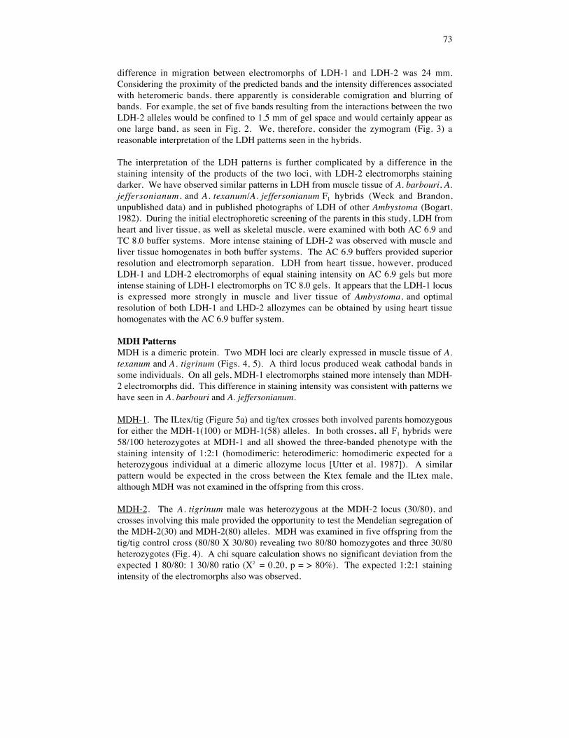
73
difference in migration between electromorphs of LDH-1 and LDH-2 was 24 mm.Considering the proximity of the predicted bands and the intensity differences associatedwith heteromeric bands, there apparently is considerable comigration and blurring ofbands. For example, the set of five bands resulting from the interactions between the twoLDH-2 alleles would be confined to 1.5 mm of gel space and would certainly appear asone large band, as seen in Fig. 2. We, therefore, consider the zymogram (Fig. 3) areasonable interpretation of the LDH patterns seen in the hybrids.
The interpretation of the LDH patterns is further complicated by a difference in thestaining intensity of the products of the two loci, with LDH-2 electromorphs stainingdarker. We have observed similar patterns in LDH from muscle tissue of A. barbouri, A.jeffersonianum, and A. texanum/A. jeffersonianum F1 hybrids (Weck and Brandon,unpublished data) and in published photographs of LDH of other Ambystoma (Bogart,1982). During the initial electrophoretic screening of the parents in this study, LDH fromheart and liver tissue, as well as skeletal muscle, were examined with both AC 6.9 andTC 8.0 buffer systems. More intense staining of LDH-2 was observed with muscle andliver tissue homogenates in both buffer systems. The AC 6.9 buffers provided superiorresolution and electromorph separation. LDH from heart tissue, however, producedLDH-1 and LDH-2 electromorphs of equal staining intensity on AC 6.9 gels but moreintense staining of LDH-1 electromorphs on TC 8.0 gels. It appears that the LDH-1 locusis expressed more strongly in muscle and liver tissue of Ambystoma, and optimalresolution of both LDH-1 and LHD-2 allozymes can be obtained by using heart tissuehomogenates with the AC 6.9 buffer system.
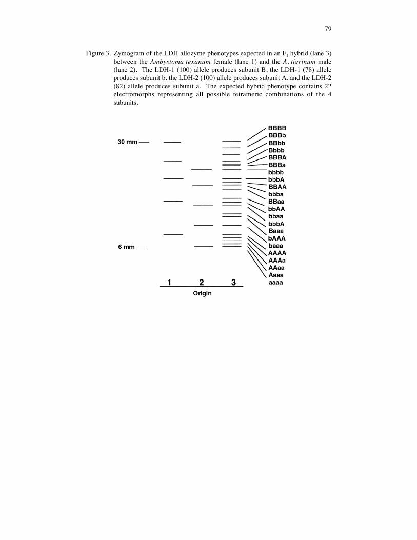
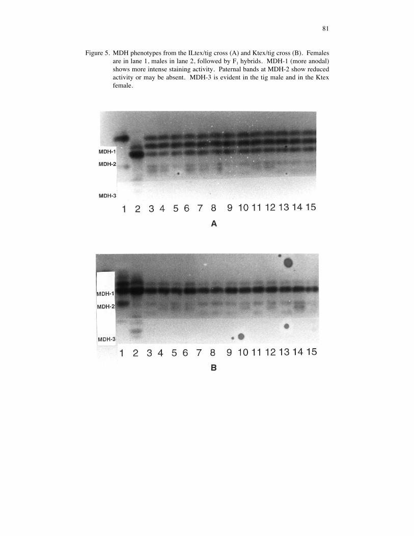
MDH PatternsMDH is a dimeric protein. Two MDH loci are clearly expressed in muscle tissue of A.texanum and A. tigrinum (Figs. 4, 5). A third locus produced weak cathodal bands insome individuals. On all gels, MDH-1 electromorphs stained more intensely than MDH-2 electromorphs did. This difference in staining intensity was consistent with patterns wehave seen in A. barbouri and A. jeffersonianum.
MDH-1. The ILtex/tig (Figure 5a) and tig/tex crosses both involved parents homozygousfor either the MDH-1(100) or MDH-1(58) alleles. In both crosses, all F1 hybrids were58/100 heterozygotes at MDH-1 and all showed the three-banded phenotype with thestaining intensity of 1:2:1 (homodimeric: heterodimeric: homodimeric expected for aheterozygous individual at a dimeric allozyme locus [Utter et al. 1987]). A similarpattern would be expected in the cross between the Ktex female and the ILtex male,although MDH was not examined in the offspring from this cross.
MDH-2. The A . tigrinum male was heterozygous at the MDH-2 locus (30/80), andcrosses involving this male provided the opportunity to test the Mendelian segregation ofthe MDH-2(30) and MDH-2(80) alleles. MDH was examined in five offspring from thetig/tig control cross (80/80 X 30/80) revealing two 80/80 homozygotes and three 30/80heterozygotes (Fig. 4). A chi square calculation shows no significant deviation from theexpected 1 80/80: 1 30/80 ratio (X2 = 0.20, p = > 80%). The expected 1:2:1 stainingintensity of the electromorphs also was observed.

74
MDH-2 patterns in three interspecific crosses were examined. Twenty F1 hybrids fromthe tig/tex cross (100/100 X 80/80) all appear to be 100/80 heterozygotes at MDH-2. TheMDH-2(100) and MDH-2(80) electromorphs were separated by only 1 mm on the gelsand the three electromorphic bands produced by the hybrids were confined to this space,forming one large fuzzy band. Reciprocal crosses (tex/tig) produced weak MDH activitywith unexpected patterns. Thirteen ILtex/tig and 13 Ktex/tig hybrids were analyzed(Figs. 5a, 5b). In both crosses, the MDH-2 alleles segregated in a Mendelian fashion(Table 3); however, the paternal electromorphs (tigrinum) were absent or were muchweaker than either the maternal (texanum) or heterodimeric bands. The presence of theheterodimeric bands indicates that the paternal MDH-2 genes were expressed in thehybrids, but it is unclear why the allozyme products of these genes produced weakerelectromorphs. MDH-2 in the tex/tig hybrids showed a pattern that would be expected intriploids. Dosage effects are predicted in triploid individuals where maternal:paternalallelic ratios are 2:1 (Seeb et al., 1988). MDH allozyme dosages have been found tocorrelate with the known ploidy level in Ambys toma jeffersonianum complexsalamanders (Bogart, 1982; Bogart et al., 1989) and, in each case, both MDH-1 andMDH-2 patterns reflected the ploidy level of the individual. Alternatively, the hybridsmay appear to be triploid because the maternal MDH-2 is expressed more strongly thanpaternal MDH-2. Wright (1975) reported expression of only maternal allozyme genes inearly embryos (through neurula) of hybrids between Rana pipiens complex frogs, butpaternal allozymes were expressed by the operculum development stage. Our hybridlarvae were relatively small (3-4 cm) when examined but well beyond limb formation.Ploidy of the hybrids remains to be confirmed by karyology.
ACKNOWLEDGMENTS
We thank Jim Seeb and Lisa Seeb for chemicals and laboratory space. Gary Miller,Harry Moeller, George Paleudus, and Steve Taylor helped collect animals. Bill Neef andAnna Kittle printed the photographs.
LITERATURE CITED
Bogart, J. P. 1982. Ploidy and genetic diversity in Ontario salamanders of the Ambystomajeffersonianum complex revealed through an electrophoretic examination of larvae. Can. J. Zool.60:848-855.
Bogart, J. P., R. P. Elinson, and L. E. Licht. 1989. Temperature and sperm incorporation inpolyploid salamanders. Science 246:1032-1034.
Bogart, J. P., L. E. Licht, M. J. Oldham, and S. J. Darbyshire. 1985. Electrophoretic identificationof Ambystoma laterale and Ambystoma texanum as well as their diploid and triploid interspecifichybrids (Amphibia: Caudata) on Pelee Island, Ontario. Can. J. Zool. 63:340-347.
Brandon, R. A. 1972. Hybridization between the Mexican salamanders Ambystoma dumerilii andAmbystoma mexicanum under laboratory conditions. Herpetologica 28:199-207.
Brandon, R. A. 1977. Interspecific hybridization among Mexican and United States salamandersof the genus Ambystoma under laboratory conditions. Herpetologica 33:133-152.
Clayton, J. W. and D. N. Tretiak. 1972. Aminecitrate buffers for pH control in starch gelelectrophoresis. J. Fish. Res. Board Canada 29:1169-1172.

75
Downs, F. L. 1978. Unisexual Ambystoma from the Bass Islands of Lake Erie. Occ. Pap. Mus.Zool. Univ. Michigan 685:1-36.
Elinson, R. P. 1977. Amphibian hybrids: a genetic approach to the analysis of their developmentalarrest. Differentiation 9:3-9.
International Union of Biochemistry. Nomenclature Committee. 1979. Enzyme Nomenclature.Academic Press, New York.
Kraus, F. 1985a. Unisexual salamander lineages in northwestern Ohio and southeastern Michigan:a study of the consequences of hybridization. Copeia 1985:309-324.
Kraus, F. 1985b. A new unisexual salamander from Ohio. Occ. Pap. Mus. Zool. Univ. Michigan709:1-24.
Kraus, F. 1988. An empirical evaluation of the use of the ontogeny polarization criterion inphylogenetic inference. Syst. Zool. 37:106-141.
Licht, L. E. and J. P. Bogart. 1989. Embryonic development and temperature tolerance in diploidand polyploid salamanders (genus Ambystoma). Am. Midl. Nat. 122:401-407.
Lowcock, L. A. and J. P. Bogart. 1989. Electrophoretic evidence for multiple origins of triploidforms in the Ambystoma laterale-jeffersonianum complex. Can. J. Zool. 67:350-356.
May, B., J. E. Wright, and M. Stoneking. 1979. Joint segregation of biochemical loci inSalmonidae: results from experiments with Salvelinus and review of the literature on otherspecies. J. Fish. Res. Board Canada 36:1114-1128.
Morris, M. A. 1985. A hybrid Ambystoma platineum x A. tigrinum from Indiana. Herpetologica41:267-271.
Morris, M. A. and R. A. Brandon. 1984. Gynogenesis and hybridization between Ambystomaplatineum and Ambystoma texanum in Illinois. Copeia 1984:324-337.
Nelson, C. E. and R. R. Humphrey. 1972. Artificial interspecific hybridization amongAmbystoma. Herpetologica 28:27-32.
Phillips, C. A., T. Uzzell, C. M. Spolsky, J. M. Serb, R. E. Szafoni, and T. R. Pollowy. 1997.Persistent high levels of tetraploidy in salamanders of the Ambystoma jeffersonianum complex.J. Herpetol. 31: 530-535.
Seeb, J. E., G. H. Thorgaard, and F. M. Utter. 1988. Survival and allozyme expression in diploidand triploid hybrids between chum, chinook, and coho salmon. Aquaculture 72:31-48.
Selander, R. K., M. H. Smith, S. Y. Yang, W. E. Johnson, and J. B. Gentry. 1971. Biochemicalpolymorphisms and systematics in the genus Peromyscus. I. Variation in the old field mouse(Peromyscus polionotus). Studies in Genetics VI. University of Texas Publication 7130:49-90.
Shaffer, H. B., J. M. Clark, and F. Kraus. 1991. When molecules and morphology clash: aphylogenetic analysis of the North American ambystomatid salamanders (Caudata:Ambystomatidae). Syst. Zool. 40:284-303.
Shaklee, J. B., F. W. Allendorf, D. C. Morizot, and G. S. Whitt. 1990. Gene nomenclature forprotein-coding loci in fish. Trans. Am. Fish. Soc. 119:2-15.
Spolsky, C., C. A. Phillips, and T. Uzzell. 1992. Gynogenetic reproduction in hybrid molesalamanders (genus Ambystoma). Evolution 46:1935-1944.
Utter, F., P. Abersold, and G. Winans. 1987. Interpretation of genetic variation detected byelectrophoresis. In Population genetics & fisheries management. N. Ryman and F. Utter (eds.),Univ. Washington Press, Seattle, pp 21-45.
Uzzell, T. 1964. Relations of the diploid and triploid species of the Ambystoma jeffersonianumcomplex (Amphibia, Caudata). Copeia 1964:257-300.
Wright, D. A. 1975. Expression of enzyme phenotypes in hybrid embryos. In Isozymes IV:genetics and evolution. C. L. Markert (ed.), Academic Press, New York, pp. 649-664.

76
Table 1. Results of laboratory crosses involving Ambystoma texanum and A. tigrinum* Indicates data from Brandon (1977) for comparison.
Cross Number of Eggs %(f/m) Inseminated Damaged Hatched Remarks
tig/ILtex 99 0 80 20% mortality duringgastrula and neurula
ILtex/tig 57 3 32 68% embryonic mortality;1 egg unfertilized
Ktex/tig 71 3 43 57% mortality duringgastrula; 1 egg unfertilized
tig/tig 84 0 77 23% mortality duringgastrula; a few survivorsdeformed
ILtex/ILtex 33 2 52 48% mortality during gastrula; a few survivorsabnormal, 2 eggs unfertilized
Ktex/ILtex 59 6 74 26% embryonic mortality;2 eggs unfertilized
tig/ILtex* 27 0 23 many abnormal
ILtex/tig* 108 ? 3 5 reached neurula; none fed
tig/tig* 21 0 2 4 died as gastrulae, 8 asneurulae, 7 died later
Iltex/ILtex* 57 57 0 none survived cleavage

77
Table 2. Allozyme genotypes of the parents at five loci.
Locus Specimen (sex) ADA-1 LDH-1 LDH-2 MDH-1 MDH-2
ILtex (F-1) 100/100 100/100 100/100 100/100 100/100ILtex (F-2) 100/100 100/100 100/100 100/100 100/100ILtex (M) 100/100 100/100 100/100 100/100 100/100Ktex (F) 100/100 100/100 100/100 58/58 100/100tig (F) 68/75 78/78 82/82 58/58 80/80tig (M) 68/68 78/78 82/82 58/58 30/80
Table 3.Observed and expected MDH-2 genotype frequencies for the ILtex/tig cross(100/100 x 30/80) and the Ktex/tig cross (100/100 x 30/80).
ILtex/tig (n = 14) Ktex/tig (n = 13)Genotype Observed Expected Observed Expected
30/100 42.9% 50% 53.8% 50%
80/100 57.1% 50% 46.2% 50%

78
Figure 1. ADA-1 phenotypes of an A. texanum female (lane 1, genotype 100/100), an A.tigrinum male (lane 2, genotype 68/68) and 13 of their F1 hybrids (lanes 3-15).All hybrids are 68/100 heterozygotes.
Figure 2. LDH phenotypes of an A. texanum female (lane 1), an A. tigrinum male (lane2) and 13 of their F1 hybrids (lanes 3-15). A diagrammatic interpretation ofthese phenotypes is shown in Figure 3.
79
Figure 3. Zymogram of the LDH allozyme phenotypes expected in an F1 hybrid (lane 3)between the Ambystoma texanum female (lane 1) and the A. tigrinum male(lane 2). The LDH-1 (100) allele produces subunit B, the LDH-1 (78) alleleproduces subunit b, the LDH-2 (100) allele produces subunit A, and the LDH-2(82) allele produces subunit a. The expected hybrid phenotype contains 22electromorphs representing all possible tetrameric combinations of the 4subunits.
80
Figure 4. MDH phenotypes for an A. tigrinum female (lane 1), an A. tigrinum male (lane2), and 5 of their F1 offspring (lanes 3-7). All individuals are homozygous forMDH-1. The male is heterozygous at MDH-2, as are the offspring in lanes 5-7.
81
Figure 5. MDH phenotypes from the ILtex/tig cross (A) and Ktex/tig cross (B). Femalesare in lane 1, males in lane 2, followed by F1 hybrids. MDH-1 (more anodal)shows more intense staining activity. Paternal bands at MDH-2 show reducedactivity or may be absent. MDH-3 is evident in the tig male and in the Ktexfemale.

82