surgical valve replacement (bioprosthetic vs....
TRANSCRIPT

Chapter 12
Surgical Valve Replacement(Bioprosthetic vs. Mechanical)
Stamenko Šušak, Lazar Velicki, Dušan Popović andIvana Burazor
Additional information is available at the end of the chapter
http://dx.doi.org/10.5772/53687
1. Introduction
The aortic valve separates the left ventricular outflow tract from the aorta. It is a tricuspid valveconsisting of three semilunar cusps and the aortic valve annulus. The aortic valve annulus isa collagenous structure lying at the level of the junction of the aortic valve and the ventricularseptum, which is the nadir of the aortic valve complex. This area is also referred to as the aorticring and serves to provide structural support to the aortic valve complex. The annulus isshaped like a crown and extends to the level of the aortic sinuses. It attaches to the aortic mediadistally and the membranous and muscular ventricular septum proximally and anteriorly.There are 3 aortic valve cusps, each half-moon shaped or semilunar in appearance. A smalldilatation of the proximal aorta is associated with each cusp; collectively, these are referred toas the sinuses of Valsalva or aortic sinuses, named after the Italian anatomist Antonio Valsalva.Their association with the respective coronary ostia identifies them: left, right, and posterior(or noncoronary).[1]
Aortic stenosis (AS) is one of the most common diseases of the aortic valve. The most commoncauses of AS are degenerative calcification, bicuspid aortic valve and rheumatic etiology. Age– related degenerative calcific AS is currently the most common cause of AS in adults and mostfrequent reason for aortic valve replacement (AVR).[2] That atherosclerosis is a cause of AS isderived primarily from five pieces of evidence: 1) that patients with familial homozygoushyperlipidemia usually develop calcific deposits on the aortic aspects of their aortic valve cuspsat a very young age, usually by the teenage years (These individuals have serum totalcholesterol levels >800 mg/dl from the time of birth.); 2) that progression of AS can be slowedby lowering total and low-density lipoprotein cholesterol levels by statins; 3) that patients >65years of age with AS involving a three-cuspid aortic valve (unassociated with mitral valve
© 2013 Šušak et al.; licensee InTech. This is an open access article distributed under the terms of the CreativeCommons Attribution License (http://creativecommons.org/licenses/by/3.0), which permits unrestricted use,distribution, and reproduction in any medium, provided the original work is properly cited.

disease) usually have extensive atherosclerosis involving the major epicardial coronaryarteries and usually other systemic arterial systems; 4) that serum total cholesterol levels andconcomitant coronary bypass grafting tend to be higher in patients with AS involving three-cuspid aortic valves than in patients of similar age and sex without AS or with congenitallybicuspid aortic valves; and 5) that histologic study of three-cuspid stenotic aortic valvedemonstrates features similar to those in atherosclerotic plaques.[2] Rare causes of aorticstenosis include obstructive, infective endocarditis, Paget’s disease, renal failure, druginduced, familial hypercholesterolemia, systemic lupus erythematosus, irradiation, andochronosis.[3] As the valves stenosis, valvular abnormality produces turbulent flow, whichtraumatizes the leaflets and eventually leads to progressive cell proliferation, extracellularmatrix production, and calcification of the valve. It is degenerative process that leads toproliferative and inflammatory changes that leading to calcification of the aortic valve.Progressive calcification leads to immobilization of the cusps.[3]
Figure 1. Normal aortic valve and stenotic aortic valve
The pathophysiology of valvular aortic stenosis is one of progressive obstruction and theresultant compensatory changes. With increasing left ventricular outflow tract obstruction,there is pressure hypertrophy of the left ventricle. Left ventricular cavity size and systolicfunction is initially maintained, as the increase in left ventricular wall thickness acts as acompensatory mechanism to normalize wall stress. The development of pressure hypertrophyis initially a beneficial adaptation. However, this hypertrophy may result in reduced coronaryflow reserve and oxygen supply–demand mismatch. These hypertrophied hearts are also more
Calcific Aortic Valve Disease362
sensitive to diffuse subendocardial ischemic injury, which may result in both systolic anddiastolic dysfunction. As the obstruction progresses to a critical level, the high afterload“overwhelms” the left ventricle and systolic function begins to decrease. With continuedsevere afterload excess, myocyte degeneration and fibrosis occurs and produces irreversibleleft ventricular systolic dysfunction. In these patients, both the high afterload and the intrinsicmyocardial disease significantly increase wall stress and a vicious cycle of deterioration inventricular function ensues.[3]
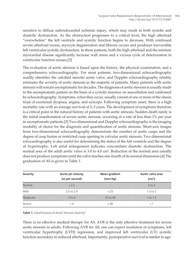
The evaluation of aortic stenosis is based upon the history, the physical examination, and acomprehensive echocardiography. For most patients, two-dimensional echocardiographyreadily identifies the calcified stenotic aortic valve, and Doppler echocardiography reliablyestimates the severity of aortic stenosis in the majority of patients. Many patients with aorticstenosis will remain asymptomatic for decades. The diagnosis of aortic stenosis is usually madein the asymptomatic patient on the basis of a systolic murmur on auscultation and confirmedby echocardiography. Symptoms, when they occur, usually consist of one or more of the classictriad of exertional dyspnea, angina, and syncope. Following symptom onset, there is a highmortality rate with an average survival of 2–3 years. The development of symptoms thereforeis a critical point in the natural history of patients with aortic stenosis. Sudden death rarely isthe initial manifestation of severe aortic stenosis, occurring at a rate of less than 1% per yearin asymptomatic patients.[3] Two-dimensional and Doppler echocardiography is the imagingmodality of choice for the diagnosis and quantification of aortic stenosis. Short-axis imagesfrom two-dimensional echocardiography demonstrate the number of aortic cusps and thedegree of cusp fusion or restricted cusp opening in valvular aortic stenosis. Two dimensionalechocardiography is also useful for determining the status of the left ventricle and the degreeof hypertrophy. Left atrial enlargement indicates concomitant diastolic dysfunction. Thenormal area of the adult aortic valve is 3.0 to 4.0 cm2. Reduction of the normal area usuallydoes not produce symptoms until the valve reaches one-fourth of its normal dimension.[4] Thegraduation of AS is given in Table 1.
Severity Aortic jet velocity
(m per second)
Mean gradient
(mm Hg)
Aortic valve area
(cm2)
Normal < 2.5 - 3 to 4
Mild 2.5 to 2.9 < 25 1.5 to 2
Moderate 3 to 4 25 to 40 1 to 1.5
Severe > 4 > 40 < 1
Table 1. Classifications of Aortic Stenosis Severity4
There is no effective medical therapy for AS. AVR is the only effective treatment for severeaortic stenosis in adults. Following AVR for AS, one can expect resolution of symptoms, leftventricular hypertrophy (LVH) regression, and improved left ventricular (LV) systolicfunction secondary to reduced afterload. Importantly, postoperative survival is similar to age-
Surgical Valve Replacement (Bioprosthetic VS Mechanical)http://dx.doi.org/10.5772/53687
363
matched controls after AVR for AS when performed prior to the development of LV dysfunc‐tion or congestive heart failure (CHF). Similarly, incomplete regression of LVH after AVR hasbeen associated with adverse outcomes such as reduced long-term survival. Contrary to theimmediate improvement in systolic performance, diastolic dysfunction may persist for severalmore years after AVR. In fact, Gjertsson et al. recently evaluated diastolic dysfunction in ASand found that the proportion of patients with moderate-to-severe diastolic dysfunctionactually increased with time after AVR despite normalization of LV mass and appropriateadjustments for senile diastolic dysfunction. Finally, AVR is associated with improved qualityof life scores, particularly among the elderly, and has been found to be similar to age-matchedindividuals without heart disease. [3,5,6]
The American College of Cardiology (ACC) and the American Heart Association (ACH) havejointly developed guidelines in which they published indications for AVR:
a. Definite indications:
• Patients who have severe AS and presented with one or more of its classical symptoms(angina, syncope, heart failure, etc.)
• Patients who have severe AS and required coronary artery bypass surgery, surgery onthe aorta or other heart valves
• Patients who have severe AS and left ventricle systolic dysfunction (ejection fractionless than 50 %)
b. Possible indications:
• Patients who have moderate AS and required coronary artery bypass surgery, surgeryon the aorta or other heart valves
• Asymptomatic patients with severe AS with abnormal exercise test, or an increase intransaortic gradient during exercise, or left ventricle systolic dysfunction (ejectionfraction less than 50 %), or left ventricular dilatation, or significantly elevated leftventricular diastolic pressure. [7]
The European Society of Cardiology (ESC) has also developed guidelines in which theypublished indications for AVR (Table 2).[8] They strongly recommended early AVR in allsymptomatic patients with severe AS.
Management of asymptomatic patients requires careful weighing of benefits against risks.Early elective surgery at these patients can only be recommended in selected patients, at lowoperative risk. This could be the case in:
• The rare asymptomatic patients with depressed LV function not due to another cause
• Those with echocardiographic predictors of poor outcome suggested by the combination ofa markedly calcified valve with a rapid increase in peak aortic velocity of ≥ 0.3 m/s per year
• If the exercise test is abnormal, particularly if it shows symptom development, which is astrong indication for surgery in physically active patients.
Calcific Aortic Valve Disease364

• However, on the other hand, breathlessness on exercise may be difficult to interpret inpatients with only low physical activity, particularly the elderly, making decisionmakingmore difficult. There is no strict age limit for performance of exercise testing and it isreasonable to propose it in patients > 70 years old who are still highly active.[8]
Patients with severe AS and any symptoms IB
Patients with severe AS undergoing coronary artery bypass surgery, surgery of the ascending aorta, or
on another valve
IC
Asymptomatic patients with severe AS and systolic LV dysfunction (LVEF < 50%) unless due to other
cause
IC
Asymptomatic patients with severe AS and abnormal exercise test showing symptoms on exercise IC
Asymptomatic patients with severe AS and abnormal exercise test showing fall in blood pressure
below baseline
IIaC
Patients with moderate AS undergoing coronary artery bypass surgery, surgery of the ascending aorta
or another valve
IIaC
Asymptomatic patients with severe AS and moderate-to-severe valve calcification, and a rate of peak
velocity progression ≥ 0.3 m/s per year
IIaC
AS with low gradient (< 40 mmHg) and LV dysfunction with contractile reserve IIaC
Asymptomatic patients with severe AS and abnormal exercise test showing complex ventricular
arrhythmias
IIbC
Asymptomatic patients with severe AS and excessive LV hypertrophy (≥ 15 mm) unless this is due to
hypertension
IIbC
AS with low gradient (< 40 mmHg) and LV dysfunction without contractile reserve IIbC
Table 2. Indications for AVR in AS
In last 50 years, the varieties of prostheses that have become available for use are numer‐ous. An ideal aortic prosthesis would be simple to implant, widely available, possesslong-term durability, would have no intrinsic thrombogenicity, would not have a predilec‐tion foe endocarditis and would have no residual transvalvular pressure gradient. Such avalve does not currently exist. Currently available options include mechanical valves,stented biological valves, stentless biological valves, allograft valves and pulmonary auto‐
Surgical Valve Replacement (Bioprosthetic VS Mechanical)http://dx.doi.org/10.5772/53687
365

graft valves. Commonly in us are mechanical and biological prostheses.[9] When selectingbetween mechanical and biologic heart valves, the surgeon and patient must balance therisks and benefits of each choice.
2. Mechanical prostheses
Charles Hufnagel in 1952. Used aortic valve ball and cage prosthesis heterotopically in thedescending aorta to treat aortic insufficiency. The first aortic valve replacement with an intracardiac mechanical prosthesis, which led to long terms survivors, was performed in 1960.Mechanical valves are classified according to their structure as caged-ball, single-tilting-diskor bileaflet-tilting-disk valves. The Starr-Edwards caged-ball valve has been available sincethe 1960’s and comprises a silastic ball, which rests on the sewing ring when closed and movesforward into the cage when the valve opens. The single-disk valves, for example, the Bjork-Shiley prosthesis and the Medtronic-Hall prosthesis, contain a disk that tilts between two strutsof the orifice housing. The most popular of the mechanical valves at present are the bileafletvalves, of which the St. Jude Medical valve and the Carbomedics valve are widely implanted.Both these devices are implanted within the aortic annulus. The two semi-circular leaflets ofthe bileaflet valve are connected to the housing by a butterfly hinge mechanism and swingapart during opening of the valve creating three outflow tracts, one central and two peripheralrespectively. In contrast to the configuration of the latter, the Carbomedics Top Hat (SulzerCarbomedics, Austin, TX) bileaflet aortic valve that was introduced in 1993 has a unique supra-annular design with all its components incorporated within the aortic sinuses.[10,11]
Mechanical valves are made from carbon, Teflon, Dacron, titanium and polyester and arevery durable. The current designs for the aortic and mitral positions include ball-and-cagevalves, single tilting disc prostheses, and bileaflet prostheses. Bileaflet mechanical valvesare the standard in current practice, with the St. Jude Medical (St. Jude Medical, Inc., St.Paul, MN) prosthesis the modern prototype, having been first implanted in 1977. Most ofthese valves are constructed using carbon strengthened with silicon carbide additives.Other examples of bileaflet mechanical valves include those manufactured by CarboMed‐ics (Austin, TX); Advancing the Standard Medical (ATS, Minneapolis, MN); Medtronic,Inc. (Minneapolis, MN); and Medical Carbon Research Institute, LLC (MCRI, Austin, TX).[10,11,12] The On-XR mechanical valve (MCRI) was introduced in Europe in 1996 and dif‐fers from other bileaflet mechanical valves in that it is made from pure pyrolytic carbon.The PROACT (Prospective Randomized On-X R Valve Anticoagulation Trial) study is anFDA-approved multicenter trial, sponsored by MCRI, currently enrolling patients to deter‐mine whether or not defined patient groups receiving AVR (low versus high risk for TEevents) with the On-X R _ valve may be safely maintained on lower doses of warfarin or,for patients in the lowrisk aortic valve arm, on antiplatelet drugs (aspirin plus clopidog‐rel) alone compared with standard anticoagulation regimens. No single mechanical valvehas shown superior patient outcomes, and all demonstrate extremely low rates of struc‐tural valve deterioration, the major advantage of mechanical valves.3
Calcific Aortic Valve Disease366

(a) (b) (c)
Figure 2. Mechanical valves: a) Starr-Edwards cage-ball valve, b) Bjork-Shiley mono-leaflet valve and c) St. Jude Medi‐cal bileaflet valve
3. Biological prosthesis
The biological prostheses include a wide variety of devices. Included within this broadcategory are the bioprostheses, a term which is used for valves with non-viable tissue ofbiological origin. The bioprostheses include the heterografts, composed of porcine (actualvalves of a pig) or bovine tissue (pericardium of a cow) and the allografts, which are preservedhuman aortic valves. The initial bioprostheses were mounted on stents to which the leafletsand sewing ring were attached but subsequently stentless valves, which are sewn in free hand,have been developed.[13] Stented bioprosthetic valves, which incorporate a semi-rigidexternal support structure for the valve leaflets, represent the majority of tissue valvesimplanted in clinical practice. The external support provides accurate valve mounting,improving ease of implantation. Two types of stented bioprosthetic valves are currentlyavailable in the United States: porcine aortic valves, which incorporate chemically stabilizedporcine valve leaflets mounted on a stented structure or frame, and bovine pericardial valves.The leaflets of the latter valve type are constructed from bovine pericardium and subsequentlymounted on a stented frame. Available porcine valves include the Medtronic Mosaic valve(Medtronic Inc., Minneapolis, MN), the St. Jude Medical Biocor and Biocor Supra valves (St.Jude Medical, Inc., St. Paul, MN), and the Carbomedics Mitroflow valve (Carbomedics, Inc.,Austin, TX). Bovine pericardial valves include the Carpentier–Edwards (C–E) Perimount(Edward Lifesciences, Irvine, CA) and the CE Perimount Magna valves as well as the SorinSoprano (Sorin Group, Saluggia, Italy) valves. At present, based on the best available data, noone bioprosthetic valve appears superior with regard to patient outcomes and none requiressystemic anticoagulation with warfarin, which is their major advantage. Their major disad‐vantage is the incidence of structural valve deterioration and subsequent need for reoperation,although the lifespan of the latest generation of tissue valves is unknown. Recent evidence alsosuggests that stentless biological valves may have better coronary flow reserve compared tostented valves. Additionally, compared with stented bovine pericardial valves, stentless valveshave been associated with increased transvalvular EOA and decreased pressure gradients
Surgical Valve Replacement (Bioprosthetic VS Mechanical)http://dx.doi.org/10.5772/53687
367
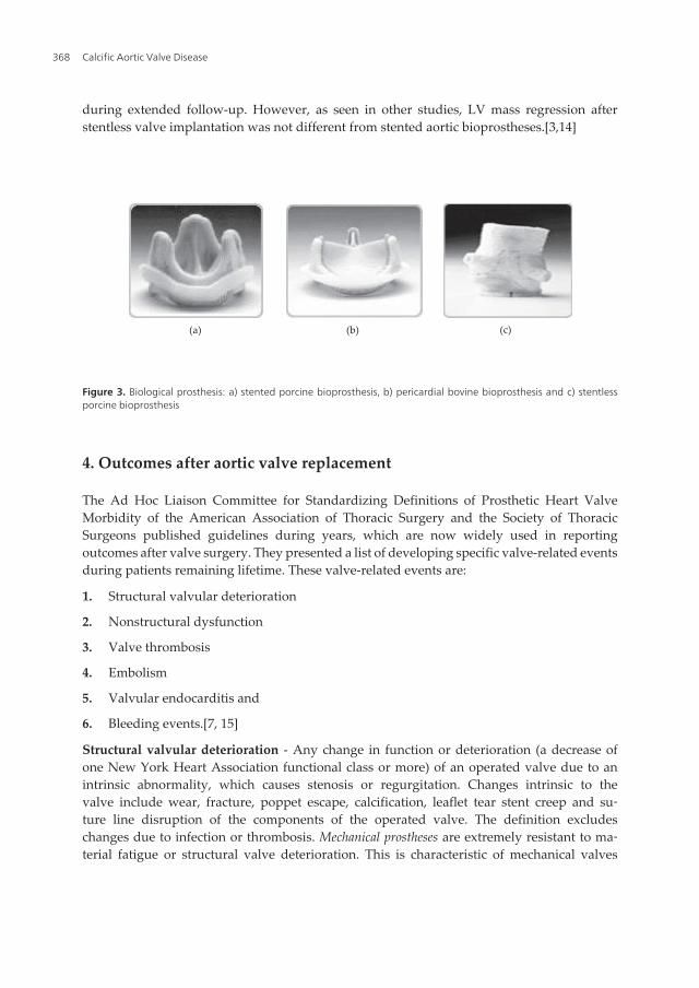
during extended follow-up. However, as seen in other studies, LV mass regression afterstentless valve implantation was not different from stented aortic bioprostheses.[3,14]
(a) (b) (c)
Figure 3. Biological prosthesis: a) stented porcine bioprosthesis, b) pericardial bovine bioprosthesis and c) stentlessporcine bioprosthesis
4. Outcomes after aortic valve replacement
The Ad Hoc Liaison Committee for Standardizing Definitions of Prosthetic Heart ValveMorbidity of the American Association of Thoracic Surgery and the Society of ThoracicSurgeons published guidelines during years, which are now widely used in reportingoutcomes after valve surgery. They presented a list of developing specific valve-related eventsduring patients remaining lifetime. These valve-related events are:
1. Structural valvular deterioration
2. Nonstructural dysfunction
3. Valve thrombosis
4. Embolism
5. Valvular endocarditis and
6. Bleeding events.[7, 15]
Structural valvular deterioration - Any change in function or deterioration (a decrease ofone New York Heart Association functional class or more) of an operated valve due to anintrinsic abnormality, which causes stenosis or regurgitation. Changes intrinsic to thevalve include wear, fracture, poppet escape, calcification, leaflet tear stent creep and su‐ture line disruption of the components of the operated valve. The definition excludeschanges due to infection or thrombosis. Mechanical prostheses are extremely resistant to ma‐terial fatigue or structural valve deterioration. This is characteristic of mechanical valves
Calcific Aortic Valve Disease368

whether they are aortic or mitral valves.[16] Because of the long-term durability of me‐chanical prostheses a valve replacement rate is less than 2% over 25 years. The most com‐mon reasons for reimplantation are pre- and postoperative endocarditis, paravalvular leakand valve thrombosis.[17] Bioprosthetic valves are not as durable, have a shorter lifespanand are more susceptible to calcification than human and mechanical valves.[13] Biopros‐theses have a significantly higher rate of reoperation due to structural valve deterioration.In large series, freedom from reoperation is > 90% at 10 years, but < 70 % at 15 years.[17,18,19] There is an important predisposition for premature bioprosthetic structuralvalve deterioration in younger patients, especially those under the age of 40 years.[17]
Nonstructural dysfunction - Any abnormality that is not intrinsic to the valve per se, whichcauses stenosis or regurgitation. Examples for this include entrapment of pannus, tissue orsuture; paravalvular leak; inappropriate sizing or positioning; residual leak and clinicallyimportant hemolytic anemia. This definition also excludes changes due to infection andthrombosis. Subvalvular pannus formation is rare with mechanical bileaflet valves.[20] Panusovergrowth and prosthetic structural degeneration interfering with normal valve opening andclosure may cause hemolysis severe enough for reoperation. Paravalvular leak is an operativecomplication and it is related to operative technique and to endocarditis.[21]
Valve thrombosis – Any thrombus, in the absence of infection, which is attached to or near anoperated valve that occludes part of the blood flow path or that interferes with function of thevalve. The incidence of prosthesis thrombosis is < 0.2 % per year and it occurs more often inmechanical prostheses.[22] It is most commonly due to inadequate anticoagulation or noncom‐pliance. Freedom from valve thrombosis at 20 years is > 97 %.[23,24]
Embolism – Any embolic event that occurs, in the absence of infection, after the immediateperi-operative period. This could be either a neurologic or peripheral embolic event. Aneurologic event includes any new, temporary or permanent focal or global neurologic deficit.A peripheral embolic event is due to an embolus that produces symptoms from obstruction ofa peripheral (non-cerebral) artery. The incidence of thromboembolic events between biopros‐theses and mechanical prostheses are the same.[25] This is a continuous risk factor that ispresent through the life of patients with mechanical valve prosthesis, so they must maintaintherapeutic anticoagulant levels. The embolic risk is highest in the first few months, before thering and valve components have fully endothelialized.[26] Acceptable thromboembolic ratesrange between 0.8 and 2.3 % per patient-year.[21,22,25] 50 % of these events are neurologic, 40% are transient and 10 % are peripheral.[21]
Valvular endocarditis – Any infection involving an operated valve diagnosed by customaryclinical criteria. It is rare case with prophylactic antibiotics. Around 60 % of events occur earlyand are associated with staphylococci. The mortality for this event is high. Freedom fromendocarditis with mechanical prosthesis is 97 to 98 % at 20 to 25 years. A number of studieshave reported a higher incidence of valvular endocarditis after mechanical valve replacementin comparison with the biologic valve replacement during the initial few months afterimplantation. Bioprostheses are less susceptible to early infection, which is often restricted tothe leaflets, making cure with antibiotics more likely but increasing the chances of late failuredue to degeneration of the cusps.[27,28,29]
Surgical Valve Replacement (Bioprosthetic VS Mechanical)http://dx.doi.org/10.5772/53687
369

Bleeding event – Formerly classified as anti-coagulant hemorrhage, a bleeding event is anepisode of major internal or external bleeding that causes death, hospitalization, and perma‐nent injury or requires transfusion. This definition applies to all patients, irrespective of anti-coagulation status. Mechanical valves are durability but anticoagulation is key of long-termsuccess. International Normalized Ratio (INR) is the standard to which anticoagulation levelsshould be targeted. Level of INR should be individual for each person. Complications occurduring fluctuations in the INR and less during steady-state levels, be they high or low.[30,31]When levels of INR increase, bleeding episodes become more common, and when levels ofINR decrease thromboembolic episodes become more common. Some studies showed thataround 40 % of the bleeding episodes occurred in the first year after surgery, when levels ofINR are more likely to fluctuate. Many studies suggested that in the early postoperative periodslowly raise the level of INR to therapeutic levels is needed, to prevent bleeding events.[21,32,33] According to ACC and ACH after mechanical AVR, the goal of antithrombotictherapy is usually to achieve an INR of 2.5 to 3.5 for the first 3 months after surgery and 2.0 to3.0 beyond that time. At that level of anticoagulation, the risk of significant hemorrhageappears to be 1% to 2% per year.7 Low-dose aspirin is also indicated in addition to warfarin toresult in a lower incidence of thromboembolic event, with a low possibility for bleeding.[34]Older patients are at higher risk for thromboembolic event because of the greater number ofrisk factors that accumulate with aging.[30] Anticoagulation-related hemorrhage (ARH) is themost common valve-related event. More often it will occur during fluctuations in INR, whichhappens most often early after valve replacement.[21,22] The most common places for ARHare gastrointestinal tract and central nervous system.[21] Acceptable ARH rates range from1.0 to 2.5% per patient-year in long term reports.[21,22,25,35] It is very dangerous complication,because mortality more often occurs in relation to bleeding events than in relation to throm‐boembolic events.[21]
Operative mortality – Operative mortality is defined as all-cause mortality within 30 days ofoperation. According to the Society of Thoracic Surgeons mortality for isolated AVR is 4.3%and for AVR with concomitant coronary artery disease is 8%.[36] Many factors have beenassociated with an increased risk of operative mortality in isolated AVR. Some of these riskfactors are age, female gender, diabetes, renal failure, and emergency status, previousoperation, advanced preoperative NYHA class, lower cardiac index, concomitant coronaryartery bypass grafting and longer aortic crossclamp and cardiopulmonary bypass timerespectively.[37] In the absence of major comorbidities and preserved ejection fraction, isolatedAVR can be performed with an expected mortality of less than 2%.[38]
Several studies have evaluated independent risk factors for operative mortality after AVR.Five variables predictive of increased mortality risk after AVR are common to each of theseanalyses: preoperative renal failure, urgency of AVR, preoperative heart failure, presence ofCAD or recent MI, and redo cardiac operation. Other factors independently associated withoperative mortality from the individual studies include preoperative atrial fibrillation, activeendocarditis, preoperative stroke, advanced age, lower body surface area, multiple valveprocedures, and hypertension. [39, 40]
Calcific Aortic Valve Disease370
5. Factors affecting long-term outcome after AVR
• Demographic
◦ Older age
◦ Male sex
• Clinical
◦ Higher pre-operative NYHA functional class
◦ Pre-operative atrial fibrillation and non-sinus rhythm
◦ Pure aortic regurgitation
◦ Hypertension
◦ Diabetes mellitus
◦ Renal failure
• Surgery-related
◦ Longer cardiopulmonary bypass time
• Morphological
◦ Previous myocardial infarction
◦ Left ventricular structure and functional abnormality
◦ Previous aortic valve surgery
◦ Coronary artery disease (CAD)
Older patients have a lot of comorbiditis and they are at higher risk for valve-related events.Atrial fibrillation is one of the risk factor for thromboembolism, because of that INR levelsmust be higher (INR 2.5 to 3.5) than regular.[30,34] The majority of patients undergoing AVRhave other cardiac lesions, most commonly CAD, and more complex pathology has beenassociated with increased risk. Combined myocardial revascularization and AVR increasescross-clamp time and has the potential to increase perioperative myocardial infarction andearly postoperative mortality compared with patients undergoing isolated AVR.7 In additionto severity of CAD and AS, the multivariate factors for late postoperative mortality includelow ejection fraction, severity of LV dysfunction, age greater than 70 years (especially inwomen), and presence of NYHA functional class IV symptoms.[36]
6. Patient selection
Propter selection of patients for valve replacement can bring us excellent long-term results,long-term survival and low incidence of valve-related complications.
Surgical Valve Replacement (Bioprosthetic VS Mechanical)http://dx.doi.org/10.5772/53687
371

In some studies of patients followed over longer time frames, freedom from all valve-relatedevents and freedom from reoperation were improved in patients with mechanical valveprostheses as compared to patients with biological prostheses. [9,16,25] Key of long-termsuccess of mechanical valve prostheses is anticoagulation. Patients that are inconsistent,noncompliant or incapable of managing medications are not good candidates for long-termchronic anticoagulation.[39,41] Also patients with higher levels of education and those fromgeographic areas with a good medical infrastructure have better compliance with necessarymedications and anticoagulant monitoring.[31]
Many centers used bioprosthetic valves for patients who are older than 70 year, based on databy Akins.[42] In patients younger than 60 years of age, the best solution would be implantationof mechanical valves, based on prosthesis durability and they have low-risk for valve-relatedevents.[21] In decade between 60 and 70 years of age, other factors have to be taken intoaccount.7 According to some studies, patients over 65 years at the time of surgery shouldreceive a biologic valve. Patients under the age of 60 should have a mechanical prosthesis tominimize the risk of structural failure requiring repeat AVR in an octogenarian. Patientsbetween 60 and 65 represent the group in whom there is still considerable debate regardingprosthesis selection. Those patients who have comorbidities such as severe CAD may be lesslikely to outlive their prosthesis and should receive a biologic valve. A detailed discussion ofthese risks and benefits of prosthesis selection should occur with all patients and their familiesprior to entering the operating room. [3,7,22,24,25,37,38]
In the early follow-up period, anticoagulation – related hemorrhage is the most commonunwanted event for mechanical valve prostheses. Over the first 10 years of follow-up there isa higher incidence of valve-related events in patients with mechanical prostheses as opposedto those with biologic valves.[32] However, in the next 10 to 20 years after AVR, the incidenceof valve failure and valve-related complications are much higher at biologic prostheses thanthose with mechanical valve prostheses. Some series showed that the time to biologic valvefailure was only 7.6 years.[43] This failure rate will increase over time. However, freedom fromvalve-related events is more strongly influenced by pre-existing comorbidities than thepresence of mechanical prostheses.[21], [22, 25, 31]
The elderly patient with severe aortic stenosis poses a therapeutic challenge. In consideringelderly patients for aortic valve replacement, important factors include the presence ofsymptoms, physiologic age, patient expectations, anticipated future activities, and comorbid‐ity. The operation itself carries a higher risk than in younger patients. Extensive calcificationof the aorta and annulus as well as fragile tissue presents significant technical difficulties forthe surgeon. In addition, particularly in women, the aortic root and annulus may be small andrequire concomitant enlargement to accommodate the valve prosthesis. Furthermore, pro‐truding arch atheroma occurs in one-fifth of patients > 65 years of age and significantlyincreases the risk of stroke and mortality during cardiac surgery. Major postoperativecomplications, nevertheless, remain high, with the incidence of permanent stroke between 4and 6%. Rehabilitation can also be a problem, as elderly patients take longer to recover fromsurgery. Survival has clearly improved in these elderly patients with severe symptomaticaortic stenosis who undergo aortic valve replacement. Survival is 80–85% at 1 year and 60–
Calcific Aortic Valve Disease372
70% at 5 years, which is similar to an age- and sex-matched population without aortic valvedisease. Most patients report improved functional capacity and quality of life, with more than90% of patients feeling better after surgery.3
A major deterrent to mechanical valve replacement in the younger patient is the impact oflong-term anticoagulation. Mechanical valves are, however, more ideal for younger patientsdue to their excellent durability characteristics. Most importantly, younger patients (i.e.,patients under the age of 50 years) are a low-risk subset for valve related events. Theseindividuals have very few risk factors for TE, and thus anticoagulation can be run at the lowerend of the therapeutic target range, decreasing the incidence of anticoagulant-related hemor‐rhage without altering the incidence of TE. In fact, many infants and children have beenmanaged with only aspirin with quite good long term results. While this is not recommendedin patients older than infancy, it is a feasible alternative. A recent study in patients under 50years of age followed 254 patients for up to 20 years and found an exceedingly low rate ofvalve related events, an exceptional long-term overall survival of nearly 88%, and event-freesurvival probability of 92% at 19 years.[3,44,45]
Patients with an absolute requirement for long-term anticoagulation such as atrial fibrillation,previous thromboembolic events, hypercoagulable state, severe LVD, another mechanicalheart valve in place, or intracardiac thrombus, should receive a mechanical valve regardlessof age. Patients in whom anticoagulation with warfarin is contraindicated, such as women ofchild-bearing age wishing to become pregnant, patients with other bleeding disorders, or thosewho refuse anticoagulation should receive a bioprosthesis. There is growing interest in usingmechanical prostheses in women of child-bearing age and providing anticoagulation withsubcutaneous low-molecular weight heparin injections. Patients with end-stage renal failurewere previously believed to have significantly elevated risk for early bioprosthetic structuralvalve deterioration. However, increased anticoagulation- related complications are also morelikely in this group, and the current ACC/AHA guidelines do not recommend routine use ofmechanical prostheses in these patients.[3,7,8,9,10]
The decision between bioprosthetic and mechanical valve should be made by the patient witheducated input regarding the pros and cons of each option from the patient’s physicians. Todaysurgeons implant bioprosthetic valves in younger patients who wish to avoid anticoagulationdue to lifestyle concerns (e.g. young, active individual, desire to become pregnant, etc.),although surgeons generally will guide patients toward a mechanical option at the time ofredo-AVR if their life expectancy exceeds 10–15 years at that time.[3]
7. Operative technique
Aortic valve replacement is most frequently done through a median sternotomy, meaningthe incision is made by cutting through the sternum. Once the pericardium has beenopened, the patient is put on a cardiopulmonary bypass machine. This machine takes overthe task of breathing for the patient and pumping their blood around while the surgeonreplaces the heart valve.
Surgical Valve Replacement (Bioprosthetic VS Mechanical)http://dx.doi.org/10.5772/53687
373

Once the patient is on bypass, a cut is made in the aorta and a crossclamp applied. The surgeonthen removes the patient`s diseased aortic valve and a mechanical or biological valve is put inits place. Once the valve is in place and aorta has been closed, the patient is taken off the heart-lung machine. Transesophageal echocardiogram can be used to verify that the new valve isfunctioning property. Pacing wires are usually put in place, so that the heart can be manuallypaced should any complications arise after surgery. Drainage tubes are also inserted to drainfluids from the chest and pericardium following surgery. These are usually removed within36-48 hours while the pacing wires are generally left in place until right before the patient isdischarged from the hospital.
8. Patient-prosthesis mismatch
Prosthesis‐patient mismatch (PPM) is that a smaller than expected effective orifice area (IEOA)in relation to the patient's body surface area (BSA) will result in higher transvalvar gradients.It is condition that occurs when the valve area of a prosthetic valve is less than the area of thatpatient’s normal valve.[46] Several authors suggest that prosthesis-patient mismatch occurs atan IEOA of 0.85 cm2/m2.[46,47] Transvalvular gradients begin to rise substantially at IEOAsbelow this value, and these elevated gradients potentially cause increased left ventricular workthat prevents adequate regression of left ventricular hypertrophy. Several factors includingage, body mass index (BMI), and pre-operative status of left ventricular function may poten‐tially influence the effect of PPM on post-operative outcomes.[46] PPM is associated with asignificant reduction in cardiac index during the postoperative course. The incidence ofcongestive heart failure was significantly higher in patients with PPM.[48] Several studiesreported that early mortality is significantly increased in patients with PPM.[47, 48, 49, 50]
The projected indexed EOA should be systematically calculated at the time of the operation toestimate the risk of PPM. PPM can be avoided by using a simple strategy at the time ofoperation. Pibarot suggested that surgeon first calculate the patient's BSA from his or herweight and height. Than multiply BSA by 0.85 cm2/m2, the result being the minimum EOA thatthe prosthesis to be implanted should have to avoid PPM, and than choose the prosthesis andthe reference values for the different types and sizes of prosthesis.[46, 47]
Due to concerns over PPM, stentless bioprosthetic valves, which generally have a largerEOA sizefor- size compared with mechanical or stented bioprosthetic valves, have beenincreasingly utilized for AVR. In initial evaluation, stentless valves had better hemody‐namics and improved survival rates relative to stented biological or mechanical valvesand were more durable than stented biological valves. Stentless valves may be preferredin patients with a small aortic root, and arguments have been made that wider utilizationof stentless valves may minimize PPM. Stentless valves also appear to have better hemo‐dynamic profiles than stented valves during exercise testing. Technical reasons for not im‐planting stentless valves include extensive aortic root calcification, coronary ostia opposedby 180, presence of the two coronary ostia in close proximity, or unusual disproportionbetween the sinotubular junction and the aortic annulus. Whereas stented valves allow
Calcific Aortic Valve Disease374
perfect valve mounting within the aortic annulus, thus reducing the risk of implanting anincompetent valve, postoperative AR and limited durability remain a concern with thefree-hand stentless valve insertion technique. This issue may be circumvented with fullaortic root replacement using a stentless porcine root.[3.49,50]
Author details
Stamenko Šušak1*, Lazar Velicki1, Dušan Popović1 and Ivana Burazor2
*Address all correspondence to: [email protected]
1 Institute of Cardiovascular Diseases Vojvodina, Sremska Kamenica, Serbia
2 Clinical Centers, Nis, Serbia
References
[1] Malouf, J. F, Edwards, W. D, Tajik, A. J, & Seward, J. Functional anatomy of theheart. In: Fuster V, O’Rourke RA, Walsh RA, Poole-Wilson P, eds. Hurst’s The Heart.12th ed. New York, NY: McGraw-Hill Companies, Inc; (2008).
[2] Otto, C. M, Lind, B. K, Kitzman, D. W, et al. Association of aortic valve sclerosis withcardiovascular mortality and morbidity in the elderly. N Eng J Med (1999).
[3] Andrew WangThomas S. Bashore. Valvular Heart Disease. Humana Press, a part ofSpringer Science Business Media, LLC (2009).
[4] Gjertsson, P, Caidahl, K, Farasati, M, et al. Preopeartive moderate to severe diastolicdysfunction: A novel Doppler echocardiographic long-term prognosis factor in pa‐tients with severe aortic stenosis. J Thorac Cardiovasc Surg (2005).
[5] Connolly, H. M, Oh, J. K, Orszulak, T. A, et al. Aortic valve replacement for aorticstenosis with severe left ventricular dysfunction: prognostic indicators. Circulation(1997). , 95, 2395-400.
[6] Gjertsson, P, Caidahl, K, & Bech-hanssen, O. Left ventricular diastolic dysfunctionlate after aortic valve replacement in patients with aortic stenosis. Am J Cardiol(2005). , 96, 722-7.
[7] Bonow, R. O, Carabello, B. A, & Chatterjee, K. de Leon AC Jr, Faxon DP, Freed MD,Gaasch WH, Lytle BW, Nishimura RA, O’Gara PT, O’Rourke RA, Otto CM, Shah PM,Shanewise JS. ACC/AHA 2006 guidelines for the management of patients with valv‐ular heart disease: a report of the American College of Cardiology/American HeartAssociation Task Force on Practice Guidelines (Writing Committee to Develop
Surgical Valve Replacement (Bioprosthetic VS Mechanical)http://dx.doi.org/10.5772/53687
375
Guidelines for the Management of Patients With Valvular Heart Disease). Circula‐tion. (2006). ee231. DOI:CIRCULATIONAHA.106.176857, 84.
[8] The European Society of Cardiology Guidelines on the management of valvularheart disease European Heart Journal (2007.
[9] Gott, V. L. Alejo DE: Mechanical heart valves: 50 years of evolution. Ann Thorac Surg(2003). S2230
[10] Bloomfield, P. Choice of heart valve prosthesis. Heart. (2002). , 87, 583-9.
[11] Dewall, R. A, Qasim, N, & Carr, L. Evolution of Mechanical Heart valves. Ann Thor‐ac Surg. (2000). , 69, 1612-21.
[12] Bonow, R. O, Carabello, B. A, Chatterjee, K. C, De Leon, J. R, Faxon, A. C, Freed, D. P,Shah, M. D, & Acc, P. M. AHA 2006 guidelines for the management of patients withvalvular heart disease. Journal of the American College of Cardiology, 48(3),1-148.doi:10.1016/j.jacc.2006.05.021
[13] YangThang, "Mechanical Versus Bioprosthetic Valve Replacement in Valvular HeartDisease: A Systematic Review" ((2011). School of Physician Assistant Studies. Paper240.http://commons.pacificu.edu/pa/240
[14] Tsialtas, D, Bolognesi, R, Beghi, C, et al. Stented versus stentless bioprostheses inaortic valve stenosis: effect on left ventricular remodeling. Heart Surg Forum (2007).E, 205-10.
[15] Golubovic, M, Mihajlovic, B, Kovacevic, P, Cemerlic-adjic, N, Pavlovic, K, Velicki, L,& Susak, S. Postoperativne neletalne komplikacije posle operacije na otvorenom srcu.Vojnosanitetski pregled (2012). , 69(1), 27-31.
[16] Stamenko, S. Susak. Mitralna regurgitacija- kardiohirurski aspekti dijagnostike i tera‐pije. Mediterran Publishing, Biblioteka Academica, knjiga 15, Novi Sad (2010).
[17] Emery, R. W, Arom, K. V, Krogh, C. C, et al. Reoperative valve replacement with theSt.Jude Medical valve prosthesis: long-term follow up. J Am Coll Cardiol (2004). A
[18] Desai, N. D, Merin, O, Cohen, G. N, et al. Long-term results of aortic valve replace‐ment with the St.Jude Toronto stentless porcine valve. Ann Thorac Surg (2004).
[19] Grunkemeier, G. L, Jamieson, W. R, & Miller, D. C. Starr A: Actuarial versus actualrisk of porcine structural valve deterioration. J Thorac Cardiovasc Surg (1994).
[20] Vongpatanasin, W, & Hills, L. D. Lange RA: Prosthetic heart valve. N Eng J Med(1996).
[21] Emery, R. W, Krogh, C. C, Arom, D. V, et al. The ST. Jude Medical cardiac valveprosthesis: A year experience with single valve replacement. Ann Thorac Surg(2005). , 25.
Calcific Aortic Valve Disease376
[22] Ikonomidis, J. S, Kratz, J. M, Crumbley, A. J, et al. Twenty-year experience with theSt.Jude Medical mechanical valve prosthesis. J Thorac Cardiovasc Surg (2003).
[23] Lengyel, M. Vandor L: The role of thrombolysis in the management of left-sidedprosthetic valve thrombosis: a study of 85 cases diagnosed by transesophageal echo‐cardiography. J Heart Valve Dis (2001).
[24] Durrleman, N, Pellerin, M, Bouchard, D, et al. Prosthetic valve thrombosis: twenty-year experience at the Montreal Heart Institute. J Thorac Cardiovasc Surg (2004).
[25] Khan, S. S, Trento, A, Derobertis, M, et al. Twenty-year comparison of tissue and me‐chanical valve replacement. J Thorac Cardiovasc Surg (2001).
[26] Heras, M, Chesebro, J. H, Fuster, V, et al. High risk of thromboemboli early after bio‐prosthetic cardiac valve replacement. J Am Coll Cardiol (1995).
[27] Mahesh, B, Angelini, G, Caputo, M, Jin, X. Y, & Bryan, A. (2005). Prosthetic valve en‐docarditis. Ann. Thorac. Surg., 0003-4975, 80(3), 1151-1158.
[28] Velicki, L, Susak, S, Cemerlic-adjic, N, & Redzek, A. Aortic valve endocarditis, Chap‐ter in: Ying-Fu Chen and Chwan-Yau Luo- Aortic valve, InTech Publishing (2011).
[29] Velicki, L, Susak, S, & Srdanovic, I. Kovacevic M; Infective endocarditis of nativeaortic valve: destruction of leaflet with an aorto-cavitary fistula to the right ventricle,Chirurgia (2010). , 23(6), 261-266.
[30] Koertke, H, Minami, K, Boethig, D, et al. INR self-management permits lower antico‐agulation levels after mechanical heart valve replacement. Circulation (2003). SupplII):II-75.
[31] Butchart, E. G, Ionescu, A, Payne, N, et al. A new scoring system to determine throm‐boembolic risk after heart valve replacement. Circulation (2003). Suppl II):II-68.
[32] Kumar, D, & Elefteriades, J. Ezekowitz MD: Anticoagulation in patients with pros‐thetic heart valves. Cardiac Surg Today (2004).
[33] Koo, S, Kucher, N, Nguyen, P. L, et al. The effect of excessive anticoagulation onmortality and morbidity in hospitalized patients with anticoagulant-related majorhemorrhage. Arch Intern Med (2004).
[34] Massel, D. Little SH: Risk and benefits of adding antiplatelet therapy to warfarinamong patients with prosthetic heart valves: a metaanalysis. J Am Coll Cardiol(2001).
[35] Lund, O, Nielsen, S. L, Arildsen, H, et al. Standard aortic St. Jude valve at 18 years:performance, profile and determinants of outcome. Ann Thorac Surg (2000).
[36] Society of Thoracic Surgeons National Cardiac Surgery DatabaseAvailable at: http://www.sts.org/documents/pdf/STS-ExecutiveSummaryFall2005.pdf. November (2005).
Surgical Valve Replacement (Bioprosthetic VS Mechanical)http://dx.doi.org/10.5772/53687
377
[37] Edwards, F. H, Peterson, E. D, Coombs, L. P, et al. Prediction of operative mortalityafter valve replacement surgery. J Am Coll Cardiol (2001).
[38] David TE: Surgery of the aortic valve. (1999). Curr Probl Surg.
[39] Rankin, J. S, Hammill, B. G, Ferguson, T. B, et al. Determinants of operative mortalityin valvular heart surgery. J Thorac Cardiovasc Surg (2006). , 131, 547-57.
[40] Kuduvalli, M, Grayson, A. D, Au, J, et al. A multi-centre additive and logistic riskmodel for in-hospital mortality following aortic valve replacement. Eur J Cardiothor‐ac Surg (2007). , 31, 607-13.
[41] Butchart, E. G, Payne, N, Li, H, et al. Better anticoagulation control improves survivalafter valve replacement. J Thorac Cardiovasc Surg (2002).
[42] Akins, C. W, Buckley, M. J, Daggett, W. M, et al. Risk of reoperative valve replace‐ment for failed mitral and aortic bioprostheses. Ann Thorac Surg (1998).
[43] Potter, D. D, & Sundt, T. M. rd, Zehr KJ, et al: Operative risk of reoperative aorticvalve replacement. J Thorac Cardiovasc Surg (2005).
[44] Cabalka, A. K, & Emery, R. W. Petersen RJ: Long-term follow-up of the St. Jude Med‐ical prosthesis in pediatric patients. Ann Thorac Surg (1995). S618
[45] Emery, R. W, Erickson, C. A, Arom, K. V, et al. Replacement of the aortic valve inpatients under 50 years old with the St. Jude Medical prosthesis. Ann Thorac Surg(2003).
[46] Dumesnil, J. G. Pibarot P: Prosthesis-patient mismatch and clinical outcomes: the evi‐dence continues to accumulate. J Thorac Cardiovasc Surg (2006).
[47] Pibarot, P, & Dumesnil, J. G. Prosthesis-patient mismatch: definition, clinical impact,and prevention. Heart. (2006). , 92, 1022-9.
[48] Milano, A D, De Carlo, M, Mecozzi, G, et al. Clinical outcome in patients with 19‐mmand 21‐mm St. Jude aortic prostheses: comparison at long‐term follow‐up, Ann Thor‐ac Surg (2002). , 7337-43.
[49] Rao, V, Jamieson, W, & Ivanov, E. J. et al Prosthesis‐patient mismatch affects survivalfollowing aortic valve replacement. Circulation (2000). IIIIII9.III9, 5.
[50] Blais, C, Dumesnil, J G, Baillot, R, et al. Impact of prosthesis‐patient mismatch onshort‐term mortality after aortic valve replacement. Circulation (2003). , 108983-988.
Calcific Aortic Valve Disease378