surgical pathology of the appendix
DESCRIPTION
Surgical pathology of the appendix. Acute appendicitis Chronic appendicitis Tumors of the appendix. Appendix. Functions – not clear in humans - it may have a significance in immune defense – abundance of lymphoid follicles - PowerPoint PPT PresentationTRANSCRIPT
Surgical pathology of the appendix
Acute appendicitisChronic appendicitisTumors of the appendix
Appendix
Functions – not clear in humans- it may have a significance in immune defense – abundance of lymphoid follicles
- removal of the appendix may be a cause for an increase in colonic cancer incidence - not supported by controlled studies
- endocrine function
“Normal” Anatomy

Typical position
2.5 cm bellow the ileo-cecal valve (base of appendix) the only fix region – important when trying to find the appendixTaeniae converge at the base of the appendix84% free mobile in any possible location16% fixed retrocecal
Acute apendicitis
Essentials of diagnosisAbdominal painAnorexia, nausea, vomitingLocalized abdominal tendernessLow grade feverLeukocytosis
General considerations= acute inflammation of the appendix wall that starts in the mucosa and may extend to adjacent organs
70% of cases present obstruction of the proximal lumen: Fibrous bands, fecaliths, foreign bodies Tumors, parasites, lymphoid hyperplasia External compression
Inflammation starts in the mucosa with ulcerations and secondary bacterial infection
Close tube
Blood supply affected as disease progresses Infection in the wall Increased pressure
Puss formation inside the lumenWall destruction: gangrene + perforationBacterial peritonitis may be limited by adhesions (plastic peritonitis)
Clinical findings
Protean manifestation: may mimic a variety of conditions
Progression of symptoms is essential
Clinical findings
Onset: vague abdominal discomfortFollowed: Nausea, anorexia, indigestionVomitingPain, mild, localized in the epigastrum
Pain: localized in RLQ +Pain or discomfort (moving, walking,
coughing)
Examination
At this moment:Tenderness on coughing, localized in RLQLocalized tenderness on palpation Slight muscular rigidityRebound tenderness referred to the same
areaRectal and pelvic examination NORMALLow fever (<38 degrees)
Examination – retrocecal appendicitis
Poorly localized pain (retrocecal position – protected from the abdominal wall)
No discomfort on coughing, walking etc.
Diarrhea
Urinary symptoms (hematuria, urinary frequency)
Pain in the flank – tenderness on one finger examination
Examination – pelvic appendicitis
May simulate gastroenteritis
Nausea, vomiting and diarrhea are more prominent (adjacent appendix to pelvic colon)
Negative abdominal examination
IMPORTANT – repeated pelvic (rectal) examination
Aberrant positions
Left side appendix – confusion with diverticulitis (malrotation)
RUQ – cecum in abnormal position may mimic cholecystitis or perforated duodenal ulcer
Normal cecum – long appendix – anything is possible
Lab workupHigh leukocyte count: average 15.000/μl, 90% more the 10.000 with more then 75% neutrophils.10% have normal formula
Urinalysis typically normal, few leukocytes or eritrocytes. Retrocecal or pelvic – special attention
X-Ray findings
Plain X-Ray films are usually not contributory Air-fluid levels or isolated ileusFecalithsFree air in the peritoneumSigns of peritonitis
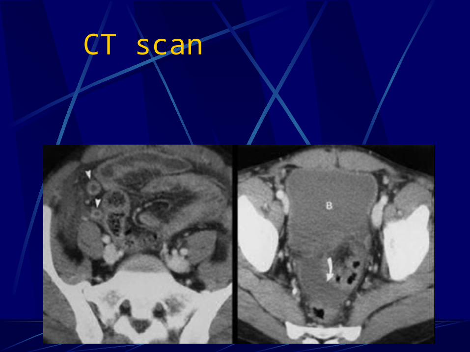
CT scan
Ultrasound scan
Appendicitis in pregnancy
Same frequency as in non-pregnant
Difficult diagnosisHigh position of the appendixAll usual signs are presentDifficult to interpret leukocytosis
Appendectomy is mandatory and urgent
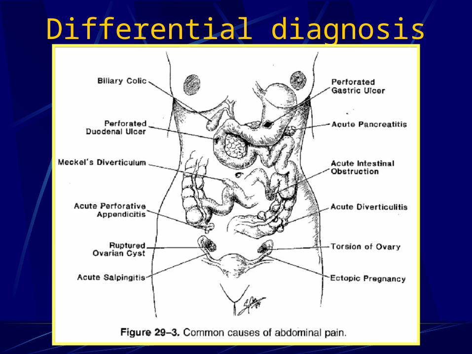
Differential diagnosis
Differential diagnosis
Difficult in young and elderly – highest incidence of perforation
High incidence of false positive appendicitis: women 20-40 PID and other genital conditions
Differential diagnosis
Local inflammatory conditions (enterocolitis, urinary infections, urinary stones, pelvic inflammatory disease)Distant digestive diseases (compliacted duodenal ulcer, billiary stones) Distant non-digestive diseases (penumonia, myocardial infarction, porphyria, lead poisoning)
Complications
PERFORATIONMore severe painFever >38Typically in the first 12 hours In 50% of patients the appendix is
perforated at the time of diagnosis
Complications
PERITONITISLocalized – microscopic perforation Increased tenderness, rigidity Abdominal distension Ileus Fever high and toxicity Douglas pouch very sensible
Generalized – classic presentation
Complications
APPENDICEAL ABSCESS (appendiceal mass)Localized peritonitisWalled off by peritoneumSymptoms of appendicitis + mass in RLQUS + CT characteristical
Complications
APPENDICEAL ABSCESSTreatment: ATB + diet low in residueDrainage of abscess +/- appendectomyPostponed appendectomy 8-12 weeks
Differential diagnosis:Carcinoma of the cecumTumors of the appendixGenital pathology
Complications
Pylephlebitis: suppurative thrombophlebitis of pportal veinChills, high fever, jaundice + hepatic
abscess formation.Serious septic problems
CT scan + US: thrombosis and gas in portal systemTreatment: ATB + surgery urgent
Treatment
CHRONIC APPENDICITIS
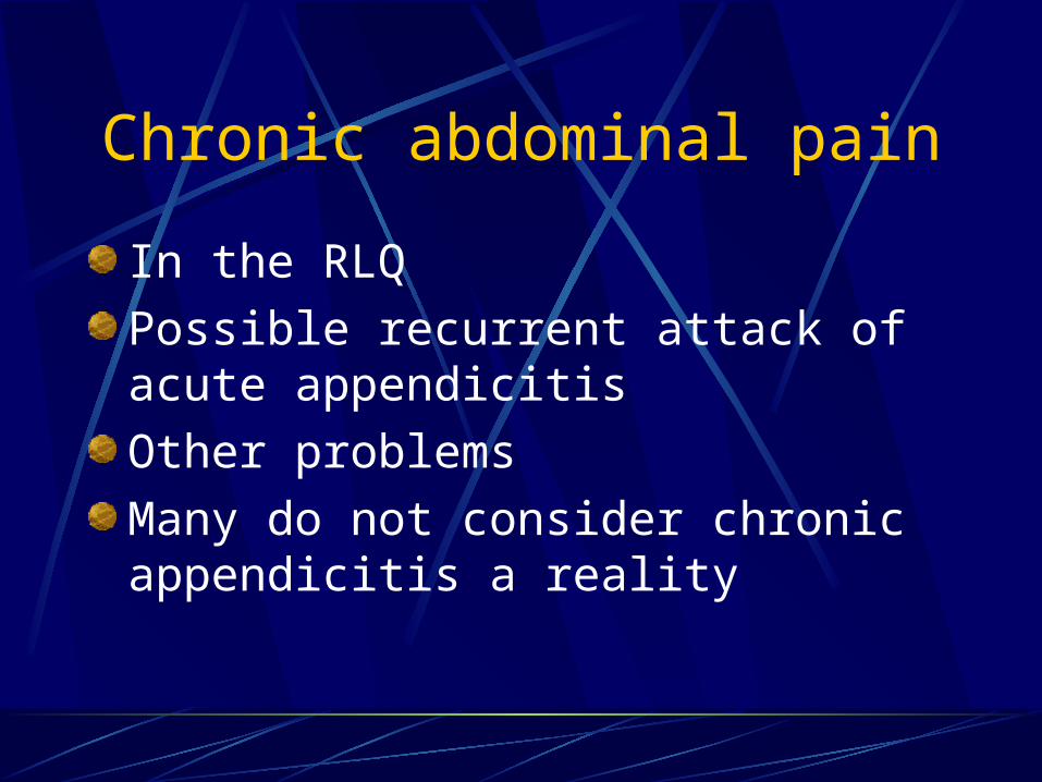
Chronic abdominal pain
In the RLQ
Possible recurrent attack of acute appendicitis
Other problems
Many do not consider chronic appendicitis a reality
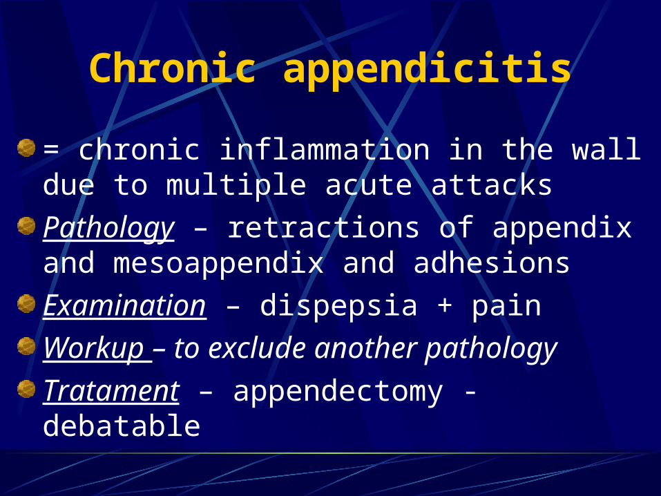
= chronic inflammation in the wall due to multiple acute attacks
Pathology – retractions of appendix and mesoappendix and adhesions
Examination – dispepsia + pain
Workup – to exclude another pathology
Tratament – appendectomy - debatable
Chronic appendicitis
Tumors of the appendix
Classification
Benign – fibroma
- leyomioma
- lypoma
Malignant – carcinoma
Bordeline - carcinoid
- mucocele
Benign tumors
Very rare
Occasionally may obstruct the lumen and cause acute apendicitis
May arise as a mass in RLQ
Carcinoma Rare and never diagnosed preoperativelyMost typical presents as acute appendicitis or RLQ abscessPrognosis: bad – 10% wide spread MTS at time of diagnosis. Rapid lymph node spread and local spread through peritoneal cavity (ovary)Treatment: right hemicolectomy + lymph node dissection
Carcinoid tumor
The most common location of carcinoid in the digestive tractSlow growth (<2 cm) and rarely MTS. 3% MTS in lymph nodesCarcinoid sdr: attacks of vasodilation, diarrhea, abdominal colical pain, tachicardia, hipotension MTSMTSExamination: RLQ pain + mass
Carcinoid
Lab workup:Urinary 5HIAUS, CT, arteriography, bronchoscopy
Treatment:AppendectomyRight hemicolectomy (>2cm, invasion of
cecum, invasion mesoappendix, nodes)MTS – enucleation (<4) +/or chemotherapy
Mucocele
Not true tumors:Chronic distension of the appendix plus continuous
mucus secretion. Flattened epithelial cellsCystadenoma – columnar epithelium (low grade
adenocarcinoma). Do not infiltrate the wall and do not produce MTS
Clinical examination:RLQ discomfortMassRupture in peritoneum: pseudomixoma peritonei
MucoceleTreatment: appendectomy
MUCINOUS CHIST-ADENOMA - APENDICULARMUCINOUS CHIST-ADENOMA - APENDICULAR