surgical management of implantation-related...
TRANSCRIPT
Listen to this manuscript’s
audio summary by JACC:
Clinical Electrophysiology
Editor-in-Chief
Dr. David J. Wilber.
J A C C : C L I N I C A L E L E C T R O P H Y S I O L O G Y V O L . 2 , N O . 1 , 2 0 1 6
ª 2 0 1 6 B Y T H E AM E R I C A N C O L L E G E O F C A R D I O L O G Y F O UN DA T I O N
P U B L I S H E D B Y E L S E V I E R
I S S N 2 4 0 5 - 5 0 0 X / $ 3 6 . 0 0
h t t p : / / d x . d o i . o r g / 1 0 . 1 0 1 6 / j . j a c e p . 2 0 1 5 . 0 9 . 0 1 1
Surgical Management ofImplantation-Related Complicationsof the Subcutaneous ImplantableCardioverter-Defibrillator
Tom F. Brouwer, MD,a Antoine H.G. Driessen, MD,b Louise R.A. Olde Nordkamp, MD, PHD,aKirsten M. Kooiman, CCDS,a Joris R. de Groot, MD, PHD,a Arthur A.M. Wilde, MD, PHD,a Reinoud E. Knops, MDa
ABSTRACT
Fro
Am
Ce
ho
NW
Ma
OBJECTIVES This study assessed outcomes in patients in whom subcutaneous implantable cardioverter-defibrillator
(S-ICD) therapy was continued after implantation-related complications, in order to avoid conversion to transvenous ICD
therapy.
BACKGROUND Patients at risk for sudden cardiac death benefit from ICD therapy, despite a significant risk for com-
plications. S-ICD has a similar complication rate as transvenous ICD therapy, but the absence of transvenous leads may
hold long-term benefits, especially in young ICD patients.
METHODS In the largest single-center cohort available to date, S-ICD patients implanted between 2009 and 2015 were
included.
RESULTS There were 123 patients at a median age of 40 years. During a median follow-up of 2 years, 10 patients
(9.4%) suffered implant-related complications. There were 5 infections, 3 erosions, and 2 implant failures for which 21
surgical procedures were needed. In 9 of 10 patients, S-ICD therapy could be continued after intervention. In 6 patients,
the period between extraction and reimplantation of the S-ICD system was bridged with a wearable cardioverter-
defibrillator (WCD). The pulse generator was reimplanted at the original site in 5 patients and in 3 underneath the serratus
anterior muscle. One patient was not reimplanted following extraction due to recurrent infections. Conversion to a
transvenous ICD was not needed in any patient.
CONCLUSIONS In most patients with a complication, S-ICD therapy could be continued after intervention, avoiding
the need to convert to a transvenous system. Bridging to recovery with a WCD and submuscular implantation of the
pulse generator are effective treatment strategies to manage S-ICD complications. (J Am Coll Cardiol EP 2016;2:89–96)
© 2016 by the American College of Cardiology Foundation.
P atients at high risk of sudden cardiacdeath benefit from implantable cardioverter-defibrillator (ICD) therapy. Conventional
transvenous ICDs have been demonstrated to in-crease survival, despite the fact that approximately10% to 15% of patients experience implant-relatedor long-term complications (1–5). A proportion of
m the aDepartment of Clinical and Experimental Cardiology, Heart
sterdam, Amsterdam, the Netherlands; and the bDepartment of Cardiot
nter, University of Amsterdam, Amsterdam, the Netherlands. Dr. Knop
noraria from Boston Scientific Inc. Dr. de Groot has received grants from
O. All other authors have reported that they have no relationships relev
nuscript received July 21, 2015; revised manuscript received September 9
these complications are related to the use of transve-nous leads, causing significant morbidity such asthrombotic events, cardiac perforation, and leadendocarditis. Failure of transvenous leads, reportedto be between 37% and 40% at 8 to 10 years’ follow-up, results in inappropriate shocks, lead-extraction-related complications, and failure to effectively detect
Center, Amsterdam Medical Center, University of
horacic Surgery, Heart Center, Amsterdam Medical
s has received an institutional research grant and
St. Jude Medical, Atricure, Medtronic, and ZonMW/
ant to the contents of this paper to disclose.
, 2015, accepted September 24, 2015.
ABBR EV I A T I ON S
AND ACRONYMS
dCMP = dilated
cardiomyopathy
DF-test = defibrillation
threshold test
DPP6 = dipeptidyl
aminopeptidase-like protein-6
mutation
ICD = implantable
cardioverter-defibrillator
iCMP = ischemic
cardiomyopathy
iVF = idiopathic ventricular
fibrillation
IQR = interquartile range
LQTS = long QT-syndrome
SAM = serratus anterior muscle
S-ICD = subcutaneous
implantable cardioverter-
defibrillator
WCD = wearable cardioverter-
defibrillator
Brouwer et al. J A C C : C L I N I C A L E L E C T R O P H Y S I O L O G Y V O L . 2 , N O . 1 , 2 0 1 6
Management of S-ICD Complications F E B R U A R Y 2 0 1 6 : 8 9 – 9 6
90
and convert ventricular arrhythmias (2,6,7).These are important limitations of transve-nous ICD therapy and are particularly relevantin young ICD patients with a life expectancy ofmany decades.
The subcutaneous implantable cardio-verter-defibrillator (S-ICD), characterized bythe extrathoracic position of the lead, wasintroduced to reduce harm associated withtransvenous leads (8). Young patients areoverrepresented in S-ICD cohorts because ofthis presumed benefit, but randomized long-term follow-up data are lacking. Drawbacksof the subcutaneous position are the largersize of the device, which is needed to deliversufficient energy to meet the defibrillationrequirements; the absence of pacing therapyat low energy levels; and the need for 1 or 2additional incisions depending on the tech-nique used for lead insertion, which mightincrease the infection risk.
When device-related complications, such
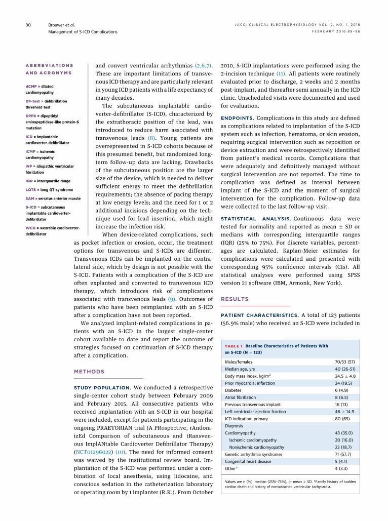
TABLE 1 Baseline Characteristics of Patients With
an S-ICD (N ¼ 123)
Males/females 70/53 (57)
Median age, yrs 40 (26-51)
Body mass index, kg/m2 24.5 � 4.8
Prior myocardial infarction 24 (19.5)
Diabetes 6 (4.9)
Atrial fibrillation 8 (6.5)
Previous transvenous implant 16 (13)
Left ventricular ejection fraction 46 � 14.9
ICD indication: primary 80 (65)
Diagnosis
Cardiomyopathy 43 (35.0)
Ischemic cardiomyopathy 20 (16.0)
Nonischemic cardiomyopathy 23 (18.7)
Genetic arrhythmia syndromes 71 (57.7)
Congenital heart disease 5 (4.1)
Other* 4 (3.3)
Values are n (%), median (25%–75%), or mean � SD. *Family history of suddencardiac death and history of nonsustained ventricular tachycardia.
as pocket infection or erosion, occur, the treatmentoptions for transvenous and S-ICDs are different.Transvenous ICDs can be implanted on the contra-lateral side, which by design is not possible with theS-ICD. Patients with a complication of the S-ICD areoften explanted and converted to transvenous ICDtherapy, which introduces risk of complicationsassociated with transvenous leads (9). Outcomes ofpatients who have been reimplanted with an S-ICDafter a complication have not been reported.
We analyzed implant-related complications in pa-tients with an S-ICD in the largest single-centercohort available to date and report the outcome ofstrategies focused on continuation of S-ICD therapyafter a complication.
METHODS
STUDY POPULATION. We conducted a retrospectivesingle-center cohort study between February 2009and February 2015. All consecutive patients whoreceived implantation with an S-ICD in our hospitalwere included, except for patients participating in theongoing PRAETORIAN trial (A PRospective, rAndom-izEd Comparison of subcutaneous and tRansven-ous ImplANtable Cardioverter Defibrillator Therapy)(NCT01296022) (10). The need for informed consentwas waived by the institutional review board. Im-plantation of the S-ICD was performed under a com-bination of local anesthesia, using lidocaine, andconscious sedation in the catheterization laboratoryor operating room by 1 implanter (R.K.). From October
2010, S-ICD implantations were performed using the2-incision technique (11). All patients were routinelyevaluated prior to discharge, 2 weeks and 2 monthspost-implant, and thereafter semi annually in the ICDclinic. Unscheduled visits were documented and usedfor evaluation.
ENDPOINTS. Complications in this study are definedas complications related to implantation of the S-ICDsystem such as infection, hematoma, or skin erosion,requiring surgical intervention such as reposition ordevice extraction and were retrospectively identifiedfrom patient’s medical records. Complications thatwere adequately and definitively managed withoutsurgical intervention are not reported. The time tocomplication was defined as interval betweenimplant of the S-ICD and the moment of surgicalintervention for the complication. Follow-up datawere collected to the last follow-up visit.
STATISTICAL ANALYSIS. Continuous data weretested for normality and reported as mean � SD ormedians with corresponding interquartile ranges(IQR) (25% to 75%). For discrete variables, percent-ages are calculated. Kaplan-Meier estimates forcomplications were calculated and presented withcorresponding 95% confidence intervals (CIs). Allstatistical analyses were performed using SPSSversion 21 software (IBM, Armonk, New York).
RESULTS
PATIENT CHARACTERISTICS. A total of 123 patients(56.9% male) who received an S-ICD were included in
J A C C : C L I N I C A L E L E C T R O P H Y S I O L O G Y V O L . 2 , N O . 1 , 2 0 1 6 Brouwer et al.F E B R U A R Y 2 0 1 6 : 8 9 – 9 6 Management of S-ICD Complications
91
the current analysis. Table 1 presents the baselinecharacteristics. The median age at implant was40 years old (IQR: 26 to 51 years), and 80 (65%) patientshad a primary prevention indication. The underlyingdiagnosis was ischemic cardiomyopathy in 16% andnonischemic cardiomyopathy in 19%, genetic arrhy-thmia syndromes in 58%, congenital heart disease in4%, and a family history of sudden cardiac death andnonsustained ventricular tachycardia in another 3%.Of the 123 patients, 92 (75%) received implants usingthe 2-incision technique. The median follow-up was27 months (IQR: 9 to 48 months).
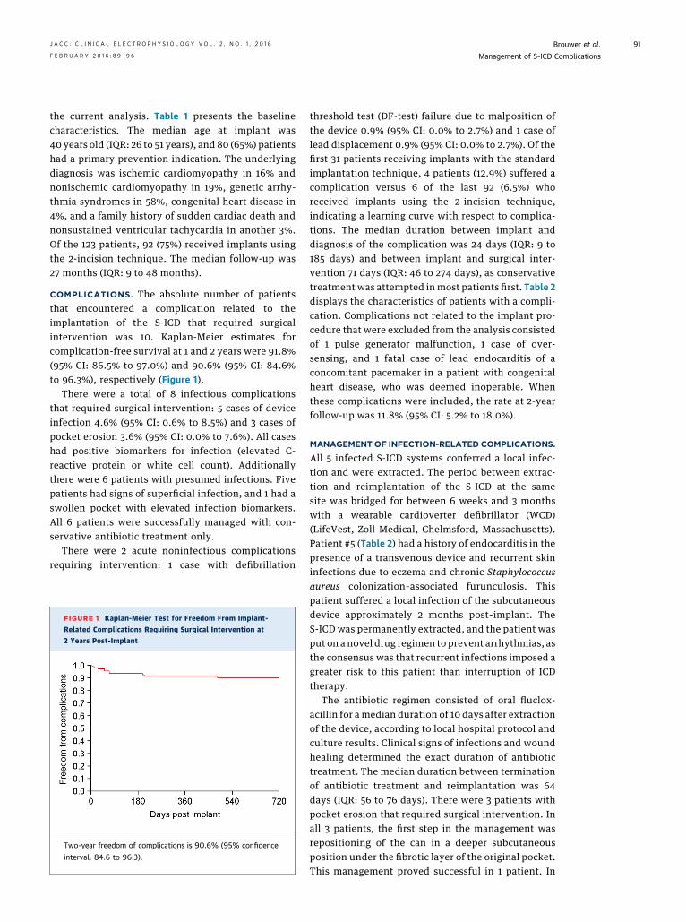
COMPLICATIONS. The absolute number of patientsthat encountered a complication related to theimplantation of the S-ICD that required surgicalintervention was 10. Kaplan-Meier estimates forcomplication-free survival at 1 and 2 years were 91.8%(95% CI: 86.5% to 97.0%) and 90.6% (95% CI: 84.6%to 96.3%), respectively (Figure 1).
There were a total of 8 infectious complicationsthat required surgical intervention: 5 cases of deviceinfection 4.6% (95% CI: 0.6% to 8.5%) and 3 cases ofpocket erosion 3.6% (95% CI: 0.0% to 7.6%). All caseshad positive biomarkers for infection (elevated C-reactive protein or white cell count). Additionallythere were 6 patients with presumed infections. Fivepatients had signs of superficial infection, and 1 had aswollen pocket with elevated infection biomarkers.All 6 patients were successfully managed with con-servative antibiotic treatment only.
There were 2 acute noninfectious complicationsrequiring intervention: 1 case with defibrillation
FIGURE 1 Kaplan-Meier Test for Freedom From Implant-
Related Complications Requiring Surgical Intervention at2 Years Post-Implant
Two-year freedom of complications is 90.6% (95% confidence
interval: 84.6 to 96.3).
threshold test (DF-test) failure due to malposition ofthe device 0.9% (95% CI: 0.0% to 2.7%) and 1 case oflead displacement 0.9% (95% CI: 0.0% to 2.7%). Of thefirst 31 patients receiving implants with the standardimplantation technique, 4 patients (12.9%) suffered acomplication versus 6 of the last 92 (6.5%) whoreceived implants using the 2-incision technique,indicating a learning curve with respect to complica-tions. The median duration between implant anddiagnosis of the complication was 24 days (IQR: 9 to185 days) and between implant and surgical inter-vention 71 days (IQR: 46 to 274 days), as conservativetreatment was attempted in most patients first. Table 2displays the characteristics of patients with a compli-cation. Complications not related to the implant pro-cedure that were excluded from the analysis consistedof 1 pulse generator malfunction, 1 case of over-sensing, and 1 fatal case of lead endocarditis of aconcomitant pacemaker in a patient with congenitalheart disease, who was deemed inoperable. Whenthese complications were included, the rate at 2-yearfollow-up was 11.8% (95% CI: 5.2% to 18.0%).
MANAGEMENTOF INFECTION-RELATED COMPLICATIONS.
All 5 infected S-ICD systems conferred a local infec-tion and were extracted. The period between extrac-tion and reimplantation of the S-ICD at the samesite was bridged for between 6 weeks and 3 monthswith a wearable cardioverter defibrillator (WCD)(LifeVest, Zoll Medical, Chelmsford, Massachusetts).Patient #5 (Table 2) had a history of endocarditis in thepresence of a transvenous device and recurrent skininfections due to eczema and chronic Staphylococcusaureus colonization–associated furunculosis. Thispatient suffered a local infection of the subcutaneousdevice approximately 2 months post–implant. TheS-ICD was permanently extracted, and the patient wasput on a novel drug regimen to prevent arrhythmias, asthe consensus was that recurrent infections imposed agreater risk to this patient than interruption of ICDtherapy.
The antibiotic regimen consisted of oral fluclox-acillin for a median duration of 10 days after extractionof the device, according to local hospital protocol andculture results. Clinical signs of infections and woundhealing determined the exact duration of antibiotictreatment. The median duration between terminationof antibiotic treatment and reimplantation was 64days (IQR: 56 to 76 days). There were 3 patients withpocket erosion that required surgical intervention. Inall 3 patients, the first step in the management wasrepositioning of the can in a deeper subcutaneousposition under the fibrotic layer of the original pocket.This management proved successful in 1 patient. In
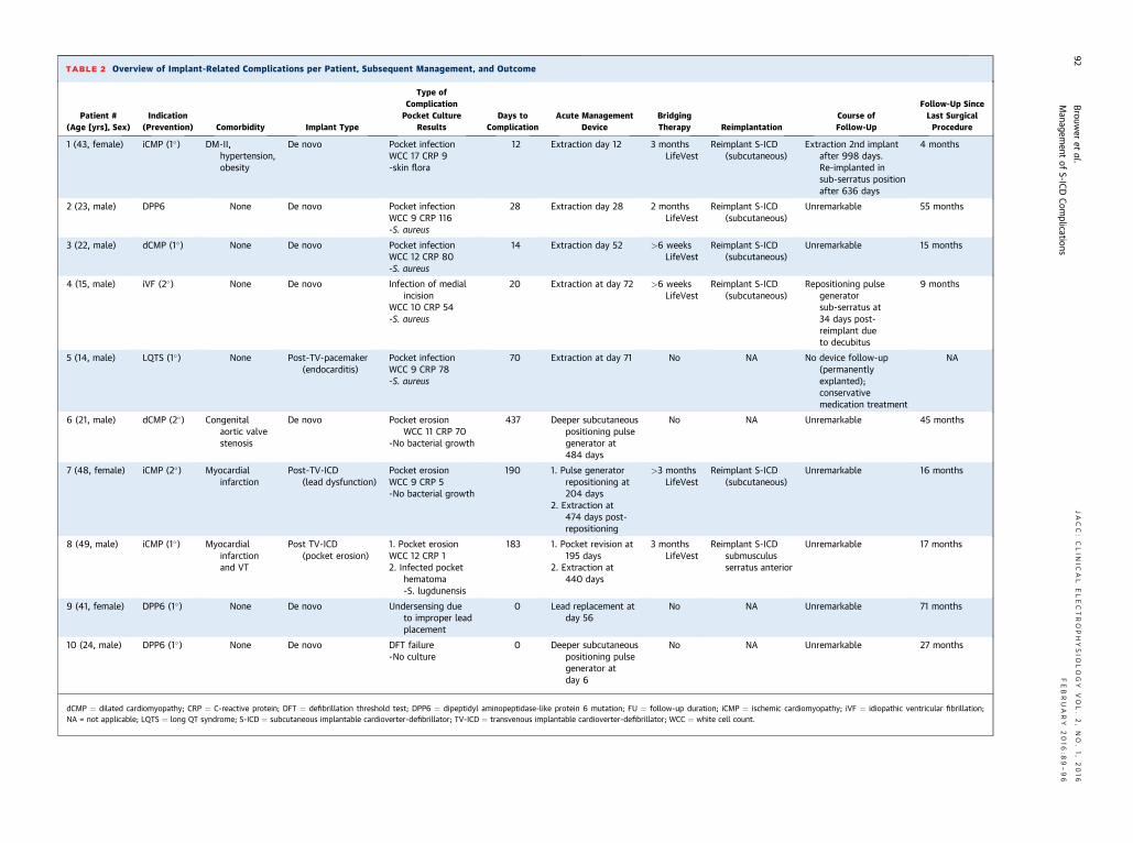
TABLE 2 Overview of Implant-Related Complications per Patient, Subsequent Management, and Outcome
Patient #(Age [yrs], Sex)
Indication(Prevention) Comorbidity Implant Type
Type ofComplicationPocket Culture
ResultsDays to
ComplicationAcute Management
DeviceBridgingTherapy Reimplantation
Course ofFollow-Up
Follow-Up SinceLast SurgicalProcedure
1 (43, female) iCMP (1�) DM-II,hypertension,obesity
De novo Pocket infectionWCC 17 CRP 9-skin flora
12 Extraction day 12 3 monthsLifeVest
Reimplant S-ICD(subcutaneous)
Extraction 2nd implantafter 998 days.Re-implanted insub-serratus positionafter 636 days
4 months
2 (23, male) DPP6 None De novo Pocket infectionWCC 9 CRP 116-S. aureus
28 Extraction day 28 2 monthsLifeVest
Reimplant S-ICD(subcutaneous)
Unremarkable 55 months
3 (22, male) dCMP (1�) None De novo Pocket infectionWCC 12 CRP 80-S. aureus
14 Extraction day 52 >6 weeksLifeVest
Reimplant S-ICD(subcutaneous)
Unremarkable 15 months
4 (15, male) iVF (2�) None De novo Infection of medialincision
WCC 10 CRP 54-S. aureus
20 Extraction at day 72 >6 weeksLifeVest
Reimplant S-ICD(subcutaneous)
Repositioning pulsegeneratorsub-serratus at34 days post-reimplant dueto decubitus
9 months
5 (14, male) LQTS (1�) None Post-TV-pacemaker(endocarditis)
Pocket infectionWCC 9 CRP 78-S. aureus
70 Extraction at day 71 No NA No device follow-up(permanentlyexplanted);conservativemedication treatment
NA
6 (21, male) dCMP (2�) Congenitalaortic valvestenosis
De novo Pocket erosionWCC 11 CRP 70
-No bacterial growth
437 Deeper subcutaneouspositioning pulsegenerator at484 days
No NA Unremarkable 45 months
7 (48, female) iCMP (2�) Myocardialinfarction
Post-TV-ICD(lead dysfunction)
Pocket erosionWCC 9 CRP 5-No bacterial growth
190 1. Pulse generatorrepositioning at204 days
2. Extraction at474 days post-repositioning
>3 monthsLifeVest
Reimplant S-ICD(subcutaneous)
Unremarkable 16 months
8 (49, male) iCMP (1�) Myocardialinfarctionand VT
Post TV-ICD(pocket erosion)
1. Pocket erosionWCC 12 CRP 12. Infected pocket
hematoma-S. lugdunensis
183 1. Pocket revision at195 days
2. Extraction at440 days
3 monthsLifeVest
Reimplant S-ICDsubmusculusserratus anterior
Unremarkable 17 months
9 (41, female) DPP6 (1�) None De novo Undersensing dueto improper leadplacement
0 Lead replacement atday 56
No NA Unremarkable 71 months
10 (24, male) DPP6 (1�) None De novo DFT failure-No culture
0 Deeper subcutaneouspositioning pulsegenerator atday 6
No NA Unremarkable 27 months
dCMP ¼ dilated cardiomyopathy; CRP ¼ C-reactive protein; DFT ¼ defibrillation threshold test; DPP6 ¼ dipeptidyl aminopeptidase-like protein 6 mutation; FU ¼ follow-up duration; iCMP ¼ ischemic cardiomyopathy; iVF ¼ idiopathic ventricular fibrillation;NA = not applicable; LQTS ¼ long QT syndrome; S-ICD ¼ subcutaneous implantable cardioverter-defibrillator; TV-ICD ¼ transvenous implantable cardioverter-defibrillator; WCC ¼ white cell count.
Brouw
eret
al.JACC:CLIN
ICAL
ELECTROPHYSIO
LOGY
VOL.2,NO.1,2016
Managem
entof
S-ICDCom
plicationsFEBRUARY
2016
:89–96
92
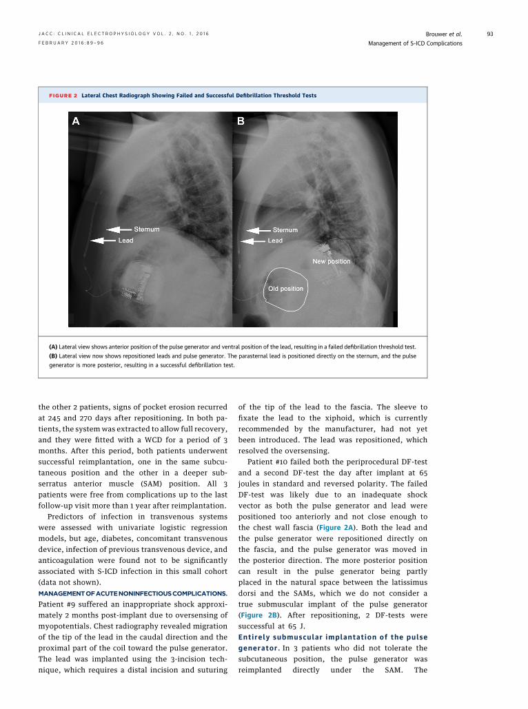
FIGURE 2 Lateral Chest Radiograph Showing Failed and Successful Defibrillation Threshold Tests
(A) Lateral view shows anterior position of the pulse generator and ventral position of the lead, resulting in a failed defibrillation threshold test.
(B) Lateral view now shows repositioned leads and pulse generator. The parasternal lead is positioned directly on the sternum, and the pulse
generator is more posterior, resulting in a successful defibrillation test.
J A C C : C L I N I C A L E L E C T R O P H Y S I O L O G Y V O L . 2 , N O . 1 , 2 0 1 6 Brouwer et al.F E B R U A R Y 2 0 1 6 : 8 9 – 9 6 Management of S-ICD Complications
93
the other 2 patients, signs of pocket erosion recurredat 245 and 270 days after repositioning. In both pa-tients, the systemwas extracted to allow full recovery,and they were fitted with a WCD for a period of 3months. After this period, both patients underwentsuccessful reimplantation, one in the same subcu-taneous position and the other in a deeper sub-serratus anterior muscle (SAM) position. All 3patients were free from complications up to the lastfollow-up visit more than 1 year after reimplantation.
Predictors of infection in transvenous systemswere assessed with univariate logistic regressionmodels, but age, diabetes, concomitant transvenousdevice, infection of previous transvenous device, andanticoagulation were found not to be significantlyassociated with S-ICD infection in this small cohort(data not shown).MANAGEMENTOFACUTENONINFECTIOUSCOMPLICATIONS.
Patient #9 suffered an inappropriate shock approxi-mately 2 months post-implant due to oversensing ofmyopotentials. Chest radiography revealed migrationof the tip of the lead in the caudal direction and theproximal part of the coil toward the pulse generator.The lead was implanted using the 3-incision tech-nique, which requires a distal incision and suturing
of the tip of the lead to the fascia. The sleeve tofixate the lead to the xiphoid, which is currentlyrecommended by the manufacturer, had not yetbeen introduced. The lead was repositioned, whichresolved the oversensing.
Patient #10 failed both the periprocedural DF-testand a second DF-test the day after implant at 65joules in standard and reversed polarity. The failedDF-test was likely due to an inadequate shockvector as both the pulse generator and lead werepositioned too anteriorly and not close enough tothe chest wall fascia (Figure 2A). Both the lead andthe pulse generator were repositioned directly onthe fascia, and the pulse generator was moved inthe posterior direction. The more posterior positioncan result in the pulse generator being partlyplaced in the natural space between the latissimusdorsi and the SAMs, which we do not consider atrue submuscular implant of the pulse generator(Figure 2B). After repositioning, 2 DF-tests weresuccessful at 65 J.Ent i re ly submuscular implantat ion of the pulsegenerator . In 3 patients who did not tolerate thesubcutaneous position, the pulse generator wasreimplanted directly under the SAM. The
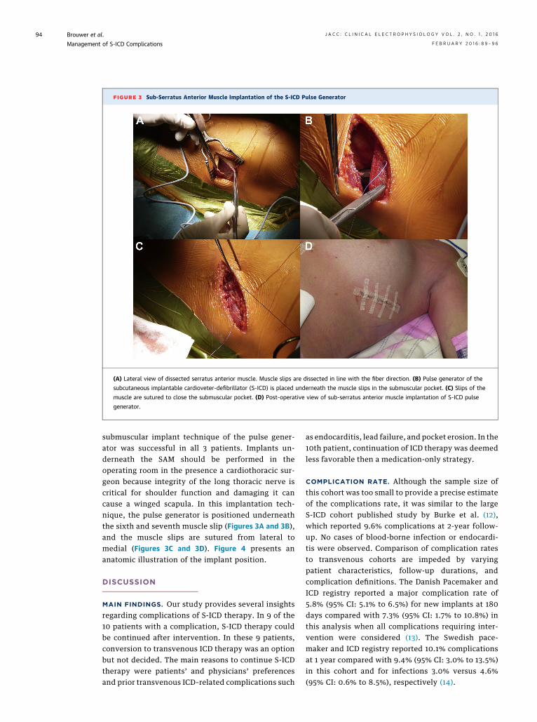
FIGURE 3 Sub-Serratus Anterior Muscle Implantation of the S-ICD Pulse Generator
(A) Lateral view of dissected serratus anterior muscle. Muscle slips are dissected in line with the fiber direction. (B) Pulse generator of the
subcutaneous implantable cardioveter-defibrillator (S-ICD) is placed underneath the muscle slips in the submuscular pocket. (C) Slips of the
muscle are sutured to close the submuscular pocket. (D) Post-operative view of sub-serratus anterior muscle implantation of S-ICD pulse
generator.
Brouwer et al. J A C C : C L I N I C A L E L E C T R O P H Y S I O L O G Y V O L . 2 , N O . 1 , 2 0 1 6
Management of S-ICD Complications F E B R U A R Y 2 0 1 6 : 8 9 – 9 6
94
submuscular implant technique of the pulse gener-ator was successful in all 3 patients. Implants un-derneath the SAM should be performed in theoperating room in the presence a cardiothoracic sur-geon because integrity of the long thoracic nerve iscritical for shoulder function and damaging it cancause a winged scapula. In this implantation tech-nique, the pulse generator is positioned underneaththe sixth and seventh muscle slip (Figures 3A and 3B),and the muscle slips are sutured from lateral tomedial (Figures 3C and 3D). Figure 4 presents ananatomic illustration of the implant position.
DISCUSSION
MAIN FINDINGS. Our study provides several insightsregarding complications of S-ICD therapy. In 9 of the10 patients with a complication, S-ICD therapy couldbe continued after intervention. In these 9 patients,conversion to transvenous ICD therapy was an optionbut not decided. The main reasons to continue S-ICDtherapy were patients’ and physicians’ preferencesand prior transvenous ICD–related complications such
as endocarditis, lead failure, and pocket erosion. In the10th patient, continuation of ICD therapy was deemedless favorable then a medication-only strategy.
COMPLICATION RATE. Although the sample size ofthis cohort was too small to provide a precise estimateof the complications rate, it was similar to the largeS-ICD cohort published study by Burke et al. (12),which reported 9.6% complications at 2-year follow-up. No cases of blood-borne infection or endocardi-tis were observed. Comparison of complication ratesto transvenous cohorts are impeded by varyingpatient characteristics, follow-up durations, andcomplication definitions. The Danish Pacemaker andICD registry reported a major complication rate of5.8% (95% CI: 5.1% to 6.5%) for new implants at 180days compared with 7.3% (95% CI: 1.7% to 10.8%) inthis analysis when all complications requiring inter-vention were considered (13). The Swedish pace-maker and ICD registry reported 10.1% complicationsat 1 year compared with 9.4% (95% CI: 3.0% to 13.5%)in this cohort and for infections 3.0% versus 4.6%(95% CI: 0.6% to 8.5%), respectively (14).
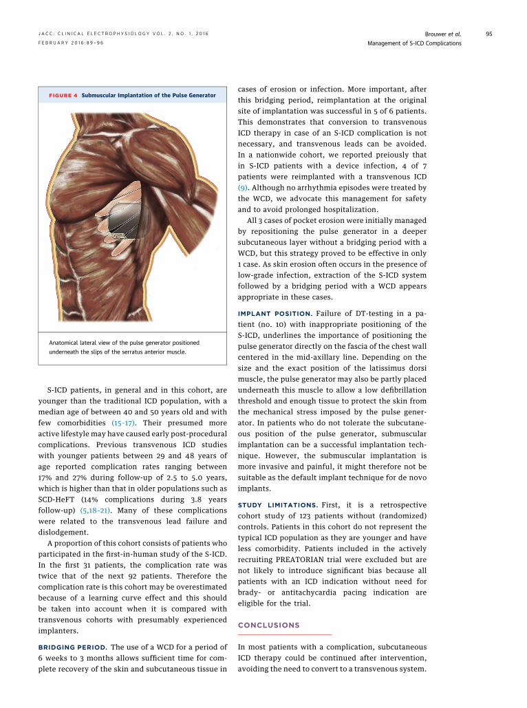
FIGURE 4 Submuscular Implantation of the Pulse Generator
Anatomical lateral view of the pulse generator positioned
underneath the slips of the serratus anterior muscle.
J A C C : C L I N I C A L E L E C T R O P H Y S I O L O G Y V O L . 2 , N O . 1 , 2 0 1 6 Brouwer et al.F E B R U A R Y 2 0 1 6 : 8 9 – 9 6 Management of S-ICD Complications
95
S-ICD patients, in general and in this cohort, areyounger than the traditional ICD population, with amedian age of between 40 and 50 years old and withfew comorbidities (15–17). Their presumed moreactive lifestyle may have caused early post-proceduralcomplications. Previous transvenous ICD studieswith younger patients between 29 and 48 years ofage reported complication rates ranging between17% and 27% during follow-up of 2.5 to 5.0 years,which is higher than that in older populations such asSCD-HeFT (14% complications during 3.8 yearsfollow-up) (5,18–21). Many of these complicationswere related to the transvenous lead failure anddislodgement.
A proportion of this cohort consists of patients whoparticipated in the first-in-human study of the S-ICD.In the first 31 patients, the complication rate wastwice that of the next 92 patients. Therefore thecomplication rate is this cohort may be overestimatedbecause of a learning curve effect and this shouldbe taken into account when it is compared withtransvenous cohorts with presumably experiencedimplanters.
BRIDGING PERIOD. The use of a WCD for a period of6 weeks to 3 months allows sufficient time for com-plete recovery of the skin and subcutaneous tissue in
cases of erosion or infection. More important, afterthis bridging period, reimplantation at the originalsite of implantation was successful in 5 of 6 patients.This demonstrates that conversion to transvenousICD therapy in case of an S-ICD complication is notnecessary, and transvenous leads can be avoided.In a nationwide cohort, we reported preiously thatin S-ICD patients with a device infection, 4 of 7patients were reimplanted with a transvenous ICD(9). Although no arrhythmia episodes were treated bythe WCD, we advocate this management for safetyand to avoid prolonged hospitalization.
All 3 cases of pocket erosion were initially managedby repositioning the pulse generator in a deepersubcutaneous layer without a bridging period with aWCD, but this strategy proved to be effective in only1 case. As skin erosion often occurs in the presence oflow-grade infection, extraction of the S-ICD systemfollowed by a bridging period with a WCD appearsappropriate in these cases.
IMPLANT POSITION. Failure of DT-testing in a pa-tient (no. 10) with inappropriate positioning of theS-ICD, underlines the importance of positioning thepulse generator directly on the fascia of the chest wallcentered in the mid-axillary line. Depending on thesize and the exact position of the latissimus dorsimuscle, the pulse generator may also be partly placedunderneath this muscle to allow a low defibrillationthreshold and enough tissue to protect the skin fromthe mechanical stress imposed by the pulse gener-ator. In patients who do not tolerate the subcutane-ous position of the pulse generator, submuscularimplantation can be a successful implantation tech-nique. However, the submuscular implantation ismore invasive and painful, it might therefore not besuitable as the default implant technique for de novoimplants.
STUDY LIMITATIONS. First, it is a retrospectivecohort study of 123 patients without (randomized)controls. Patients in this cohort do not represent thetypical ICD population as they are younger and haveless comorbidity. Patients included in the activelyrecruiting PREATORIAN trial were excluded but arenot likely to introduce significant bias because allpatients with an ICD indication without need forbrady- or antitachycardia pacing indication areeligible for the trial.
CONCLUSIONS
In most patients with a complication, subcutaneousICD therapy could be continued after intervention,avoiding the need to convert to a transvenous system.
PERSPECTIVES
COMPETENCY IN MEDICAL KNOWLEDGE: The
complication rate of S-ICDs is similar to that of
transvenous ICD therapy. S-ICD therapy can be
continued after a complication such as infection or
pocket erosion, avoiding the need to convert to
transvenous ICD therapy.
TRANSLATIONAL OUTLOOK: Additional research
is needed to evaluate the potential long-term benefits
of subcutaneous ICD therapy compared with trans-
venous ICD therapy.
Brouwer et al. J A C C : C L I N I C A L E L E C T R O P H Y S I O L O G Y V O L . 2 , N O . 1 , 2 0 1 6
Management of S-ICD Complications F E B R U A R Y 2 0 1 6 : 8 9 – 9 6
96
The complication rate in this cohort is similar to whathas been reported for transvenous ICDs, albeit thatthe complication rate in this cohort may be over-estimated because of a learning curve. Bridging timewith a wearable cardioverter-defibrillator allowed re-implantation at the original site of implantation. Inpatients who do not tolerate subcutaneous placementof the pulse generator, implantation of the pulsegenerator underneath the SAM can be a viable treat-ment option.
REPRINT REQUESTS AND CORRESPONDENCE: Dr.Tom F. Brouwer, Department of Cardiology, AcademicMedical Center, P.O. Box 22700, 1100 DE Amsterdam,the Netherlands. E-mail: [email protected].
RE F E RENCE S
1. Alter P, Waldhans S, Plachta E, Moosdorf R,Grimm W. Complications of implantable car-dioverter defibrillator therapy in 440 consecutivepatients. Pacing Clin Electrophysiol 2005;28:926–32.
2. Borleffs CJW, van Erven L, van Bommel RJ,et al. Risk of failure of transvenous implantablecardioverter-defibrillator leads. Circ ArrhythmElectrophysiol 2009;2:411–6.
3. Moss A, Zareba W, Hall W, et al. Prophylacticimplantation of a defibrillator in patients withmyocardial infarction and reduced ejection frac-tion. N Engl J Med 2002;346:877–83.
4. Moss A, Hall W, Cannom D, et al. Improvedsurvival with an implanted defibrillator in patientswith coronary disease at high risk for ventriculararrhythmia. Multicenter Automatic DefibrillatorImplantation Trial Investigators. N Engl J Med1996;335:1933–40.
5. Bardy GH, Lee KL, Mark DB, et al. Amiodaroneor an implantable cardioverter-defibrillator forcongestive heart failure. N Engl J Med 2005;352:225–37.
6. Kleemann T, Becker T, Doenges K, et al. Annualrate of transvenous defibrillation lead defects inimplantable cardioverter-defibrillators over aperiod of >10 years. Circulation 2007;115:2474–80.
7. Dorwarth U, Frey B, Dugas M, et al. Trans-venous defibrillation leads: high incidence offailure during long-term follow-up. J CardiovascElectrophysiol 2003;14:38–43.
8. Bardy G, Smith W, Hood M, et al. An entirelysubcutaneous implantable cardioverter–defibril-lator. N Engl J Med 2010;363:36–44.
9. Olde Nordkamp LR, Dabiri Abkenari L,Boersma LV, et al. The entirely subcutaneousimplantable cardioverter-defibrillator: initial clin-ical experience in a large Dutch cohort. J Am CollCardiol 2012;60:1933–9.
10. Olde Nordkamp LR, Knops RE, Bardy GH, et al.Rationale and design of the PRAETORIAN trial: aProspective, RAndomizEd comparison of subcu-TaneOus and tRansvenous ImplANtablecardioverter-defibrillator therapy. Am Heart J2012;163:753–60.
11. Knops RE, Olde Nordkamp LR, de Groot JR,Wilde AA. Two-incision technique for implantationof the subcutaneous implantable cardioverter-defibrillator. Heart Rhythm 2013;10:1240–3.
12. BurkeMC, Gold MR, Knight BP, et al. Safety andEfficacy of the totally subcutaneous implantabledefibrillator. J Am Coll Cardiol 2015;65:1605–15.
13. Kirkfeldt RE, Johansen JB, Nohr EA,Jorgensen OD, Nielsen JC. Complications aftercardiac implantable electronic device implanta-tions: an analysis of a complete, nationwidecohort in Denmark. Eur Heart J 2014;35:1186–94.
14. Gadler F, Valzania C, Linde C. Current use ofimplantable electrical devices in Sweden: datafrom the Swedish pacemaker and implantablecardioverter-defibrillator registry. Europace 2014;17:69–77.
15. Aydin A, Hartel F, Schlüter M, et al. Shock ef-ficacy of subcutaneous implantable cardioverter-defibrillator for prevention of sudden cardiacdeath: initial multicenter experience. CircArrhythm Electrophysiol 2012;5:913–9.
16. Köbe J, Reinke F, Meyer C, et al. Implantationand follow-up of totally subcutaneous versus
conventional implantable cardioverter-defibrillators:a multicenter case-control study. Heart Rhythm2013;10:29–36.
17. Lambiase PD, Barr C, Theuns DAMJ, et al.Worldwide experience with a totally subcutaneousimplantable defibrillator: early results from theEFFORTLESS S-ICD registry. Eur Heart J 2014;35:1657–65.
18. Schwartz PJ, Spazzolini C, Priori SG, et al. Whoare the long-QT syndrome patients who receive animplantable cardioverter-defibrillator and whathappens to them? Data from the European Long-QT Syndrome Implantable Cardioverter-Defibrillator (LQTS ICD) registry. Circulation2010;122:1272–82.
19. O’Mahony C, Lambiase PD, Quarta G, et al. Thelong-term survival and the risks and benefits ofimplantable cardioverter defibrillators in patientswith hypertrophic cardiomyopathy. Heart 2012;98:116–25.
20. Corrado D, Calkins H, Link MS, et al. Prophy-lactic implantable defibrillator in patients witharrhythmogenic right ventricular cardiomyopathy/dysplasia and no prior ventricular fibrillation orsustained ventricular tachycardia. Circulation2010;122:1144–52.
21. Olde Nordkamp LR, Wilde AA, Tijssen JGP,Knops RE, Van Dessel PFHM, De Groot JR. The ICDfor primary prevention in patients with inheritedcardiac diseases: indications, use, and outcome: acomparison with secondary prevention. CircArrhythmia Electrophysiol 2013;6:91–100.
KEY WORDS complication, erosion, ICD,infection, S-ICD