surface morphology of the human ciliary body during prenatal development
TRANSCRIPT

Graefe's Arch Clin Exp Ophthalmol (1988)226:78 83 Graefe's Archive lot Clinical and Experimental
Ophthalmology © Springer-Verlag 1988
Surface morphology of the human ciliary body during prenatal development* A scanning electron microscopic study
K. Sellheyer and M. Spitznas Universit/its-Augenklinik, Sigmund-Freud-Strasse 25, D-5300 Bonn, Federal Republic of Germany
Abstract. The development of the ciliary body was exam- ined by scanning electron microscopy in human embryos and fetuses with a gestation age of 9.5 to 24 weeks. During this period it was possible to follow up the main morpho- genetical events, beginning with the appearance of the first radial folds up to the occurrence of ciliary processes with a rather adultlike arrangement. The ciliary processes ob- served during week 24 differed from those of the adult eye only by their dimensions and the lack of surface infoldings. A primitive pars plana could only be identified during week 24. The morphological basis for aqueous humor pro- duction is discussed.
Introduction
Knowledge of ocular morphogenetical events contributes to a better understanding of the functional and anatomical implications of the adult eye. This especially holds true for such a complex stucture as the ciliary body, which is in- volved in the accommodation of the lens and secretion of the aqueous humor as well as of the vitreous glycosamino- glycans. These various functions are the reasons why a com- plex developmental process takes place in the differentiation of this ocular tissue. In the human embryo and fetus, Wulle [l 3-15] studied the ultrastructure of the differentiating cili- ary processes extensively using transmission electron mi- croscopy (TEM). In this paper, we attempt to complement his findings by presenting data on the surface aspect of the developing human ciliary body, as viewed under the scanning electron microscope (SEM).
Materials and methods
For our study we obtained seven eyes from human embryos and fetuses after legal abortions, which were performed be- cause of social medical indications. The aspiration-curet- tage technique or extraction with forceps was used. The age of the embryos (up to week 12) was determined by measuring the length of one or more of the following bones:
* This study was performed under the support of a training grant in ophthahnic electron microscopy from Deutsche Forschungsge- meinschaft
Offprint requests to." K. Sellheyer
tibia, femur, humerus, and radius [6]. The age at later gesta- tion weeks was determined by ultrasonographic measure- ment of the biparietal diameter. The following gestation ages (in weeks after conception) were examined: 9.5, 10.3, 12 (2 eyes), 16, 21, and 24. The maximal postmortem delay for fixation was 20 rain.
Up to week 12, the intact eyeballs were fixed in 2% formaldehyde and 2% glutaraldehyde in 0.1 M phosphate buffer, pH 7.2, for 5 h to 4 weeks and postfixed in 2% os- mium tetroxide in phosphate buffer, pH 7.2, for 2 h. The globes from later gestation weeks were opened for fixation by means of a small incision made in the equatorial region. These eyes were dissected before postosmication. All globes were bisected in the equatorial region. The anterior eye segments without lenses (and occasionally without both the lens and cornea) were dehydrated in a graded series of ace- tone, dried in a critical-point-drying apparatus, and sputter- coated with gold or palladium. The specimens were then examined in a Philips SEM 505 or in a Leitz AMR 1000 scanning electron microscope.
Results
SEM showed no time-dependent differences between speci- mens stored for different time periods in the fixative. The gradual transformation of the ciliary folds (radial folds) into ciliary processes can be observed in Figs. 1-3. In Fig. 1 (week 10.3), the ciliary folds are rarely recognizable. In Fig. 2 (week 12), the future location of the ciliary processes is distinct. In Fig. 3 (week 24) their definitive form is almost complete.
Week 9.5
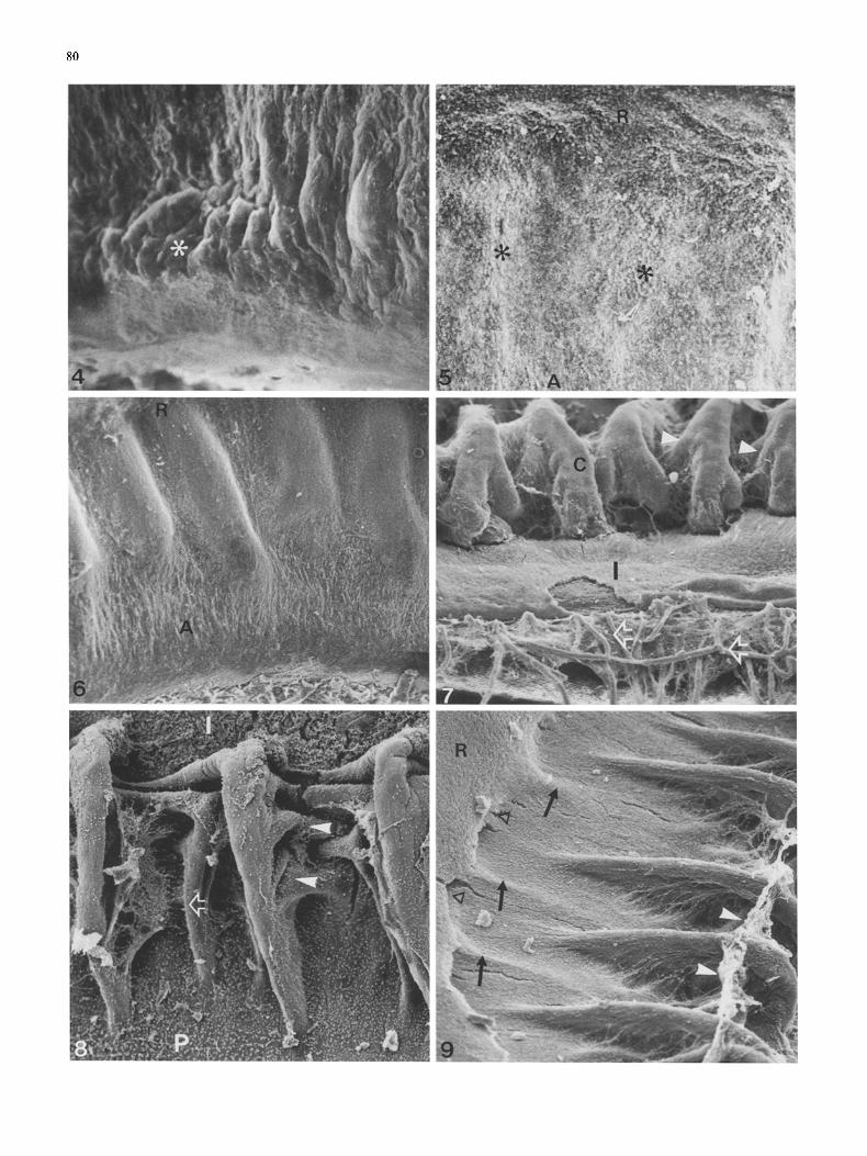
No ciliary folds were discernible (Fig. 4). The inner surface of the anterior eye segment exhibited small radial undula- tions but was never smooth.
Week 10.3
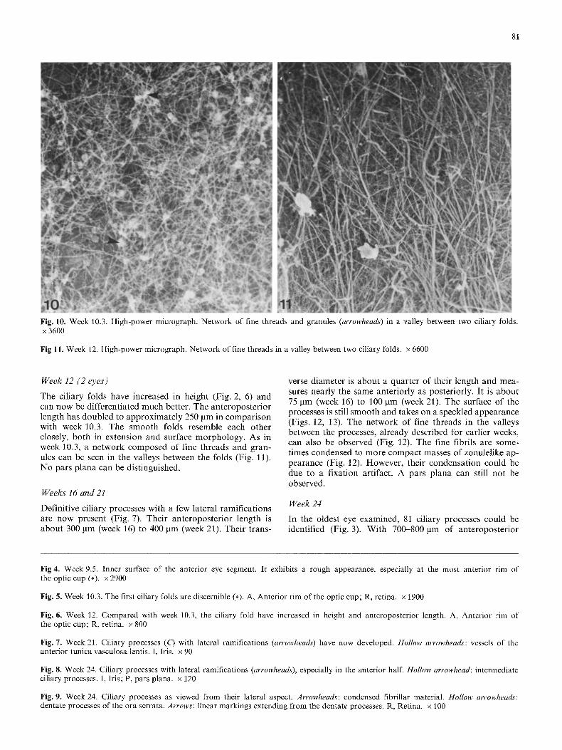
The first ciliary folds could be identified (Figs. 1, 5). They started as simple nonramified elevations that were approxi- mately 100-150 gm from the anterior rim of the optic cup; their posterior ends were directly adjacent to the neurore- tina. The anteroposterior length of these primitive folds was about 120 l~m. In the valleys between them a network of fine threads and granules could be observed (Fig. 10). No pars plana was discernible.
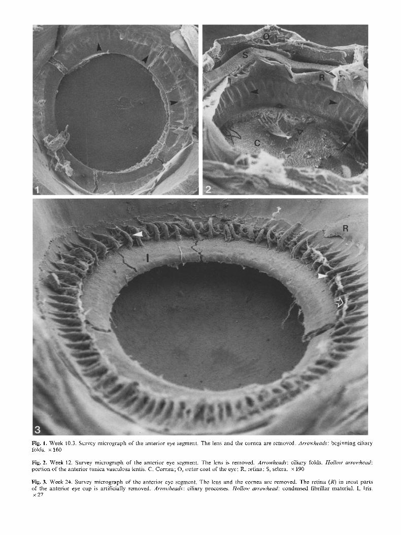
Fig. 1. Week 10.3, Survey micrograph of the anterior eye segment. The lens and the cornea are removed. Arrowhead~: beginning ciliary folds, x 160
Fig. 2. Week 12. Survey micrograph of the anterior eye segment. The lens is removed. Arrowheads: ciliary folds. Hollow arrowhead: port ion of the anterior tunica vasculosa lentis. C, Cornea; O, outer coat of the eye; R, retina; S, sclera, x 190
Fig. 3. Week 24. Survey micrograph of the anterior eye segment. The lens and the cornea are removed. The retina (R) in most parts of the anterior eye cup is artificially removed. Arrowheads: ciliary processes. Hollow arrowhead: condensed fibrillar material. I, Iris. ×27
80
81
Fig. 10. Week 10.3. High-power micrograph. Network of fine threads and granules (arrowheads) in a valley between two ciliary folds. x 3600
Fig l l . Week 12. High-power micrograph. Network of fine threads in a valley between two ciliary folds, x 6600
Week 12 (2 eyes)
The ciliary folds have increased in height (Fig. 2, 6) and can now be differentiated much better. The anteroposterior length has doubled to approximately 250 gm in comparison with week 10.3. The smooth folds resemble each other closely, both in extension and surface morphology. As in week 10.3, a network composed of fine threads and gran- ules can be seen in the valleys between the folds (Fig. 11). No pars plana can be distinguished.
Weeks 16 and 21
Definitive ciliary processes with a few lateral ramifications are now present (Fig. 7). Their anteroposterior length is about 300 gm (week 16) to 400 gm (week 21). Their trans-
verse diameter is about a quarter of their length and mea- sures nearly the same anteriorly as posteriorly. It is about 75 gm (week 16) to 100 ~tm (week 21). The surface of the processes is still smooth and takes on a speckled appearance (Figs. 12, 13). The network of fine threads in the valleys between the processes, already described for earlier weeks, can also be observed (Fig. 12). The fine fibrils are some- times condensed to more compact masses of zonulelike ap- pearance (Fig. 12). However, their condensat ion could be due to a fixation artifact. A pars plana can still not be observed.
Week 24
In the oldest eye examined, 81 ciliary processes could be identified (Fig. 3). With 700-800 ~tm of anteroposterior
Fig 4. Week 9.5. Inner surface of the anterior eye segment. It exhibits a rough appearance, especially at the most anterior rim of the optic cup (*). x 2900
Fig. g. Week 10.3. The first ciliary folds are discernible (*). A, Anterior rim of the optic cup; R, retina, x 1900
Fig. 6. Week 12. Compared with week 10.3, the ciliary fold have increased in height and anteroposterior length. A, Anterior rim of the optic cup; R, retina, x 800
Fig. 7. Week 21. Ciliary processes (C) with lateral ramifications (arrowheads) have now developed. Hollow arrowheads: vessels of the anterior tunica vasculosa lentis. I, Iris. x 90
Fig. 8. Week 24. Ciliary processes with lateral ramifications (arrowheads), especially in the anterior half. Hollow arrowhead: intermediate ciliary processes. I, Iris; P, pars plana, x 120
Fig. 9. Week 24. Ciliary processes as viewed from their lateral aspect. Arrowheads: condensed fibrillar material. Hollow arrowheads: dentate processes of the ora serrata. Arrows: linear markings extending from the dentate processes. R, Retina. x 100

82
Fig. 12. Week 21. Part of the anterior half of two ciliary processes (C). The surface takes a smooth appearance. Arrowhead: condensed fibrillar material, x 500
Fig. 13. Week 21. High-power micrograph of the surface of a ciliary process, which has a speckled appearance. Arrowheads: fine fibrillar material, x 2600
length, their meridional extension has doubled in compari- son with week 21. The processes have developed much more lateral ramifications, which are mostly localized in the ante- rior half of the pars plicata (Fig. 8). In addition to the major ciliary processes, smaller intermediate processes could be distinguished (Fig. 8). The condensed threadlike material in the valleys between the processes can be seen much better than in week 21 (Fig. 9). A narrow pars plana has now developed (Fig. 9), which is up to 200 gm in an- teroposterior length. Posteriorly, it adjoins the ora serrata where dentate processes and scalloped bays can be distin- guished. As in the adult, the dentate processes exhibit linear markings that extend toward the valleys o f the pars plicata.
Discussion
The only SEM study on the development of the ciliary body was performed on the chick eye [3], but the authors did not study this aspect in detail. With the data given in this publication, we were able to cover the main events in the morphogenesis of this ocular tissue in man, starting with the appearance of radial folds up to the beginning of the development of the pars plana.
The first radial folds could be observed during week 10.3. This is in agreement with light microscopical data [2, 4, 8, 9, 12, 15]. Wulle [12, 15] has definitely shown that the folding process starts in the pigmented layer during

83
week 10. The nonpigmented epithelium part icipates in the elevation during week 11. His findings are in agreement with earlier observat ions [1, 2], but are in contras t to the descriptions of Duke-Elder and Cook [4] and M a n n [8]. These authors describe the folding process as beginning in the nonpigmented epithelium. On our own light microscopi- cal studies (Sellheyer and Spitznas, unpubl ished observa- tions) we were able to confirm the findings o f Wulle [12, 15].
The network, which can be observed in the valleys be- tween the early folds, consists of fine threads and granules. I t could represent primit ive zonule fibers or merely second- ary vitreous fibrils. The zonule fibers have been identified by light microscopy during week 12 [8, 9]. T E M studies on this subject are now in progress.
Judging f rom the surface morphology, the principle structure of the ciliary body is established during week 24. However, there are still two main differences compared with the adul t arrangement . Firs t the pars p lana has jus t arisen. The time point is in agreement with the da ta found in the l i terature [2, 4, 8]. However, the pars p lana extends over a distance comparab le to a quar ter of the anteropos ter ior length of the ciliary processes. In the adul t eye the length rat io between the pars p lana and pars pl icata is about 2:1 [5, 10]. Second the surface o f the ciliary processes is still smooth during week 24 and is not comparab le to the SEM appearance of the newborn pars pl icata exhibiting surface infoldings [10]. In T E M studies, Wulle [14, 15] did not ob- serve surface infoldings of the pars pl icata until the 9th month of gestation. I t has a l ready been shown by Kinsey and Jackson [7] that the secretion of aqueous humor begins during the second trimester. As our SEM studies have shown that the surface infoldings do not occur in week 24 (at the end of the second trimester), they could not be re- sponsible for aqueous humor product ion as supposed by Streeten [10]. Wulle [14, 15] considered them to be involved in the resorpt ion of the aqueous and, therefore, control l ing its composi t ion. However, as a l ready pointed out by Weale [11], it is not clear why this control mechanism should mate- rialize only 3 months after aqueous humor is first secreted [71.
Acknowledgements. The authors are grateful to Professor E. Reale, Hannover, and Professor M.H. Blessing, Cologne, for letting them use their scanning electron microscope facilities; we are also grate- ful to Dr. H. Konitz for taking the SEM photographs on the
Philips SEM 505 and to Dipl.-Biol. D. von Kortzfleisch for helping with the Leitz AMR 1000. We also wish to acknowledge the help of Professor H. Wartenberg, Bonn, in measuring the bone length of the embryos for determination of the gestation age.
References
1. Bach L, Seefelder R (1911) Atlas zur Entwicklungsgeschichte des menschlichen Auges. Engelmann, Leipzig Berlin
2. Barber AN (1955) Embryology of the human eye. Mosby, St. Louis
3. Bard JBL, Ross ASA (1982) The morphogenesis of the ciliary body of the avian eye. Dev Biol 92 : 73-86
4. Duke-Elder S, Cook CH (1963) Normal and abnormal develop- ment. In: Duke-Elder S (ed) System of ophthalmology, vol 3, part 1, Embryology. Mosby, St. Louis
5. Hogan MJ, Alvarado JA, Wedell JE (1971) Histology of the human eye. Saunders, Philadelphia
6. Kelemen E, Janossa M, Calvo W, Fliedner TM (1984) Develop- mental age estimated by bone-length measurement in human fetuses. Anat Rec 209 : 547-552
7. Kinsey VE, Jackson B (1949) Investigation of the blood-aque- ous barrier in the newborn to ascorbic acid. Am J Ophthalmol 32 : 374-378
8. Mann I (1964) The development of the human eye, 3rd edn. Grune & Stratton, New York
9. Ozanics V, Jakobiec FA (1983) Prenatal development of the eye and its adnexa. In: Duane TD, Jaeger EA (eds) Biomedical foundations of ophthalmology, vol 1. Harper & Row, Philadel- phia
10. Streeten BW (1982) Ciliary body. In: Jakobiec FA (ed) Ocular anatomy, embryology and teratology. Harper & Row, Philadel- phia, pp 303-330
11. Weale RA (1982) A biography of the eye. Lewis, London 12. Wulle K-G (1966) Frfihentwicklung der Ziliarforts/itze im
menschlichen Auge. Phasenkontrast- und elektronenmikros- kopische Untersuchungen. Z Zellforsch 71 : 545-561
13. Wulle K-G (1967) Elektronenmikroskopie der Kapillarent- wicklung in menschlichen Ziliarfortsfitzen. Verh Anat Ges 61 : 465-473
14. Wulle K-G (1967) Zelldifferenzierungen im Ziliarepithel wfih- rend der menschlichen Fetalentwicklung und ihre Beziehungen zur Kammerwasserbildung. Graefe's Arch Clin Exp Ophthal- mol 172:170-179
15. Wulle K-G (1972) The development of the productive and drainage system of the aqueous humor in the human eye. Adv Ophthalmol 26 : 296-355
Received July 16, 1987 / Accepted September 11, 1987