surface-modified nanoparticles enhance transurothelial penetration and … · large molecule...
TRANSCRIPT

Large Molecule Therapeutics
Surface-Modified Nanoparticles Enhance TransurothelialPenetration and Delivery of Survivin siRNA in TreatingBladder Cancer
Darryl T. Martin1, Jill M. Steinbach2, Jingchun Liu1, Shogo Shimizu1, Hristos Z. Kaimakliotis1,Marcia A. Wheeler1, Adam B. Hittelman1, W. Mark Saltzman2, and Robert M. Weiss1
AbstractPenetration of the bladder permeability barrier (BPB) is a major challenge when treating bladder diseases
via intravesical delivery. To increase transurothelial migration and tissue and tumor cell uptake, poly
(lactic-co-glycolic acid; PLGA) nanoparticles (NP) were modified by addition of a low molecular weight
(2.5 or 20 kDa) positively charged mucoadhesive polysaccharide, chitosan, to the NP surface. In designing
these NPs, we balanced the adhesive properties of chitosan with the release and bioactivity of the siRNA.
Chitosan-functionalized NPs demonstrated increased binding to and uptake in intravesically instilled
mouse bladders and human ureter at 10 times the level of unmodified NPs. Furthermore, we extended the
bioactivity of survivin siRNA in vitro for up to 9 days and demonstrated a decrease in proliferation when
using chitosan-modified NPs relative to unmodified NPs. In addition, treatment of xenograft tumors with
chitosan-modified NPs that encapsulate survivin siRNA (NP-siSUR-CH2.5) resulted in a 65% reduction in
tumor volume and a 75% decrease in survivin expression relative to tumors treated with blank chitosan
NPs (NP-Bk-CH2.5). Our lowmolecular weight chitosan delivery system has the capacity to transport large
amounts of siRNA across the urothelium and/or to the tumor site, thus increasing therapeutic response.
Mol Cancer Ther; 13(1); 71–81. �2013 AACR.
IntroductionOral or systemic administration of drugs for the treat-
ment of pathologic conditions affecting the bladder isoften only partially effective because a limited portion ofdrug reaches the affected site. The remainder of thedrug isdistributed to other tissues, decreasing efficacy and result-ing in adverse side effects (1, 2). Localized, intravesicalinstillation of therapeutic agents avoids first-pass metab-olismassociatedwith systemic andoral delivery, allowingthe drugs/siRNAs to be delivered more efficiently and athigher dosage (3), thus minimizing adverse effects. How-ever, the challenges of localized delivery of drugs andsiRNAto the bladder include lowurotheliumpenetration,short half-life, increased dilutions (3, 4), and short reten-tion times (5). To circumvent the preceding caveats offreely delivered drug/siRNA, we encapsulated siRNA inpoly(lactic-co-glycolic acid; PLGA) nanoparticles (NP) forbladder administration (6).
PLGA is a biocompatible, degradable polymer app-roved by the U.S. Food and Drug Administration (FDA)that has been used to encapsulate and deliver drugs,siRNAs, DNAs, peptides, and proteins (7–11). PLGANPscan protect biologic agents from degradation during sys-temic circulation (12), thereby extending drug efficacy.Our group previously has used the solvent emulsionmethod (water/oil/water) to successfully and efficientlyentrap a variety of agents. Our NPs can efficiently encap-sulate siRNAs (9) and drugs (13), provide controlledrelease of biologic agents (8), and can be modified fortargeted delivery (14). The main focus of this study is toimprove the application of these NPs as a more effectivedelivery modality.
The bladder permeability barrier (BPB) or urothelium isan effective biomembrane barrier, which consists ofumbrella cells, tight junctions, and plaques (1). Althoughthe BPB is impermeable tomost substances,modificationsto the urothelial cells or tight junctions can increase barrierpermeability (1, 15), which is critical for drug/siRNApenetration through the bladder wall. Uptake of drugs/siRNAs can be improved by modifying NPs to enhancebinding and internalization. Surface modifications withnatural polysaccharides and cell-penetrating peptides(CPP) are known to provide increased cell adhesion,internalization, and tissue penetration (1). Penetratin (AP)is derived from the Drosophila melanogaster transcriptionfactor of the antennapedia homeodomain and has been
Authors' Affiliations: Departments of 1Urology and 2Biomedical Engi-neering, Yale University, New Haven, Connecticut
Corresponding Author: Darryl T. Martin, Department of Urology, YaleUniversity School of Medicine, 789 Howard Avenue, Fitkin 3, P.O. Box208058, New Haven, CT 06520-8058. Phone: 203-785-2863; Fax: 203-785-4043; E-mail: [email protected]
doi: 10.1158/1535-7163.MCT-13-0502
�2013 American Association for Cancer Research.
MolecularCancer
Therapeutics
www.aacrjournals.org 71
on June 14, 2020. © 2014 American Association for Cancer Research. mct.aacrjournals.org Downloaded from
Published OnlineFirst November 12, 2013; DOI: 10.1158/1535-7163.MCT-13-0502
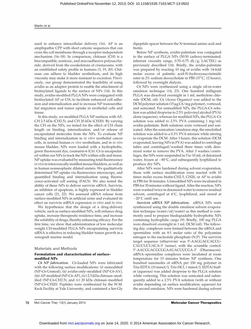
used to enhance intracellular delivery (16). AP is anamphipathic CPP with short cationic sequences that cancross the cell membrane through a receptor-independentmechanism (16–18). In comparison, chitosan (CH) is abiocompatible, nontoxic, and mucoadhesive polysaccha-ride, derived from the exoskeletons of crustaceans, withan established safety profile in humans (3, 19, 20). Chit-osan can adhere to bladder urothelium, and its highviscosity may make it more resistant to excretion. Previ-ously, our group demonstrated the feasibility of usingavidin as an adaptor protein to enable the attachment ofbiotinylated ligands to the surface of NPs (14). In thisstudy, avidin-modified PLGANPswere conjugatedwithbiotinylated AP or CH, to facilitate enhanced cell adhe-sion and internalization and to increase NP transurothe-lial migration and tumor uptake in urothelial cells andtissue.
In this study, we modified PLGANP surfaces with AP,CH 2.5 kDa (CH2.5), and CH 20 kDa (CH20). By varyingthe CH on the NPs, we tested for the effect of CH chainlength on binding, internalization, and/or release ofencapsulated molecules from the NPs. To evaluate NPbinding and internalization in in vitro urothelial cancercells, in normal human ex vivo urothelium, and in in vivomouse bladder, NPs were loaded with a hydrophobic,green fluorescent dye, coumarin-6 (C6). C6 is encapsulat-edwithin ourNPs to track theNPswithin cells and tissue.NPuptakewas evaluated bymeasuring total fluorescencein vivo in intravesically instilledmousebladders, aswell asin human nonneoplastic dilated ureters. We qualitativelydetermined NP uptake via fluorescence microscopy, andquantified binding and internalization using fluores-cence-activated cell sorting (FACS). We also tested theability of these NPs to deliver survivin siRNA. Survivin,an inhibitor of apoptosis, is highly expressed in bladdercancer cells (21, 22). We assessed siRNA release fromsurface-modified NPs in artificial urine and evaluated itseffect on survivin mRNA expression in vitro and in vivo.
We hypothesize that the design of a drug-deliveryvehicle, such as surface-modified NPs, will enhance druguptake, increase therapeutic residence time, and increasethe solubility of drugs, thereby enhancing efficacy. For thefirst time, we show that administration of low molecularweight CH-modified PLGA NPs encapsulating survivinsiRNA is effective in reducing bladder tumor growth in axenograft murine model.
Materials and MethodsFormulation and characterization of surface-modified NPs
C6 NP fabrication. C6-loaded NPs were fabricatedwith the following surface modifications: (i) unmodified(NP-C6-Unmod), (ii) avidin-only–modified (NP-C6-AV),(iii) AP-modified (NP-C6-AP), (iv) 2.5 kDa chitosan–mod-ified (NP-C6-CH2.5), and (v) 20 kDa chitosan–modified(NP-C6-CH20). Peptides were synthesized by the W.M.Keck Facility at Yale University, and contained a Ser-Gly
dipeptide spacer between the N-terminal amino acid andbiotin.
Before NP synthesis, avidin-palmitate was conjugatedto the surface of PLGA NPs (50:50 carboxy-terminated;inherent viscosity range, 0.55–0.75 dL/g; LACTEL) aspreviously described (14). Briefly, the avidin-palmitatewas prepared by reacting 10 mg of avidin with 14-foldmolar excess of palmitic acid-N-hydroxysuccinimideester in 2% sodium deoxycholate in PBS (37�C, 12 hours),followed by overnight dialysis.
C6 NPs were synthesized using a single oil-in-wateremulsion technique (14, 23). One hundred milligramPLGA was dissolved overnight in 1 mL methylene chlo-ride (DCM; oil). C6 (Acros Organics) was added to theDCMpolymer solution (15mgC6/mgpolymer), vortexed,and sonicated. For unmodified NPs, the PLGA-C6 solu-tionwas added dropwise to 2.5%polyvinyl alcohol (PVA)alone (aqueous), whereas formodifiedNPs, the PLGA-C6solution was added to 2.5% PVA containing 1 mg/mLavidin-palmitate. Both solutions were vortexed and son-icated. After the sonication/emulsion step, the emulsifiedsolution was added to a 0.3% PVAmixture while stirringto evaporate the DCM. After 3 hours, this solution (DCMevaporated, leavingNPs in PVA)was added to centrifugetubes and centrifuged/washed three times with deio-nized water to remove the PVA. After 3 washes/centri-fuges, the NPswere suspended in 5 to 10mL of deionizedwater, frozen at �80�C, and subsequently lyophilized toproduce dry NPs.
After NPs were hardened during solvent evaporation,those with surface modification were reacted with 10times molar excess biotin-CH2.5, CH20, or AP to avidinin PBS for 30minutes. UnmodifiedNPswere incubated inPBS for 30minuteswithout ligand. After the reaction,NPswerewashed twice in deionizedwater to remove residualsolvent, centrifuged at 4�C, lyophilized, and stored at�20�C until use.
Survivin siRNA NP fabrication. siRNA NPs weresynthesized using the double emulsion solvent evapora-tion technique (water/oil/water) as this method is com-monly used to prepare biodegradable hydrophobic NPscontaining hydrophilic cargo (9). Briefly, 100 mg PLGAwere dissolved overnight in 1 mL DCM (oil). The follow-ing day, complexes were formed between the siRNA andspermidine with an 8:1 molar ratio of the polyaminenitrogen to the nucleotide phosphate (N:P). The survivintarget sequence (siSurvivin) was 50-AAGGACCACCG-CAUCUCUACA-30 (sense), with the scramble control:50-AACGUACGCGGAAUACUUCGA-30 (Dharmacon).siRNA–spermidine complexes were incubated at roomtemperature for 15 minutes before NP synthesis. Onehundred nanomoles of siRNA per 100 mg polymer inTris-EDTA (10mmol/L Tris-HCl, 1 mmol/L EDTA) buff-er (aqueous) was added dropwise to the PLGA solutionwhile vortexing. This solution was sonicated and subse-quently added to a 2.5% PVA solution (with or withoutavidin depending on surface modification; aqueous) forthe second emulsion. NPs were hardened during solvent
Martin et al.
Mol Cancer Ther; 13(1) January 2014 Molecular Cancer Therapeutics72
on June 14, 2020. © 2014 American Association for Cancer Research. mct.aacrjournals.org Downloaded from
Published OnlineFirst November 12, 2013; DOI: 10.1158/1535-7163.MCT-13-0502

evaporation in 0.3% PVA for 3 hours. The hardened NPswere washed three times in deionized water to removeresidual solvent, centrifuged at 4�C, lyophilized, andstored at �20�C until use.
NP characterization: siRNA loading, encapsulationefficiency, and sizeTo determine siRNA loading and encapsulation effi-
ciency, NPs were incubated with DCM, and siRNA wasextracted into aqueous buffer as described previously (9).Briefly, 3 to 5mgof siRNANPsweredissolved in 0.5mLofDCM for 30 minutes, and the siRNA was extracted intoTris-EDTA buffer. The quantity of extracted double-stranded siRNAwasdetermined using theQuant-iT Pico-Green assay (Invitrogen). Fluorescence was comparedwith a known siRNA standard. The unmodified NPsencapsulated 600 pmol siRNA/mg NP, corresponding toan encapsulation efficiency of 60%. Our theoretical load-ing of theNPswas 1,000 pmol (1 nmol). Both formulationsof surface-modified chitosan NPs encapsulated 700 pmolsurvivin siRNA/mg NP, with a corresponding encapsu-lation efficiency of 70%. NP morphology, diameter, andsize distribution were determined from scanning electronmicroscope images using ImageJ.
Fluorescence-activated cell sortingT-24 and UM-UC-3 bladder cancer cells were obtained
directly from the American Type Culture Collection in2007 and 2010, respectively. The cells were authenticatedand underwent quality control using short tandem repeatDNA profiles, morphology verification, growth assays,and mycoplasma testing during these experiments. Inaddition, only low-passage cells were used. UM-UC-3andT-24 cellsweremaintained inEaglesMinimumEssen-tial and McCoy’s medium, respectively. Both cell lineswere supplemented with 10% FBS, 1% penicillin–strep-tomycin, and 1% glutamine. UM-UC-3 or T-24 cells wereincubated with NP-C6-AV, NP-C6-AP, NP-C6-CH2.5,and NP-C6-CH20 at 1 mg/mL for 2 hours in a 37�Chumidified chamber, in the appropriate medium. Theavidin control group represents NPs with no CPP (ortargeting ligand), as we wanted to ensure that the avidinmodification was not the reason for enhanced binding/uptake. Then, ice-cold medium was added to stop endo-cytosis. Cells were washed with PBS, detached withtrypsin-EDTA, and either fixed in FACS solution contain-ing 1% (v/v) of FBS and paraformaldehyde (PFA) in PBSor exposed to 0.3% trypan blue for 2 minutes before beingfixed in FACS solution (24, 25). Trypan blue quenchesextracellular surface fluorescence of living cells, allowingfor quantification of internalized fluorescence (25). Thecells thenwere sortedusing aBDLSR II, located in theCellSorter Facility at Yale University. Data were analyzedusing FlowJo software, and a minimum of 20,000 cell-events were analyzed per sample. To overlay histogramscontaining different number of events collected, theFlowJo software would use percentage of maximum (%Max) for normalization. The %Max is the number of cells
in each bin divided by the number of cells in the bin thatcontains the greatest number of cells (26).
Fluorescence microscopyUM-UC-3 or T-24 cells were grown on coverslips for 48
hours until 80% to 90% confluence. Cells were incubatedwith NP-C6-AV, NP-C6-AP, NP-C6-CH2.5, and NP-C6-CH20 at 1 mg/mL for 2 hours at 37�C in a CO2 incubator.The cells were incubated with or without 0.3% trypanblue, washed in PBS, and fixed with 3.7% PFA. The cellswere then incubated with 0.1% Triton X-100 before beingtreated with Texas Red phalloidin (Invitrogen), to visu-alize the F-actin. The coverslips were mounted on slideswith VECTASHIELD containing 40,6-diamidino-2-pheny-lindole (DAPI) to visualize nuclei.
Ex vivo human ureter systemThe use of biologic specimens for human research
was approved by the Human Investigation Committeeat Yale University (Protocol number, 0710003157). Theuptake of surface-modified NPs was measured in an exvivo binding assay, using human nonneoplastic dilatedureters. The ureters were used within 4 hours of remov-al. After removing external connective tissue and wash-ing in sterile PBS, the ureter was placed in an autoclaved96-well dot blot chamber (Bio-Rad) with the luminalurothelium facing upward. Unmodified and surface-modified NPs, loaded with C6 (100 mL of 2 mg/mL),were suspended in artificial urine and added to indi-vidual wells. Artificial urine alone was used as a control.The apparatus was incubated at 37�C in a humidifiedincubator for up to 2 hours. After incubation, wells werewashed four times to remove nonadherent NPs, coredwith a biopsy punch, and weighed. The samples werefrozen for extraction of C6.
In vivo mouse intravesical instillationAll animal studies were approved by the Institutional
Animal Care and Use Committee of Yale University.Femalemicewere intraperitoneally sedatedwithketamine(100 mg/kg) and xylazine (10 mg/kg). External genitaliawere cleansed. Then, the mice were catheterized with alubricated 24G catheter (BD Biosciences), and their blad-ders were emptied. The catheter was then attached to aHamilton syringe filled with 100 mL of artificial urine withor without surface-modified, C6-loaded NPs (2 mg/mL).The solution was instilled into the bladder, the catheterwas removed, and an ultra-small clampwas placed on theexternal urethra (Natsume Seisakusho Co., Ltd), whichremained in place for 2 hours, the typical amount of timeused for clinical intravesical treatments. Mice then weresacrificed; bladders were washed extensively with PBS toremove nonadherent NPs and weighed. Samples werefrozen for extraction of C6.
Extraction of C6 from human and mouse tissueDistilled water (750 mL) was added to frozen human
ureteral tissue cores or mouse bladders and the tissue
Modified NPs for Enhanced siRNA Bladder Delivery
www.aacrjournals.org Mol Cancer Ther; 13(1) January 2014 73
on June 14, 2020. © 2014 American Association for Cancer Research. mct.aacrjournals.org Downloaded from
Published OnlineFirst November 12, 2013; DOI: 10.1158/1535-7163.MCT-13-0502

was homogenized on ice using a Polytron homogenizer(Brinkman). The homogenate was vortexed for 1 hour atroom temperature using theVX-2500Multi TubeVortexer(VWR) at the highest speed (setting 10) and then centri-fuged (16,000�g; 10minutes). After removal of the super-natant, dimethyl sulfoxide (DMSO; a solvent that dis-solves the PLGA NPs) was added so that the weight(g)/volume (mL) ratio was 1:10. The samples were againvortexed and centrifuged. C6 fluorescence in the samples(100-mL aliquots) was measured on a 96-well plate readerat 460 nm excitation, 540 nm emission. Fluorescence in theDMSO supernatant was divided by total (input) fluores-cence to determine percentage fluorescence or by mousebladder or tissue weight.
Quantitative real-time PCRTotal RNA was extracted from cells or murine tumors
using TRizol (Invitrogen). cDNA was synthesized usingiScript (Bio-Rad). Quantitative real-time PCR (qRT-PCR)was performedusing iQ SYBRGreen Supermix (Bio-Rad),100 nmol/L specific primers and equivalent amounts ofcDNA. Melting curves for all primers produced a singlepeak. For mRNA analysis, the relative level of geneexpression was normalized to glyceraldehyde-3-phos-phate dehydrogenase (GAPDH) expression, using thecycle thresholdmethod.The followingprimerswereused:GAPDH sense, 50-CCACTAGGCGCTCACTGTTCT-30;antisense, 50-GCGAACTCACCCGTTGACT-30; survivinsense, 50-GACCACCGCATCTCTACATTC-30; and anti-sense; 50-TGCTTTTTATGTTCCTCTATGGG-30.
In vitro siRNA transfection and cell proliferationassay
UM-UC-3 cells were seeded in a six-well plate over-night until they reached a cell density of 20%. The cellswere transfectedwith siSurvivin to afinal concentration of100 nmol/L with Lipofectamine RNAiMAX (Invitrogen).After 3 and 9 days at 37�C in a 5%CO2 incubator, the cellswere harvested to determine gene expression. For the 9-day siSurvivin study, UM-UC-3 cells were trypsinizedand replatedwithnewmedia after the fourthday.Also, onday 9, a cell proliferation assay was performed in whichbladder cancer cells that were treated with NP-siSUR-Unmod, NP-siSUR-AV, NP-siSUR-CH2.5, or NP-siSUR-CH20were trypsinized and counted in a TC10 automatedcell counter (Bio-Rad).
Controlled release of siRNA from NPsNPs (4 mg) containing fluorescein-labeled siSurvivin
(F-siSurvivin) were suspended in 1 mL of artificial urineand incubated at 37�Cwith gentle shaking over 13 days ina single conical tube. Cumulative release of siRNA wasmeasured at predetermined time points. At each timepoint, the sample was centrifuged at 12,000 �g and thesupernatant was collected. Once the supernatants werecollected, the extracted F-siSurvivin was measured on a96-well plate reader at 488 nmexcitation, 520 nmemissionwavelengths.
Xenograft modelEight-week-old female Foxn1 nu/nu mice were sub-
cutaneously injected in the left flank with 5 � 106 UM-UC-3 cells, which have high metastatic potential and arecommonly used in xenograft models. The tumor volumewas estimated using the formula (tumor length � tumorwidth2) � p/6. The mice were randomly divided intothree groups: PBS (control), NP-Bk-CH2.5 (empty NPs),or NP-siSUR-CH2.5, when the tumor volumes reached100 � 15 mm3. Mice were intratumorally injected with:PBS, 1 mg/100 mL of NP-Bk-CH2.5 diluted in PBS or 0.7mg/100 mL NP-siSUR-CH2.5 (500 pmol siSUR) dilutedin PBS. Mouse tumors were intratumorally injected onday 0, 4, and 7, with day 0 being 4 days after inoculationof bladder cancer cells. Tumor volumes were measuredbefore each injection. All animals were sacrificed on day11 (15 days after cell inoculation). Xenograft mousetumors were weighed, snap-frozen, and stored at�80�C.
Statistical analysisData are presented as mean � SEM from five to six
samples for each condition unless noted. Significance isdetermined by ANOVA (P < 0.05).
ResultsMicroscopic assessment unmodified and modifiedNPs
Scanning electron microscopy was used to assess thesize and morphology of both unmodified and surface-modified NPs (Fig. 1). The average size for each batch ofNPs was comparable (not statistically different) withmean particle diameters of 168 � 64 nm (NP-Unmod),137� 51nm (NP-CH2.5), and 130nm� 56 nm (NP-CH20).
NP uptake using in vitro, ex vivo, and in vivomodelsUsingfluorescencemicroscopy,wevisualized the inter-
nalization ofNPs in bladder cancer cells after 2 hours (Fig.2). Surface-modifiedNPswith chitosan (CH2.5 andCH20)had higher internalization than control NP-AV. Weobserved NPs outside the nucleus in the cytoplasm (Fig.2A). FACS analysis verified that cells bind and internalizesurface-modified NP-C6 after 2 hours. Avidin-modifiedNPs associated and internalized relative to untreatedsamples; however, the CH-modified NPs had superiorinternalization/binding. The internalization of modifiedC6 NPs also was quantified for T-24 and UM-UC-3bladder cancer cells (Fig. 2B). The mean fluorescenceintensity was the lowest for the cells treated with controlNPs (modified only with avidin) and greatest for thecells treated with chitosan-modified NPs. Representa-tive histograms illustrated a higher fluorescence inten-sity for UM-UC-3 cells treated with NP-C6-CH2.5 andNP-C6-CH20 compared with that of NP-C6-AV control(Fig. 2C).
During ex vivo experiments,NP formulations (2mg/mL)were incubated with human ureter tissue for 30 to 120minutes. At 60 minutes, the uptake of NP-C6-CH20,
Martin et al.
Mol Cancer Ther; 13(1) January 2014 Molecular Cancer Therapeutics74
on June 14, 2020. © 2014 American Association for Cancer Research. mct.aacrjournals.org Downloaded from
Published OnlineFirst November 12, 2013; DOI: 10.1158/1535-7163.MCT-13-0502
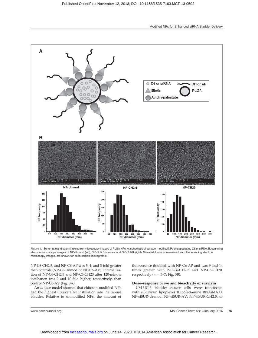
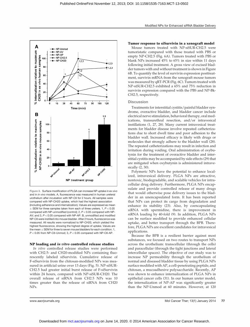
NP-C6-CH2.5, and NP-C6-AP was 5, 4, and 3-fold greaterthan controls (NP-C6-Unmod or NP-C6-AV). Internaliza-tion of NP-C6-CH2.5 and NP-C6-CH20 after 120-minuteincubation was 9 and 10-fold higher, respectively, thancontrol NP-C6-AV (Fig. 3A).An in vivo model showed that chitosan-modified NPs
had the highest uptake after instillation into the mousebladder. Relative to unmodified NPs, the amount of
fluorescence doubled with NP-C6-AP and was 9 and 14times greater with NP-C6-CH2.5 and NP-C6-CH20,respectively (n ¼ 3–7; Fig. 3B).
Dose–response curve and bioactivity of survivinUM-UC-3 bladder cancer cells were transfected
with siSurvivin lipoplexes (Lipofectamine RNAiMAX),NP-siSUR-Unmod, NP-siSUR-AV, NP-siSUR-CH2.5, or
A
NP diameter (mm)
B
NP diameter (mm) NP diameter (mm)
NP
fre
qu
ency
NP
fre
qu
ency
NP
fre
qu
ency
Figure 1. Schematic and scanning electronmicroscopy images of PLGANPs. A, schematic of surface-modified NPs encapsulating C6 or siRNA. B, scanningelectron microscopy images of NP-Unmod (left), NP-CH2.5 (center), and NP-CH20 (right). Size distributions, measured from the scanning electronmicroscopy images, are shown for each sample (histograms).
Modified NPs for Enhanced siRNA Bladder Delivery
www.aacrjournals.org Mol Cancer Ther; 13(1) January 2014 75
on June 14, 2020. © 2014 American Association for Cancer Research. mct.aacrjournals.org Downloaded from
Published OnlineFirst November 12, 2013; DOI: 10.1158/1535-7163.MCT-13-0502

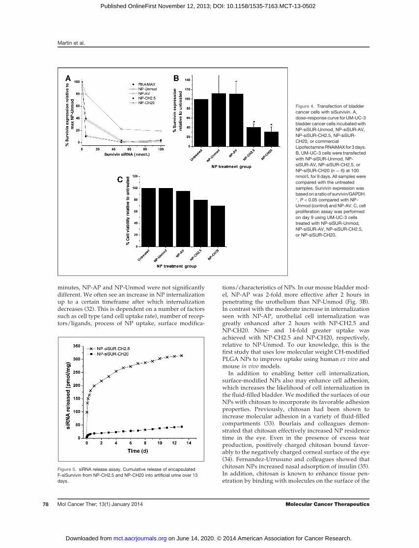
NP-siSUR-CH20.We tested three different concentrationsof siSurvivin (5, 50, and 100 nmol/L) to determine themost effective concentration needed to achieve survivinknockdown. Three days after administration ofNP-siSUR(5 nmol/L), survivin mRNA expression decreased from20% (NP-siSUR-Unmod; control) to 90% (NP-siSUR-CH20). Following administration of all surface-modifiedNPs containing siSUR at 50 and 100 nmol/L, survivinmRNA expression decreased by more than 95%. Theseresults were comparable with the knockdown demon-strated using lipoplexes (Fig. 4A). To demonstrate pro-
longed knockdown, transfection of UM-UC-3 cells with100 nmol/L siSurvivin, NP-siSUR-CH2.5, or NP-siSUR-CH20 resulted in 7-fold reduction in survivin mRNAexpression relative to the controls, NP-siSUR-Unmod,or NP-siSUR-AV, after 9 days (Fig. 4B). Cell prolifera-tion was measured in UM-UC-3 bladder cancer cells,which were treated for 2 hours with NP-siSUR-Unmod,NP-siSUR-AV, NP-siSUR-CH2.5, or NP-siSUR-CH20. Cellproliferation was reduced by 20% to 30% in the bladdercancer cells when treated with the chitosan-modified NPscompared with the NP-siSUR-Unmod control (Fig. 4C).
Fluorescence intensityNP treatment group
NP treatment group
% M
FI r
elat
ive
to c
on
tro
l%
MF
I rel
ativ
e to
co
ntr
ol
% M
ax
A
B C
Figure 2. In vitro internalization ofmodified PLGA NPs. A, humanbladder cancer cells wereincubated with 1 mg/mL of NP-AV,NP-AP, NP-CH2.5, or NP-CH20loaded with C6 (green) for 2 hours.NPs were "associated" (includingadherent and internalized) or"internalized" into the cancer cells.The nucleus was stained with DAPI(blue) and the actin cytoskeletonwas stained with Texas Red X-phalloidin (red). B, FACS analysisshows that modified NPs loadedwith C6 were internalized within T-24 and UM-UC-3 bladder cancercells more efficiently than NP-AV(con). All results were normalizedwith NP-AV, which was set to100%mean fluorescence intensity(MFI). �, P < 0.05 from control NPs.Results represent the average ofthree experiments. C,representative histogram of UM-UC-3 cells treated with NP-CH2.5(solid line) and NP-CH20 (dashedline) show a greater C6 peak shiftcompared with NP-AV (avidincontrol; dotted line). The untreatedcells were negative for C6 (dash–dotted line).
Martin et al.
Mol Cancer Ther; 13(1) January 2014 Molecular Cancer Therapeutics76
on June 14, 2020. © 2014 American Association for Cancer Research. mct.aacrjournals.org Downloaded from
Published OnlineFirst November 12, 2013; DOI: 10.1158/1535-7163.MCT-13-0502
NP loading and in vitro controlled release studiesIn vitro controlled release studies were performed
with CH2.5- and CH20-modified NPs containing fluo-rescently labeled siSurvivin. Cumulative release ofF-siSurvivin from the chitosan-modified NPs was mea-sured in artificial urine over 13 days (Fig. 5). NP-siSUR-CH2.5 had greater initial burst release of F-siSurvivinwithin 24 hours, compared with NP-siSUR-CH20. Theoverall release of siRNA from CH2.5 NPs was 10times greater than the release of siRNA from CH20NPs.
Tumor response to siSurvivin in a xenograft modelMouse tumors treated with NP-siSUR-CH2.5 were
tumoristatic compared with those treated with PBS orempty NP-CH2.5 (Fig. 6A). Tumors treated with PBS orblank NPs increased 45% to 65% in size within 11 daysfollowing initial treatment. A gross view of excised blad-der tumorswith andwithout treatment is shown in Figure6B. To quantify the level of survivin expression posttreat-ment, survivin mRNA from the xenograft mouse tumorswasmeasured by qRT-PCR (Fig. 6C). Tumors treatedwithNP-siSUR-CH2.5 exhibited a 65% and 75% reduction insurvivin expression compared with the PBS and NP-Bk-CH2.5, respectively.
DiscussionTreatments for interstitial cystitis/painful bladder syn-
drome, overactive bladder, and bladder cancer includeelectrical nerve stimulation, behavioral therapy, oralmed-ications, transurethral resection, and/or intravesicalinstillations (1, 27, 28). Many current intravesical treat-ments for bladder disease involve repeated catheteriza-tions due to short dwell time and poor adhesion to thebladder wall. Increased efficacy is likely with drugs ormolecules that strongly adhere to the bladder wall (28).The repeated catheterizations may result in infection andirritation during voiding. Oral administration of oxybu-tynin for the treatment of overactive bladder and inter-stitial cystitismay be accompanied by side effects (29) thatare mitigated when oxybutynin is administered intrave-sically (2, 30).
Polymeric NPs have the potential to enhance local-ized, intravesical delivery. PLGA NPs are attractive,nontoxic, biodegradable, and scalable vehicles for intra-cellular drug delivery. Furthermore, PLGA NPs encap-sulate and provide controlled release of many drugsthat would otherwise pose delivery issues to the blad-der in an unencapsulated form. It has been reportedthat NPs can protect its cargo from degradation andenhance its stability (23). Also, by coencapsulatingsiRNA with spermidine, there is improvement insiRNA loading by 40-fold (9). In addition, PLGA NPscan be surface modified to provide enhanced cellularuptake, and better transport through the BPB. There-fore, PLGA NPs are excellent candidates for intravesicalapplications.
Because the BPB is a resilient barrier against mostsubstances, we focused on two routes to transport NPsacross the urothelium: transcellular (through the cells)and paracellular (through the tight junctions and lateralintercellular spaces). The objective of our study was toincrease NP permeability through the urothelium ofnormal and diseased bladder tissue by using PLGA NPssurfacemodifiedwithAP, a cell-penetrating peptide, andchitosan, a mucoadhesive polysaccharide. Recently, APwas shown to enhance internalization of PLGA NPs inepithelial cancer cells (31). In our human ureter model,the internalization of NP-AP was significantly greaterthan the NP-Unmod at 60 minutes. However, at 120
Figure 3. Surface modification of PLGA can increase NP uptake in ex vivoand in in vivo models. A, fluorescence was measured in human ureteralurothelium after incubation with NP-C6 for 2 hours. All samples werecompared with NP-CH20 uptake, which had the highest association(including adherence and internalization). Values are expressed as mean� SEM for three samples taken from each of three ureters. 1, P < 0.05compared with NP-unmodified (control); 2, P < 0.05 compared with NP-AV; and 3, P < 0.05 compared with NP-AP. B, unmodified and modifiedNP-C6were instilled intomouse bladder. After 2 hours, fluorescencewasmeasured. All results were normalized to NP-CH20, which yielded thehighest fluorescence, showing the highest degree of uptake. Values arethemean�SEM for three to sevenmouse bladders for each condition. 1,P < 0.05 from NP-C6-Unmod; 3, P < 0.05 compared with NP-C6-AP.
Modified NPs for Enhanced siRNA Bladder Delivery
www.aacrjournals.org Mol Cancer Ther; 13(1) January 2014 77
on June 14, 2020. © 2014 American Association for Cancer Research. mct.aacrjournals.org Downloaded from
Published OnlineFirst November 12, 2013; DOI: 10.1158/1535-7163.MCT-13-0502
minutes, NP-AP and NP-Unmod were not significantlydifferent. We often see an increase in NP internalizationup to a certain timeframe after which internalizationdecreases (32). This is dependent on a number of factorssuch as cell type (and cell uptake rate), number of recep-tors/ligands, process of NP uptake, surface modifica-
tions/characteristics of NPs. In our mouse bladder mod-el, NP-AP was 2-fold more effective after 2 hours inpenetrating the urothelium than NP-Unmod (Fig. 3B).In contrast with the moderate increase in internalizationseen with NP-AP, urothelial cell internalization wasgreatly enhanced after 2 hours with NP-CH2.5 andNP-CH20. Nine- and 14-fold greater uptake wasachieved with NP-CH2.5 and NP-CH20, respectively,relative to NP-Unmod. To our knowledge, this is thefirst study that uses low molecular weight CH-modifiedPLGA NPs to improve uptake using human ex vivo andmouse in vivo models.
In addition to enabling better cell internalization,surface-modified NPs also may enhance cell adhesion,which increases the likelihood of cell internalization inthe fluid-filled bladder. We modified the surfaces of ourNPs with chitosan to incorporate its favorable adhesionproperties. Previously, chitosan had been shown toincrease molecular adhesion in a variety of fluid-filledcompartments (33). Bourlais and colleagues demon-strated that chitosan effectively increased NP residencetime in the eye. Even in the presence of excess tearproduction, positively charged chitosan bound favor-ably to the negatively charged corneal surface of the eye(34). Fernandez-Urrusuno and colleagues showed thatchitosan NPs increased nasal adsorption of insulin (35).In addition, chitosan is known to enhance tissue pen-etration by binding with molecules on the surface of the
Figure 4. Transfection of bladdercancer cells with siSurvivin. A,dose–response curve for UM-UC-3bladder cancer cells incubated withNP-siSUR-Unmod, NP-siSUR-AV,NP-siSUR-CH2.5, NP-siSUR-CH20, or commercialLipofectamineRNAiMAX for 3days.B, UM-UC-3 cells were transfectedwith NP-siSUR-Unmod, NP-siSUR-AV, NP-siSUR-CH2.5, orNP-siSUR-CH20 (n ¼ 6) at 100nmol/L for 9 days. All samples werecompared with the untreatedsamples. Survivin expression wasbasedona ratio of survivin/GAPDH.�, P < 0.05 compared with NP-Unmod (control) and NP-AV. C, cellproliferation assay was performedon day 9 using UM-UC-3 cellstreated with NP-siSUR-Unmod,NP-siSUR-AV, NP-siSUR-CH2.5,or NP-siSUR-CH20.
Figure 5. siRNA release assay. Cumulative release of encapsulatedF-siSurvivin from NP-CH2.5 and NP-CH20 into artificial urine over 13days.
Martin et al.
Mol Cancer Ther; 13(1) January 2014 Molecular Cancer Therapeutics78
on June 14, 2020. © 2014 American Association for Cancer Research. mct.aacrjournals.org Downloaded from
Published OnlineFirst November 12, 2013; DOI: 10.1158/1535-7163.MCT-13-0502

urothelium, and disrupting the tight junctions betweenumbrella cells (36). In fact, by inducing desquamation ofpig urothelium, chitosan removes all diffusion barriers,including glycosaminoglycans (GAG), membrane pla-ques, and umbrella cell tight junctions, thereby enablingdeeper permeation. Because of its ability to increaseresidence time within fluid-filled compartments andenhance transport through urothelial tissues, chitosan
is an attractive ligand for surface modification of NPsfor intravesical instillation.
Indesigning theseNPs, therewas abalance between theadhesive properties of chitosan and the release of siRNAfrom the NPs, which are both affected by the molecularweight of chitosan (37). Eroglu and colleagues showedthat higher molecular weight chitosan (650 kDa) had agreater bioadhesive force to sheep bladder than lowermolecular weight chitosan (150 kDa; ref. 38). Althoughboth chitosan NP formulations (CH2.5 and CH20) wereeffective in penetrating the urothelium of our human exvivo andmouse in vivo systems,NPswith the longer CH20chain showed stronger cellular uptake relative to NPswith the shorter CH2.5 chain. At acidic pH, chitosan, withabundant hydroxyl and amine groups, becomes proton-ated and forms electrostatic interactions with the GAGsfoundon the surface of theurothelium (1, 39, 40) or humancancer cells, thereby increasing chitosan’s binding poten-tial. Moreover, Amoozgar and colleagues demonstratedthat the longer chitosan chain had a five times greaterpositive surface charge than the shorter chitosan chain(40). The exact mechanism by which chitosan enhancespenetration is unknown (1); however, enhanced internal-ization using chitosan-modified NPs is attributed to acombination of increased mucoadhesion and the tempo-rary loss of the tight junctions (19, 36, 41, 42). Although thehigh molecular weight chitosan likely enhances bindingand internalization, the positively charged chitosan canbind to the negatively charged siRNA preventing itsrelease, so this must be considered in NP design.
The bioactivity of our NP-siSUR was measured byknockdown of survivin mRNA, an inhibitor of apoptosis.Previously, we showed that survivin is highly expressedin bladder cancer cells (22). In addition, we showed thatsurvivin could be detected in urine of patients withbladder cancer, but was absent from the urine of patientswithout cancer (21). In fact, survivin expression is elevat-ed inmany cancers and absent inmany noncancer tissues,making it a good target for cancer therapy (43). Yang andcolleagues showed a 30% decrease in cell viability andproliferation of T-24 bladder cancer cells in vitro for 3dayswhen using survivin siRNA (44).Wewere interestedin decreasing survivin expression by extending the bio-activity of siSurvivin. Previous studies showed that siSur-vivin degradation typically occurs at 3 days when usingcommercially available transfection reagents (45). To testthe efficacy of our NP-siSUR, we analyzed survivinexpression in human bladder cancer cell models (Fig.4). We initially varied the siSurvivin concentration todetermine an efficacious dose. At higher concentrationsof siRNA (100 nmol/L; ref. 46) in vitro, there was littledifference between the chitosan formulations. Afteradministration of 100nmol/L siRNA, at day 9,we showeda 60% to 70% decrease in the expression of survivin usingboth chitosan formulations.
We then assessed the release of siRNA from NP-CH2.5and NP-CH20 to determine the effect of surface modifi-cation on siRNA release. Although our in vitro data
Figure 6. In vivo treatment of xenograft bladder tumors with siSurvivin. A,four days after flank injection of UM-UC-3 cells, tumors were injected atotal of three times (arrows) with PBS-Con (n¼ 5), NP-Bk-CH2.5 (n¼ 4),or NP-siSUR-CH2.5 (n ¼ 5). Tumor volumes were measured beforeinjections and harvested on day 11. B, gross view of excised bladdertumors treated with PBS, NP-Bk-CH2.5, or NP-siSUR-CH2.5. C, qRT-PCR analysis of survivin expression in excised mouse bladder tumorstreated with PBS, NP-Bk-CH2.5, or NP-siSUR-CH2.5. �, P < 0.05 fromPBS-control and NP-Bk-CH2.5.
Modified NPs for Enhanced siRNA Bladder Delivery
www.aacrjournals.org Mol Cancer Ther; 13(1) January 2014 79
on June 14, 2020. © 2014 American Association for Cancer Research. mct.aacrjournals.org Downloaded from
Published OnlineFirst November 12, 2013; DOI: 10.1158/1535-7163.MCT-13-0502
suggest that both NP-siSUR-CH2.5 and NP-siSUR-CH20decrease survivin expression, likely due to enhancedinternalization and subsequent release of siSurvivin, theoverall release of survivin siRNA from NP-siSUR-CH2.5was 10 times greater than that fromNP-siSUR-CH20. Thisis in agreement with previous studies that show that thehigher molecular weight (long chain) chitosan entanglesand traps more free-siRNA than the lower molecularweight (short chain) chitosan (47), resulting in a hindranceof siRNA migration. Lower release of F-siSurvivin fromNP-siSUR-CH20 was expected, relative to NP-siSUR-CH2.5, as the longer positively charged chains hindersiRNA release. Furthermore, longer chitosan chains canform bulkier surface layers and may affect siRNA release(38). Conversely, the short positively charged chitosan,NP-siSUR-CH2.5, demonstrated an initial burst release ofthe F-siSurvivin, followed by a steady release of siRNA. Ithas been reported that lower molecular weight chitosanhas resulted in better transfection efficiencies, degrada-tion, and intracellular release (48, 49). Because of thisdramatically different release profile and transfectionefficacy, NP-CH2.5 was used in the in vivo bladder tumorgrowth studies.
Using a xenograft mouse model, we demonstrated asignificant reduction in tumor growth, up to 65%,withNP-siSUR-CH2.5, which was comparable with previousreports that used antisense oligonucleotide survivin ordominant-negative mutant survivin to reduce tumorgrowth by up to 50% (50). Kunze and colleagues delivered4.6mg/kg of free survivin siRNA3 times perweek in vivo,but did not observe a reduction in tumor growth (46),whereas we were able to deliver 20 times less siRNA insurface-modified NPs (only 2 times per week), with asignificant reduction in tumor growth. We were able toshow a greater decrease in tumor growth using ourchitosan-functionalized NPs compared with other knock-down methods.
For the first time, we demonstrate that PLGA NPs,functionalizedwith lowmolecularweight chitosan encap-
sulating siRNA (and unmodified siRNA PLGA NPs)improves binding in human ex vivo specimens and inmouse in vivo specimens. Furthermore, the low molec-ular weight CH2.5-modified PLGA NPs encapsulatingsiRNA prolonged survivin knockdown and reducedtumor growth. We demonstrated that chitosan-modi-fied PLGA NPs have many advantages, including asufficient amount of siRNA release to reduce survivinmRNA expression and an increase in siSurvivin bioac-tivity. We now have a versatile NP system that iscapable of carrying large amounts of bioactive siRNAacross the urothelium, thus increasing and improvingtherapeutic response.
Disclosure of Potential Conflicts of InterestNo potential conflicts of interest were disclosed.
Authors' ContributionsConception and design: D.T. Martin, J.M. Steinbach, M.A. Wheeler, A.B.Hittelman, W.M. Saltzman, R.M. WeissDevelopment of methodology: D.T. Martin, J.M. Steinbach, J. Liu, H.Z.Kaimakliotis, M.A. Wheeler, A.B. Hittelman, R.M. WeissAcquisition of data (provided animals, acquired and managed patients,provided facilities, etc.):D.T.Martin, J.M. Steinbach, J. Liu,M.A.Wheeler,R.M. WeissAnalysis and interpretation of data (e.g., statistical analysis, biostatis-tics, computational analysis): D.T. Martin, J.M. Steinbach, J. Liu, H.Z.Kaimakliotis, M.A. Wheeler, W.M. Saltzman, R.M. WeissWriting, review, and/or revision of the manuscript: D.T. Martin, J.M.Steinbach, H.Z. Kaimakliotis, M.A. Wheeler, A.B. Hittelman, W.M. Saltz-man, R.M. WeissAdministrative, technical, or material support (i.e., reporting or orga-nizing data, constructing databases): D.T. Martin, S. Shimizu, M.A.Wheeler, R.M. WeissStudy supervision: D.T. Martin, R.M. Weiss
Grant SupportThis study was supported in part by the NIH grant 5RC1DK087015
(to R.M. Weiss) from the National Institute of Diabetes and Digestiveand Kidney Diseases (NIDDK), NIH grant EB000487 (to W.M. Saltz-man), and a Pilot Grant from the Yale Comprehensive Cancer Center (toR.M. Weiss).
Received June 26, 2013; revised October 9, 2013; accepted October 30,2013; published OnlineFirst November 12, 2013.
References1. GuhaSarkar S, Banerjee R. Intravesical drug delivery: Challenges,
current status, opportunities and novel strategies. J Control Release2010;148:147–59.
2. Tyagi P,WuPC,ChancellorM,YoshimuraN,HuangL.Recent advancesin intravesical drug/gene delivery. Mol Pharm 2006;3:369–79.
3. Giannantoni A, Di Stasi SM, Chancellor MB, Costantini E, Porena M.New frontiers in intravesical therapies and drug delivery. Eur Urol2006;50:1183–93; discussion 93.
4. Le VisageC, Rioux-LeclercqN, HallerM, Breton P,MalavaudB, LeongK. Efficacy of paclitaxel released from bio-adhesive polymer micro-spheres onmodel superficial bladder cancer. J Urol 2004;171:1324–9.
5. Highley MS, van Oosterom AT, Maes RA, De Bruijn EA. Intravesicaldrug delivery. Pharmacokinetic and clinical considerations. Clin Phar-macokinet 1999;37:59–73.
6. Behlke MA. Chemical modification of siRNAs for in vivo use. Oligo-nucleotides 2008;18:305–19.
7. Khan A, Benboubetra M, Sayyed PZ, Ng KW, Fox S, Beck G, et al.Sustained polymeric delivery of gene silencing antisense ODNs,
siRNA, DNAzymes and ribozymes: in vitro and in vivo studies. J DrugTarget 2004;12:393–404.
8. Anthony T, Fong P, Goyal A, Saltzman WM, Moss RL, Breuer C.Development of a parathyroid hormone-controlled release system asa potential surgical treatment for hypoparathyroidism. J Pediatr Surg2005;40:81–5.
9. Woodrow KA, Cu Y, Booth CJ, Saucier-Sawyer JK, Wood MJ, Saltz-man WM. Intravaginal gene silencing using biodegradable polymernanoparticles densely loaded with small-interfering RNA. Nat Mater2009;8:526–33.
10. Sirianni RW, Olausson P, Chiu AS, Taylor JR, Saltzman WM. Thebehavioral and biochemical effects of BDNF containing polymersimplanted in the hippocampus of rats. Brain Res 2010;1321:40–50.
11. McNeer NA, Chin JY, Schleifman EB, Fields RJ, Glazer PM, SaltzmanWM.Nanoparticles deliver triplex-formingPNAs for site-specificgeno-mic recombination in CD34þ human hematopoietic progenitors. MolTher 2011;19:172–80.
Martin et al.
Mol Cancer Ther; 13(1) January 2014 Molecular Cancer Therapeutics80
on June 14, 2020. © 2014 American Association for Cancer Research. mct.aacrjournals.org Downloaded from
Published OnlineFirst November 12, 2013; DOI: 10.1158/1535-7163.MCT-13-0502

12. Zhou J, Patel TR, Fu M, Bertram JP, Saltzman WM. Octa-functionalPLGA nanoparticles for targeted and efficient siRNA delivery totumors. Biomaterials 2012;33:583–91.
13. Martin DT, Hoimes CJ, Kaimakliotis HZ, Cheng CJ, Zhang K, Liu J,et al. Nanoparticles for urothelium penetration and delivery of thehistone deacetylase inhibitor belinostat for treatment of bladdercancer. Nanomedicine 2013;9:1124–34.
14. Fahmy TM, Samstein RM, Harness CC, Saltzman WM. Surfacemodification of biodegradable polyesters with fatty acidconjugates for improved drug targeting. Biomaterials 2005;26:5727–36.
15. Lewis SA. Everything you wanted to know about the bladderepithelium but were afraid to ask. Am J Physiol Renal Physiol 2000;278:F867–74.
16. Dupont E, Prochiantz A, Joliot A. Penetratin story: an overview.Methods Mol Biol 2011;683:21–9.
17. Dietz GP, Bahr M. Delivery of bioactive molecules into the cell: theTrojan horse approach. Mol Cell Neurosci 2004;27:85–131.
18. Gratton JP, Yu J, Griffith JW, Babbitt RW, Scotland RS, Hickey R, et al.Cell-permeable peptides improve cellular uptake and therapeutic genedelivery of replication-deficient viruses in cells and in vivo. Nat Med2003;9:357–62.
19. Artursson P, Lindmark T, Davis SS, Illum L. Effect of chitosan on thepermeability ofmonolayers of intestinal epithelial cells (Caco-2). PharmRes 1994;11:1358–61.
20. Wang JJ, Zeng ZW, Xiao RZ, Xie T, Zhou GL, Zhan XR, et al. Recentadvances of chitosan nanoparticles as drug carriers. Int J Nanome-dicine 2011;6:765–74.
21. Smith SD, Wheeler MA, Plescia J, Colberg JW, Weiss RM, Altieri DC.Urine detection of survivin and diagnosis of bladder cancer. JAMA2001;285:324–8.
22. Swana HS, Grossman D, Anthony JN, Weiss RM, Altieri DC. Tumorcontent of the antiapoptosis molecule survivin and recurrence ofbladder cancer. N Engl J Med 1999;341:452–3.
23. Cu Y, Saltzman WM. Controlled surface modification with poly(ethyl-ene)glycol enhances diffusion of PLGA nanoparticles in human cervi-cal mucus. Mol Pharm 2009;6:173–81.
24. Yoo JW, Doshi N, Mitragotri S. Endocytosis and Intracellular Distri-bution of PLGA Particles in Endothelial Cells: Effect of Particle Geom-etry. Macromol Rapid Commun 2010;31:142–8.
25. Rejman J, Oberle V, Zuhorn IS, Hoekstra D. Size-dependent internal-ization of particles via the pathways of clathrin- and caveolae-medi-ated endocytosis. Biochem J 2004;377:159–69.
26. Quintino L, Baudet A, Larsson J, Lundberg C. FACS bindingassay for analysing GDNF interactions. J Neurosci Methods 2013;218:25–8.
27. Moutzouris DA, Falagas ME. Interstitial cystitis: an unsolved enigma.Clin J Am Soc Nephrol 2009;4:1844–57.
28. Kaufman J, Tyagi V, Anthony M, Chancellor MB, Tyagi P. State of theart in intravesical therapy for lower urinary tract symptoms. Rev Urol2010;12:e181–9.
29. Wein AJ. Practical uropharmacology. Urol Clin North Am 1991;18:269–81.
30. BarbaliasGA, LiatsikosEN,AthanasopoulosA,NikiforidisG. Interstitialcystitis: bladder training with intravesical oxybutynin. J Urol 2000;163:1818–22.
31. Cheng CJ, Saltzman WM. Enhanced siRNA delivery into cells byexploiting the synergy between targeting ligands and cell-penetratingpeptides. Biomaterials 2011;32:6194–203.
32. Cartiera MS, Johnson KM, Rajendran V, Caplan MJ, Saltzman WM.The uptake and intracellular fate of PLGA nanoparticles in epithelialcells. Biomaterials 2009;30:2790–8.
33. Kim WJ. Chitin, Chitosan, Oligosaccharides and Their Derivatives:Biological Activities and Applications. In:Kim S-K, editor. ElectrostaticProperties of Chitosan: CRC Press; 2010. p. 117–25.
34. Bourlais CL, Acar L, Zia H, Sado PA, Needham T, Leverge R. Oph-thalmic drug delivery systems–recent advances. Prog Retin Eye Res1998;17:33–58.
35. Fernandez-Urrusuno R, Calvo P, Remunan-Lopez C, Vila-Jato JL,AlonsoMJ. Enhancement of nasal absorption of insulin using chitosannanoparticles. Pharm Res 1999;16:1576–81.
36. Kos MK, Bogataj M, Veranic P, Mrhar A. Permeability of pig urinarybladderwall: timeand concentration dependent effect of chitosan. BiolPharm Bull 2006;29:1685–91.
37. Duceppe N, Tabrizian M. Advances in using chitosan-based nano-particles for in vitro and in vivo drug and gene delivery. Expert OpinDrug Deliv 2010;7:1191–207.
38. Eroglu M, Irmak S, Acar A, Denkbas EB. Design and evaluation of amucoadhesive therapeutic agent delivery system for postoperativechemotherapy in superficial bladder cancer. Int J Pharm 2002;235:51–9.
39. Weecharangsan W, Opanasopit P, Ngawhirunpat T, ApirakaramwongA, Rojanarata T, Ruktanonchai U, et al. Evaluation of chitosan salts asnon-viral gene vectors in CHO-K1 cells. Int J Pharm 2008;348:161–8.
40. Amoozgar Z, Park J, Lin Q, Yeo Y. Lowmolecular-weight chitosan as apH-sensitive stealth coating for tumor-specific drug delivery. MolPharm 2012;9:1262–70.
41. Dodane V, Amin Khan M, Merwin JR. Effect of chitosan on epithelialpermeability and structure. Int J Pharm 1999;182:21–32.
42. Smith J, Wood E, Dornish M. Effect of chitosan on epithelial cell tightjunctions. Pharm Res 2004;21:43–9.
43. Lladser A, Sanhueza C, Kiessling R, Quest AF. Is survivin the potentialAchilles' heel of cancer? Adv Cancer Res 2011;111:1–37.
44. Yang D, Song X, Zhang J, Ye L, Wang S, Che X, et al. Therapeuticpotential of siRNA-mediated combined knockdown of the IAP genes(Livin, XIAP, and Survivin) on human bladder cancer T24 cells. ActaBiochim Biophys Sin 2010;42:137–44.
45. Xue HY, Wong HL. Tailoring nanostructured solid-lipid carriers fortime-controlled intracellular siRNA kinetics to sustain RNAi-mediatedchemosensitization. Biomaterials 2011;32:2662–72.
46. Kunze D, Wuttig D, Kausch I, Blietz C, Blumhoff L, Burmeister Y, et al.Antisense-mediated inhibition of survivin, hTERT and VEGF in bladdercancer cells in vitro and in vivo. Int J Oncol 2008;32:1049–56.
47. Techaarpornkul S, Wongkupasert S, Opanasopit P, ApirakaramwongA, Nunthanid J, Ruktanonchai U. Chitosan-mediated siRNA delivery invitro: effect of polymermolecular weight, concentration and salt forms.AAPS PharmSciTech 2010;11:64–72.
48. Lavertu M, Methot S, Tran-Khanh N, Buschmann MD. High efficiencygene transfer using chitosan/DNA nanoparticles with specific combi-nations of molecular weight and degree of deacetylation. Biomaterials2006;27:4815–24.
49. Koping-Hoggard M, Varum KM, Issa M, Danielsen S, Christensen BE,Stokke BT, et al. Improved chitosan-mediated gene delivery based oneasily dissociated chitosan polyplexes of highly defined chitosanoligomers. Gene Ther 2004;11:1441–52.
50. Kanwar JR,ShenWP,KanwarRK,BergRW,KrissansenGW.Effects ofsurvivin antagonists on growth of established tumors and B7-1 immu-nogene therapy. J Natl Cancer Inst 2001;93:1541–52.
Modified NPs for Enhanced siRNA Bladder Delivery
www.aacrjournals.org Mol Cancer Ther; 13(1) January 2014 81
on June 14, 2020. © 2014 American Association for Cancer Research. mct.aacrjournals.org Downloaded from
Published OnlineFirst November 12, 2013; DOI: 10.1158/1535-7163.MCT-13-0502

2014;13:71-81. Published OnlineFirst November 12, 2013.Mol Cancer Ther Darryl T. Martin, Jill M. Steinbach, Jingchun Liu, et al. CancerPenetration and Delivery of Survivin siRNA in Treating Bladder Surface-Modified Nanoparticles Enhance Transurothelial
Updated version
10.1158/1535-7163.MCT-13-0502doi:
Access the most recent version of this article at:
Cited articles
http://mct.aacrjournals.org/content/13/1/71.full#ref-list-1
This article cites 49 articles, 1 of which you can access for free at:
Citing articles
http://mct.aacrjournals.org/content/13/1/71.full#related-urls
This article has been cited by 1 HighWire-hosted articles. Access the articles at:
E-mail alerts related to this article or journal.Sign up to receive free email-alerts
Subscriptions
Reprints and
To order reprints of this article or to subscribe to the journal, contact the AACR Publications Department at
Permissions
Rightslink site. Click on "Request Permissions" which will take you to the Copyright Clearance Center's (CCC)
.http://mct.aacrjournals.org/content/13/1/71To request permission to re-use all or part of this article, use this link
on June 14, 2020. © 2014 American Association for Cancer Research. mct.aacrjournals.org Downloaded from
Published OnlineFirst November 12, 2013; DOI: 10.1158/1535-7163.MCT-13-0502