surface & coatings technology -...
TRANSCRIPT

Surface & Coatings Technology 312 (2017) 19–24
Contents lists available at ScienceDirect
Surface & Coatings Technology
j ourna l homepage: www.e lsev ie r .com/ locate /sur fcoat
Non-thermal atmospheric pressure plasma jet applied to inactivation ofdifferent microorganisms
T.M.C. Nishime a,⁎, A.C. Borges b, C.Y. Koga-Ito b, M. Machida c, L.R.O. Hein a, K.G. Kostov a
a São Paulo State University – UNESP, Campus in Guaratinguetá – FEG, Guaratinguetá, SP 12516-410, Brazilb São Paulo State University – UNESP, Institute of Science and Technology – ICT, São José dos Campos, SP 12247-004, Brazilc Campinas State University –UNICAMP, Physics Institute – IFGW, Campinas, SP 13083–859, Brazil
⁎ Corresponding author.E-mail address: [email protected] (T.M.C. N
http://dx.doi.org/10.1016/j.surfcoat.2016.07.0760257-8972/© 2016 Elsevier B.V. All rights reserved.
a b s t r a c t
a r t i c l e i n f oArticle history:Received 14 March 2016Revised 22 July 2016Accepted in revised form 26 July 2016Available online 27 July 2016
Non-thermal atmospheric pressure plasma jets (APPJs) are capable of generating cold plasma plumes thatare not confined by electrodes, which makes them very attractive for bio-medical applications. In thepresent work, the inactivation efficiency of cold APPJ was evaluated against three pathogenic microorgan-isms with different cell wall characteristics. The Gram-positive bacterium Enterococcus faecalis (ATCC29212), the Gram-negative bacterium Pseudomonas aeruginosa (ATCC 15442) and the fungus Candidaalbicans (SC 5314) were plated on standard Petri dishes filled with specific culture media. The plasma jetwith mean power of 1.8 W was directed perpendicularly on agar plates and the system was flushed withpure helium at two different flows, 2.0 and 4.0 SLM. During the treatments, time and distance betweennozzle and agar were varied. The presence of excited reactive species was confirmed by optical emissionspectroscopy. Scanning electron microscopy (SEM) was applied for investigation of cell morphology. Themicrobicidal efficiency was evaluated by measuring the area of inhibition zone (where there was no cellgrowth). For different flows of helium, no significant difference of inhibition zone size was noted for thesame microbial species. However, high flows led to formation of non-homogenous inhibition zones,presenting microcolonies distributed through the inactivated region. The Gram-positive bacterium wasmore susceptible to the plasma antimicrobial effects than the other microorganisms.
© 2016 Elsevier B.V. All rights reserved.
Keywords:Plasma jetCold atmospheric plasmaDecontamination
1. Introduction
Non-thermal atmospheric pressure plasmas have attractedmuch at-tention due to their innumerous advantages over low-pressure plasmas[1–4]. Since they can be generated under ambient pressure and temper-ature conditions there is no limitation in the size of the treated objects[3,4]. Cold atmospheric pressure plasmas are also characterized by reac-tive chemistry at close-to-room temperature, which allows the treat-ment of heat sensitive materials [3].
Among other plasma sources, the atmospheric pressure plasma jethas emerged as a promising tool capable of generating cold plasmaplumes that are not spatially confined by electrodes. Depending onthe operation conditions, the resulting plasma jet can propagate up toseveral centimeters into surrounding ambient, which makes it appro-priate for treating irregular surfaces or 3D objects [4]. Plasma jets pres-ent relatively low operational cost and when launched into air generatelarge amount of reactive and exited species under ambient temperatureconditions. Because of their rich chemistry and the possibility for easy
ishime).
application to any target, APPJs have become very attractive for bio-medical applications [5–8]. They have drawn significant attention in ap-plications such as microbial inactivation [9–11], blood coagulation [12],decontamination of medical equipment [13], wound healing [14], formedical therapy and applications in dentistry [15,16].
Most APPJ systems consist of a high voltage electrode embedded in adielectric tube or a capillary. The device is fed with a noble gas or a mix-ture of a noble and a reactive gas. The plasma is generated inside thetube and expands into the open air where the plasma plume interactswith air molecules forming reactive oxygen (ROS) and nitrogen species(RNS) [17]. These reactive species combined with UV photons andcharged particles can cause microbial inactivation. Besides, humiditypresent in air can enhance the production of ROS by the plasma jet[18]. The exact mechanism of cell inactivation by cold plasma jet aswell as the precise contribution of each plasma component to theprocess are not well-understood [19]. The damaging effects of reactiveoxygen and nitrogen species (RONS) on cells can be attributed to theirability of reacting with some cellular biomacromolecules such asproteins, lipids and DNA [20]. Some ROS can cause oxidative damageto microorganisms [21]. However, it is important to consider thesynergetic effect of multiple species presented in APPJs. It was shown
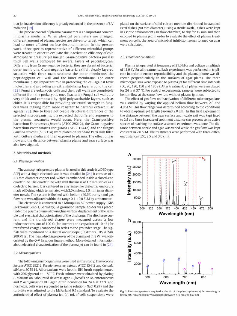
Fig. 1. Emission spectrum acquired at the tip of the plasma plume (a) for wavelengthsbelow 500 nm and (b) for wavelengths between 475 nm and 850 nm.
20 T.M.C. Nishime et al. / Surface & Coatings Technology 312 (2017) 19–24
that jet inactivation efficiency is greatly enhanced in the presence of UVradiation [19].
The precise control of plasma parameters is an important concernin plasma medicine. When physical parameters are changed,different amount of plasma species are driven to target, which canlead to more efficient surface decontamination. In the presentwork, three species representative of different microbial groupswere treated in order to evaluate the inactivation efficiency of coldatmospheric pressure plasma jet. Gram-positive bacteria possessthick cell walls composed by several layers of peptidoglycan.Differently from Gram-negative bacteria, they are absent of bacterialouter membrane. Gram-negative bacteria exhibit clearly layeredstructure with three main sections: the outer membrane, thepeptidoglycan cell wall and the inner membrane. The outermembrane plays important role in protecting the cell against toxicmolecules and providing an extra stabilizing layer around the cell[22]. Fungi are eukaryotic cells and their cell walls are completelydifferent from the prokaryotic ones (bacteria). Fungal cell walls arevery thick and composed by rigid polysaccharide layers, such aschitin. It is responsible for providing structural strength to fungicell walls making them more resistant to harmful extracellularagents [23]. Due to these noticeable structural differences of theselected microorganisms, it is expected that different responses tothe plasma treatment would occur. Here, the Gram-positivebacterium Enterococcus faecalis (ATCC 29212), the Gram-negativebacterium Pseudomonas aeruginosa (ATCC 15442) and the fungusCandida albicans (SC 5314) were plated on standard Petri dish filledwith culture media and then exposed to plasma. The effect of gasflow and the distance between plasma plume and agar surface wasalso investigated.
2. Materials and methods
2.1. Plasma generation
The atmospheric pressure plasma jet used in this study is a DBD typeAPPJ with a single electrode and it was detailed in [24]. It consists of a2.3 mm diameter copper rod, which is embedded inside a closed-endquartz tube. The quartz tube with wall thickness of 1.7 mm serves as adielectric barrier. It is centered in a syringe-like dielectric enclosuremade of Delrin,which terminatedwith 2.0 cm long, 1.5mm inner diam-eter nozzle. The system is flushed with helium (99.5% purity) and gasflow rate was adjusted within the range 0.1–10.0 SLM by a rotameter.
The electrode is connected to a Minupuls4 AC power supply (GBSElektronik GmbH, Germany). A grounded sample holder was placedunder the plasma plume allowingfine vertical displacement of the sam-ple and electrical characterization of the discharge. The discharge cur-rent and the transferred charge were measured across a lowinductance resistor of 100 Ω (for current) or a capacitor of 10 nF (fortransferred charge) connected in series to the grounded stage. The sig-nals were monitored on a digital oscilloscope (Tektronix TDS 2024B,200MHz). Themeandischarge power of the plasma jet (1.8W)was cal-culated by the Q-V Lissajous figure method. More detailed informationabout electrical characterization of the plasma jet can be found in [24].
2.2. Microorganisms
The followingmicroorganisms were used in this study: Enterococcusfaecalis ATCC 29212, Pseudomonas aeruginosa ATCC 15442 and Candidaalbicans SC 5314. All organisms were kept in BHI broth supplementedwith 20% glycerol at −80 °C. Fresh cultures were obtained by platingC. albicans on Sabouraud dextrose agar, E. faecalis on M-enterococcusand P. aeruginosa on BHI agar. After incubation for 24 h at 37 °C andnormoxia, cells were suspended in saline solution (NaCl 0.9%) and theturbidity was adjusted to the McFarland 0.5 standard. To evaluate theantimicrobial effect of plasma jet, 0.1 mL of cells suspensions were
plated on the surface of solid culture medium distributed in standardPetri dishes (90 mm diameter) using a sterile swab. Dishes were keptin aseptic environment (air flow chamber) to dry for 15 min and thenexposed to plasma jet. In order to evaluate the effect of plasma treat-ment on cells, the area of microbial inhibition zones formed on agarwere calculated.
2.3. Treatment conditions
Plasma jet operated at frequency of 31.0 kHz and voltage amplitudeof 13.0 kV for all treatments. Each experiment was performed in tripli-cate in order to ensure reproducibility and the plasma plume was di-rected perpendicularly to the surfaces of agar plates. The threemicroorganisms were exposed to plasma jet for different time intervals(60, 90, 120, 150 and 180 s). After treatment, all plates were incubatedfor 24 h at 37 °C. For control experiments, samples were subjected tohelium flow at the same flow rate without plasma ignition.
The effect of gas flow on inactivation of different microorganismswas studied by varying the applied helium flow between 2.0 and4.0 SLM. This flow range was determined according to the conditionsto obtain optimal jet length (around 2.0 cm). In this first experiment,the distance between the agar surface and nozzle exit was kept fixedto 2.5 cm. Since increase of treatment distance can prevent some activespecies of reaching the surface, a second experiment was done. The dis-tance between nozzle and agar was varied while the gas flow was keptconstant in 2.0 SLM. The treatments were performed with three differ-ent distances (2.0, 2.5 and 3.0 cm).
21T.M.C. Nishime et al. / Surface & Coatings Technology 312 (2017) 19–24
2.4. Scanning electron microscopy analysis (SEM)
After exposure to plasma all microorganismswere subjected to SEManalysis in order to investigate possible cell damages. Ten microliters ofcells suspensionswere transferred to glass slides and exposed to plasmajet for 120 s. After treatment, glass slides were maintained in 2% glutar-aldehyde solution for 2 h. Then, the slides were washed with saline so-lution, after that exposed consequently to 25%, 50% and 75% ethanolsolutions for 20 min in each and finally immersed in 100% ethanol for1 h. After drying for 24 h at 28 °C, a thin golden layer was deposited
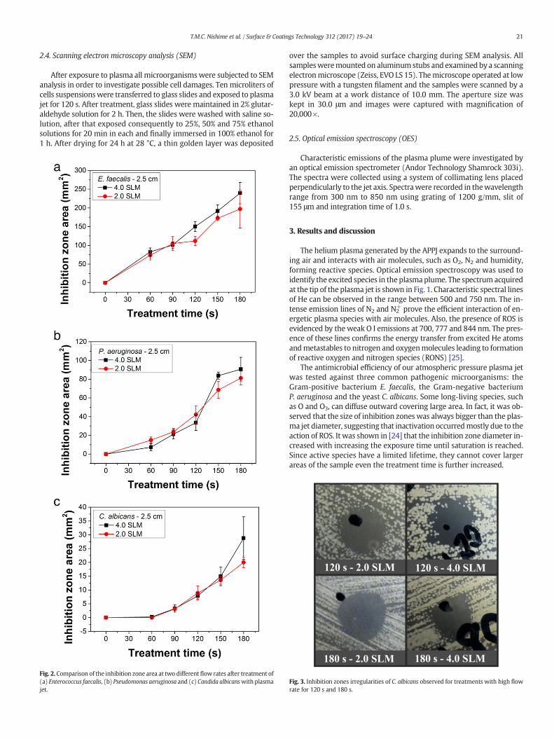
Fig. 2. Comparison of the inhibition zone area at two different flow rates after treatment of(a) Enterococcus faecalis, (b) Pseudomonas aeruginosa and (c) Candida albicanswith plasmajet.
over the samples to avoid surface charging during SEM analysis. Allsamplesweremounted on aluminumstubs and examined by a scanningelectronmicroscope (Zeiss, EVO LS 15). Themicroscope operated at lowpressure with a tungsten filament and the samples were scanned by a3.0 kV beam at a work distance of 10.0 mm. The aperture size waskept in 30.0 μm and images were captured with magnification of20,000×.
2.5. Optical emission spectroscopy (OES)
Characteristic emissions of the plasma plume were investigated byan optical emission spectrometer (Andor Technology Shamrock 303i).The spectra were collected using a system of collimating lens placedperpendicularly to the jet axis. Spectrawere recorded in thewavelengthrange from 300 nm to 850 nm using grating of 1200 g/mm, slit of155 μm and integration time of 1.0 s.
3. Results and discussion
The helium plasma generated by the APPJ expands to the surround-ing air and interacts with air molecules, such as O2, N2 and humidity,forming reactive species. Optical emission spectroscopy was used toidentify the excited species in the plasmaplume. The spectrum acquiredat the tip of the plasma jet is shown in Fig. 1. Characteristic spectral linesof He can be observed in the range between 500 and 750 nm. The in-tense emission lines of N2 and N2
+ prove the efficient interaction of en-ergetic plasma species with air molecules. Also, the presence of ROS isevidenced by theweak O I emissions at 700, 777 and 844 nm. The pres-ence of these lines confirms the energy transfer from excited He atomsandmetastables to nitrogen and oxygenmolecules leading to formationof reactive oxygen and nitrogen species (RONS) [25].
The antimicrobial efficiency of our atmospheric pressure plasma jetwas tested against three common pathogenic microorganisms: theGram-positive bacterium E. faecalis, the Gram-negative bacteriumP. aeruginosa and the yeast C. albicans. Some long-living species, suchas O and O3, can diffuse outward covering large area. In fact, it was ob-served that the size of inhibition zones was always bigger than the plas-ma jet diameter, suggesting that inactivation occurredmostly due to theaction of ROS. It was shown in [24] that the inhibition zone diameter in-creased with increasing the exposure time until saturation is reached.Since active species have a limited lifetime, they cannot cover largerareas of the sample even the treatment time is further increased.
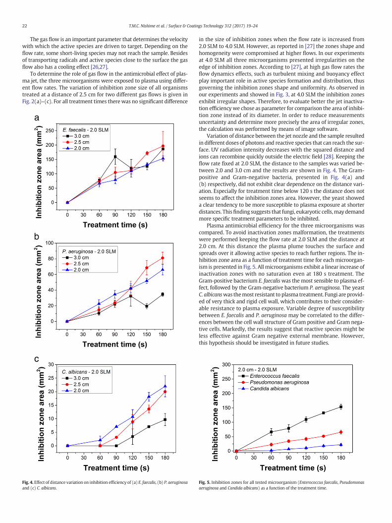
Fig. 3. Inhibition zones irregularities of C. albicans observed for treatments with high flowrate for 120 s and 180 s.
22 T.M.C. Nishime et al. / Surface & Coatings Technology 312 (2017) 19–24
The gas flow is an important parameter that determines the velocitywith which the active species are driven to target. Depending on theflow rate, some short-living species may not reach the sample. Besidesof transporting radicals and active species close to the surface the gasflow also has a cooling effect [26,27].
To determine the role of gas flow in the antimicrobial effect of plas-ma jet, the three microorganisms were exposed to plasma using differ-ent flow rates. The variation of inhibition zone size of all organismstreated at a distance of 2.5 cm for two different gas flows is given inFig. 2(a)–(c). For all treatment times there was no significant difference
Fig. 4. Effect of distance variation on inhibition efficiency of (a) E. faecalis, (b) P. aeruginosaand (c) C. albicans.
in the size of inhibition zones when the flow rate is increased from2.0 SLM to 4.0 SLM. However, as reported in [27] the zones shape andhomogeneity were compromised at higher flows. In our experimentsat 4.0 SLM all three microorganisms presented irregularities on theedge of inhibition zones. According to [27], at high gas flow rates theflow dynamics effects, such as turbulent mixing and buoyancy effectplay important role in active species formation and distribution, thusgoverning the inhibition zones shape and uniformity. As observed inour experiments and showed in Fig. 3, at 4.0 SLM the inhibition zonesexhibit irregular shapes. Therefore, to evaluate better the jet inactiva-tion efficiencywe chose as parameter for comparison the area of inhibi-tion zone instead of its diameter. In order to reduce measurementsuncertainty and determine more precisely the area of irregular zones,the calculation was performed by means of image software.
Variation of distance between the jet nozzle and the sample resultedin different doses of photons and reactive species that can reach the sur-face. UV radiation intensity decreases with the squared distance andions can recombine quickly outside the electric field [28]. Keeping theflow rate fixed at 2.0 SLM, the distance to the samples was varied be-tween 2.0 and 3.0 cm and the results are shown in Fig. 4. The Gram-positive and Gram-negative bacteria, presented in Fig. 4(a) and(b) respectively, did not exhibit clear dependence on the distance vari-ation. Especially for treatment time below 120 s the distance does notseems to affect the inhibition zones area. However, the yeast showeda clear tendency to be more susceptible to plasma exposure at shorterdistances. Thisfinding suggests that fungi, eukaryotic cells,may demandmore specific treatment parameters to be inhibited.
Plasma antimicrobial efficiency for the three microorganisms wascompared. To avoid inactivation zones malformation, the treatmentswere performed keeping the flow rate at 2.0 SLM and the distance at2.0 cm. At this distance the plasma plume touches the surface andspreads over it allowing active species to reach further regions. The in-hibition zone area as a function of treatment time for each microorgan-ism is presented in Fig. 5. All microorganisms exhibit a linear increase ofinactivation zones with no saturation even at 180 s treatment. TheGram-positive bacterium E. faecaliswas the most sensible to plasma ef-fect, followed by the Gram-negative bacterium P. aeruginosa. The yeastC. albicanswas themost resistant to plasma treatment. Fungi are provid-ed of very thick and rigid cell wall, which contributes to their consider-able resistance to plasma exposure. Variable degree of susceptibilitybetween E. faecalis and P. aeruginosa may be correlated to the differ-ences between the cell wall structure of Gram positive and Gram nega-tive cells. Markedly, the results suggest that reactive species might beless effective against Gram negative external membrane. However,this hypothesis should be investigated in future studies.
Fig. 5. Inhibition zones for all tested microorganism (Enterococcus faecalis, Pseudomonasaeruginosa and Candida albicans) as a function of the treatment time.
23T.M.C. Nishime et al. / Surface & Coatings Technology 312 (2017) 19–24
Several mechanisms are thought to be responsible for microbial in-hibition in plasma. Some authors showed that the reactive plasmaagents target proteins, DNA, cell wall andmembrane. For instance, oxy-gen species, such as O andO3 can physically etch the cell membrane andinduce DNA deterioration [13]. UV radiation in the wavelength range of250–500 nm can also induce damage to DNA [28]. Excessive charge ac-cumulation onmicroorganisms can lead to cell disruption [29]. After ex-position to all active species present in the plasma plume,microorganisms are mostly affected by cell wall damage [30]. Thus,cell morphology of each microorganism was investigated by SEM andthe results are presented in Figs. 6–8.
The SEM images showed that plasma jet induced damage toC. albicans, E. faecalis and P. aeruginosa cells. Surface damage and intra-cellular material loss are evidenced in C. albicans shown in Fig. 6(b).Fig. 7(b) shows changes in cell morphology of E. faecalis. Also, majormodifications in P. aeruginosa cells structure can be observed in Fig.8(b), suggesting loss of cellular content.
4. Conclusions
The He plasma jet investigated in this study was found to be anefficient microbicide, when tested against three different microbialspecies. The size of inhibition zones on Petri dishes was much larger
Fig. 7. Scanning electronmicroscopy of E. faecalis cells. (a) Cells not exposed to plasma jet.(b) Cells exposed to plasma jet indicating change in cell morphology. The arrow indicatesloss of cellular integrity.
Fig. 6. Scanning electronmicroscopy of C. albicans cells. (a) Cells not exposed to plasma jet(20,000×). (b) Cells exposed to plasma jetwith arrows indicating surface damage and lossof cellular material.
than the jet diameter, suggesting that ozone, as a long-living species,is the major inactivation agent. Although the gas flow does notsignificantly influence the size of inhibition zones, their shape andhomogeneity were affected. The 2.0 SLM flow presented more uniformzones with circular shape. The Gram-positive bacterium presented thegreatest susceptibility to plasma exposure compared to the othersmicroorganisms, while the fungus C. albicans showed to be the mostresistant. The amount of active species and UV radiation that reach thesample can be controlled varying the distance between the plasmaplume and the substrate. The presented results reveal that the distanceis an essential parameter for fungi inactivation especially for longtreatment time. SEM analysis revealed loss of cells integrity after plasmaexposure, suggesting that the process induces membrane damage andleak of cellular content.
Acknowledgments
The authors acknowledge financial support from Conselho Nacionalde Desenvolvimento Científico e Tecnológico (CNPq) under researchgrant 470995/2013-0 and from São Paulo State Research Foundation(FAPESP) under grant 2014/02354-7.

Fig. 8. Scanning electronmicroscopy of P. aeruginosa cells. (a) Cells not exposed to plasmajet. (b) Cells exposed to plasma jet exhibiting change in cell morphology.
24 T.M.C. Nishime et al. / Surface & Coatings Technology 312 (2017) 19–24
References
[1] C. Tendero, C. Tixier, P. Tristant, J. Desmaison, P. Leprince, Atmospheric pressureplasmas: a review, Spectrochim. Acta B 61 (2006) 2–30.
[2] V. Nehra, A. Kumar, H.K. Dwivedi, Atmospheric non-thermal plasma sources, Int. J.Eng. 2 (2008) 53–68.
[3] A. Schutze, J.Y. Jeong, S.E. Babayan, J. Park, G.S. Selwyn, R.F. Hicks, The atmospheric-pressure plasma jet: a review and comparison to other plasma sources, IEEE Trans.Plasma Sci. 26 (1998) 1685–1694.
[4] X. Lu, M. Laroussi, V. Puech, On atmospheric-pressure non-equilibrium plasma jetsand plasma bullets, Plasma Sources Sci. Technol. 21 (2012) 1–17.
[5] I. Koban, R. Matthes, N.-O. Hübner, A. Welk, P. Meisel, B. Holtfreter, R. Sietmann, E.Kindel, K.-D. Weltmann, A. Kramer, T. Kocher, Treatment of Candida albicansbiofilms with low-temperature plasma induced by dielectric barrier discharge andatmospheric pressure plasma jet, New J. Phys. 12 (2010) 1–16.
[6] K. Fricke, I. Koban, H. Tresp, L. Jablonowski, K. Schröder, A. Kramer, K.-D. Weltmann,T. von Woedtke, T. Kocher, Atmospheric pressure plasma: a high-performance toolfor the efficient removal of biofilms, PLoS One 7 (2012) 1–8.
[7] M.Y. Alkawareek, S.P. Gorman, W.G. Grahan, B.F. Gilmore, Potential cellular targetsand antibacterial efficacy of atmospheric pressure non-thermal plasma, Int. J.Antimicrob. Ag. 43 (2014) 154–160.
[8] Y. Nasruddin, K. Nakajima, E. Mukai, H.S.E. Komatsu, M. Rahayu, T. Nur, H. Ishijima,Y. Enomoto, J. Uesugi, T. Sugama, Nakatani, a simple technique to improve contrac-tile effect of cold plasma jet on acute mouse wound by dropping water, Plasma Pro-cess. Polym. 12 (2015) 1–12.
[9] H. Kuwahata, T. Yamaguchi, R.-I. Ohyama, A. Ito, Inactivation of Escherichia coli usingatmospheric-pressure plasma jet, Jpn. J. Appl. Phys. 54 (2015) 1–6.
[10] M.Y. Alkawareek, Q.T. Algwari, G. Laverty, S.P. Gorman, W.G. Graham, D. O'Connell,B.F. Gilmore, Eradication of Pseudomonas aeruginosa biofilms by atmospheric pres-sure non-thermal plasma, PLoS One 7 (2012) 1–7.
[11] P.P. Sedghizadeh, M.T. Chen, C. Schaudinn, A. Gorur, C. Jiang, Inactivation kineticsstudy of an atmospheric-pressure cold-plasma jet against pathogenic microorgan-isms, IEEE Trans. Plasma Sci. 40 (2012) 2879–2882.
[12] M. Lee, H. Kim, Y. Kim, W.-Y. Lee, K.Y. Baik, N.K. Kaushik, G. Cho, Blood coagulationwith atmospheric-plasma jets, IEEE Int. Conf. Plasma Sci. 2P-159 (2012).
[13] A. Mai-Prochnow, A.B. Murphy, K.M. McLean, M.G. Kong, K. Ostrikov, Atmosphericpressure plasmas: infection control and bacterial response, Int. J. Antimicrob. Ag.43 (2014) 508–517.
[14] G.M. Xu, X.M. Shi, J.F. Cai, S.L. Chen, P. Li, C.W. Yao, Z.S. Chang, G.J. Zhang, Dual effectsof atmospheric pressure plasma jet on skin wound healing of mice, Wound RepairRegen. 23 (2015) 878–884.
[15] K.-D. Weltmann, E. Kindel, T. von Woedtke, M. Hähnel, M. Stieber, R. Brandenburg,Atmospheric-pressure plasma sources: prospective tools for plasma medicine, PureAppl. Chem. 82 (2010) 1223–1237.
[16] H. Yamazaki, T. Ohshima, Y. Tsubota, H. Yamaguchi, J. Jayawardena, Y. Nishimura,Microbicidal activities of low frequency atmospheric pressure plasma jets on oralpathogens, Dent. Mater. J. 30 (2011) 384–391.
[17] K. Wende, P. Williams, J. Dalluge, W. van Gaens, H. Aboubakr, J. Bischof, T. vonWoedtke, S.M. Goyal, K.-D. Weltmann, A. Bogaerts, K. Masur, P.J. Bruggeman, Iden-tification of the biologically active liquid chemistry induced by a nonthermal atmo-spheric pressure plasma jet, Biointerphases 10 (2015) 1–16.
[18] S. Reuter, J. Winter, S. Iséni, A. Schmidt-Bleker, M. Dünnbier, K. Masur, K. Wende, K.-D.Weltmann, The influence of feed gas humidity versus ambient humidity on atmo-spheric pressure plasma jet-effluent chemistry and skin cell viability, IEEE Trans.Plasma Sci. 43 (2014) 3185–3192.
[19] J.W. Lackman, S. Schneider, E. Edengeiser, F. Jarzina, S. Brinckmann, E. Steinborn, M.Havenith, J. Benedikt, J.E. Bandow, Photons and particles emitted from coldatmospheric-pressure plasma inactivate bacteria and biomolecules independentlyand synergistically, J. R. Soc. Interface 10 (2014) 1–12.
[20] M.Y. Alkawareek, Q.T. Algwari, S.P. Gorman, W.G. Graham, D. O'Connell, B.F.Gilmore, Application of atmospheric pressure nonthermal plasma for the in vitroeradication of bacterial biofilms, FEMS Immunol. Med. Mic. 65 (2012) 381–384.
[21] E. Dolezalova, P. Lukes, Membrane damage and active but nonculturable state in liq-uid cultures of Escherichia coli treated with an atmospheric pressure plasma jet,Bioelectrochemistry 103 (2015) 7–14.
[22] T.J. Silhavy, D. Kahne, S. Walker, The bacterial cell envelope, Cold Spring Harb.Perspect. Biol. 2 (2010) 1–16.
[23] J. Ruiz-Herrera, Fungal CellWall: Structure, Synthesis, and Assembly, second ed. CRCPress, Boca Raton, 2012.
[24] K.G. Kostov, A.C. Borges, C.Y. Koga-Ito, T.M.C. Nishime, V. Prysiazhnyi, R.Y. Honda, In-activation of Candida albicans by cold atmospheric pressure plasma jet, IEEE Trans.Plasma Sci. 43 (2015) 770–775.
[25] X. Pei, X. Lu, J. Liu, D. Liu, Y. Yang, Inactivation of a 25.5 μm Enterococcus faecalis bio-film by a room-temperature, battery-operated, handheld air plasma jet, J. Phys. D.Appl. Phys. 45 (2012) 1–5.
[26] C.-T. Liu, C.-J. Wu, Y.-W. Yang, Z.-H. Lin, J.-S. Wu, S.-C. Hsiao, C.-P. Lin, Atomic oxygenand hydroxyl radical generation in round helium-based atmospheric-pressure plas-ma jets by various electrode arrangements and its application in sterilizing Strepto-coccus mutans, IEEE Trans. Plasma Sci. 42 (2014) 3830–3836.
[27] J. Goree, B. Liu, D. Drake, Gas flow dependence for plasma-needle disinfection ofS. mutans bacteria, J. Physics D Appl. Phys. 39 (2006) 3479–3486.
[28] J.-W. Lackmann, J.E. Bandow, Inactivation of microbes and macromolecules byatmospheric-pressure plasma jets, Appl. Microbiol. Biotechnol. 98 (2014)6205–6213.
[29] E. Stoffels, “Tissue processing” with atmospheric plasmas, Contrib. Plasma Phys. 47(2007) 40–48.
[30] K. Kim, H.J. Ahn, J.-H. Lee, J.-H. Kim, S.S. Yang, J.-S. Lee, Cellular membrane collapseby atmospheric-pressure plasma jet, Appl. Phys. Lett. 104 (2014) 1–3.