suppression of inflammation delays hair cell regeneration and … · abstract: background: human...
TRANSCRIPT

1
Suppression of Inflammation Delays Hair Cell 1
Regeneration and Functional Recovery Following 2
Lateral Line Damage in Zebrafish Larvae 3
Ru Zhang2,3, Xiao-Peng Liu3,4, Ya-Juan Li2, Ming Wang1,3, Lin Chen1,3*, 4
Bing Hu1,2* 5
6
1 CAS Key Laboratory of Brain Function and Diseases, School of Life 7
Sciences, University of Science and Technology of China, Hefei 230027, 8
China. 9
2 Lab of Neurodevelopment and Repair, University of Science and 10
Technology of China, Hefei 230027, China. 11
3 Auditory Research Laboratory, University of Science and Technology of 12
China, Hefei 230027, China. 13
4 Center for Hearing and Deafness, State University of New York at Buffalo, 14
NY14214, USA. 15
16
17
18
*Corresponding authors. 19
Bing Hu, PhD, Lab of Neurodevelopment and Repair, University of Science 20
and Technology of China, Hefei 230027, China. 21
Tel.: +86 (551) 6360-2489 22
E-mail: [email protected] 23
Lin Chen, PhD, Auditory Research Laboratory, University of Science and 24
Technology of China, Hefei 230027, China. 25
Tel.: +86 (551) 6360-7623 26
E-mail: [email protected] 27
28

2
Abstract: 29
Background: Human cochlear hair cells cannot regenerate after loss. In 30
contrast, those in fish and amphibians have a remarkable ability to regenerate 31
after damaged. Previous studies focus on mechanisms of hair cell 32
regeneration, such as Wnt and Notch signals. These studies ignore the fact 33
that the beginning of regeneration is accompanied by a large number of 34
inflammatory responses. The role of this inflammation in hair cell regeneration 35
is still unknown. In addition, there is no appropriate behavioral method to 36
quantitatively evaluate the functional recovery of lateral line hair cells after 37
regeneration. 38
Results: In this study, we found that when inflammation was suppressed, the 39
regeneration of lateral line hair cells and the recovery of the rheotaxis of the 40
larvae were significantly delayed. Calcium imaging showed that the function of 41
the neuromasts in the inflammation-inhibited group was weaker than that in the 42
non-inflammation-inhibited group at the Early Stage of regeneration, and 43
returned to normal at the Late Stage. Calcium imaging also revealed the cause 44
of the mismatch between the function and quantity during regeneration. 45
Conclusions: Our results, meanwhile, suggest that suppressing 46
inflammation delays hair cell regeneration and functional recovery when hair 47
cells are damaged. This study may provide a new idea for how to promote hair 48
cell regeneration and functional recovery in adult mammals. 49
50
51
Keywords: inflammation, hair cell regeneration, neuromast, lateral line, 52
zebrafish larva, rheotaxis, calcium imaging. 53
54
55

3
Background: 56
Deafness and hearing defects are usually caused by loss of sensory hair 57
cells or defect of auditory function. The loss of hair cells is result of aging, 58
infection, genetic factors, hypoxia, autoimmune disorder, ototoxic drugs or 59
noise exposure. Unfortunately, including humans, hair cells cannot regenerate 60
in mammals (Oesterle and Stone, 2008; Yorgason. et al., 2006). In contrast, 61
hair cells in some non-mammalian vertebrates have a remarkable ability to 62
regenerate, such as birds, reptiles, amphibians and fish (Matsui. and 63
Cotanchea., 2004; Popper and Hoxter, 1984; Stone. and Rubel., 2000). It 64
could suggest that if we figure out the mechanism of hair cell regeneration in 65
these species, we probably can promote hair cell regeneration in mammals. 66
When hair cells are damaged, support cells proliferate into both hair cells 67
and support cells, or convert into hair cells directly(Baird et al., 1996; 68
Lopez-Schier and Hudspeth, 2006; Raphael, 1992; Roberson et al., 2004). 69
Hair cell regeneration is finely regulated by the interaction of multiple signaling 70
pathways, such as Notch signaling(Ma et al., 2008; Mizutari et al., 2013), 71
Wnt/b-catenin signaling(Aman and Piotrowski, 2008; Chai et al., 2012; Shimizu 72
et al., 2012), Fgf signaling(Aman and Piotrowski, 2008; Nechiporuk and Raible, 73
2008), retinoic acid(Rubbini et al., 2015) and so on. In the process of hair cell 74
damaged, it is accompanied by a lot of inflammatory reaction, which has been 75
found to play a role in tissue regeneration in recent years(Mescher, 2017). For 76
example, macrophages are considered having main function in the 77
inflammatory resolution stage and being required for fin regeneration(Li et al., 78
2012) and hair cell regeneration in zebrafish(Carrillo et al., 2016). In addition, it 79
has been confirmed that neutrophils in mice play a central role in 80
inflammation-induced optic nerve regeneration(Kurimoto et al., 2013). 81
In recent years, zebrafish (Danio rerio) has become an ideal model for 82
studying inflammation and hair cell regeneration because it has conservative 83

4
innate immunity (Renshaw and Trede, 2012) and strong regeneration ability in 84
lateral line system(Lush and Piotrowski, 2014) which makes zebrafish larvae 85
to perceive the change of surrounding flow, detect their prey and avoid 86
predators(Coombs. et al., 2014; Dijkgraaf, 1962). The lateral system of a larva 87
is composed of neuromasts which located on the surface of the body. 88
The neuromasts on the head consist of the anterior lateral line system (aLL) 89
and the ones along the body comprise the posterior lateral line system 90
(pLL)(Thomas et al., 2015). The center of the neuromast is composed of hair 91
cells and they are surrounded by support cells and mantle cells. At the top of 92
the hair cells, rows of short stereocilia and a long kinocilium extend out of the 93
body called the hair bundle and are covered in a gelatinous cupula. The 94
arrangement of stereocilia and kinocilium determines the polarity of hair cells 95
and the polarity of the hair cells is planar cell polarity (PCP), which is arranged 96
symmetrically (Flock and Wersall, 1962), half in each direction. 97
When hair bundles are deflected, hair cells release transmitters and cause 98
exciting spikes in afferent neurons(Dijkgraaf, 1962). And then, larvae show a 99
robust behavior called rheotaxis(Olszewski et al., 2012). This behavior can be 100
applied to evaluate the function of hair cells (Suli et al., 2012). 101
In recent years, calcium imaging has become a popular method to 102
measure the function of neural cells in detail and quantitatively (Zhang et al., 103
2016). When the mechanical hair bundle deflected, calcium and other cations 104
enter into cytoplasm through mechanotransduction channels. It changes the 105
membrane potential and activates voltage-gated calcium channels which allow 106
rapid calcium inflow to trigger synaptic transmission. GCaMPs, a 107
genetically-encoded calcium indicator(GECIs), are single fluorescent proteins, 108
which can bind calcium directly and alter conformation to respond the change 109
of calcium concentration(Tian et al., 2012). These significant, 110
activity-dependent signals can reflect the function of hair cells in a single 111

5
neuromast (Zhang et al., 2018; Zhang et al., 2016). 112
Previous research has found that the deletion of macrophages by 113
morpholino leads to the delay of hair cell regeneration(Carrillo et al., 2016). 114
However, does it still cause the delay of hair cell regeneration when the 115
macrophages are intact, and the pro-inflammatory factors are suppressed as 116
the hair cells are damaged? Is there any delay in the functional recovery of the 117
lateral line? 118
In order to figure out the above problems, we used an anti-inflammatory 119
agent, BRS-28, to suppress the inflammation when hair cells are damaged by 120
copper. BRS-28 is a derivative of 5α-cholestan-6-one, which was confirmed to 121
be a remarkably suppressor of the production of pro-inflammatory factors, 122
such as NO, TNF-α、IL-1β、iNOS and cox-2(Yang et al., 2014). We count the 123
number of neutrophils and macrophages in Tg(corola-eGFP; lyz-Dsred) 124
transgenic line. Then, AB/WT zebrafish larvae were used to count the number 125
of regenerated hair cells. Since there is no appropriate behavioral method to 126
quantitatively evaluate the function of lateral line hair cells, we designed and 127
built devices to test rheotaxis behavior in AB/WT larvae. A behavioral analysis 128
software was applied for quantitative evaluation of rheotaxis, so as to reflect 129
the holistic functional recovery of the posterior lateral line. Finally, the function 130
of the regenerated hair cells in a single neuromast was evaluated by the 131
method of calcium imaging in Huc:h2b-gcamp6f transgenic line. 132
133
Results 134
CuSO4 damaged hair cells in lateral line of zebrafish. 135
Sensory hair cells in a 6-day post fertilization (dpf) AB/WT zebrafish larva 136
were labeled with 0.05% DASPEI clearly (Fig. 1A). L2、LII2、L3 neuromasts 137
(circles in Fig.1 A) were three of the posterior lateral neuromasts, which 138

6
located along the flat truck body and easily to be observed. A lateral view of the 139
neuromasts showed the elongated kinocilia extending from the body (Fig. 1B). 140
The neuromasts are consisted of hair cells surrounded by support cells, which 141
are surrounded by mantle cells (Fig. 1C). In order to study the effects of 142
inflammation on hair cell regeneration, we established a hair-cell-damaged 143
model. Hair cells were damaged completely, when treated with 5 μM CuSO4 144
for 1 h (Fig. 1D). Labeled with 0.05% DASPEI, hair cells displayed close 145
arrangement and clear boundary. Only treated with CuSO4 solution for 20 min, 146
hair cells became loose and unclear which suggested that they were already 147
injured. The number of hair cells decreased with weaker fluorescence intensity 148
and obscure cell boundary at 40 min. Hair cells were completely disappeared 149
at 60 min, indicating that they had been completely damaged. TUNEL assay 150
revealed the missing hair cells underwent apoptosis (Supplementary Fig. 1). 151
After being transferred to embryo medium (EM), the number of hair cells 152
quickly returned to normal (Fig. 1E). 153
BRS-28 reduced the number of neutrophils and macrophages migrating 154
to the injured neuromasts. 155
Neutrophils (Fig. 2B, C, blue arrows) and macrophages (Fig. 2B, C, white 156
arrows) could be marked and distinguished in larvae of Tg(corola-eGFP; 157
lyz-Dsred) transgenic line (Supplementary Fig. 2). Normally, neutrophils and 158
macrophages were almost absent from the neuromasts (example, Fig. 2A). 159
When treated with CuSO4 solution, hair cells were damaged. Neutrophils and 160
macrophages migrated to neuromasts within 1 hours (example, Fig. 2B). 161
When larvae were immerged in BRS-28, an anti-inflammatory agent, before 162
treated with CuSO4 solution, less neutrophils and macrophages migrated to 163
the damaged neuromasts (example, Fig. 2C). When the inflammation 164
suppressed, the numbers of neutrophils appeared around the damaged 165
neuromasts were lower at 0.5,1,3 and 4 h after add the CuSO4 solution in 166

7
BRS+CuSO4 group than in CuSO4 group (Fig. 2D). In addition, we observed 167
BRS+CuSO4 group had fewer macrophages at 0.5, 1, 2 and 3 h than CuSO4 168
group (Fig. 2E). Collectively, the data strongly suggested that BRS-28 reduced 169
the number of neutrophils and macrophages migrating to the injured 170
neuromasts. It was worth noting that compared with control, there was no 171
significant difference in the numbers of neutrophils and macrophages between 172
CuSO4 group and BRS+CuSO4 group at 5 and 6 h, indicating that the 173
inflammation was resolved. 174
Suppressing inflammation delayed hair cell regeneration. 175
In order to investigate whether the regeneration of hair cells were delayed 176
after suppressing inflammation, we observed hair cells in the L2, LII2 and L3 177
neuromasts. We found that the regeneration of hair cells was delayed after the 178
inflammation was suppressed by the inflammatory inhibitor, BRS-28. Live 179
imaging showed regenerated hair cells in CuSO4, BRS+CuSO4 group at 24, 48 180
and 96 hours post injured (hpi) by CuSO4(Fig. 3A). Control group was showed 181
at the same time point. Further analysis revealed that the numbers of 182
regenerated hair cells were significantly decreased in BRS+CuSO4 group than 183
that in CuSO4 group at 16 hpi (P=0.0061), 24 hpi (P=0.0021) and 48 hpi 184
(P<0.0001) (Fig. 3B, n = 30 neuromasts). These results indicated that the 185
regeneration of hair cells was delayed in BRS+CuSO4 group within 48 hpi. 186
Compared with Control group, there was no difference in the number of hair 187
cells between CuSO4 group and BRS+CuSO4 group at 96 hpi, suggesting that 188
hair cells were regenerated to the normal level at 96 hpi. We also analyzed the 189
number of hair cells when only teated with BRS-28 (BRS group). As expected, 190
BRS group had no difference with Control group at any time point, excluding 191
the effect of BRS-28 on hair cells. 192
Since hair cells did not regenerate at a uniform rate, we defined the time 193
of regeneration into two periods: the Early Stage which includes the time from 194

8
0 to 48 hpi and the Late Stage which includes the time after 48 hpi. The 195
regeneration of hair cells was fast in the Early Stage and slow in the Late Stage. 196
Linear analysis was conducted on the number of hair cell regeneration in the 197
Early Stage. The slope in CuSO4 group (0.1879) was higher than that in 198
BRS+CuSO4 group (0.148) , meanwhile, x-intercept in CuSO4 group (4.16) 199
was higher than that in BRS+CuSO4 group (8.287) (Fig. 3C, D). These implied 200
that the hair cell regeneration in BRS+CuSO4 group may begin later and 201
slower than that in CuSO4 group. 202
To explore whether the time window of inflammatory suppression had 203
contribute to delayed regeneration, we changed the start time of BRS-28 204
treatment. We found that compared with the CuSO4 group, whether BRS-28 205
was added at the same time as CuSO4 (CuSO4+BRS 0 h group), or 30 minutes 206
after the addition of CuSO4 (CuSO4+BRS 0.5 h group), or 1 hour after the 207
addition of CuSO4(CuSO4+BRS 1 h group) (Fig. 3E), there was no statistical 208
difference on the number of regenerated hair cells. 209
To sum up, the regeneration of hair cells in lateral line was delayed after 210
the inflammation was suppressed by the inflammatory inhibitor BRS-28. 211
The functional recovery of the lateral line system was delayed when 212
inflammation was suppressed. 213
Since the rheotaxis could reflect the function of the lateral line, we 214
designed a behavioral device to test the rheotaxis of zebrafish (Fig. 4A, see 215
details in Materials and Methods). Larvae were placed from the right platform, 216
and they sense the water flow from right to left. Figure 4B, C were two 217
examples of larval rheotaxis processed by behavioral analysis software: the 218
former was a larva with excellent rheotaxis(Fig. 4B) and the latter was a larva 219
performed failure in the rheotaxis test(Fig. 4C). The left panels in these two 220
examples showed the track of this larva. The behavioral analysis software 221
mapped the movement path of larvae by line segment. The color of the line 222

9
segment represented the direction of movement of the larvae. All the 223
movements from right to left were represented by purplish or red segments, 224
where purple indicated that the velocity along the flow direction was greater 225
than or equal to the flow velocity, and red indicated that the velocity along the 226
flow direction was less than the flow velocity. All the movements from left to 227
right were represented by green segments, and the higher the brightness was, 228
the faster the speed was. The right panels displayed the motion vector. The 229
lengths of the blue segments represented the distance of each movement, and 230
the direction of the blue segment represented the direction of that movement. 231
The length of the red line segment was the ratio of motion vectors sum to the 232
motion arithmetic sum and the direction was the direction of the sum of the 233
vectors. 234
When the red segment was long and had a small angle of 0 degree, it 235
indicated that the motion of the larva was consistent with the opposite direction 236
of flow. It represented that the larva had good rheotaxis, indicating its lateral 237
line system had good function. Therefore, the software reported a high score. 238
On the contrary, when the red segment was short or had a small angle of 180 239
degree, it indicated that the larva moved randomly and had a poor rheotaxis, 240
indicating its lateral line system had poor function. In this case, the software 241
reported a low score. The scores reported by the software were plotted into bar 242
charts and showed in Figure 4D. After the hair cells were damaged by CuSO4, 243
there was little rheotaxis in both CuSO4 group and BRS+CuSO4 group. At 24 244
and 48 hpi, the rheotaxis of BRS+CuSO4 group was significantly lower than 245
that of control. On the contrary, the rheotaxis of CuSO4 group was not 246
significantly different from that of control within 24 hpi. Therefore, it indicated 247
that the functional recovery of lateral line system was delayed in BRS+CuSO4 248
group. The rheotaxis of BRS group at each time point was not different from 249
that of control, suggesting that BRS-28 had no significant effect on the 250

10
rheotaxis. In addition, we noted that the speed and distance of each 251
movements were consistent within different times and between different 252
groups: both were stable at around 22 mm/s (Fig. 4E, F), which indicated that 253
BRS-28 or CuSO4 did not affect the movement of zebrafish. 254
The conclusion was that the regenerated hair cells still had the ability to 255
sense water flow, but the functional recovery of lateral line system was delayed 256
when inflammation was suppressed. 257
Calcium imaging revealed the function of a single neuromast after hair 258
cell regeneration 259
Since we found a mismatch between the function of the lateral line and the 260
amount of hair cell regeneration, that is, after the zebrafish lateral line was 261
damaged by copper sulfate, it took 96 h for the hair cells to return to normal, 262
while the flow ability returned to normal at 24 h. The function of a single 263
neuromast can be evaluated by observing its calcium activity (Zhang et al., 264
2016). The L3 neuromast, located in flat trunk, was stimulated by water flow 265
from an electrode (Fig. 5A). Since hair cells had polarities, the yellow and 266
green hair cells represented opposite polarities. Chou et al. reported that the 267
polarity of the L3 neuromast is parallel to the anterior-posterior body axis(Chou 268
et al., 2017). Thus, by adjusting the direction of the electrode, water was 269
controlled to flow in two directions: anterior to posterior (A-P) direction or 270
posterior to anterior (P-A) direction. We found that not all hair cells responded 271
to the water flow, and only some hair cells were active (example, Fig. 5B, 272
circled cells). These active cells only responded to stimulus in one direction: 273
P-A direction (Fig. 5C, yellow ones and yellow circles in Fig. 5B) or A–P 274
direction (Fig. 5D, green ones and green circles in Fig. 5B). Because the 275
neuromasts were stereoscopic, some of the active hair cells were far from this 276
focal plane (dashed circles in Fig. 5B) and were not included in subsequent 277
fluorescence intensity analysis. 278
11
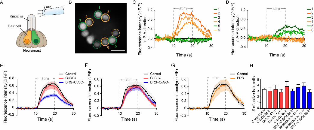
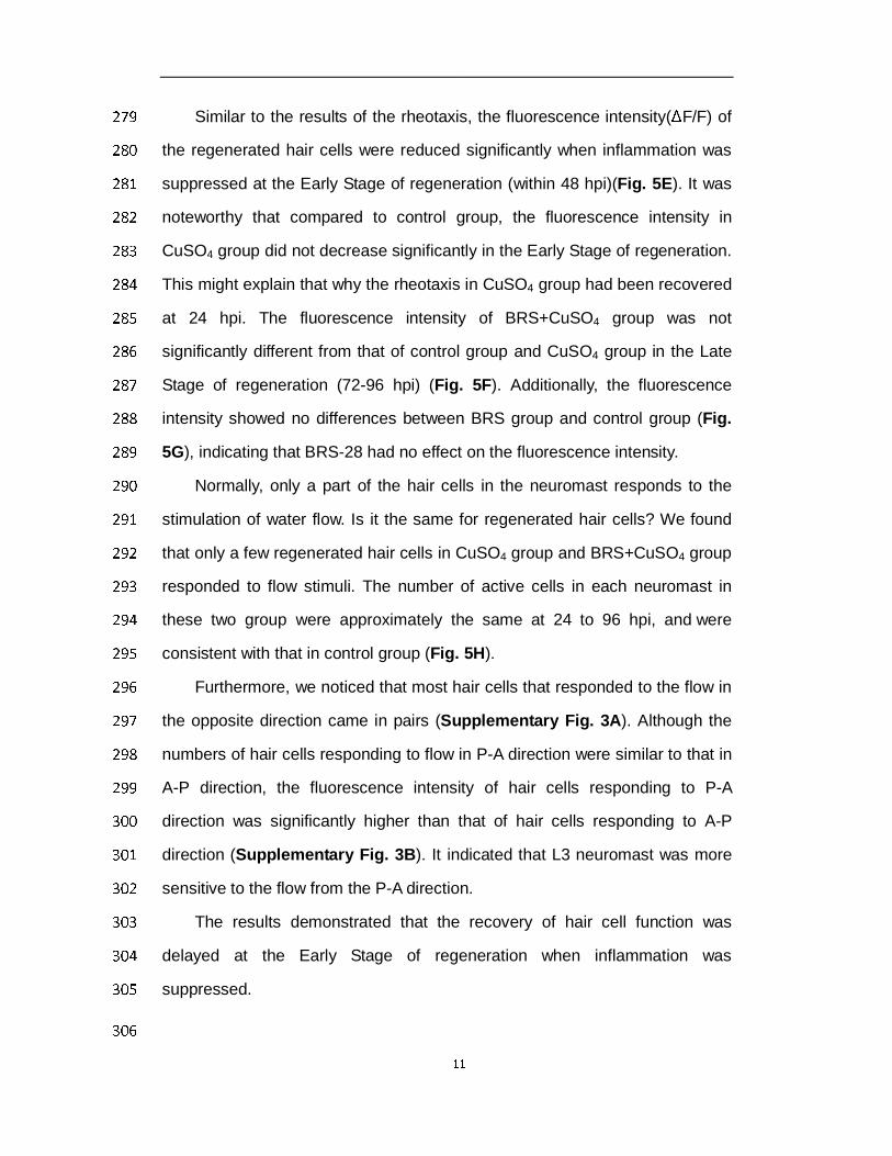
Similar to the results of the rheotaxis, the fluorescence intensity(ΔF/F) of 279
the regenerated hair cells were reduced significantly when inflammation was 280
suppressed at the Early Stage of regeneration (within 48 hpi)(Fig. 5E). It was 281
noteworthy that compared to control group, the fluorescence intensity in 282
CuSO4 group did not decrease significantly in the Early Stage of regeneration. 283
This might explain that why the rheotaxis in CuSO4 group had been recovered 284
at 24 hpi. The fluorescence intensity of BRS+CuSO4 group was not 285
significantly different from that of control group and CuSO4 group in the Late 286
Stage of regeneration (72-96 hpi) (Fig. 5F). Additionally, the fluorescence 287
intensity showed no differences between BRS group and control group (Fig. 288
5G), indicating that BRS-28 had no effect on the fluorescence intensity. 289
Normally, only a part of the hair cells in the neuromast responds to the 290
stimulation of water flow. Is it the same for regenerated hair cells? We found 291
that only a few regenerated hair cells in CuSO4 group and BRS+CuSO4 group 292
responded to flow stimuli. The number of active cells in each neuromast in 293
these two group were approximately the same at 24 to 96 hpi, and were 294
consistent with that in control group (Fig. 5H). 295
Furthermore, we noticed that most hair cells that responded to the flow in 296
the opposite direction came in pairs (Supplementary Fig. 3A). Although the 297
numbers of hair cells responding to flow in P-A direction were similar to that in 298
A-P direction, the fluorescence intensity of hair cells responding to P-A 299
direction was significantly higher than that of hair cells responding to A-P 300
direction (Supplementary Fig. 3B). It indicated that L3 neuromast was more 301
sensitive to the flow from the P-A direction. 302
The results demonstrated that the recovery of hair cell function was 303
delayed at the Early Stage of regeneration when inflammation was 304
suppressed. 305
306

12
Discussion 307
BRS-28 suppresses inflammation and delays the initiation of hair cell 308
regeneration. 309
Although the downregulation of Notch signal during lateral line 310
regeneration induces the proliferation of support cells by activating 311
Wnt/b-Catenin signal (Romero-Carvajal et al., 2015), it is still unknown how the 312
downregulation of Notch signal is triggered after the death of hair cells. Kniss 313
and his colleague proposed a hypothesis of triggering hair cell regeneration in 314
2016 (Kniss et al., 2016). They believed that apoptosis initiated the 315
proliferation of peripheral cells to promote tissue repair (Fan and Bergmann, 316
2008; Mollereau et al., 2013) and they called this process as compensatory 317
proliferation. They assumed that a similar process may be involved in the 318
regeneration of hair cells. On the basis of their hypothesis , we speculate that 319
when hair cells are damaged by CuSO4, it cause apoptosis in lateral line hair 320
cells, trigger the rise of reactive oxygen species (ROS) and reactive nitrogen 321
species (RNS), and induce the oxidative stress. This process may improve 322
AP-1,HIF-1α and NF-κB activity, and thus increase pro-inflammatory cytokines 323
and chemokines, such as NO, IL-1β, TNF-α, cox-2,iNOS and so on (Pereira et 324
al., 2016). BRS-28, suppress the production of NO、IL-1β、TNF-α、cox-2、iNOS 325
(Yang et al., 2014), reducing the number of neutrophils and macrophages 326
migrating to the damage of neuromasts. Besides that, the decrease of 327
pro-inflammatory factors may reduce the activation of macrophages. These 328
processes would decrease the production of TNF ligands and inhibit the JNK 329
signal, which contributes to initiating cells regeneration, and eventually leads 330
to delay initiation of compensatory proliferation and delay regeneration of hair 331
cells. 332
When the initiate time of inflammatory inhibitors changed, there is no 333
delay in the amount of regeneration. This also suggests that the timing of 334
13
inflammation suppression is important: when inflammation occurs, 335
compensated proliferation of the support cells has been triggered and hair cell 336
regeneration has been initiated. If inflammation suppression does not take 337
effect, regeneration seems to be unaffected. 338
In addition, neutrophils can also remove dead cell debris, and 339
macrophages can absorb apoptotic neutrophils or fragments of dead cells. We 340
suggest that neutrophils and macrophages with reduced number and activity 341
become slow in cleaning up damaged tissue areas, so as to have a good 342
regeneration environment. Since the damaged neuromasts need more time to 343
clean up these cell fragments, it may also delay the regeneration of hair cells. 344
Suppression of inflammation delays functional recovery of regenerated 345
hair cells 346
In this study, we have found that the number of hair cells decreased when 347
inflammation was suppressed which is the same as the functional recovery. 348
Finally, the quantity and the function of hair cells will be consistent with the 349
recovery of the normal level. Therefore, although the suppression of 350
inflammation delayed the regeneration of hair cells, it did not affect the overall 351
process of hair cell regeneration, and the function of hair cells regenerated in 352
the state of low inflammation eventually tended to be intact. These phenomena 353
are also consistent with the hypothesis I mentioned earlier. However, the effect 354
of inflammation on the regeneration of lateral hair cells seems to be different 355
from that of the fin. In 2012, Li and his colleague found that when zebrafish 356
larvae lacked macrophages, vacuoles appeared in the regenerated fin, which 357
suggests that macrophages may also be involved in the regeneration of the fin. 358
In our research, although the suppression of inflammation has delayed hair cell 359
regeneration and its recovery of function at the Early Stage of regeneration, 360
they eventually return to the normal status at the Late Stage of regeneration. 361
This is not because inflammation is not suppressed sufficiently, as Carrillo et al. 362
14
found in their study that the number of hair cells finally completed regeneration 363
even when macrophages was knockout(Carrillo et al., 2016). But that, this may 364
be because the injured organs are different, and the intact function of lateral 365
hair cells is crucial for the survival of zebrafish. We suggest that the hair cells 366
in lateral line may have more complex regulation in regeneration. 367
The functional recovery of hair cells is much faster than its quantity 368
Previous studies have focused on the morphological and quantitative 369
recovery of regenerated hair cells in zebrafish (Carrillo et al., 2016; 370
Romero-Carvajal et al., 2015). Since the regeneration takes 3-4 days post 371
injured, it is easy to assume that the recovery of the function of the neuromasts 372
may be proportional to the number of regenerated hair cells. In this study, for 373
the first time, we performed a method for evaluating the function of 374
regenerated hair cells. We found that the CuSO4 group already performed 375
excellent rheotaxis at 24 hpi (Fig. 4 C), although the average number of hair 376
cells was only 3.667 at that time (Fig. 3 B). Thus, the functional recovery of hair 377
cells is much faster than its quantity. In other words, although it takes 72-96 h 378
to complete regeneration, the function of hair cells can be recovered at 24 h. It 379
is critical to the survival of zebrafish. When BRS-28 is used to suppress the 380
inflammation, the amplitude of calcium activity of hair cells was significantly 381
lower than not only that of control group but that of CuSO4 group which makes 382
larvae show poor rheotaxis at the Early Stage of regeneration. Therefore, the 383
suppression of inflammation not only delays the hair cell regeneration, but also 384
delays the functional recovery. 385
We noticed that there is a mismatch between the function and quantity 386
during regeneration. Calcium image has revealed that only a part of hair cells 387
in one neuromast respond to the flow. This result is consistent with previous 388
study(Zhang et al., 2018). We have found that this phenomenon also exists in 389
regeneration group (CuSO4 and BRS+CuSO4 group). No matter what the 390

15
amount of regenerated hair cells is, the number of hair cells responding to the 391
flow remained stable during regeneration and has no differences with that of 392
the controls (Fig5 H). Besides that, the magnitude of fluorescence intensity 393
and reaction time is also consistent with that of the controls. This explains 394
why the number of regeneration in the CuSO4 group at 24 h is only 3.667 on 395
average, but the function of the lateral line has been restored to the level very 396
close to that of the control group. 397
In this study, We only performed calcium imaging on the L3 neuromast, 398
which was confirmed as the polarity of the A-P body axis in the study of Chou 399
et al(Chou et al., 2017). Consistent with their results, we also found this 400
neuromast is insensitive to the flow in the dorsal-ventral (D-V) body axis. 401
Therefore, this study only focused on the stimulus response in the direction of 402
the A-P body axis, and did not further analyze the stimulus data of the D-V 403
body axis. We have observed that most of hair cells that responded to the flow 404
from the direction P to A were more active than that from A to P (Fig5 J; sample, 405
Fig5 C,D) and that those responded to the opposite flow came in pairs(Fig 5 I). 406
All these suggest that it looks like being arranged beforehand rather than at 407
random though only a part of cells in one neuromast responded to flow 408
stimulation. 409
In summary, our research suggests that suppression of inflammation 410
delays functional regeneration of lateral hair cells in zebrafish larvae. The 411
inflammation plays positive and permissive roles in regeneration of hair cells. 412
413
Materials and Methods 414
Zebrafish strains and maintenance 415
AB/Wild-type strain, Tg(corola-eGFP;lyz-Dsred) and Huc:h2b-gcamp6f 416
transgenic line were used in this study. Embryos were generated by paired 417
mating and maintained at 28.5℃ in EM and on a 14/10 h light/dark cycle 418

16
according to the standard protocols. 419
All animal manipulations were conducted strictly in accordance with the 420
guidelines and regulations set forth by the University of Science and 421
Technology of China (USTC) Animal Resources Center and the University 422
Animal Care and Use Committee. The protocol was approved by the 423
Committee on the Ethics of Animal Experiments of the USTC (Permit Number: 424
USTCACUC1103013). 425
Hair cell damage and inflammation inhibition 426
In order to damage hair cells in lateral line, 4 dpf Larvae were treated with 427
5 μM CuSO4(Sangon, China) diluted in embro medium (EM) for 1 h. Then, they 428
were washed three times and recovered in EM. 429
To suppress inflammation, 4 dpf larvae were immersed in 0.1% BRS-28, 430
an anti-inflammatory agent, for 3 h before being moved into CuSO4 to damage 431
hair cells. 432
Live imaging 433
AB/Wild-type larvae were used to count the number of regenerated hair 434
cells in L2、LII2、L3 neuromasts (Fig. 1A). Hair cells were marked by 435
0.01 %DAPI for 5 minutes. Being anesthetized in 0.02% MS-222 (Tricaine 436
mesylate, Sigma,USA), larvae were imaged under a fluorescence microscope 437
(Olympus BX-60, Japan). 438
In order to exhibit the damage of hair cells in copper sulfate solution and 439
the regeneration of hair cell in different phases, hair cells were labeled by 440
0.05 % DASPEI (Sigma, USA), and larvae were anesthetized in MS-222 and 441
imaged under a confocal microscopy (Zeiss LSM 880 +Airyscan, Germany). 442
Tg(corola-eGFP; lyz-Dsred) transgenic line was used to observe the 443
number of neutrophils and macrophages migrating to the injured neuromasts 444
in vivo. This transgenic line expressed the enhanced green fluorescent protein 445
(eGFP) in macrophages and expressed both eGFP and Dsred in neutrophils, 446

17
which shows yellow fluorescent when merged. To show the neutrophils and 447
macrophages migrating to damaged neuromasts, larvae were anesthetized in 448
MS-222 and imaged under a confocal microscopy (Zeiss LSM 880 +Airyscan). 449
For neutrophils and macrophages counting, we determined the area within 100 450
μm around the L2、LII2、L3 neuromasts as the target. Zebrafish larvae were 451
anesthetized and imaged by the fluorescence microscope (Olympus BX-60) 452
with a green and a red channel. 453
Rheotaxis behavior experiments 454
A U-shaped tank was designed to test the rheotaxis behavior of larvae 455
(Fig 4A). The bottom of the two cubic tanks (7 cm length *8 cm width*8 cm 456
height) were connected by a platform (10 cm length *8 cm width*0.5 cm height). 457
A peristaltic pump (Longer Pump YZ1515x,China) was used to move EM 458
solution from the left tank to the right one, so that, a stable reverse flow was 459
formed (right to left, v=10 mm/s). AB/WT zebrafish larvae were applied to 460
detect the ability of rheotaxis. Larvae were released at the right side of the 461
platform with early velocity almost equals 0. To avoid visual cues, experiments 462
were operated in the dark and rheotaxis performs were recorded by an infrared 463
CCD (IR850, weixinshijie, China). 464
Rheotaxis data were analyzed by our own rheotaxis software edited in 465
Matlab (2015a, MathWorks, USA). This software can plot the movement track 466
of zebrafish larvae in the platform, measure the direction and distance of each 467
swimming and calculate the speed of each movement. Finally, it reports scores 468
based on the magnitude in the horizontal direction of the ratio of motion 469
vectors sum to the motion arithmetic sum. 470
Calcium imaging and data analyses 471
Huc: h2b-GCamp6f transgenic line was used in calcium imaging which 472
expressed pan-neuronal nucleus-labelled GCamp6f. Larvae were 473
anesthetized and fixed by a net pressure. The one-step pulled micropipette 474

18
had a long, wispy tip which must be trimmed by rubbing it against with another 475
pulled micropipette to generate a tip with an outer diameter of approximately 476
40 μm. The micropipette was filled with MS-222 and fixed to the holder of a 477
micromanipulator (MX7500, Scientific Design Company, USA). The tip of the 478
micropipette should be positioned at a distance of approximately 100 μm from 479
the top of the kinocilia (Fig.5 A). The duration of flow was controlled by 480
three direct links which were linked with a syringe. 481
Calcium imaging was collected by a confocal microscopy (FV 1000, 482
Olympus, Japan). In order to make as many hair cells as possible in the 483
observation area at the same time, a single z-axis was adjusted. Region of 484
interest (ROI) was set to 110*108. We took 100 time-lapse images for each 485
neuromast, and the total capture time was 29.7 s (0.297 s per slice). Flow 486
stimulation occured at the period from 10.098 to 19.899 s. 487
Since the neuromasts are three-dimensional, different hair cells have 488
different levels of fluorescence intensity. Namely, they have different levels of F 489
prime. The relative fluorescence intensity change (ΔF/F0) is more commonly 490
used. For each hair cell, the average fluorescence intensity before flow stimuli 491
(0-10 s) was set as F0. The data would be excluded when F0<95, which means 492
these hair cells were too far from the focal plane. If neuromasts had more than 493
two hair cells that responded to the flow stimulus, we took only the two that 494
responded the most in the fluorescence intensity change curve. 495
Statistical analysis 496
All data were shown as mean ± S.E.M. or as relative proportions of 100 % as 497
indicated in the appropriate legends. The data were analyzed in either 498
one-way ANOVA with Tukey's multiple comparisons test or two-way ANOVA 499
with Tukey's multiple comparisons test by GraphPad Prism version 7.0 (Prism, 500
San Diego, CA, USA). The level of significance was set to P < 0.05. *, **and 501
***represent P < 0.05, P < 0.01 and P < 0.001, respectively. 502

19
503
Acknowledgments 504
The authors thank Drs. Wen Zilong for providing the Tg(corola-eGFP; 505
lyz-Dsred) transgenic fish line, Drs. Wen Quan for providing the 506
Huc:h2b-gcamp6f transgenic fish line. The authors thank Drs. Zhen Xuechu for 507
providing BRS-28 and the compound-26 in their study is the BRS-28 508
mentioned in this study. 509
510
References 511
Aman, A., and Piotrowski, T. (2008). Wnt/beta-catenin and Fgf signaling control 512
collective cell migration by restricting chemokine receptor expression. Developmental 513
cell 15, 749-761. 514
Baird, R.A., Steyger, P.S., and Schuff, N.R. (1996). Mitotic and nonmitotic hair cell 515
regeneration in the bullfrog vestibular otolith organs. Annals of the New York Academy 516
of Sciences 781, 59-70. 517
Carrillo, S.A., Anguita-Salinas, C., Pena, O.A., Morales, R.A., Munoz-Sanchez, S., 518
Munoz-Montecinos, C., Paredes-Zuniga, S., Tapia, K., and Allende, M.L. (2016). 519
Macrophage Recruitment Contributes to Regeneration of Mechanosensory Hair Cells in 520
the Zebrafish Lateral Line. Journal of cellular biochemistry 117, 1880-1889. 521
Chai, R., Kuo, B., Wang, T., Liaw, E.J., Xia, A., Jan, T.A., Liu, Z., Taketo, M.M., Oghalai, 522
J.S., Nusse, R., et al. (2012). Wnt signaling induces proliferation of sensory precursors 523
in the postnatal mouse cochlea. Proceedings of the National Academy of Sciences of 524
the United States of America 109, 8167-8172. 525
Chou, S.W., Chen, Z., Zhu, S., Davis, R.W., Hu, J., Liu, L., Fernando, C.A., Kindig, K., 526
Brown, W.C., Stepanyan, R., et al. (2017). A molecular basis for water motion detection 527
by the mechanosensory lateral line of zebrafish. Nature communications 8, 2234. 528
Coombs., S., Bleckmann., H., Fay., R.R., and Popper, A.N. (2014). The Lateral Line 529
System (New York: Springer). 530
Dijkgraaf, S. (1962). The functioning and significance of the lateral-line organs. 531
Biological Reviews of the Cambridge Philosophical Society 38: 51-105. 532
Fan, Y., and Bergmann, A. (2008). Apoptosis-induced compensatory proliferation. 533
The Cell is dead. Long live the Cell! Trends in cell biology 18, 467-473. 534
Flock, A., and Wersall, J. (1962). A study of the orientation of the sensory hairs of 535
the receptor cells in the lateral line organ of fish, with special reference to the function 536
of the receptors. The Journal of cell biology 15, 19-27. 537
Kniss, J.S., Jiang, L., and Piotrowski, T. (2016). Insights into sensory hair cell 538

20
regeneration from the zebrafish lateral line. Current opinion in genetics & development 539
40, 32-40. 540
Kurimoto, T., Yin, Y., Habboub, G., Gilbert, H.Y., Li, Y., Nakao, S., Hafezi-Moghadam, 541
A., and Benowitz, L.I. (2013). Neutrophils express oncomodulin and promote optic 542
nerve regeneration. The Journal of neuroscience : the official journal of the Society for 543
Neuroscience 33, 14816-14824. 544
Li, L., Yan, B., Shi, Y.Q., Zhang, W.Q., and Wen, Z.L. (2012). Live imaging reveals 545
differing roles of macrophages and neutrophils during zebrafish tail fin regeneration. 546
The Journal of biological chemistry 287, 25353-25360. 547
Lopez-Schier, H., and Hudspeth, A.J. (2006). A two-step mechanism underlies the 548
planar polarization of regenerating sensory hair cells. Proceedings of the National 549
Academy of Sciences of the United States of America 103, 18615-18620. 550
Lush, M.E., and Piotrowski, T. (2014). Sensory hair cell regeneration in the zebrafish 551
lateral line. Developmental dynamics : an official publication of the American 552
Association of Anatomists 243, 1187-1202. 553
Ma, E.Y., Rubel, E.W., and Raible, D.W. (2008). Notch signaling regulates the extent 554
of hair cell regeneration in the zebrafish lateral line. The Journal of neuroscience : the 555
official journal of the Society for Neuroscience 28, 2261-2273. 556
Matsui., J.I., and Cotanchea., D.A. (2004). Sensory hair cell death and regeneration 557
two halves of the same equation. Hearing Science. 558
Mescher, A.L. (2017). Macrophages and fibroblasts during inflammation and tissue 559
repair in models of organ regeneration. Regeneration 4, 39-53. 560
Mizutari, K., Fujioka, M., Hosoya, M., Bramhall, N., Okano, H.J., Okano, H., and Edge, 561
A.S. (2013). Notch inhibition induces cochlear hair cell regeneration and recovery of 562
hearing after acoustic trauma. Neuron 77, 58-69. 563
Mollereau, B., Perez-Garijo, A., Bergmann, A., Miura, M., Gerlitz, O., Ryoo, H.D., 564
Steller, H., and Morata, G. (2013). Compensatory proliferation and apoptosis-induced 565
proliferation: a need for clarification. Cell death and differentiation 20, 181. 566
Nechiporuk, A., and Raible, D.W. (2008). FGF-Dependent Mechanosensory Organ 567
Patterning in Zebrafish. Science 320, 1774-1777. 568
Oesterle, E.C., and Stone, J.S. (2008). Hair Cell Regeneration: Mechanisms Guiding 569
Cellular Proliferation and Differentiation. In Hair Cell Regeneration, Repair and 570
Protection, RJ Salvi , A. Popper, and R. Fay, eds. (New York: Springer), pp. 141-197. 571
Olszewski, J., Haehnel, M., Taguchi, M., and Liao, J.C. (2012). Zebrafish Larvae 572
Exhibit Rheotaxis and Can Escape a Continuous Suction Source Using Their Lateral 573
Line. PloS one 7, e36661. 574
Pereira, T.C., Campos, M.M., and Bogo, M.R. (2016). Copper toxicology, oxidative 575
stress and inflammation using zebrafish as experimental model. Journal of applied 576
toxicology : JAT 36, 876-885. 577
Popper, A.N., and Hoxter, B. (1984). Growth of a fish ear: 1. Quantitative analysis of 578
hair cell and ganglion cell proliferation. Hearing research 15, 133-142. 579
Raphael, Y. (1992). Evidence for supporting cell mitosis in response to acoustic 580

21
trauma in the avian inner ear. J Neurocytol 21, 663-671. 581
Renshaw, S.A., and Trede, N.S. (2012). A model 450 million years in the making: 582
zebrafish and vertebrate immunity. Disease models & mechanisms 5, 38-47. 583
Roberson, D.W., Alosi, J.A., and Cotanche, D.A. (2004). Direct transdifferentiation 584
gives rise to the earliest new hair cells in regenerating avian auditory epithelium. 585
Journal of Neuroscience Research 78, 461-471. 586
Romero-Carvajal, A., Navajas Acedo, J., Jiang, L., Kozlovskaja-Gumbriene, A., 587
Alexander, R., Li, H., and Piotrowski, T. (2015). Regeneration of Sensory Hair Cells 588
Requires Localized Interactions between the Notch and Wnt Pathways. Developmental 589
cell 34, 267-282. 590
Rubbini, D., Robert-Moreno, A., Hoijman, E., and Alsina, B. (2015). Retinoic Acid 591
Signaling Mediates Hair Cell Regeneration by Repressing p27kip and sox2 in 592
Supporting Cells. The Journal of neuroscience : the official journal of the Society for 593
Neuroscience 35, 15752-15766. 594
Shimizu, N., Kawakami, K., and Ishitani, T. (2012). Visualization and exploration of 595
Tcf/Lef function using a highly responsive Wnt/β-catenin signaling-reporter transgenic 596
zebrafish. Developmental Biology 370, 71-85. 597
Stone., J.S., and Rubel., E.W. (2000). Cellular studies of auditory hair cell 598
regeneration in birds. PNAS. 599
Suli, A., Watson, G.M., Rubel, E.W., and Raible, D.W. (2012). Rheotaxis in Larval 600
Zebrafish Is Mediated by Lateral Line Mechanosensory Hair Cells. PloS one 7, e29727. 601
Thomas, E.D., Cruz, I.A., Hailey, D.W., and Raible, D.W. (2015). There and back again: 602
development and regeneration of the zebrafish lateral line system. Wiley Interdiscip Rev 603
Dev Biol 4, 1-16. 604
Tian, L., Hires, S.A., and Looger, L.L. (2012). Imaging neuronal activity with 605
genetically encoded calcium indicators. Cold Spring Harbor protocols 2012, 647-656. 606
Yang, Y.X., Zheng, L.T., Shi, J.J., Gao, B., Chen, Y.K., Yang, H.C., Chen, H.L., Li, Y.C., 607
and Zhen, X.C. (2014). Synthesis of 5alpha-cholestan-6-one derivatives and their 608
inhibitory activities of NO production in activated microglia: discovery of a novel 609
neuroinflammation inhibitor. Bioorganic & medicinal chemistry letters 24, 1222-1227. 610
Yorgason., J.G., Fayad., J.N., and Kalinec., F. (2006). Understanding drug ototoxicity: 611
molecular insights for preventionand clinical management. Biological Reviews. 612
Zhang, Q., Li, S., Wong, H.C., He, X.J., Beirl, A., Petralia, R.S., Wang, Y.X., and Kindt, 613
K.S. (2018). Synaptically silent sensory hair cells in zebrafish are recruited after damage. 614
Nature communications 9, 1388. 615
Zhang, Q.X., He, X.J., Wong, H.C., and Kindt, K.S. (2016). Functional calcium 616
imaging in zebrafish lateral-line hair cells. Methods in cell biology 133, 229-252. 617
618
619
620

22
Fig. 1 CuSO4 damaged hair cells in lateral line of zebrafish. 621
(A) Lateral line hair cells in a 6 day post fertilization (dpf) AB/WT zebrafish 622
larvae are labeled with 0.05% DASPEI. L2, LII3 and L3 neuromasts are 623
marked with circles. Scale bar represents 500 μm. 624
(B) Lateral view of a neuromast shows sensory hair cells in the center labeled 625
with DASPEI and a bundle of kinocilia (arrow) extend out of the periderm. 626
Scale bar represents 50 μm. 627
(C) A cartoon illustrating the structure of the neuromast. 628
(D)Time lapse imaging shows that when merged in 5 μM CuSO4 solution, hair 629
cells were gradually injured and damaged within 60 min. Scale bar 630
represents 10 μm. 631
(E) DASPEI staining displays that hair cells regenerate completely within 632
96 hours post injured (hpi). Scale bar represents 10 μm. 633
634

23
635
636
Fig. 2 BRS-28 reduces the number of neutrophils and macrophages 637
migrating to the injured neuromasts. 638
(A-C) Live imaging (×40) displays the regions of L3 neuromasts of larvae 639
at GFP channel, Dsred channel, and bright field (BF) channel and 640
superimposed image in different group. Neutrophils (show both green and 641
yollow fluorescence, indicated by white arrows) and macrophages (show olny 642
green fluorescence, indicated by blue arrows) around the neuromasts can be 643
observed in Tg(corola-eGFP; lyz-Dsred) larvae. They are almost absent from 644
the neuromasts in control (A). Many neutrophils and macrophages migrate to 645
injured neuromasts in CuSO4 group (B) while fewer neutrophils and 646
macrophages migrate to injured neuromasts in BRS+CuSO4 group (C). The 647
image is captured after adding CuSO4 solution for 1 h. Scale bar represents 50 648
μm. 649
(D-E) Line charts reveal decreased numbers of neutrophils (D) and 650
macrophages (E) within a radius of 50 μm from the center of neuromasts at 651
different time points after adding CuSO4 in BRS+CuSO4 group (n≥16) than 652
CuSO4 group (n≥15). Control (n≥11) is observed at the same time points. 653
To (D) and (E), comparisons were performed by using two-way ANOVA, 654
with Tukey's multiple comparisons test. All Error bars show mean ± S.E.M., *** 655
P < 0.001, **P < 0.01,*P < 0.05. 656
657
658

24
659
Fig. 3 Suppressing inflammation delays hair cell regeneration. 660
(A) Real-time imaging (×40) displays regenerated hair cells in the CuSO4 661
and BRS+CuSO4 group at 24, 48 and 96 hpi. Control group is taken at the 662
same time point. Scale bar represents 10 μm. 663
(B) The numbers of regenerated hair cells were significantly decreased in 664
BRS+CuSO4 group than that in CuSO4 group at 16 (P=0.0061), 24 (P=0.0021) 665
and 48(P<0.0001) hpi. At 96 hpi, hair cells in both CuSO4 group and 666
BRS+CuSO4 group regenerated to normal levels. 667
Linear analysis in CuSO4 group (C) and BRS+CuSO4 group (D) were 668
conducted on the number of regeneration within 48 hours. The slope in CuSO4 669
group (0.1879) is higher than that in BRS+CuSO4 group (0.148) and 670
x-intercept in CuSO4 group (4.16) is higher than that in BRS+CuSO4 group 671
(8.287). 672
(E) When delay the time window of inflammatory suppression, there is no 673
delay in the regeneration of hair cells. BRS-28 was added at the same time as 674
CuSO4 (CuSO4+BRS 0 h group), or 30 minutes after the addition of CuSO4 675
(CuSO4+BRS 0.5 h group), or 1 hour after the addition of CuSO4 (CuSO4+BRS 676
1 h group)(n≥27 neuromasts in each time point of each group). 677
To (B) and (E), comparisons were performed by using two-way ANOVA, 678
with Tukey's multiple comparisons test. All Error bars show mean ± S.E.M., *** 679
P < 0.001, **P < 0.01,*P < 0.05. 680
681

25
682
Fig.4 The recovery of the functional of lateral line system was 683
delayed when inflammation was suppressed. 684
(A) A U-shaped tank was designed to test the rheotaxis behavior of larvae. 685
A peristaltic pump was used to form flow at the bottom of the tank. Larvae were 686
placed from the right platform, and they sense the water flow from right to left. 687
Rheotaxis perform was recorded by an infrared CCD. 688
A larva with excellent rheotaxis (B) and a larva with poor rheotaxis (C) 689
were analyzed by behavioral analysis software. Moving traces were plotted in 690
left panels and the motion vector were displayed in right panels. The lengths of 691
the blue segments represented the distance of each movement, and the 692
direction of the blue segment represented the direction of that movement. The 693
length of the red line segment was the ratio of motion vectors sum to the 694
motion arithmetic sum and the direction was the direction of the sum of the 695
vectors. 696
(D) Rheotaxis score revealed that at 24 and 48 hpi, the rheotaxis of 697
BRS+CuSO4 group was significantly lower than that of control. On the contrary, 698
the rheotaxis of CuSO4 group was not significantly different from that of control 699
within 24 hpi. 700
The speed (E) and distance (F) of larvae swimming at each time were 701
consistent within different times and between different groups. 702
To (D-F), comparisons were performed by using two-way ANOVA, with 703
Tukey's multiple comparisons test. All Error bars show mean ± S.E.M., **P < 704
0.01,*P < 0.05. 705
706
707

26
708
Fig.5 Calcium imaging revealed the function of a single neuromast 709
after hair cell regeneration 710
(A) Schematic diagram shows an electrode filled with fluid is located about 711
100 μm away from the top of kinocilia to stimulate the neuromast. The yellow 712
and green hair cells represent different polarities. 713
(B) When stimulated by the flow, only a part of hair cells respond in this 714
focal plane (circled cells), and some are far from this focal plane (dashed 715
circled cells). The No. 2, 4, and 6 active hair cells (yellow circles) only respond 716
to the flow in P-A direction (C). At the same time, the No. 1, 3, and 5 active hair 717
cells (green circles) only respond to the flow in A-P direction (D). Scale bar in 718
(B) represents 10 μm. 719
(E) The fluorescence intensity (ΔF/F) of the BRS + CuSO4 group is 720
significantly lower than that of the CuSO4 group in the Early Stage of 721
regeneration (within 48 hpi)( P < 0.001). 722
(F) The ΔF/F of BRS+CuSO4 group is not significantly different from that 723
of control group and CuSO4 group in the Late Stage of regeneration (72-96 724
hpi) 725
(G) There is no difference in ΔF/F between the BRS group and the control 726
group. 727
(H) During the regeneration process, the number of active hair cells in 728
CuSO4 and BRS+CuSO4 group is basically the same, and did not increase 729
with the total number of regenerated hair cells. 730
To (E-H), comparisons were performed by using one-way ANOVA, with 731
Tukey’s multiple comparisons test. 732
733
734
735

27
736
Supplementary Fig. 1 CuSO4 caused apoptosis in hair cells. 737
TUNEL assay revealed hair cells occurred apoptosis when treated with 738
CuSO4. Nuclei were stained with DAPI. BF: Bright Field. Scale bar represents 739
20 μm. 740
741
742

28
743
Supplementary Fig. 2 Tg(corola-eGFP; lyz-Dsred) transgenic line 744
could mark both neutrophils and macrophages. 745
This transgenic line expresses the enhanced green fluorescent protein 746
(eGFP) in macrophages and expresses both eGFP and the enhanced red 747
fluorescent protein (DsRed) in neutrophils, which shows yellow fluorescent 748
when merged. Scale bar represents 200 μm. 749
750
751
752

29
753
Supplementary Fig. 3 Most active hair cells are polar in pairs and are 754
sensitive to flow in the P-A direction. 755
(A) Most hair cells that responded to the flow in the opposite direction 756
come in pairs. 757
(B) The fluorescence intensity of hair cells responding to P-A direction is 758
significantly higher than that of hair cells responding to A-P direction. 759
To (A,B), comparisons were performed by using one-way ANOVA, with 760
Tukey’s multiple comparisons test. All Error bars show mean ± S.E.M. 761
762
763
764