supporting information materials and methods
TRANSCRIPT

1
Supporting Information
Materials and Methods
Structural analysis of C. elegans N- and O-glycans
Processing of samples to obtain N- and O-glycans. Extracts of about 600 mg of C.
elegans pmk-1(km25) and C. elegans samt-1(op532)pmk-1(km25) were analyzed.
Both samples were subjected to reduction, carboxymethylation, and tryptic digestion:
they were reduced in 1 ml of 50 mM Tris-HCl buffer, pH 8.5, containing 2 mg/ml
dithiothreitol. Reduction was performed at 37 °C in a water bath for 1 h.
Carboxymethylation was carried out by the addition of iodoacetic acid (5-fold molar
excess over dithiothreitol), and the reaction was allowed to proceed at room
temperature in the dark for 1.5 h. Carboxymethylation was terminated by dialysis
against 4 times 4.5 liters of 50 mM ammonium bicarbonate, pH 8.5, at 4 °C for 48 h.
After dialysis, the samples were lyophilized. The reduced carboxymethylated
proteins were then digested with N-p-tosyl-l-phenylalanine chloromethyl ketone-
pretreated bovine pancreas trypsin (Sigma) for 16 h at 37 °C in 50 mM ammonium
bicarbonate buffer, pH 8.4. The products were purified by C18 Sep-Pak® (Waters)
as described previously (1).
N-Glycans were enzymatically released from the peptide backbone by sequential
digestion with PNGase F and PNGase A. PNGase F (Roche Applied Science)
digestion was carried out in 50 mM ammonium hydrogen carbonate, pH 8.5, for 24 h
at 37 °C with 5 units of enzyme. The reaction was terminated by lyophilization, and
the products were purified using a propanol, 5% (v/v) acetic acid reverse-phase C18
Sep-Pak system (Waters Corp.). Glycopeptides remaining after the PNGase F
digestion were further digested with 0.2 milliunits of PNGase A (Roche Applied
Science) for 24 h at 37 °C, and products were purified on a C18 Sep-Pak (Waters
Corp.) as described previously (1). The released N-glycans were purified from
glycopeptides and peptides by chromatography on a Sep-Pak C18 cartridge (Waters
Corp., Milford, MA).

2
Reductive elimination of O-glycans was performed as explained previously (2). Four
hundred microliters of 0.1 M potassium hydroxide (Sigma-Aldrich, UK) containing
potassium borohydride (54 mg/ml) (Sigma-Aldrich, UK) was added to dried samples
and incubated at 45 °C for 14 to 16 h. The reaction was terminated by adding a few
drops of 5% (v/v) acetic acid followed by purification with Dowex 1-X8 desalting
column (Sigma-Aldrich, UK). The columns were first washed with 15 ml of 5% (v/v)
acetic acid. Next, the samples were loaded and eluted with 5 ml of 5% (v/v) acetic
acid. The volume of the eluents was reduced with a Savant SpeedVac followed by
lyophilization for 16 h. Excess borates in the samples were removed by co-
evaporating with 10% (v/v) acetic acid in methanol (4 times 0.5 ml) under a stream of
nitrogen at room temperature.
The purified N- and O-glycans were subsequently deuteromethylated using the
sodium hydroxide permethylation procedure as described previously (3). Briefly, 5 to
7 NaOH pellets were ground to fine powder and mixed with 2 to 3 ml anhydrous
dimethylsulfoxide (Romil) before adding to each dried sample. This was followed by
the addition of 0.6 ml of d3-methyl iodide (Sigma-Aldrich) and vigorous shaking at
room temperature for 15 min. Deuteropermethylated glycans were extracted with
chloroform and then purified by using Sep-Pak C18 cartridges. The cartridges were
successively conditioned with methanol (5 ml), water (5 ml), acetonitrile (5 ml) and
water (15 ml). Each sample was dissolved in 200 μl of methanol:water (1:1) solution
before loading onto the cartridges. The cartridges were washed with 5 ml of water
and then eluted sequentially with 3 ml of each 15%, 35%, 50% and 75% acetonitrile
solution in water (v/v). 35%, 50% and 75% acetonitrile/water fractions were collected
and then concentrated with a Savant SpeedVac and subsequently lyophilized.
MS and MS/MS analyses of permethylated glycans. MALDI-TOF data were acquired
on a Voyager-DE STR mass spectrometer (Applied Biosystems, Foster City, CA) in
the reflectron mode with delayed extraction. Permethylated samples were dissolved
in 10 μl of 70% (v/v) aqueous methanol, and 1 μl of dissolved sample was premixed
with 1 μl of matrix (20 mg/ml 2,5-dihydroxybenzoic acid in 80% (v/v) aqueous
methanol), spotted onto a target plate, and dried under vacuum. Further MS/MS
analyses of peaks observed in the MS spectra were carried out using a 4800 MALDI-
TOF/TOF (Applied Biosystems) mass spectrometer in the positive ion mode

3
producing [M+Na]+ molecular ions. The collision energy was set to 1 kV, and argon
was used as collision gas. Samples were dissolved in 10 μl of methanol, and 1 μl
was mixed at a 1:1 ratio (v/v) with 2,5-dihydroxybenzoic acid (20 mg/ml in 70%
methanol in water) as matrix.
Analyses of MALDI data. The MS and MS/MS data were processed using Data
Explorer 4.9 Software (Applied Biosystems). The mass spectra were baseline
corrected (default settings) and noise filtered (with correction factor of 0.7), and then
converted to ASCII format. The processed spectra were then subjected to manual
assignment and annotation with the aid of a glycobioinformatics tool known as
GlycoWorkBench (4). Peak picking was done manually, and proposed assignments
for the selected peaks were based on molecular mass composition of the 12C
isotope together with knowledge of the biosynthetic pathways. Some of the proposed
structures were then confirmed by data obtained from MS/MS experiment.
Monosaccharide analysis of C. elegans N-glycans
Proteins were extracted from 150 mg of nematodes with 150 µl of extraction buffer
as described above. To precipitate proteins trichloroacetic acid was added to a final
concentration of 10%. Samples were incubated for 5 min on ice before 5 min
centrifugation at 20000 x g at 4 °C. The pellet was washed with acetone (-20 °C)
twice, dissolved in 700 µl of PBS pH 7.4 and proteins were digested with 1 mg/ml
trypsin (Sigma) at 37 °C for 16 h shaking. For release of N-glycans samples were
acidified with sodium acetate buffer to pH 5 to 6. Three microliters of PNGase A
(Roche Diagnostics) were added and samples were incubated at 37 °C for 16 h
shaking. For purification of glycans a C18 cartridge (C18 Sep Pack, Waters) was
placed on top of a column packed with 250 µl of ENVI-Carb 120/400 resin (Sigma-
Aldrich). The combined columns were washed with 5 ml of methanol, 5 ml of
acetonitrile, 5 ml of 50% acetonitrile (in water), and equilibrated with 10 ml of 2%
acetonitrile. The sample was adjusted to 2% acetonitrile and loaded onto the
columns. Columns were washed with 10 ml of 2% acetonitrile and glycans were
eluted twice with 750 µl of 25% acetonitrile. The eluate was collected in a 1.5 ml
screw-cap tube and the solvent was evaporated under vacuum. The dried pellet was
resuspended in 100 µl of ultra-pure water and 100 µl of freshly prepared 5 M
trifluoroacetic acid (TFA) were added. The tube was sealed with teflon tape, wrapped

4
with aluminum foil, and incubated on an Eppendorf Thermomixer (100 °C; 750 rpm)
for 5 h. The solution was transferred to a new 1.5 ml screw-cap tube and TFA was
evaporated under a stream of air at 45 °C. The residue was dissolved in 50 µl of 1%
NaOAc and 50 µl of 2-AA labeling mix (30 mg/ml 2-aminobenzoic acid, 20 mg/ml
sodium cyanoborohydride, 2.4% NaOAC, 2% boric acid in methanol) was added.
The tube was sealed with teflon tape and wrapped with aluminum foil. After
incubation on an Eppendorf Thermomixer (80 °C; 750 rpm) for 1 h, the sample was
cooled to room temperature, diluted to 1 ml with eluent A (0.3% 1-amino butane,
0.5% phosphoric acid, 1% tetrahydrofuran in water) and passed through a 0.45 µm
filter. Samples were finally diluted 20-fold in eluent A and 90 µl were loaded on a
C18 column (YMC, C18 ODS-A, 5 µm particle size, 12 nm pore size, 46x150 mm).
The following elution program was used: 0-35 min 6% eluent B (50% eluent A, 50%
acetonitrile), 35-90 min linear gradient 6-25% B; 90-115 min 100% B; 115-130 min
6% B; detection parameters were: excitation 360 nm, emission 425 nm. Hundred µl
of a solution of standard monosaccharides (concentration of each monosaccharide
was 10 mM) were treated with TFA, labelled with 2-AA, and diluted 100-fold; 50 µl of
the resulting solution were analysed by HPLC as described. Retention times of the
standard monosacharides were used to identify the respective monosaccharides in
the test samples. The abundance of individual monosaccharides was calculated as
follows: the corresponding peak area in the HPLC chromatogram was determined
using the software Chromeleon (Dionex) and divided by the total peak area of all
monosaccharides identified. Ratios were calculated from four independent
experiments.
Chemical synthesis
General Experimental Details
Nuclear magnetic resonance (NMR) spectroscopy was performed on a Bruker
Avance III 400 UltraShield spectrometer at 400 MHz (1H) or 101 MHz (13C).
Chemical shifts are given in ppm and were calibrated on residual solvent peaks as
internal standard. Multiplicities were specified as s (singlet), m (multiplet) or
interpreted according to 1st order where possible. ArH denotes aromatic protons and
ArC or ArCH denotes aromatic quaternary carbons or aromatic CH carbon atoms.
The signals were assigned with the help of 1H,1H-COSY, DEPT-135-edited 1H,13C-
HSQC and 1H,13C-HMBC experiments. Spectra are supplied as Dataset S2. High

5
resolution mass spectra were obtained on an ESI Bruker micrOTOF II spectrometer.
Data were analyzed using DataAnalysis from Bruker. Thin layer chromatography
(TLC) was performed using silica gel 60 coated aluminum sheets containing
fluorescence indicator (Merck KGaA, Darmstadt, Germany) using UV light (254 nm)
and by charring in a molybdate solution (a 0.02 M solution of ammonium cerium
sulfate dihydrate and ammonium molybdate tetrahydrate in aqueous 10% H2SO4)
with heating. The microwave reactions were carried out in a Biotage Initiator
Microwave Synthesizer. Medium pressure liquid chromatography (MPLC) was
performed on a Teledyne Isco Combiflash Rf200 system using pre-packed silica gel
60 columns from Teledyne Isco, SiliCycle or Macherey-Nagel. Commercial
chemicals and solvents were used without further purification. D-mannose and L-
fucose was purchased from Dextra Laboratories (Reading, UK). MeOH-d4 and
CDCl3 was purchased from Eurisotop (Saarbrücken, Germany).
Overview
Chemical synthesis scheme of allyl 3-O-methyl α-D-mannopyranoside (3), allyl 2-O-
methyl α-L-fucopyranoside (8) and allyl 3-O-methyl α-L-fucopyranoside (9).

6
Allyl α-D-mannopyranoside (2) was synthesized by Fischer glycosylation of D-
mannose (1). 1 (2.0 g, 11.1 mmol) was suspended in allyl alcohol (18 ml) and
Amberlite IR120/H+ (910 mg) was added under argon. The mixture was heated to 70
°C for 26 h and filtered hot through a pad of celite. The volatiles were removed in
vacuo and the residue was purified by column chromatography (SiO2/CH2Cl2/EtOH
gradient of 3-20%) to give pure 2 (1.41 g, 58%). 1H NMR (400 MHz, MeOH-d4) δ
6.00-5.88 (m, 1H, allyl-CH), 5.33-5.26 (m, 1H, allyl-CH2), 5.20-5.14 (m, 1H, allyl-
CH2), 4.80 (d, J = 1.7 Hz, 1H, H-1), 4.25-4.18 (m, 1H, allyl-CH2), 4.05-3.95 (m, 1H,
allyl-CH2), 3.87-3.80 (m, 2H, H-2,-6a), 3.75-3.68 (m, 2H, H-3, -6b), 3.62 (dd, J1=J2 =
9.5 Hz, 1H, H-4), 3.53 (ddd, J = 9.8, 5.7, 2.4 Hz, 1H, H-5). 13C NMR (101 MHz,
MeOH-d4) δ 135.39 (allyl-CH), 117.26 (allyl-CH2), 100.60 (C-1), 74.60 (C-5), 72.54
(C-3), 72.09 (C-2), 68.77 (allyl-CH2), 68.50 (C-4), 62.78 (C-6). HRMS: [C9H16O6+Na]+
calcd: 243.08391 found: 243.08513. The 1H NMR corresponds to the one reported
by Winnik et al. (5).
Allyl 3-O-methyl-α-D-mannopyranoside (3) was synthesized from 2 in analogy to
Liao et al. (6). A microwave tube was charged with 2 (627 mg, 2.85 mmol), di-n-
butyltinoxide (780 mg, 3.13 mmol), a stirring bar and was dried under vacuo. The vial
was flushed with argon, dry PhMe (5.8 ml) and dry MeCN (1.2 ml) was added and
the vial was sealed with a rubber septum cap. The suspension was heated to 150 °C
(2 x 10 min) under microwave irradiation. After cooling to room temperature, MeI (4.5
ml, 71 mmol) was added to the clear solution, which was then stirred for 72 h at 40
°C. The volatiles were removed in vacuo and the residue was purified by column
chromatography (SiO2/CH2Cl2/EtOH gradient of 3-20%) to give pure 3 (267 mg,
40%) and recovered starting material 2 (305 mg, 49%). 1H NMR (400 MHz, MeOH-
d4) δ 6.00-5.88 (m, 1H, allyl-CH), 5.35-5.26 (m, 1H, allyl-CH2), 5.21-5.15 (m, 1H,
allyl-CH2), 4.83 (d, J = 1.8 Hz, 1H, H-1), 4.26-4.17 (m, 1H, allyl-CH2), 4.05-3.97 (m,
2H, H-2, allyl-CH2), 3.83 (dd, J = 11.8, 2.4 Hz, 1H, H-6a), 3.76-3.63 (m, 2H, H-4, -
6b), 3.55 (ddd, J = 9.9, 5.8, 2.3 Hz, 1H, H-5), 3.45 (s, 3H, OCH3), 3.37 (dd, J = 9.4,
3.3 Hz, 1H, H-3). 13C NMR (101 MHz, MeOH-d4) δ 135.38 (allyl-CH), 117.43 (allyl-
CH2), 100.54 (C-1), 82.31 (C-3), 74.67 (C-5), 68.84 (allyl-CH2), 67.91 (C-2), 67.38
(C-4), 62.83 (C-6), 57.35 (OCH3). HRMS: [C10H18O6+Na]+ calcd: 257.09956 found:
257.10046. The 1H NMR corresponds to the selected signals reported by Liao et al.
(6).

7
Allyl α-L-fucopyranoside (5) was synthesized by Fischer glycosylation of L-fucose (4)
according to Unverzagt et al. (7). 4 (2.0 g, 12.2 mmol) was suspended in allyl alcohol
(24 ml) and Amberlite IR120/H+ (1.0 g) was added under argon. The mixture was
heated to 70 °C for 3.5 h and filtered hot through a pad of celite. Upon cooling of the
reaction mixture, pure 5 (986 mg, 40%) was obtained by crystallization.
Concentration of the mother liquor and recrystallization from allyl alcohol (5 ml)
yielded additional 5 which was contaminated with the β-anomer and furanosides. 1H
NMR (400 MHz, MeOH-d4) δ 6.02-5.90 (m, 1H, allyl-CH), 5.36-5.27 (m, 1H, allyl-
CH2), 5.20-5.13 (m, 1H, allyl-CH2), 4.80 (d, J = 3.0 Hz, 1H, H-1), 4.20-4.13 (m, 1H,
allyl-CH2), 4.06-3.99 (m, 1H, allyl-CH2), 3.95 (q, J = 6.9 Hz, 1H, H-5), 3.79-3.71 (m,
2H, H-2, -3), 3.68-3.65 (m, 1H, H4), 1.21 (d, J = 6.6 Hz, 3H, H-6). 13C NMR (101
MHz, MeOH-d4) δ 135.73 (allyl-CH), 117.35 (allyl-CH2), 99.60 (C-1), 73.61 (C-4),
71.65 (C-2/3), 69.95 (allyl-CH2), 69.50 (C-2/3), 67.60 (C-5), 16.57 (C-6). HRMS:
[C9H16O5+Na]+ calcd: 227.08899 found: 227.09026. The 1H NMR corresponds to the
one reported by Unverzagt et al. (7), the 13C NMR corresponds to the selected
signals reported in the same work.
Allyl 3,4-O-benzylidene-α-L-fucopyranoside (6). Fucoside 5 (600 mg, 2.94 mmol) and
camphorsulfonic acid monohydrate (73 mg, 0.29 mmol) was dissolved in dry DMF (6
ml) under argon. PhCH(OMe)2 (1.32 ml, 8.81 mmol) was added dropwise under
stirring at room temperature and the mixture was stirred for 22 h. The reaction was
neutralized with NEt3 (82 μl, 0.59 mmol) and the volatiles were removed in vacuo.
Purification by column chromatography (SiO2/petrol ether/EtOAc gradient of 3-40%)
gave a endo-/exo-diastereomeric mixture of 6 (532 mg, 62%, d.r. 1:1). 1H NMR (400
MHz, CDCl3) δ 7.56-7.51 (m, 2H, ArH), 7.48-7.44 (m, 2H, ArH), 7.41-7.34 (m, 6H,
ArH), 6.18 (s, 1H, PhCH(OR)2), 5.99-5.88 (m, 2H, allyl-CH), 5.89 (s, 1H,
PhCH(OR)2), 5.39-5.15 (m, 4H, allyl-CH2), 4.96 (d, J = 4.0 Hz, 1H, H-1), 4.91 (d, J =
3.9 Hz, 1H, H-1), 4.45 (dd, J = 6.8, 5.8 Hz, 1H, H-3), 4.38 (dd, J1 = J2 = 6.3, 1H, H-
3), 4.33-4.19 (m, 3H), 4.19-4.13 (m, 2H), 4.13-4.02 (m, 3H), 3.95 (dd, J = 6.9, 4.0 Hz,
1H, H-2), 3.90 (dd, J = 6.0, 4.0 Hz, 1H, H-2), 1.38 (d, J = 6.6 Hz, 3H, H-6), 1.37 (d, J
= 6.7 Hz, 3H, H-6). 13C NMR (101 MHz, CDCl3) δ 139.21 (ArC), 137.43 (ArC),
133.90 (allyl-CH), 133.73 (allyl-CH), 129.53, 129.12, 128.51, 128.49, 126.96, 126.25
(10C, ArCH), 117.98 (allyl-CH2), 117.80 (allyl-CH2), 104.10 (PhCH(OR)2), 103.22
(PhCH(OR)2), 96.93 (C-1), 96.58 (C-1), 77.51, 76.27, 76.10 (4C, C-3/4), 69.26 (C-2),
68.76 (allyl-CH2), 68.67 (allyl-CH2), 67.75 (C-2), 64.22 (C-5), 64.17 (C-5), 16.44 (C-
6), 16.36 (C-6).

8
Allyl 2-O-methyl-3,4-O-benzylidene-α-L-fucopyranoside (7). Benzylidene 6 (194 mg,
0.66 mmol) was dissolved in dry DMF (3 ml) under argon and cooled to 0 °C. NaH
(48 mg, 1.2 mmol, 60% in mineral oil) was added and subsequently, MeI (124 μl, 2.0
mmol) was added dropwise. After stirring at 0 °C for 1 h, the reaction was heated to
40 °C and stirred for 4 d. Then, NaH (24 mg, 0.6 mmol, 60% in mineral oil) was
added followed by MeI (62 μl, 1.0 mmol) and the reaction was keep at 40 °C for
further 24 h. Then, the reaction was cooled to 0 °C, quenched with EtOH (1 ml) and
diluted with EtOAc (10 ml). The organic layer was washed with aqueous NaHCO3 (3
x 3 ml), dried over Na2SO4, filtered and the volatiles removed in vacuo. The
diastereomeric mixture (1:1) of crude 7 (179 mg, 88%) which contained 15% 6 as
judged by 1H NMR, was used in the next step without further purification. 1H NMR
(400 MHz, CDCl3) δ 7.58-7.49 (m, 2H, ArH), 7.49-7.43 (m, 2H, ArH), 7.43-7.31 (m,
6H, ArH), 6.19 (s, 1H, PhCH(OR)2), 6.01-5.88 (m, 2H, allyl-CH), 5.92 (s, 1H,
PhCH(OR)2), 5.40-5.28 (m, 2H, allyl-CH2), 5.28-5.15 (m, 2H, allyl-CH2), 5.04 (d, J =
3.6 Hz, 1H, H-1), 4.95 (d, J = 3.5, 1H, H-1), 4.56 (dd, J = 8.0, 5.3, 1H, H-3), 4.41 (dd,
J = 7.6, 6.0 Hz, 1H, H-3), 4.27-4.17 (m, 3H), 4.17-3.97 (m, 5H), 3.57 (s, 3H, OCH3),
3.53 (dd, J = 8.0, 3.6 Hz, 1H, H-2), 3.44 (s, 3H, OCH3), 3.38 (dd, J = 7.6, 3.6 Hz, 1H,
H-2), 1.42 (d, J = 6.7 Hz, 3H, H-6), 1.37 (d, J = 6.7 Hz, 3H, H-6). 13C NMR (101 MHz,
CDCl3) δ 139.45 (ArC), 138.01 (ArC), 133.80 (allyl-CH), 133.72 (allyl-CH), 129.26,
129.05, 128.49, 128.46, 128.43, 126.94, 126.72, 126.25 (10C, ArCH), 118.15 (allyl-
CH2), 118.08 (allyl-CH2), 103.81 (PhCH(OR)2), 102.62 (PhCH(OR)2), 95.66 (C-1),
95.36 (C-1), 79.68 (C-2), 78.72, 77.04, 76.70, 76.12, 75.63 (5C, C-2, 2 x C-3, 2 x C-
4), 68.51 (2C, allyl-CH2), 63.30 (C-5), 63.23 (C-5), 58.66 (OCH3), 58.59 (OCH3),
16.54 (C-6), 16.36 (C-6).
Allyl 2-O-methyl-α-L-fucopyranoside (8). Crude 7 (75.4 mg) was dissolved in
aqueous HOAc (60%, 5 ml) and stirred at room temperature for 7 h. The reaction
was neutralized with saturated aqueous NaHCO3 (15 ml) and extracted with EtOAc
(5 x 10 ml). The combined organic layers were dried over Na2SO4, filtered and the
volatiles were removed in vacuo. The residue (35.7 mg) was purified by column
chromatography (SiO2/petrol ether/EtOAc gradient of 35-75%) to give pure 8 (11.8
mg, 19% over 2 steps). 1H NMR (400 MHz, MeOH-d4) δ 6.00-5.89 (m, 1H, allyl-CH),
5.36-5.28 (m, 1H, allyl-CH2), 5.20-5.16 (m, 1H, allyl-CH2), 4.99 (d, J = 3.8 Hz, 1H, H-
1), 4.19-4.12 (m, 1H, allyl-CH2), 4.05-3.98 (m, 1H, allyl-CH2), 3.93 (dq, J = 6.6, 0.9
Hz, 1H, H-5), 3.82 (dd, J = 10.1, 3.4 Hz, 1H, H-3), 3.65 (dd, J = 3.5, 1.2 Hz, 1H, H-4),

9
3.47 (dd, J = 10.0, 3.8 Hz, 1H, H-2), 3.46 (s, 3H, OMe), 1.21 (d, J = 6.6 Hz, 3H, H-
6).13C NMR (101 MHz, MeOH-d4) δ 135.59 (allyl-CH), 117.56 (allyl-CH2), 96.81 (C-
1), 79.32 (C-2), 73.63 (C-4), 70.80 (C-3), 69.28 (allyl-CH2), 67.44 (C-5), 58.49
(OCH3), 16.54 (C-6). HRMS: [C10H18O5+Na]+ calcd: 241.10464 found: 241.10553.
The 1H NMR corresponds to the selected signals reported by Takeo et al. (8).
Allyl 3-O-methyl-α-L-fucopyranoside (9) was synthesized from 5 in analogy to the
synthesis of 3. A microwave tube was charged with 5 (245 mg, 1.20 mmol), di-n-
butyltinoxide (329 mg, 1.32 mmol), a stirring bar and was dried under vacuo. The vial
was flushed with argon, dry PhMe (2.5 ml) and dry MeCN (0.5 ml) was added and
the vial was sealed with a rubber septum cap. The suspension was heated to 150 °C
(2 x 5 min) under microwave irradiation. The vial was moved to an oil bath at 50 °C,
MeI (1.88 ml, 30.1 mmol) was added and the reaction was stirred for 66 h. The
volatiles were removed in vacuo and the residue was purified by column
chromatography (SiO2/CH2Cl2/EtOH gradient of 3-20%) to give pure 9 (121 mg,
46%) and recovered starting material 5 (74 mg, 30%). 1H NMR (400 MHz, MeOH-d4)
δ 6.02-5.90 (m, 1H, allyl-CH), 5.37-5.28 (m, 1H, allyl-CH2), 5.19-5.13 (m, 1H, allyl-
CH2), 4.79 (d, J = 4.0 Hz, 1H, H-1), 4.20-4.13 (m, 1H, allyl-CH2), 4.06-4.00 (m, 1H,
allyl-CH2), 3.94 (q, J = 6.8 Hz, 1H, H-5), 3.89 (d, J = 3.0 Hz, 1H, H-4), 3.82 (dd, J =
10.1, 4.0 Hz, 1H, H-2), 3.45 (s, 3H, OMe), 3.42 (dd, J = 10.1, 3.2 Hz, 1H, H-3), 1.22
(d, J = 6.6 Hz, 3H, H-6).13C NMR (101 MHz, MeOH-d4) δ 135.71 (allyl-CH), 117.38
(allyl-CH2), 99.49 (C-1), 81.23 (C-3), 69.49 (allyl-CH2), 69.43 (C-4), 68.88 (C-2),
67.46 (C-5), 57.17 (OCH3), 16.63 (C-6). HRMS: [C10H18O5+Na]+ calcd: 241.10464
found: 241.10562.
Microcalorimetry titrations
The titration was performed with a solution of 2, 3, 5, 8 or 9 (12 - 28 mM) in the same
buffer. After one preinjection (0.2 μl), 19 injections of 2 μl and 4 s each were
performed with a spacing of 240 s. At least two independent titrations were run.
Heats of dilution of the ligands (2, 3, 5, 8 or 9) were measured by titrating the ligand
into buffer and were insignificant.

10
Figure S1. Toxicity assays of Lb-Tec2 (Tectonin) towards insects. The assays
against Aedes aegypti (A) and Drosophila melanogaster (B) were performed as
described previously (9, 10). The fungal lectins CGL2 and XCL were used as
positive and vector-containing bacteria (Empty vector: BL21(DE3)/pET24) and
bovine serum albumin (BSA) as negative controls, respectively. The protein
concentration in the D. melanogaster assays was 100 μg/ml. Error bars indicate the
standard deviations (N=5).
A
B

11
Figures S2. Toxicity of Lb-Tec2 towards C. elegans fucosylation mutants.
Development of C. elegans wild-type (N2), pmk-1(km25), pmk-1(km25)samt-
1(op532) and various fucosylation mutants (the specificities of the encoded
fucosyltransferases are not known yet) feeding on E. coli BL21(DE3) containing the
empty vector (pET24) or expressing Lb-Tec2 (n = 5). Error bars indicate the standard
deviations.

12
Figure S3. Binding of TAMRA-labeled Lb-Tec2 to C. elegans intestine. Lb-Tec2
was labeled with TAMRA and fed to larvae of C. elegans strain pmk-1(km25) as
previously described (11). The picture shows an overlay of a phase contrast and
fluorescent micrograph which were acquired as described in Materials and Methods.

13
Figure S4. Chromatogram of 2-AA labeled monosaccharides of C. elegans N-
glycans and monosaccharide standards. Proteins were extracted from C. elegans
pmk-1(km25) and samt-1(op532)pmk-1(km25) and digested with trypsin. N-glycans
were released with PNGase A, purified, and hydrolyzed. Monosaccharides were
labeled with 2-AA and separated by reversed-phase HPLC. Elution profiles were
recorded with a fluorescence detector. The retention times of monosaccharides were
determined by comparison to standards. 1, GlcNAc; 2, GalNAc; 3, galactose; 4,
mannose; 5, glucose; 6, xylose; 7, glucuronic acid; 8, fucose; 9, 3-O-methyl-
galactose; 10, 3-O-methyl-mannose; 11, 2-O-methyl-fucose; the asterisk indicates
label peaks.

14
Figure S5. MALDI-TOF spectra of deuteromethylated wild-type and mutant C.
elegans N-glycans released by PNGase digestion. N-Glycans were released from
C. elegans tryptic glycopeptides by PNGase F and subsequent PNGase A digestion.
Released glycans were deuteromethylated prior to analysis, all molecular ions are
[M+Na]+. Structural assignments are based on monosaccharide composition, MS/MS
fragmentation analyses and knowledge of the glycan biosynthetic pathways. (A) N-
glycans released from C. elegans pmk-1(km25), m/z 1000-2000 by initial PNGase F
digestion. (B) N-glycans released from C. elegans pmk-1(km25) by initial PNGase F
digestion, m/z 2000-3000. (C) N-glycans released from C. elegans samt-
1(op532)pmk-1(km25) by initial PNGase F digestion, m/z 1000-2000. (D) N-glycans
released from C. elegans samt-1(op532)pmk-1(km25) by initial PNGase F digestion,
m/z 2000-3000. (E) N-glycans released from C. elegans pmk-1(km25) by PNGase A
after initial PNGase F digestion, m/z 1100-2000. (F) N-glycans released from C.
elegans samt-1(op532)pmk-1(km25) by PNGase A after initial PNGase F digestion,
m/z 1100-2000.

15

16

17
Figure S6. MALDI-TOF spectra of deuteromethylated wild-type and mutant C.
elegans O-glycans released by reductive elimination. O-glycans were released
from C. elegans tryptic glycopeptides by reductive elimination after release of N-
glycans by digestion with PNGase F and PNGase A. (A) O-glycans released from C.
elegans pmk-1(km25), m/z 500-2000. (B) O-glycans released from C. elegans samt-
1(op532)pmk-1(km25), m/z 500-2000.
A
B
18
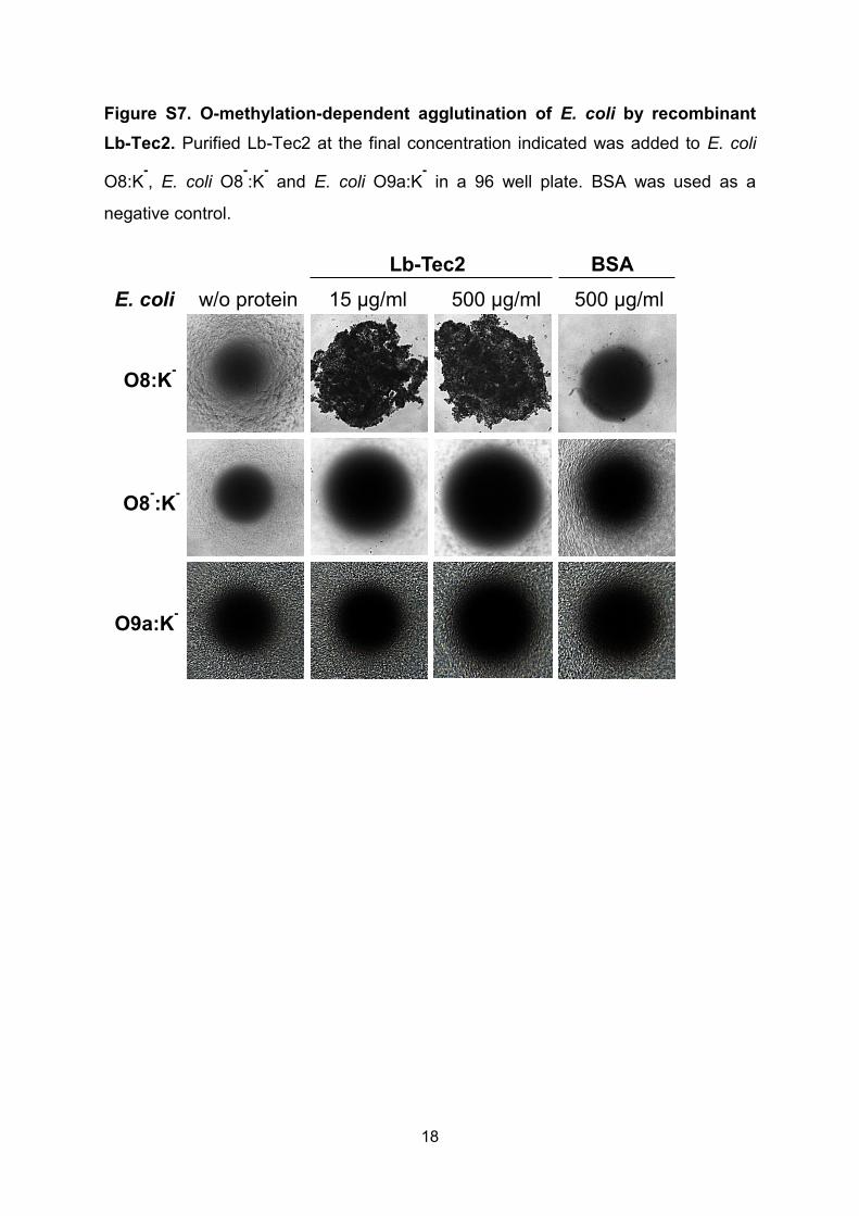
Figure S7. O-methylation-dependent agglutination of E. coli by recombinant
Lb-Tec2. Purified Lb-Tec2 at the final concentration indicated was added to E. coli
O8:K-, E. coli O8
-:K
- and E. coli O9a:K
- in a 96 well plate. BSA was used as a
negative control.
w/o protein
O8:K-
15 µg/ml 500 µg/ml 500 µg/ml
O8-:K
-
E. coli
Lb-Tec2 BSA
O9a:K-

19
Table S1. Genbank Identifiers (GI) of analyzed Tectonin sequences shown in
Fig 1.
Designation in Fig. 1 GI
L.bicolor_Tec2 170090420
L.bicolor_Tec1 170093369
N.dassonvillei 297560837
S.roseosporus 493083551
S.zinciresistens 494763290
Frankia_sp. 497423668
P.polycephalum_II 8134732
P.polycephalum_I 8134731
E.fluviatilis 83715952
T.tridentatus_L6 8134531
C.rotundicauda 55775500
T.tridentatus_TLP 6778723
T.tridentatus_GBP 8347764
S.domuncula 34368388
C.carpio 55976183
O.fasciatus 375331860
X.laevis 379046837
H.sapiens_Leuko 223016919
C.mydas 465976228
H.sapiens_TCPR1 32698704

20
Table S2. Microcalorimetry titration data for the binding of Lb-Tec2 to allyl
monosaccharides. The stoichiometry was fixed (N = 6), due to the low solubility of
Lb-Tec2 and the low affinity nature of its interaction with the ligands tested.
Therefore, the thermodynamic parameters entropy (S) and enthalpy (H) should be
considered with care (12) and are completely omitted for c-values below 0.01, (c =
[protein]/Kd). Individual titrations of Lb-Tec2 with O-methylated and unmethylated
ligands are shown in Fig. 6.
Lb-Tec2 [μM] ligand [mM] N Ka (M
-1) H (kcal/mol) TdS (kcal/mol) Kd (mM) c
Allyl mannoside (2) 134 10 6 no binding
134 28 6 no binding
Allyl 3-O-methyl mannoside (3) 134 28 6 52.0 -5.54 -3.19 19.2 0.007
300 28 6 45.7 -5.63 -3.37 21.9 0.014
40 28 6 48.6 - - 20.6 0.002
average: 6 48.8 -5.59 -3.28 20.6
st.dev.: 1.7 - - 0.8
Allyl fucoside (5) 134 28 6 10.0 - - 100.2 0.001
328 28 6 20.2 - - 49.5 0.007
average: 6 15.1 - - 74.9
Allyl 2-O-methyl-fucoside (8) 134 28 6 248 -4.13 -0.86 4.03 0.033
252 28 6 234 -4.58 -1.34 4.27 0.059
300 12 6 346 -3.91 -0.45 2.89 0.104
40 28 6 155 - - 6.45 0.006
average: 6 246 -4.21 -0.88 4.41
st.dev.: 78 0.34 0.45 1.49
Allyl 3-O-methyl-fucoside (9) 239 28 6 48 -5.14 -2.83 20.75 0.012
191 28 6 49 -3.79 -1.48 20.41 0.009
average: 6 48.6 -4.47 -2.16 20.6

21
Table S3. Characteristics of C. elegans glycosylation mutants used in this
study.
Designation Characteristics Reference
Bristol N2 wildtype (13)
fut-8(ok2558) Defective in α1,6- fucosylation of proximal core GlcNAc residue in N-glycans
(14)
fut-1(ok892);fut-8(ok2558) Defective in α1,3- and α1,6- fucosylation of proximal core GlcNAc residue in N-glycans
(14)
fut-6(ok475);fut-8(ok2558) Defective in α1,3-fucosylation of distal and α1,6-fucosylation of proximal core GlcNAc residue in N-glycans
(14)
bre-3(ye26) Defective in β1,4-mannosylation of core glucose in arthroseries of glycosphingolipids (Egghead activity)
(15)
gly-14(id48);gly-12(id47)gly-13(ok712) Defective in β1,4-GlcNAcylation of α1,3-branch of N-glycans (GNTI-activity) and thus the buildup of complex N-glycans
(16)
aman-2(tm1078) Defective in removing α1,3- and α1,6-linked mannoses from the α1,6-branch of N-glycans (Golgi-mannosidase II activity) and thus the buildup of complex N-glycans
(17)
hex-3(tm2725);hex-2(tm2530) Defective in removing GlcNAc from α1,3-branch of N-glycans (Hexosaminidase activity) and hypersensitive to lectins targeting N-glycan core modifications
(18)
pmk-1(km25) Defective in p38 MAPK pathway and hypersensitive to many abiotic and biotic stresses
(19)
samt-1(op532)pmk-1(km25) Defective in hypothetical Golgi-SAM transporter necessary for O-methylation of glycans in pmk-1(km25) background
This study
fut-6(ok475)fut-1(ok892);pmk-1(km25) Defective in α1,3- fucosylation of proximal and distal core GlcNAc residue in N-glycans in pmk-1(km25) background
(14, 20)
ger-1(op499);pmk-1(km25) Defective in the conversion of GDP-mannose to GDP-fucose in pmk-1(km25) background
(11, 21)
pmk-1(km25)bre-1(op509) Defective in the conversion of GDP-mannose to GDP-fucose in pmk-1(km25) background
(21, 22)
pmk-1(km25);galt-1(op497) Defective in the β1,4-galactosylation of the α1,6-linked fucose on the proximal core GlcNAc of N-glycans in pmk-1(km25) background
(11, 23)

22
REFERENCES
1. Dell A, et al. (1994) Mass spectrometry of carbohydrate-containing biopolymers. Methods Enzymol 230:108-132.
2. North SJ, et al. (2010) Mass spectrometric analysis of mutant mice. Methods Enzymol 478:27-77.
3. Jang-Lee J, et al. (2006) Glycomic profiling of cells and tissues by mass spectrometry: fingerprinting and sequencing methodologies. Methods Enzymol 415:59-86.
4. Ceroni A, et al. (2008) GlycoWorkbench: a tool for the computer-assisted annotation of mass spectra of glycans. J Proteome Res 7(4):1650-1659.
5. Winnik FM, Brisson JR, Carver JP, & Krepinsky JJ (1982) Syntheses of model oligosaccharides of biological significance. Synthesis of methyl 3,6-di-O-(alpha-D-mannopyranosyl)-alpha-d-mannopyranoside and the corresponding mannobiosides. Carbohydr Res 103(1):15-28.
6. Liao W & Lu D (1996) Synthesis of a hexasaccharide acceptor corresponding to the reducing terminus of mycobacterial 3-O-methylmannose polysaccharide (MMP). Carbohydr Res 296:171-182.
7. Unverzagt C & Kunz H (1992) Stereoselective Synthesis of Glycosides and Anomeric Azides of Glucosamine. J Prak Chem-Chem Ztg 334(7):570-578.
8. Takeo K, Aspinall GO, Brennan PJ, & Chatterjee D (1986) Synthesis of tetrasaccharides related to the antigenic determinants from the glycopeptidolipid antigens of serovars 9 and 25 in the Mycobacterium avium-M. intracellulare-M. scrofulaceum serocomplex. Carbohydr Res 150:133-150.
9. Sabotic J, et al. (2012) Structural basis of trypsin inhibition and entomotoxicity of cospin, serine protease inhibitor involved in defense of Coprinopsis cinerea fruiting bodies. J Biol Chem 287(6):3898-3907.
10. Kunzler M, et al. (2010) Biotoxicity assays for fruiting body lectins and other cytoplasmic proteins. Methods Enzymol 480:141-150.
11. Butschi A, et al. (2009) Caenorhabditis elegans N-glycan Core -galactoside Confers Sensitivity towards Nematotoxic Fungal Galectin CGL2. PLoS Pathog 6(1):e1000717.
12. Turnbull WB & Daranas AH (2003) On the value of c: can low affinity systems be studied by isothermal titration calorimetry? J Am Chem Soc 125(48):14859-14866.
13. Brenner S (1974) The genetics of Caenorhabditis elegans. Genetics 77(1):71-94.
14. Yan S, Serna S, Reichardt NC, Paschinger K, & Wilson IB (2013) Array-assisted characterization of a fucosyltransferase required for the biosynthesis of complex core modifications of nematode N-glycans. J Biol Chem 288(29):21015-21028.
15. Griffitts JS, et al. (2003) Resistance to a bacterial toxin is mediated by removal of a conserved glycosylation pathway required for toxin-host interactions. J Biol Chem 278(46):45594-45602.
16. Chen S, Spence AM, & Schachter H (2003) Isolation of null alleles of the Caenorhabditis elegans gly-12, gly-13 and gly-14 genes, all of which encode UDP-GlcNAc: alpha-3-D-mannoside beta1,2-N-acetylglucosaminyltransferase I activity. Biochimie 85(3-4):391-401.

23
17. Paschinger K, et al. (2006) A deletion in the golgi alpha-mannosidase II gene of Caenorhabditis elegans results in unexpected non-wild-type N-glycan structures. J Biol Chem 281(38):28265-28277.
18. Yan S, et al. (2012) Galactosylated fucose epitopes in nematodes: increased expression in a Caenorhabditis mutant associated with altered lectin sensitivity and occurrence in parasitic species. J Biol Chem 287(34):28276-28290.
19. Kim DH, et al. (2002) A conserved p38 MAP kinase pathway in Caenorhabditis elegans innate immunity. Science 297(5581):623-626.
20. Schubert M, et al. (2012) Plasticity of the beta-trefoil protein fold in the recognition and control of invertebrate predators and parasites by a fungal defence system. PLoS Pathog 8(5):e1002706.
21. Rhomberg S, et al. (2006) Reconstitution in vitro of the GDP-fucose biosynthetic pathways of Caenorhabditis elegans and Drosophila melanogaster. FEBS J 273(10):2244-2256.
22. Barrows BD, et al. (2007) Resistance to Bacillus thuringiensis toxin in Caenorhabditis elegans from loss of fucose. J Biol Chem 282(5):3302-3311.
23. Titz A, et al. (2009) Molecular basis for galactosylation of core fucose residues in invertebrates: identification of caenorhabditis elegans N-glycan core alpha1,6-fucoside beta1,4-galactosyltransferase GALT-1 as a member of a novel glycosyltransferase family. J Biol Chem 284(52):36223-36233.