supporting information · 1 supporting information establishing reactivity descriptors for platinum...
TRANSCRIPT

1
Supporting Information
Establishing Reactivity Descriptors for Platinum Group Metal
(PGM)-free Fe-N-C Catalysts for PEM Fuel Cells
Mathias Primbsa‡, Yanyan Suna‡, Aaron Royb, Daniel Malkoc, Asad Mehmoodc, Moulay-Tahar
Sougratib2, Pierre-Yves Blanchardb, Gaetano Granozzid, Tomasz Kosmalad,Giorgia Danield,
Plamen Atanassove, Jonathan Sharmanf*, Christian Duranted*, Anthony Kucernakc*, Deborah
Jonesb*, Frédéric Jaouenb*, Peter Strassera*
aDepartment of Chemistry, Chemical Engineering Division, Technical University of Berlin, 10623
Berlin, Germany
bICGM, Univ. Montpellier, CNRS, ENSCM, Montpellier, France
cDepartment of Chemistry, Imperial College London, South Kensington SW7 2AZ London, United
Kingdom
dDepartment of Chemical Sciences, University of Padova Via Marzolo 1, 35131 Padova, Italy
eDepartment of Chemical & Biomolecular Engineering and National Fuel Cell Research Center,
University of California, Irvine, CA 92697, USA
fJohnson Matthey Technology Center, Blount’s Court, Sonning Common, Reading RG4 9NH,
United Kingdom
‡These authors contributed equally.
*E-mail: [email protected];
Electronic Supplementary Material (ESI) for Energy & Environmental Science.This journal is © The Royal Society of Chemistry 2020

2
Figure S1. (a) XPS survey spectra and (b) high-resolution N 1s XPS spectrum for the benchmark catalysts.

3
Figure S2. TEM images of the catalysts: (a) CNRS, (b) ICL, (c) PAJ and (d) UNM.
TEM measurements. TEM measurements were performed using a FEI Tecnai G2 transmission electron microscopy
operating at 100 kV. The samples were dispersed in milli-Q water, sonicated for few minutes and dropped cast on a
gold grid.
PAJ
ICLCNRS
UNM
a) b)
d)c)

4
Figure S3. HAADF-STEM images of (a) ) a catalyst prepared similarly to the CNRS catalyst of the present study but
with less Fe content and containing only atomically dispersed Fe sites , (b) ICL, (c) UNM, and (d) PAJ catalysts. (a)
Reprinted with permission ref. 1 Copyright 2018, The Royal Society of Chemistry; (b) reprinted with permission ref. 2 Copyright 2019 Elsevier Inc.; (c) reprinted with permission ref. 3 Copyright 2019, American Chemical Society; (d)
reprinted with permission ref. 4 Copyright 2016, Wiley-VCH.

5
Figure S4. Initial ORR mass activity of the four catalysts as measured by RRDE for different catalyst loadings and
applied potentials: (a) 0.2 mg cm-2 and 0.80 VRHE, (b) 0.8 mg cm-2 and 0.80 VRHE, (c) 0.2 mg cm-2 and 0.85 VRHE, and
(d) 0.8 mg cm-2 and 0.85 VRHE. The electrolyte was 0.5 M H2SO4.
0.0
0.2
0.4
0.6
0.8
1.0
1.20.8 mg cm-2
0.85 VRHE
Mass A
ctivity (
A g
-1)
CNRS ICL PAJ UNM
0.0
0.5
1.0
1.5
2.0
2.5
3.0
3.5
4.0
4.5
5.00.8 mg cm-2
0.80 VRHE
Mass A
ctivity (
A g
-1)
CNRS ICL PAJ UNM
0.0
0.2
0.4
0.6
0.8
1.0
1.2 0.2 mg cm-2
0.85 VRHE
Mass A
ctivity (
A g
-1)
CNRS ICL PAJ UNM
0.0
0.5
1.0
1.5
2.0
2.5
3.0
3.5
4.0
4.5
5.00.2 mg cm-2
0.80 VRHE
Mass A
ctivity (
A g
-1)
CNRS ICL PAJ UNM
a) b)
d)c)c)

6
Figure S5. ORR mass activity before and after a load-cycling AST for a catalyst loading of 0.2 mg cm-2: (a) mass
activities at 0.80 VRHE and (b) mass activities at 0.85 VRHE. The filled columns represent the initial activity and the
hatched ones represent the activity after the AST (5,000 cycles between 0.6 and 0.9 V vs. RHE).
Figure S6. Hydrogen peroxide production (H2O2 %) during ORR of the four catalysts as measured by RRDE for two
different catalyst loadings and two applied potentials: (a) 0.2 mg cm-2 and 0.20 VRHE, (b) 0.8 mg cm-2 and 0.20 VRHE,
(c) 0.2 mg cm-2 and 0.70 VRHE, and (d) 0.8 mg cm-2 and 0.70 VRHE.
0
2
4
6
8
10
12
14
16
18
20
22
24
260.8 mg cm-2
0.70 VRHE
H2O
2 (
%)
CNRS ICL PAJ UNM
0
2
4
6
8
10
12
14
160.8 mg cm-2
0.20 VRHE
H2O
2 (
%)
CNRS ICL PAJ UNM
a) b)
d)c)
0
2
4
6
8
10
12
14
160.2 mg cm-2
0.20 VRHE
H2O
2 (
%)
CNRS ICL PAJ UNM
0
2
4
6
8
10
12
14
16
18
20
22
24
260.2 mg cm-2
0.70 VRHE
H2O
2 (
%)
CNRS ICL PAJ UNM

7
Figure S7. Repetitive CO pulse chemisorption profiles of the four benchmarking catalysts: (a) CNRS (3 repeats), (b)
ICL (3 repeats), (c) PAJ (4 repeats), and (d) UNM (4 repeats). Surface CO saturation was achieved after ca 3 pulses.
Reduced pulse peak area indicate CO uptake.
0 20 40 60 80 100 120 140 160
No
rma
lize
d I
nte
nsity
Time (min)
2
1
3
4
UNM
0 20 40 60 80 100 120 140 160
No
rma
lize
d I
nte
nsity
Time (min)
2
1
3
ICLa) b)
d)c)
0 20 40 60 80 100 120 140 160
No
rma
lize
d I
nte
nsity
Time (min)
2
1
3
4
PAJ
0 20 40 60 80 100 120 140 160
No
rma
lize
d I
nte
nsity
Time (min)
2
1
3
CNRS

8
Figure S8. Temperature-programmed desorption (TPD) profiles in the range from -80 to 600 °C after saturation of
the Fe surface sites with CO. TPD was conducted to get more insights about the chemical bonding between CO and
catalytic active sites, where two obvious peaks are observed. It should be here highlighted that only the second
desorption peak at high temperature corresponds to the binding of CO molecule on FeNx centres in the Fe-N-C
catalysts whereas the first peak at lower temperature should be attributed to the desorption of pre-adsorbed air resulting
from the porous structure of the Fe-N-C catalysts. Interestingly, the CO desorption temperature of peak 2 follows the
increasing order of UNM, PAJ, ICL, and CNRS, which may be explained by the different average Fe coordination
geometry or different average oxidation state and/or spin state of the iron sites.
-100 0 100 200 300 400 500 600
0.0
0.5
1.0
1.5
2.0
2.5
3.0
3.5
4.0
4.5
5.0
2
UNM
PAJ
ICL
Norm
aliz
ed Inte
nsi
ty
Sample temperature [°C]
CNRS
1

9
Figure S9. Rotating disk electrode measurement of CNRS catalyst before and after nitrite poisoning and its recovery.
(a) ORR measurement in O2-saturated electrolyte at the scan rate of 5 mV s-1. (b) Stripping voltammetry in N2-
saturated electrolyte at 10 mV s-1. (c) The kinetic current densities derived from the ORR measurement. (d) Excess
current associated with the reductive stripping of nitrite. All measurements were performed in 0.5 M acetate buffer at
pH 5.2 at 1600 rpm and the catalyst loading was 0.2 mg cm−2.
a) b)
d)c)
-0.8 -0.6 -0.4 -0.2-0.8
-0.6
-0.4
-0.2
0.0
0.2
0.4
0.6
E / V vs SCE
i /
mA
cm
-2
CNRS unpoisoned
CNRS poisoned
CNRS recovered
-0.3 -0.2 -0.1 0.0 0.1 0.2 0.3 0.4Potential / V vs RHE
-0.9 -0.8 -0.7 -0.6 -0.5 -0.4 -0.3 -0.2 -0.1 0.0-0.000018
-0.000016
-0.000014
-0.000012
-0.000010
-0.000008
-0.000006
-0.000004
-0.000002
0.000000
0.000002
0.000004 CNRS Difference
Potential applied (V)
Dif
fere
nc
e (
A)
Area=4.53209E-6
FWHM=0.31361
-0.2 0.0 0.2 0.4 0.6-3.5
-3.0
-2.5
-2.0
-1.5
-1.0
-0.5
0.0
0.5
CNRS unpoisoned
CNRS poisoned
CNRS recovered
E / V vs SCE
i /
mA
cm
-2
0.3 0.4 0.5 0.6 0.7 0.8 0.9 1.0E / V vs RHE
0.01 0.1 10.20
0.25
0.30
0.35
0.40
0.45
EIR
-fre
e /
V v
s R
HE
ikin
/ A g-1
EIR
-fre
e /
V v
s S
CE
ikin
/ mA cm-2
CNRS unpoisoned
CNRS poisoned
CNRS recovered
0.1 1 10
0.70
0.75
0.80
0.85
0.90

10
Figure S10. Rotating disk electrode measurement of ICL catalyst before and after poisoning and recovery. (a) ORR
measurement in O2-saturated electrolyte at the scan rate of 5 mV s-1. (b) Stripping voltammetry in N2-saturated
electrolyte at 10 mV s-1. (c) The kinetic current densities derived from the ORR measurement. (d) Excess current
associated with the reductive stripping of nitrite. All measurements were performed in 0.5 M acetate buffer at pH 5.2
at 1600 rpm and the catalyst loading was 0.2 mg cm−2.
a) b)
d)c)
-0.9 -0.8 -0.7 -0.6 -0.5 -0.4 -0.3 -0.2 -0.1 0.0
-0.000008
-0.000006
-0.000004
-0.000002
0.000000
0.000002
0.000004
Diffe
rence
(A
)
Potential applied (V)
ICL Difference
Area=-2.69534E-6
FWHM=0.42942
-0.2 0.0 0.2 0.4
-4.0
-3.5
-3.0
-2.5
-2.0
-1.5
-1.0
-0.5
0.0
i /
mA
cm
-2
E / V vs SCE
ICL unpoisoned
ICL poisoned
ICL recovered
0.3 0.4 0.5 0.6 0.7 0.8 0.9 1.0E / V vs RHE
-0.8 -0.6 -0.4 -0.2-0.8
-0.6
-0.4
-0.2
0.0
0.2
0.4
0.6
0.8
i / m
A c
m-2
E / V vs SCE
ICL unpoisoned
ICL poisoned
ICL recovered
-0.3 -0.2 -0.1 0.0 0.1 0.2 0.3 0.4E / V vs RHE
0.01 0.1 10.20
0.25
0.30
0.35
0.40
0.45
ikin / A g-1
EiR
-fre
e /
V v
s R
HE unpoisoned
poisoned
recovered
EiR
-fre
e /
V v
s S
CE
ikin / mA cm-2
0.70
0.75
0.80
0.85
0.90
0.1 1 10

11
Figure S11. Rotating disk electrode measurement of PAJ catalyst before and after poisoning and recovery. (a) ORR
measurement in O2-saturated electrolyte at the scan rate of 5 mV s-1. (b) Stripping voltammetry in N2-saturated
electrolyte at 10 mV s-1. (c) The kinetic current densities derived from the ORR measurement. (d) Excess current
associated with the reductive stripping of nitrite. All measurements were performed in 0.5 M acetate buffer at pH 5.2
at 1600 rpm and the catalyst loading was 0.2 mg cm−2.
a) b)
d)c)
-0.9 -0.8 -0.7 -0.6 -0.5 -0.4 -0.3 -0.2 -0.1 0.0
-0.000004
-0.000003
-0.000002
-0.000001
0.000000
0.000001
0.000002
Diffe
rence (
A)
Potential applied (V)
PAJ Difference
Area=-7.88448E-7
FWHM=0.25038
-0.2 0.0 0.2 0.4
-4.5
-4.0
-3.5
-3.0
-2.5
-2.0
-1.5
-1.0
-0.5
0.0
i / m
A c
m-2
E / V vs SCE
PAJ unpoisoned
PAJ poisoned
PAJ recovered
0.3 0.4 0.5 0.6 0.7 0.8 0.9 1.0E / V vs RHE
-0.8 -0.6 -0.4 -0.2-0.6
-0.4
-0.2
0.0
0.2
0.4
0.6
i / m
A c
m-2
E / V vs SCE
PAJ unpoisoned
PAJ poisoned
PAJ recovered
-0.3 -0.2 -0.1 0.0 0.1 0.2 0.3 0.4E / V vs RHE
0.01 0.1 10.20
0.25
0.30
0.35
0.40
0.45
ikin
/ A g-1
EiR
-fre
e / V
vs R
HE unpoisoned
poisoned
recovered
EiR
-fre
e / V
vs S
CE
ikin
/ mA cm-2
0.70
0.75
0.80
0.85
0.90
0.1 1 10

12
Figure S12. Rotating disk electrode measurement of UNM catalyst before and after poisoning and recovery. (a) ORR
measurement in O2-saturated electrolyte at the scan rate of 5 mV s-1. (b) Stripping voltammetry in N2-saturated
electrolyte at 10 mV s-1. (c) The kinetic current densities derived from the ORR measurement. (d) Excess current
associated with the reductive stripping of nitrite. All measurements were performed in 0.5 M acetate buffer at pH 5.2
at 1600 rpm and the catalyst loading was 0.2 mg cm−2.
a) b)
d)c)
-0.9 -0.8 -0.7 -0.6 -0.5 -0.4 -0.3 -0.2 -0.1 0.0
-0.000008
-0.000006
-0.000004
-0.000002
0.000000
0.000002
0.000004
0.000006
Diffe
rence
(A
)
Potential applied (V)
UNM Difference
Area=-1.98949E-6
FWHM=0.33134
-0.2 0.0 0.2 0.4
-4.5
-4.0
-3.5
-3.0
-2.5
-2.0
-1.5
-1.0
-0.5
0.0
i /
mA
cm
-2
E / V vs SCE
UNM poisoned
UNM unpoisoned
UNM recovered
0.3 0.4 0.5 0.6 0.7 0.8 0.9 1.0E / V vs RHE
-0.8 -0.6 -0.4 -0.2-0.8
-0.6
-0.4
-0.2
0.0
0.2
0.4
0.6
0.8
i / m
A c
m-2
E / V vs SCE
UNM unpoisoned
UNM poisoned
UNM recovered
-0.3 -0.2 -0.1 0.0 0.1 0.2 0.3 0.4E / V vs RHE
0.01 0.1 10.20
0.25
0.30
0.35
0.40
0.45
EiR
-fre
e /
V v
s R
HE unpoisoned
poisoned
recovered
EiR
-fre
e /
V v
s S
CE
ikin
/ mA cm-2
0.70
0.75
0.80
0.85
0.90
0.1 1 10ikin
/ A g-1

13
Figure S13. Mass SD-TOF reactivity maps at 0.85 VRHE: The mass-based Fe surface site density (SDmass) plotted
against the TOF for each catalyst. Points of constant activity (iso activity curves) shown in grey with units of A g-1 at
0.85 VRHE: (a) SDmass(CO) – TOF reactivity map, derived from CO chemisorption and (b) SDmass(NO2-) – TOF
reactivity map derived from nitrite stripping values.
The correlation between the catalytic active site density and TOF at the potential of 0.85 VRHE was
also plotted (Figure S14). Different rankings in the mass activity are observed, that is, ICL< UNM
≈ PAJ < CNRS with the CO chemisorption method whereas CNRS ≈ PAJ < ICL ≈ UNM in
the nitrite stripping method, which may be due to different calculation methods for kinetic current
in the CO chemisorption and nitrite stripping method. Nevertheless, with both methods, the
CNRS and PAJ catalysts were located the extreme place in terms of catalytic active site density
and TOF. Further, the correlation of surface area corrected catalytic active site density with TOF
was also investigated, and the different trend in regards of areal catalytic activity is also observed
in the CO chemisorption and nitrite stripping method (Figure S15). Meanwhile, the ICL catalyst
exhibited more active sites (more kinetic current per surface area) compared to the CNRS catalyst
despite the fact that both catalysts possessed almost the same areal catalytic site density.

14
Figure S14. Areal SD-TOF reactivity maps at 0.85 VRHE: The areal Fe surface site density (SD) plotted against the
TOF for each catalyst. Points of constant activity (iso activity curves) shown in grey with units of mA m -2 at 0.85
VRHE: (a) SDBET (CO) – TOF reactivity map, derived from CO chemisorption and (b) SDBET (NO2-) – TOF reactivity
map derived from nitrite stripping.
Figure S15 Correlation of catalytic active site density via (a, c) CO chemisorption and (b, d) nitrite stripping method
with (a, b) pore volume and (c, d) nitrogen species from XPS results.

15
Figure S16 Correlation of the bulk weight ratio of the molecular Fe D1 moiety, derived from 57Fe Mößbauer spectra,
with the micropore volume, and the Fe surface site density, SDmass (CO) derived from CO-chemisorption method.
Figure S17 Correlation of the areal catalytic active site density via (a) CO chemisorption and (b) nitrite stripping
method with nitrogen species including pyridinic-N and pyrrolic-N.

16
Figure S18. Correlation of experimental TOF values with the ratio of Fe-Nx and Pyrrolic nitrogen species. TOF values
were derived from (a) CO cryo-adsorption and (b) nitrite stripping. N species ratios were determined from N1s XPS.
Data measured at at 0.80 VRHE.
Figure S19. Correlations between TOF values at 0.80 VRHE derived from CO chemisorption and nitrite stripping and
the Mössbauer-derived weight of Fe sites a) D1, b) sum of D1 and D2

17
Quantitative correlation between SD (CO) and SD (NO2-).
The x-intercept of the regression line in Figure 4b, here reproduced as Figure S20a, is about
3.1×10¹⁶ sites m-2 and may be interpreted as the amount of BET-normalized Fe surface site density,
SDBET(CO), that remained inaccessible to in-situ reductive NO2- / NO stripping. The inaccessibility
is in part based on the lower probe molecule chemisorption energy (see CO-TPD analysis of the
four catalysts in Figure S8), which makes sampling of these sites under in-situ ambient conditions
in the presence of liquid electrolyte impossible. Subtracting the NO-inaccessible portion of
SDBET(CO) from the experimental SDBET(CO) values then yields corrected SDBET. corr (CO) values
(Figure S20b), which display a perfect quantitative 1:1 correlation with the corresponding
SDBET(NO2-) values for catalysts with a balanced ratio of meso- and micropore volumes. The
quantitative deconvolution and correlation of independent, experimental in-situ and ex-situ SD
values represents an important step forward in the accurate evaluation of catalytically active
surface single-Fe sites.
Figure S20. Correlations of ex-situ and in-situ Fe surface site density (SD) values of four PGM-free Fe-N-C ORR
catalysts obtained using CO-chemisorption and nitrite electrochemical reductive stripping (a) SDBET values
measured in situ vs. SDBET values measured ex situ with a regression line in points PAJ, UNM and ICL (b) SDBET
(NO2-) values versus x-intercept-subtracted SDBET,corr (CO) values. The x=y line are given as grey dashed lines.

18
Table S1. Mössbauer analysis results of the four selected benchmarking catalysts: CNRS, ICL, PAJ, and UNM.
Material Assignment IS
mm/s
QS
mm/s
LW
mm/s
H
Tesla
Area
%
Lamb-
Mössbauer
factor
Relative amount
of each component
to all Fe (1) %
Wt % Fe of each
component (2) %
wt
CNRS
-Fe -0.08 - 0.34 - 10 0.78 14 0.36
Fe3C 0.185 - 0.35 20 3 0.67 4 0.09
α-Fe 0 - 0.36 33 18 0.67 22 0.55
D1 0.34 1.03 0.72 - 42 0.46 35 0.88
D2 0.45 2.48 1.50 - 27 0.52 25 0.62
ICL D1 0.36 0.96 0.73 - 38 0.46 35 0.35
D2 0.55 2.2 1.75 - 62 0.52 65 0.65
PAJ
-Fe -0.12 - 0.35 - 36 0.78 45 0.27
α-Fe 0 - 0.21 33.2 15 0.67 16 0.1
D1 0.37 0.75 0.9 - 11 0.46 8 0.05
D2 0.40 2.34 1.81 - 38 0.52 31 0.19
UNM
-Fe -0.08 - 0.37 - 11 0.78 16.36 0.13
D1 0.36 1.1 0.72 - 40 0.46 35.07 0.28
D2 0.41 2.77 1.53 - 49 0.52 48.57 0.39
Note: IS is the isomer shift (reported vs. an α-Fe foil reference at room temperature), QS the quadrupole splitting, LW
the line width and H the hyperfine field, describing the singlet, doublet and sextet components used for the fittings.
Area is the relative % of area corresponding to each fitted spectral component relative to the total absorption area. The
column “relative amount of each component to all Fe” is derived from the knowledge of the area % of each spectral
component of a given Fe-N-C catalyst (column “Area”) and of the Lamb Mössbauer factors of each Fe species. It
therefore includes the necessary correction of the -ray absorption area in order to account for the different probability
of the recoilless absorption event for Fe atoms in different coordination environments. For each catalyst, the last
column is derived from the multiplication of the next-to-last column by the total wt % Fe bulk content (measured by
ICP).

19
Table S2. CO chemisorption and NO2- stripping data and derived quantities such as mass- and BET-normalized Fe
surface site densities (SDmass and SDBET) and the turnover frequencies (TOF) at applied electrode potentials of +0.80
and +0.85 VRHE for the four benchmarking catalysts.
Catalyst
Method
CNRS ICL PAJ UNM
NO2- CO NO2
- CO NO2- CO NO2
- CO
CO uptake
[nmol g-1] - 96 ± 1 - 36 ± 10 -
33.6 ±
0.6 - 52 ± 4
SDmass
[1019 sites g-1] 1.44
5.80 ±
0.08 0.86
2.20 ±
0.6 0.25
2.02 ±
0.03 0.63
3.12 ±
0.2
BET surface area
[m2 g-1] 840 463 593 763
SDBET
[1016 sites m-2] 1.71 6.90 1.86 4.76 0.42 3.41 0.83 4.10
TOF
at 0.80 VRHE
[electrons site-1 s-1]
0.65 0.20 0.96 0.36 7.23 0.71 3.45 0.47
TOF
at 0.85 VRHE
[electrons site-1 s-1]
0.14 0.06 0.34 0.08 0.80 0.16 0.46 0.10
20
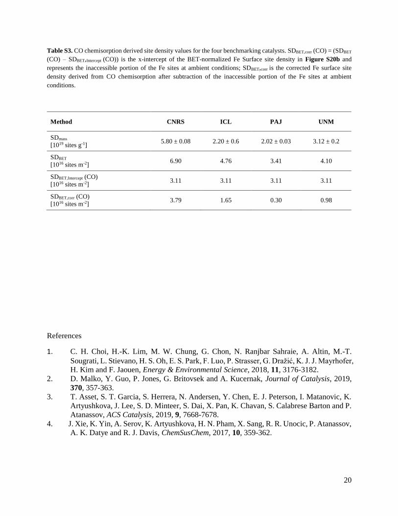
Table S3. CO chemisorption derived site density values for the four benchmarking catalysts. SDBET,corr (CO) = (SDBET
(CO) – SDBET,Intercept (CO)) is the x-intercept of the BET-normalized Fe Surface site density in Figure S20b and
represents the inaccessible portion of the Fe sites at ambient conditions; SDBET,corr is the corrected Fe surface site
density derived from CO chemisorption after subtraction of the inaccessible portion of the Fe sites at ambient
conditions.
Method CNRS ICL PAJ UNM
SDmass
[1019 sites g-1] 5.80 ± 0.08 2.20 ± 0.6 2.02 ± 0.03 3.12 ± 0.2
SDBET
[1016 sites m-2] 6.90 4.76 3.41 4.10
SDBET,Intercept (CO)
[1016 sites m-2] 3.11 3.11 3.11 3.11
SDBET,corr (CO)
[1016 sites m-2] 3.79 1.65 0.30 0.98
References
1. C. H. Choi, H.-K. Lim, M. W. Chung, G. Chon, N. Ranjbar Sahraie, A. Altin, M.-T.
Sougrati, L. Stievano, H. S. Oh, E. S. Park, F. Luo, P. Strasser, G. Dražić, K. J. J. Mayrhofer, H. Kim and F. Jaouen, Energy & Environmental Science, 2018, 11, 3176-3182.
2. D. Malko, Y. Guo, P. Jones, G. Britovsek and A. Kucernak, Journal of Catalysis, 2019,
370, 357-363.
3. T. Asset, S. T. Garcia, S. Herrera, N. Andersen, Y. Chen, E. J. Peterson, I. Matanovic, K.
Artyushkova, J. Lee, S. D. Minteer, S. Dai, X. Pan, K. Chavan, S. Calabrese Barton and P.
Atanassov, ACS Catalysis, 2019, 9, 7668-7678.
4. J. Xie, K. Yin, A. Serov, K. Artyushkova, H. N. Pham, X. Sang, R. R. Unocic, P. Atanassov,
A. K. Datye and R. J. Davis, ChemSusChem, 2017, 10, 359-362.