supplementary materials for - science · supplementary materials for cyclin-dependent kinase...
TRANSCRIPT

www.sciencemag.org/cgi/content/full/science.aad3925/DC1
Supplementary Materials for
Cyclin-dependent kinase 1–dependent activation of APC/C ubiquitin ligase
Kazuyuki Fujimitsu, Margaret Grimaldi, Hiroyuki Yamano*
*Corresponding author. E-mail: [email protected]
Published 21 April 2016 on Science First Release
DOI: 10.1126/science.aad3925
This PDF file includes
Materials and Methods Supplementary Text Figs. S1 to S15 Table S1 Full References

2
Supplementary Materials: Materials and Methods
Cloning and expression of recombinant Xenopus APC/C (APC/C) The approach used in this study is based on the modified MultiBac pFBDM and pUCDM system allowing USER ligation-independent cloning (17, 18). Xenopus APC/C genes, apart from Apc1, were amplified by PCR using Xenopus laevis cDNA library. For Apc1, Xenopus tropicalis Apc1 was amplified from the IMAGE clone of Xenopus tropicalis Apc1 (Source BioScience). All APC/C genes amplified were cloned into pOENmyc vector with polH promoter and SV40 terminator for Apc2, Apc3, Apc7, Apc8, Apc11 and Apc13 or p10 promoter and HSVtk terminator for Apc1, Apc4, Apc5, Apc6, Apc10, Apc12 and Apc16. TEV cleavable tandem Strep II-tag was fused to Apc6 at C-terminus (Apc6-strep) for affinity purification. Mutant APC/Cs were generated by PCR-based mutagenesis at this step and then all APC/C genes were further cloned into MultiBac vectors creating one pF1 vector-derivative pFUBB carrying Apc3, Apc8 and Apc7 at MUM1 site and Apc5, Apc4 and Apc6-strep at MUM2 site and a second pF1 vector-derivative pFUBB carrying Apc2 and Apc11 at MUM1 site and Apc1 and Apc10 at MUM2 site. A pU1 vector-derivative pUUBB carrying Apc13 at MUM1 site, and Apc12 and Apc16 at MUM2 site was also created. The resultant recombinant transfer vectors were transformed into MultiBacDH10cre cells to generate bacmids by in vivo recombination using the Tn7 sites of the pFUBB vector and the Cre-Lox site of the pUUBB vector. Bacmid 1 incorporated Apc3, Apc8, Apc7, Apc5, Apc4, Apc6-strep, Apc13, Apc12 and Apc16 and Bacmid 2 incorporated Apc2, Apc11, Apc1 and Apc10. Sf9 insect cells were transfected with bacmids 1 and 2 to generate recombinant baculoviruses. To express APC/C complex, High Five insect cells (Invitrogen) at a cell density 1.5 x 10⁶ were co-infected with the two recombinant baculoviruses at an MOI (multiplicity of infection) of 1 for each virus and incubated at 27 °C for 48h with shaking (150 rev/min). The cells were harvested, frozen in liquid nitrogen and stored at -80 °C. Purification of APC/C All purification steps were performed at 4 °C. Cell pellets were thawed on ice and resuspended in APC/C lysis buffer [50 mM Tris-HCl pH 8.0, 250 mM NaCl, 5% glycerol, 2 mM DTT, 0.5 mM EDTA, 10 µg/ml leupeptin, 10 µg/ml pepstatin A, 10 µg/ml chymostatin, 30 units/ml benzonase (Novagen) ] and lysed by sonication. The lysate was centrifuged at 48,400 g for 60 min, the supernatant centrifuged again for 20 min and the final soluble supernatant filtered through a 0.45 µm filter. This cleared lysate was loaded onto a 5 ml Strep-Tactin Superflow Cartridge (Qiagen) at a flow rate of 0.5 ml/min. The column was washed with APC/C wash buffer [50 mM Tris-HCl pH 8.0, 250 mM NaCl, 5% glycerol, 2 mM DTT] at a flow rate of 1.5 ml/min and recombinant APC/C was eluted with APC/C wash buffer containing 2.5 mM desthiobiotin (Sigma). Peak fractions were pooled and concentrated using Amicon Ultra (Millipore), and loaded onto Superose 6 HR 10/30 (GE Healthcare) equilibrated with APC/C SE-D buffer [20 mM Hepes-NaOH pH 7.9, 500 mM NaCl, 2mM DTT, 0.01% n-Dodecyl b-D-maltoside, 10 % glycerol]. Mutant APC/C activity was tested by incubating 1ml lysates with Strep-Tactin Superflow (Qiagen) beads (100 µl) at 4 °C for 1 h, washing with 3 x 1 ml of APC/C wash

3
buffer and elution with 2 x 300 µl of APC/C wash buffer containing 2.5 mM desthiobiotin. The eluted APC/C proteins were incubated with Dynabeads-Protein A conjugated to anti-Apc3 antibodies (AF3.1) and the bound-APC/C was washed with XBCSF buffer [10mM Hepes-KOH pH7.8, 50mM Sucrose, 100mM KCl, 2mM MgCl2, 5mM EGTA] containing 0.01% NP-40, followed by XBCSF buffer alone. The APC/C bound beads were flash-frozen and stored at -80 °C. Preparation of Xenopus egg cell-free extracts Meiotic metaphase II-arrested (CSF) Xenopus laevis egg extracts were prepared as described (31). To prepare interphase extracts, CSF extracts were incubated at 23 °C for 1.5 hr in the presence of 0.4 mM CaCl2 and 10 µg/ml cycloheximide, a protein synthesis inhibitor. Anaphase extracts were prepared by adding nondegradable GST-fused Xenopus cyclinB∆167 (a truncated cyclin B lacking the N-terminal 167 amino acids) to interphase extracts and incubating for 30 min at 23 °C. APC/C depleted (∆APC/C) or Cdc20 depleted (∆Cdc20) extracts were prepared as reported previously (32, 33). Fresh cycling egg extracts were prepared as described (34). Cell-free destruction and ubiquitylation assays Destruction assays were performed essentially as described previously (33). Substrates were labelled with [35S]methionine (Hartmann Analytic, UK) in a coupled in vitro transcription-translation system (Promega, UK) and destruction assays were carried out using Xenopus egg cell-free extracts (anaphase or interphase extracts). The samples were taken at the indicated time points and analysed by SDS-PAGE and autoradiography. The images were analysed using Image J (NIH, USA) and the statistical analysis was performed using Prism GraphPad Software (CA, USA). Ubiquitylation assays were essentially performed as described (35). Reactions were carried out at 23 °C in 10 µl of buffer (20 mM Tris-HCl pH 7.5, 100 mM KCl, 2.5 mM MgCl2 , 2 mM ATP, 0.3 mM DTT) containing 10 ng/µl APC/C, 0.05 mg/ml E1, 0.025 mg/ml UbcX, 0.75 mg/ml ubiquitin, 1 µM ubiquitin-aldehyde, 150 µM MG132, purified His-Cdh1 protein (or Strep-tag Cdc20), and 1 µl of 35S-labeled cyclin B (fission yeast Cdc13). The reactions were stopped at the indicated time points with SDS sample buffer and analysed by SDS-PAGE and autoradiography. To prepare the APC/C phosphorylated by CDK1-cyclin B/p9, the APC/C was incubated in the XBCSF buffer containing 100 µM ATP in the presence of 10 ng/µl human CDK1-cyclin B (a kind gift of Dr. Endicott, Newcastle University, UK) (36) and 32 ng/µl Xenopus p9 (Cks2) at 23˚C for 40 min. The resultant APC/C was immunoprecipitated and washed by buffer (20 mM Tris-HCl pH7.5, 100 mM KCl, 2.5 mM MgCl2, 0.01% NP-40), and subjected to ubiquitylation. Phosphorylation assay For in vitro phosphorylation using purified p9-CDK1-cyclin B, WT or mutant recombinant APC/Cs bound to Dynabeads-Protein A conjugated to anti-Apc3 antibodies (AF3.1) were incubated in the XBCSF buffer containing 100 µM cold ATP and 1 µCi/nmol of [γ-32P] ATP (Hartmann Analytic, UK) in the presence of 10 ng/µl human CDK1-cyclin B (36) and 32 ng/µl Xenopus p9 (Cks2) at 23˚C for 20 min or 40 min. For phosphorylation assay in Xenopus egg extracts, the APC/C was incubated with APC/C-depleted egg extract in the presence of [γ-32P] ATP and cyclinB∆167 at 23˚C for 45 min.
4
The APC/C was immuno-precipitated and boiled in SDS sample buffer and analysed by SDS-PAGE and autoradiography. Immunoprecipitation of APC/C The APC/C was immunoprecipitated using Apc3 MAb (AF3.1) immobilized Dynabeads protein A. The bound proteins were washed twice with XBCSF_HS [XBCSF containing 500 mM KCl and 0.01% NP-40], eluted with SDS sample buffer and analysed by SDS-PAGE and immunoblotting. Cloning and purification of Xenopus Apc1 fragments The fragment (294-399) of Xenopus tropicalis Apc1 was fused to 3xFlag tag at N-terminus and 6xHis tag at C-terminus, and subcloned into pET vector. Mutants were generated by PCR-based mutagenesis. The resultant plasmids were introduced into BL21-CodonPlus (DE3) and the fusion proteins were expressed at 37 °C for 1hr in the presence of 1 mM IPTG. The cells were lysed by 0.3 mg/ml lysozyme, sonicated in lysis buffer [20 mM Hepes-NaOH pH 7.9, 500 mM NaCl, 5mM EGTA, 10 µg/ml leupeptin, 10 µg/ml pepstatin A, 10 µg/ml chymostatin, 0.1 % Triton X-100, 10mM imidazole]. The proteins were purified from clarified lysate on Ni-NTA agarose beads (Qiagen). Binding assay using Xenopus Apc1 fragments Purified Apc1 fragments were incubated with Anti-Flag Affinity M2 beads (Sigma) at 4 °C for 0.5-1h. Beads were washed with XBCSF buffer and incubated with interphase extract in the presence or absence of cyclin B∆167 at 23 °C for 45 min, separated from extract on Micro Bio-Spin columns (Bio-Rad), and washed twice with XBCSF buffer containing 0.1% NP-40. The bound proteins were eluted with SDS-sample buffer and analysed by SDS-PAGE and immunoblotting. Antibodies Antibodies used are as follows: anti-Apc1 (RbAb 4853, 1:100), Apc2 (RbAb 7577, 1:500), Apc3/Cdc27 (1:200; BD Transduction Laboratories), Apc5 (RbAb 3445, 1:500), Apc6 (RbAb 3446, 1:500), Cdc20 (MAb BA8, 1:100), Xe-p9 (RbAb CE/R2, 1:50), phospho-Plx1 (MAb AZ44, 1:1000), Cdc2 (MAb A17, 1:3000). Apc1 phosphopeptide antibody (anti-pS314pS318, 1:1000) was raised in rabbits against LPH (the horseshoe crab Limulus polyphemus hemocyanin) conjugated phosphopeptide CVSKGEpSPTApSPFQN (BioGenes, Germany). Antibodies were affinity purified using a phosphopeptide column prepared with SulfoLink Kit (Pierce). After elution with ImmunoPure Gentle Ag/Ab Elution Buffer (Pierce), antibodies were dialysed against TBS and non-phosphospecific antibody was affinity depleted by passing through a column cross-linked with non-phosphopeptide CVSKGESPTASPFQN. The eluted phospho-specific antibodies were then enriched by dialysis against TBS containing 50% glycerol. Author Contributions K.F and H.Y. designed and performed the experiments, analysed and interpreted data, and wrote the manuscript. M.G. contributed towards the cloning and expression of recombinant APC/C. H.Y. conceived and supervised the overall research.

5
Fig. S1. Multi-subunit APC/C reconstitution and cell-free APC/C functional assays. (A) Schematic illustration of the expression of recombinant Xenopus apo-APC/C (APC/C). APC/C subunit genes were cloned into plasmid, and transferred into bacmid by recombination as shown. Red and blue circles are Tn7 transposition site and Cre-LoxP sites, respectively. The resultant two baculoviruses were used to co-infect High Five

6
insect cells and the APC/C was purified by affinity chromatography using Strept-Tactin resin. (B) Reconstituted WT APC/C was bound to a Strept-Tactin column, eluted by desthiobiotin, concentrated and applied to a Superose 6 column. The peak fraction was run on SDS-PAGE gradient gel, and stained with Coomassie Brilliant Blue (CBB). The CBB staining pattern confirms the high-purity and quality of expression of APC/C. (C) Xenopus egg cell-free extract based APC/C functional assay. APC/C substrates such as cyclin B can be degraded in Xenopus egg extract but if endogenous APC/C is depleted (∆APC/C), cyclin B becomes stable. Reconstituted APC/Cs that contain mutation(s) in any subunit(s) can be created and their activity tested in ∆APC/C extracts (∆APC/C+APC/C). (D) Reconstituted WT APC/C supports cyclin B destruction. Cdc20-dependent cyclin destruction assay in Xenopus egg cell-free extracts. Mock or WT APC/C was incubated in ∆APC/C anaphase extract. 35S-labelled cyclin B and a version of cyclin B lacking the N-terminal 67 residues (∆67, stable control) were used as substrates. Samples taken at indicated time points after addition of substrates were analysed by SDS–PAGE and autoradiography. A lack of cyclin destruction in ∆APC/C anaphase extract was restored by adding back WT APC/C (lanes 15-21). (E) Reconstituted WT APC/C is a functional ubiquitin ligase. WT APC/C (lanes 1-15) or none (lanes 16-30) was incubated with ubiquitylation factors (E1, E2, energy generating mix, ubiquitin) at 23°C. 35S-labelled cyclin B (fission yeast Cdc13) and purified Cdh1 were used as substrate and co-activator, respectively. Samples taken at indicated time points were analysed by SDS–PAGE and autoradiography. (F) Cdh1 can activate APC/C in interphase and trigger cyclin B destruction (lanes 1-6). Mock or WT APC/C was incubated in ∆APC/C interphase extract supplemented with Cdh1 (lanes 7-18). Please note that Xenopus eggs and early embryos lack Cdh1 until MBT (midblastula transition). 35S-labelled cyclin B and a version of cyclin B lacking the N-terminal 67 residues (∆67, stable control) were used as substrates. A lack of cyclin destruction in ∆APC/C interphase extract was restored by adding back WT APC/C (lanes 13-18).

7
Fig. S2. A screening of APC/Cs harbouring CDK site mutations. (A) Cyclin destruction assay in Xenopus egg cell-free extracts. Mock or endogenous APC/C-depleted anaphase extracts (∆APC/C) were supplemented with a range of reconstituted APC/Cs (table S1). 35S-labelled cyclin B and a version of cyclin B lacking the N-terminal 67 residues (∆67, stable control) were used as substrates. Samples taken at indicated time points after addition of substrates were analysed by SDS–PAGE and autoradiography. Quantification of the destruction assays is shown in Fig. 1A. (B) The activity of APC/C3-12A in interphase. Left panel: Mock, APC/CWT or APC/C3-12A mutant complex was incubated in APC/C-depleted (∆APC/C) interphase extract supplemented with Cdh1 and 35S-labelled APC/C substrates (cyclin B and ∆67). Samples taken at indicated time points after addition of substrates were analysed by SDS–PAGE and autoradiography. Right panel: Quantification of Left panel. Relative levels of cyclin B are shown. (C) The activity of APC/C1-15A in interphase. Same as (B), but APC/CWT or APC/C1-15A mutant complex was used.

8
Fig. S3 The activities of a series of mutant APC/Cs carrying mutations in Apc3 in Xenopus egg anaphase extracts. (A) Schematic diagram of Apc3 and its phosphorylation site mutants. SP/TP sites shared between Xenopus and Human Apc3 are shown in black, mutated sites in red. (B) The activities of the indicated Apc3 mutant APC/Cs were assessed using cyclin destruction assay, like fig. S2A. (C) Quantification of the destruction assays (B). (D) The impact of S567 phosphorylation on APC activity was carefully investigated as S567 is located close to the IR-binding domain. WT or APC/Cs carrying S567 mutation (substitution to alanine, aspartic acid or glutamic acid) in the TPR domain (Apc3-S567A, Apc3-S567D, Apc3-S567E) were examined. The N576A mutant was used as a control because this residue is highly conserved among species and is known to be important for APC/C activity (19). (E) Quantification of the destruction assays (D).
9
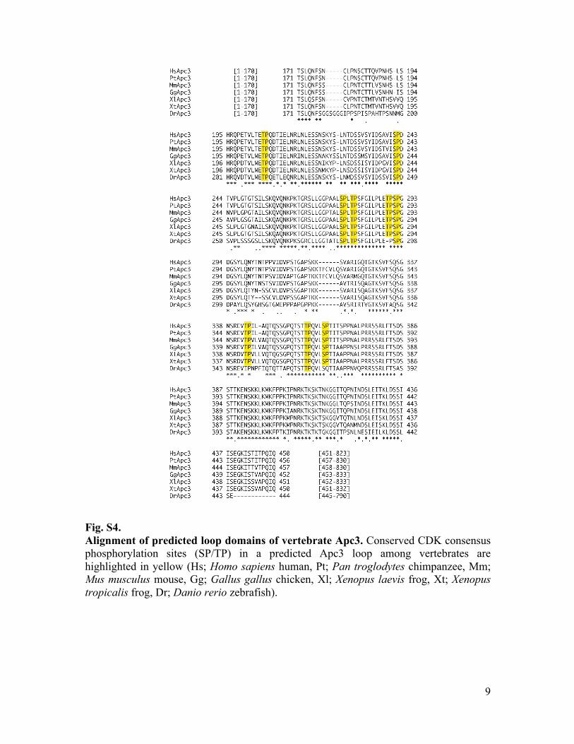
Fig. S4. Alignment of predicted loop domains of vertebrate Apc3. Conserved CDK consensus phosphorylation sites (SP/TP) in a predicted Apc3 loop among vertebrates are highlighted in yellow (Hs; Homo sapiens human, Pt; Pan troglodytes chimpanzee, Mm; Mus musculus mouse, Gg; Gallus gallus chicken, Xl; Xenopus laevis frog, Xt; Xenopus tropicalis frog, Dr; Danio rerio zebrafish).

10
Fig. S5. The loop domain of Apc3 binds p9-CDK1 in a phosphorylation-dependent manner. (A) WT APC/C or APC/CApc3-12A was incubated with ∆APC/C interphase or anaphase extracts. The APC/C was recovered with anti-Apc3 beads and bound proteins were analysed by immunoblotting. Different volumes of APC/Cs were loaded to quantitatively examine levels of Cdk1 associated and Apc1 phosphorylation on APC/CApc3-12A, compared with WT APC/C. LC; IgG light chain. (B) Left panel: Schematic diagram of Apc3 fragments used. Right panel: Bacterially purified MBP-tagged Apc3 fragments were incubated with Xenopus egg CSF-arrested extracts for 20 min and Apc3 bound proteins were analysed by SDS–PAGE and immunoblotting. The loop domain fragment (residues 202-342) binds p9 in mitotic egg extracts, whereas the same fragment with CDK mutations (residues 202-342 6A) abolishes p9-binding activity. A similar length fragment from the N-terminal domain (residues 61-215) does not bind whereas a second fragment (residues 234-374) containing the loop domain is able to bind to p9 and CDK1, albeit with a weaker interaction than the loop domain fragment (residues 202-342).

11
Fig. S6. The activities of a series of mutant APC/Cs carrying mutations in Apc1 in Xenopus egg extracts. (A) Schematic diagram of Apc1 and its phosphorylation site mutants used. SP/TP sites shared between Xenopus and Human Apc1 are shown in black, mutated sites in red. (B) Cyclin destruction assay in anaphase extracts. The indicated mutant APC/C were incubated with ∆APC/C anaphase extracts and then substrates 35S-labelled cyclin B and ∆67 (as a stable control) were added. Samples taken at indicated time points after addition of substrates, were analysed by SDS–PAGE and autoradiography. (C) Quantification of the destruction assays (B). (D) Cyclin destruction assay in interphase extracts. Using the same APC/Cs as (B), the activities of indicated mutant APC/Cs were assessed by destruction assay in APC/C-depleted (∆APC/C) interphase extract supplemented with Cdh1 and 35S-labelled APC/C substrates (cyclin B and ∆67). Samples

12
taken at indicated time points after addition of substrates were analysed by SDS–PAGE and autoradiography. (E) Quantification of the destruction assays (D).

13
Fig. S7. Alignment of predicted loop domains of vertebrate Apc1. Conserved CDK consensus phosphorylation sites (SP/TP) in a predicted Apc1 loop among vertebrates are highlighted (Hs; Homo sapiens human, Pt; Pan troglodytes chimpanzee, Mm; Mus musculus mouse, Gg; Gallus gallus chicken, Xt; Xenopus tropicalis frog, Dr; Danio rerio zebrafish). Highly conserved (Yellow), Conserved apart from Mus musculus (Green), Conserved between Xenopus tropicalis and Danio rerio (Blue)

14
Fig. S8. The specificity of phospho-specific Apc1 antibody. (A) Schematic diagram of Apc1 full length (FL) and its flexible loop domain fragments (residues 294-399): Apc1loop and Apc1loop-7A. The mutated CDK sites are in red. (B) Antibody binding to synthetic Apc1 peptides with no phosphorylation (NP), phosphorylation at S314 and S318 (S314/S318-Pho) or single site phosphorylation at S314 (S314-Pho). Peptides were spotted onto a nitrocellulose membrane and blotted with phospho-S314-S318 antibody (pS314pS318). (C) The pS314pS318 antibody specifically recognises Apc1 only when they are phosphorylated in anaphase. Bacterially purified Flag-tagged WT Apc1loop fragment was incubated in anaphase or interphase extracts for 45 min, purified using anti-FLAG affinity beads and analysed by immunoblotting with anti-Flag (Left: lanes 1-4) or anti-pS314pS318 (Right: lanes 5-8) antibodies. An excess of the corresponding epitope phospho-peptide (lane 7) or the same non-phosphorylated peptide (lane 8: nonphospho-peptide competitor) was incubated with the pS314pS318 antibody during immunoblotting. The pS314pS318 antibody recognises Apc1loop only when it is phosphorylated in anaphase (lanes 5 and 6) and the band disappears if the antibody is neutralized by the blocking peptide (lane 7).

15
Fig. S9. Phosphorylation of Apc1 and Apc3 subunits is crucial for APC/C activation. Upper panel: Cyclin destruction assay in mock or ∆APC/C anaphase extract or ∆APC/C anaphase extract supplemented with WT, Apc1-7A, Apc3-9A single or Apc3-9A/Apc1-7A double mutant APC/Cs. 35S-labelled cyclin B and cyclin B lacking the N-terminal 67 residues (∆67, stable control) were used as substrates. Samples taken at indicated time points after addition of substrates were analysed by SDS–PAGE and autoradiography. Quantification of the destruction assays is shown in Fig. 3A. Lower panel: 3D images of the APC/C showing WT Apc1 (cyan), Apc3 (green) and mutated subunits in red.

16
Fig. S10. The APC/CApc3-9A/Apc1-7A is inactive and unable to support cyclin B or securin proteolysis. Fresh “cycling” frog extracts and endogenous APC/C-depleted cycling extracts (∆APC) were prepared. WT or Apc3-9A/Apc1-7A double mutant APC/C (3-9A/1-7A) was added back to ∆APC extracts. Egg extracts were incubated at 23˚C and samples were taken every 10 min for analysis. Apc3, cyclin B2, Cdk1 and securin were detected by immunoblotting. The control (mock) cycling extracts performed cycles of entry into and exit from mitosis, judging from band shifts of Apc3 and disappearance of cyclin B and securin whereas ∆APC never degraded cyclin B nor securin. WT APC/C, but not APC/CApc3-9A/Apc1-7A rescued ∆APC extracts, allowing cyclin B and securin destruction.
17
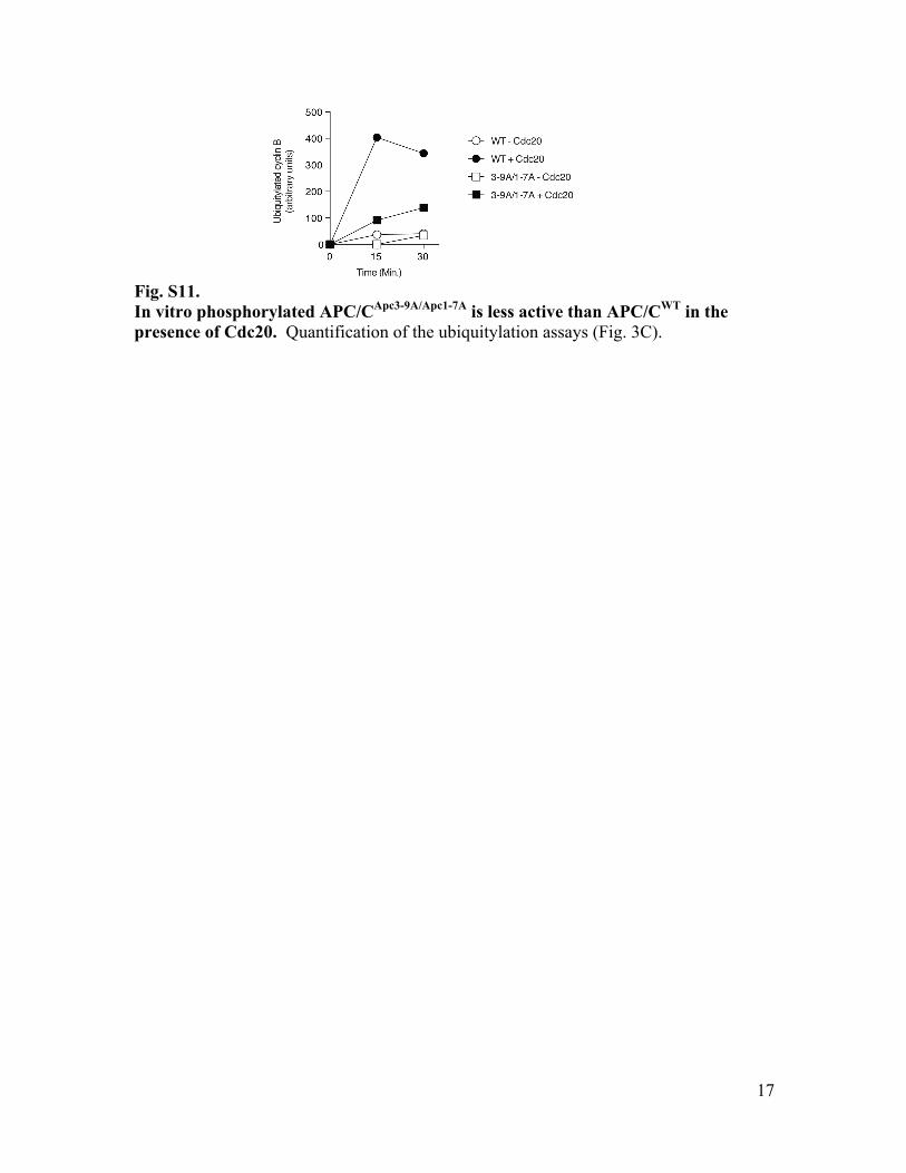
Fig. S11. In vitro phosphorylated APC/CApc3-9A/Apc1-7A is less active than APC/CWT in the presence of Cdc20. Quantification of the ubiquitylation assays (Fig. 3C).

18
Fig. S12. Phosphorylation status of WT or CDK site mutant APC/Cs in anaphase egg extract and during in vitro phosphorylation by CDK-cyclin B. (A) WT APC/C, APC/CApc3-9A, APC/CApc1-7A or APC/CApc3-9A/Apc1-7A was incubated in ∆APC egg extract in the presence of [γ-32P] ATP and cyclinB∆167 at 23 ˚C for 45 min (‘anaphase’ extract). The APC/C was recovered using anti-Apc3 affinity beads as Fig. 1B and phosphorylation of APC/Cs was analysed by SDS-PAGE and autoradiography. The phosphorylation levels of Apc3-9A and Apc1-7A mutant APC/Cs were decreased, compared with WT. Note that both Apc3-9A and Apc1-7A still contain CDK phosphorylation sites located in the non-loop domain and also under these conditions add-back APC/C can be phosphorylated by any mitotic kinases in Xenopus egg extracts. In anaphase extract, phosphorylation of Apc1 is reduced if phosphorylation of Apc3 is blocked (see also Fig. 2E). (B) WT APC/C, APC/CApc3-9A, APC/CApc1-7A or APC/CApc3-9A/Apc1-7A was incubated with CDK1-cyclin B and p9 in the presence of [γ-32P] ATP at 23˚C for 40 min. The APC/C was recovered using anti-Apc3 affinity beads, and analysed by SDS-PAGE and autoradiography. In purified p9-CDK1-cyclin B-dependent assay, phosphorylation of Apc1 is observed even if phosphorylation of Apc3 is blocked. The mechanism behind the difference between egg extracts and the purified CDK system remains unknown.

19
Fig. S13. The APC/CApc1-7D is more active than WT APC/C in interphase. (A) Cyclin destruction assay in interphase extracts. WT APC/C, APC/CApc3-9A or APC/CApc3-9A/Apc1-7D was incubated with ∆APC/C interphase extract and samples taken at indicated time points after addition of substrates were analysed by SDS–PAGE and autoradiography. Quantification of the destruction assays is shown in Fig. 3D. (B)
20
Endogenous APC/C was immunodepleted from fresh cycling extract (∆APC) and either WT or Apc1-7D mutant APC/C added back. Egg extracts were incubated at 23˚C and samples were taken every 10 min for analysis. Apc3, cyclin B2, phosphorylated Plx1 (pPlx1:AZ44) and Cdk1 were detected by immunoblotting. As controls, mock and ∆APC extracts are shown. In the presence of APC/CApc1-7D, cyclin B does not sufficiently accumulate after the first mitotic exit (see between 60 min and 110 min) compared with WT APC add back and thus fails to enter the second mitosis which occurs at 120 min with WT APC/C. C) Unphosphorylated WT APC/C or APC/CApc1-7D was incubated with ubiquitylation factors (E1, E2, energy generating mix, ubiquitin) in the presence/absence of Cdc20 at 23°C. 35S-labelled cyclin B (fission yeast Cdc13) was used as substrate. Samples taken at indicated time points were analysed by SDS–PAGE and autoradiography. The amounts of APC/C used were monitored by anti-Apc3 immunoblotting.

21
Fig. S14. Apc1loop binds to the APC/C depending on its phosphorylation status. Phospho-mimetic mutation into Apc1loop abolishes the ability to interact with APC/C. Seven CDK sites in Flag-tagged Apc1 loop fragment were substituted to alanine, aspartic acid or glutamic acid with an additional mutation in 7D1E and 8E (T378E). Wild type Apc1 fragment (WT) or its derivatives carrying mutations in CDK sites (7A, 7D, 7D1E or 8E) was incubated in anaphase extract and immunopurified with anti-FLAG affinity beads. Bound proteins were analysed by immunoblotting. M; molecular marker, staining pattern is a result of crossreaction with anti-Apc3 and anti-Apc6 antibodies.

22
Fig. S15. Location of Apc1, Apc3, Apc6, Apc8 and C box in human APC/C-Cdh1-Emi1 complex. Top: The image was generated using the Protein Data Bank file (4UI9: APC/CCdh1.Emi1)(25) and PyMOL, highlighting Apc1 (cyan), Apc3A (green), Apc3B (light green), Apc6A (orange), Apc6B (light orange), Apc8A (blue), Apc8B (light blue) and the C box region (SKHGDRFIPSRAG, yellow). The PC domain of Apc1 (PC) and the N- and C- terminal residues of predicted loop domains of Apc1 and Apc3 are indicated. Note that each of Apc3, Apc6 and Apc8 forms symmetric V-shaped homo-dimers through their N-terminal regions. Bottom: Closed view of the interaction interface of three subunits, Apc1, Apc8 and the C-box from Cdh1/Cdc20. The location of Apc1loop is not determined, but the assigned adjacent sites such as G290 (red) and P402 (orange) in Apc1 are very close to the interface between the C-box (yellow) and Apc8.

23
Table S1. CDK phosphorylation site mutant APC/C APC/C subunit
No. of conserved CDK sites (SP/TP)
APC/C mutant
CDK mutation sites
APC1 15 Apc1-15A S121A, T223A, T292A, S314A, S318A, S335A, S344A, S358A, S380A, S389A, T532A, T539A, S558A, S691A, S1352A
APC2 2 Apc2-2A T303A, S648A APC3 12 Apc3-12A T68A, S150A, T206A, S242A, S277A, T280A, T290A, S292A,
T343A, T360A, S365A, S567A APC4 3 Apc4-3A S22A, S481A, S503A APC5 2 Apc5-2A T31A, T235A APC6 1 Apc6-1A S558A APC7 4 Apc7-4A S38A, T92A, T134A, S523A APC8 4 Apc8-4A S544A, T565A, S571A, T579A APC10 4 Apc10-4A T3A, T7A, T90A, T160A APC11 0 APC12 0 APC13 0 APC16 1

24
References
1. V. Sudakin, D. Ganoth, A. Dahan, H. Heller, J. Hershko, F. C. Luca, J. V. Ruderman, A. Hershko, The cyclosome, a large complex containing cyclin-selective ubiquitin ligase activity, targets cyclins for destruction at the end of mitosis. Mol. Biol. Cell 6, 185–197 (1995). Medline doi:10.1091/mbc.6.2.185
2. S. Irniger, S. Piatti, C. Michaelis, K. Nasmyth, Genes involved in sister chromatid separation are needed for B-type cyclin proteolysis in budding yeast. Cell 81, 269–277 (1995). Medline doi:10.1016/0092-8674(95)90337-2
3. R. W. King, J. M. Peters, S. Tugendreich, M. Rolfe, P. Hieter, M. W. Kirschner, A 20S complex containing CDC27 and CDC16 catalyzes the mitosis-specific conjugation of ubiquitin to cyclin B. Cell 81, 279–288 (1995). Medline doi:10.1016/0092-8674(95)90338-0
4. I. Primorac, A. Musacchio, Panta rhei: The APC/C at steady state. J. Cell Biol. 201, 177–189 (2013). Medline
5. L. Chang, D. Barford, Insights into the anaphase-promoting complex: A molecular machine that regulates mitosis. Curr. Opin. Struct. Biol. 29, 1–9 (2014). Medline doi:10.1016/j.sbi.2014.08.003
6. J. Pines, Cubism and the cell cycle: The many faces of the APC/C. Nat. Rev. Mol. Cell Biol. 12, 427–438 (2011). Medline doi:10.1038/nrm3132
7. H. C. Huang, J. Shi, J. D. Orth, T. J. Mitchison, Evidence that mitotic exit is a better cancer therapeutic target than spindle assembly. Cancer Cell 16, 347–358 (2009). Medline doi:10.1016/j.ccr.2009.08.020
8. E. Manchado, M. Guillamot, G. de Cárcer, M. Eguren, M. Trickey, I. García-Higuera, S. Moreno, H. Yamano, M. Cañamero, M. Malumbres, Targeting mitotic exit leads to tumor regression in vivo: Modulation by Cdk1, Mastl, and the PP2A/B55α,δ phosphatase. Cancer Cell 18, 641–654 (2010). Medline doi:10.1016/j.ccr.2010.10.028
9. X. Zeng, F. Sigoillot, S. Gaur, S. Choi, K. L. Pfaff, D. C. Oh, N. Hathaway, N. Dimova, G. D. Cuny, R. W. King, Pharmacologic inhibition of the anaphase-promoting complex induces a spindle checkpoint-dependent mitotic arrest in the absence of spindle damage. Cancer Cell 18, 382–395 (2010). Medline doi:10.1016/j.ccr.2010.08.010
10. K. L. Sackton, N. Dimova, X. Zeng, W. Tian, M. Zhang, T. B. Sackton, J. Meaders, K. L. Pfaff, F. Sigoillot, H. Yu, X. Luo, R. W. King, Synergistic blockade of mitotic exit by two chemical inhibitors of the APC/C. Nature 514, 646–649 (2014). Medline doi:10.1038/nature13660
11. K. E. Gascoigne, S. S. Taylor, Cancer cells display profound intra- and interline variation following prolonged exposure to antimitotic drugs. Cancer Cell 14, 111–122 (2008). Medline doi:10.1016/j.ccr.2008.07.002
12. J. A. Steen, H. Steen, A. Georgi, K. Parker, M. Springer, M. Kirchner, F. Hamprecht, M. W. Kirschner, Different phosphorylation states of the anaphase promoting complex in response to antimitotic drugs: A quantitative proteomic analysis. Proc. Natl. Acad. Sci. U.S.A. 105, 6069–6074 (2008). Medline doi:10.1073/pnas.0709807104

25
13. C. Kraft, F. Herzog, C. Gieffers, K. Mechtler, A. Hagting, J. Pines, J. M. Peters, Mitotic regulation of the human anaphase-promoting complex by phosphorylation. EMBO J. 22, 6598–6609 (2003). Medline doi:10.1093/emboj/cdg627
14. E. R. Kramer, N. Scheuringer, A. V. Podtelejnikov, M. Mann, J. M. Peters, Mitotic regulation of the APC activator proteins CDC20 and CDH1. Mol. Biol. Cell 11, 1555–1569 (2000). Medline doi:10.1091/mbc.11.5.1555
15. M. Shteinberg, A. Hershko, Role of Suc1 in the activation of the cyclosome by protein kinase Cdk1/cyclin B. Biochem. Biophys. Res. Commun. 257, 12–18 (1999). Medline doi:10.1006/bbrc.1999.0409
16. G. Fang, H. Yu, M. W. Kirschner, Direct binding of CDC20 protein family members activates the anaphase-promoting complex in mitosis and G1. Mol. Cell 2, 163–171 (1998). Medline doi:10.1016/S1097-2765(00)80126-4
17. Z. Zhang, J. Yang, E. H. Kong, W. C. Chao, E. P. Morris, P. C. da Fonseca, D. Barford, Recombinant expression, reconstitution and structure of human anaphase-promoting complex (APC/C). Biochem. J. 449, 365–371 (2013). Medline doi:10.1042/BJ20121374
18. I. Berger, D. J. Fitzgerald, T. J. Richmond, Baculovirus expression system for heterologous multiprotein complexes. Nat. Biotechnol. 22, 1583–1587 (2004). Medline doi:10.1038/nbt1036
19. M. E. Matyskiela, D. O. Morgan, Analysis of activator-binding sites on the APC/C supports a cooperative substrate-binding mechanism. Mol. Cell 34, 68–80 (2009). Medline doi:10.1016/j.molcel.2009.02.027
20. D. Patra, W. G. Dunphy, Xe-p9, a Xenopus Suc1/Cks homolog, has multiple essential roles in cell cycle control. Genes Dev. 10, 1503–1515 (1996). Medline doi:10.1101/gad.10.12.1503
21. D. Patra, W. G. Dunphy, Xe-p9, a Xenopus Suc1/Cks protein, is essential for the Cdc2-dependent phosphorylation of the anaphase- promoting complex at mitosis. Genes Dev. 12, 2549–2559 (1998). Medline doi:10.1101/gad.12.16.2549
22. A. D. Rudner, A. W. Murray, Phosphorylation by Cdc28 activates the Cdc20-dependent activity of the anaphase-promoting complex. J. Cell Biol. 149, 1377–1390 (2000). Medline doi:10.1083/jcb.149.7.1377
23. M. Kõivomägi, M. Ord, A. Iofik, E. Valk, R. Venta, I. Faustova, R. Kivi, E. R. Balog, S. M. Rubin, M. Loog, Multisite phosphorylation networks as signal processors for Cdk1. Nat. Struct. Mol. Biol. 20, 1415–1424 (2013). Medline doi:10.1038/nsmb.2706
24. D. A. McGrath, E. R. Balog, M. Kõivomägi, R. Lucena, M. V. Mai, A. Hirschi, D. R. Kellogg, M. Loog, S. M. Rubin, Cks confers specificity to phosphorylation-dependent CDK signaling pathways. Nat. Struct. Mol. Biol. 20, 1407–1414 (2013). Medline doi:10.1038/nsmb.2707
25. L. Chang, Z. Zhang, J. Yang, S. H. McLaughlin, D. Barford, Atomic structure of the APC/C and its mechanism of protein ubiquitination. Nature 522, 450–454 (2015). Medline doi:10.1038/nature14471

26
26. H. Labit, K. Fujimitsu, N. S. Bayin, T. Takaki, J. Gannon, H. Yamano, Dephosphorylation of Cdc20 is required for its C-box-dependent activation of the APC/C. EMBO J. 31, 3351–3362 (2012). Medline doi:10.1038/emboj.2012.168
27. N. Lianga, E. C. Williams, E. K. Kennedy, C. Doré, S. Pilon, S. L. Girard, J. S. Deneault, A. D. Rudner, A Wee1 checkpoint inhibits anaphase onset. J. Cell Biol. 201, 843–862 (2013). Medline doi:10.1083/jcb.201212038
28. J. Y. Huang, G. Morley, D. Li, M. Whitaker, Cdk1 phosphorylation sites on Cdc27 are required for correct chromosomal localisation and APC/C function in syncytial Drosophila embryos. J. Cell Sci. 120, 1990–1997 (2007). Medline doi:10.1242/jcs.006833
29. I. García-Higuera, E. Manchado, P. Dubus, M. Cañamero, J. Méndez, S. Moreno, M. Malumbres, Genomic stability and tumour suppression by the APC/C cofactor Cdh1. Nat. Cell Biol. 10, 802–811 (2008). Medline doi:10.1038/ncb1742
30. K. E. Gascoigne, S. S. Taylor, How do anti-mitotic drugs kill cancer cells? J. Cell Sci. 122, 2579–2585 (2009). Medline doi:10.1242/jcs.039719
31. A. W. Murray, Cell cycle extracts. Methods Cell Biol. 36, 581–605 (1991). Medline doi:10.1016/S0091-679X(08)60298-8
32. M. J. Hayes, Y. Kimata, S. L. Wattam, C. Lindon, G. Mao, H. Yamano, A. M. Fry, Early mitotic degradation of Nek2A depends on Cdc20-independent interaction with the APC/C. Nat. Cell Biol. 8, 607–614 (2006). Medline doi:10.1038/ncb1410
33. H. Yamano, M. Trickey, M. Grimaldi, Y. Kimata, In vitro assays for the anaphase-promoting complex/cyclosome (APC/C) in Xenopus egg extracts. Methods Mol. Biol. 545, 287–300 (2009). Medline doi:10.1007/978-1-60327-993-2_18
34. K. Ohsumi, T. M. Yamamoto, M. Iwabuchi, Oocyte extracts for the study of meiotic M-M transition. Methods Mol. Biol. 322, 445–458 (2006). Medline doi:10.1007/978-1-59745-000-3_32
35. Y. Kimata, J. E. Baxter, A. M. Fry, H. Yamano, A role for the Fizzy/Cdc20 family of proteins in activation of the APC/C distinct from substrate recruitment. Mol. Cell 32, 576–583 (2008). Medline doi:10.1016/j.molcel.2008.09.023
36. N. R. Brown, S. Korolchuk, M. P. Martin, W. A. Stanley, R. Moukhametzianov, M. E. Noble, J. A. Endicott, CDK1 structures reveal conserved and unique features of the essential cell cycle CDK. Nat. Commun. 6, 6769 (2015). Medline doi:10.1038/ncomms7769