supplemental description of paraphelenchus acontioides (tylenchida
TRANSCRIPT

Nematology, 2011, Vol. 13(8), 887-899
Supplemental description of Paraphelenchus acontioides(Tylenchida: Aphelenchidae, Paraphelenchinae), with ribosomal
DNA trees and a morphometric compendiumof female Paraphelenchus
Lynn K. CARTA 1,∗, Andrea M. SKANTAR 1, Zafar A. HANDOO 1 and Melissa A. BAYNES 2
1 United States Department of Agriculture, ARS-BARC, Nematology Laboratory, Beltsville, MD 20705, USA2 Department of Forest Ecology and Biogeosciences, University of Idaho, Moscow, ID 83844, USA
Received: 30 September 2010; revised: 7 February 2011Accepted for publication: 7 February 2011; available online: 5 April 2011
Summary – Nematodes were isolated from surface-sterilised stems of cheatgrass, Bromus tectorum (Poaceae), in Colorado, grown onFusarium (Hypocreaceae) fungus culture, and identified as Paraphelenchus acontioides. Morphometrics and micrographic morphologyof this species are given to supplement the original description and expand the comparative species diagnosis. A tabular morphometriccompendium of the females of the 23 species of Paraphelenchus is provided as the last diagnostic compilation was in 1984. Variationsin the oviduct within the genus are reviewed to evaluate the taxonomic assignment of P. deckeri, a morphologically transitionalspecies between Aphelenchus and Paraphelenchus. Sequences were generated for both 18S and 28S ribosomal DNA, representingthe first identified species within Paraphelenchus so characterised. These sequences were incorporated into phylogenetic trees withrelated species of Aphelenchidae and Tylenchidae. Aphelenchus avenae isolates formed a well supported monophyletic sister group toParaphelenchus. The ecology of Paraphelenchus, cheat grass and Fusarium is also discussed.
Keywords – fungivorous nematode, invasive species, key, morphology, molecular, phylogeny, taxonomy.
Nematodes of the genus Paraphelenchus Micoletzky,1922 are generally regarded as fungivorous (Hunt, 1993),and are frequently found in association with plants.Twenty-three species are known, including Paraphelen-chus acontioides Taylor & Pillai, 1967; P. alii (Ali, Fa-rooqui & Suryawanshi, 1970) Fortuner, 1985 (= P. afsiHunt, 1993) and P. micoletzkyi Ali, Farooqui & Suryawan-shi, 1970 (= junior homonym) nec P. micoletzkyi Steiner,1941 (= junior synonym of Aphelenchus avenae); P. am-blyurus Steiner, 1934; P. basili Das, 1960; P. batavicusFilipjev, 1934; P. crenatus Das & Singh, 1968; P. deckeri(Zeidan & Geraert, 1989) Andrássy, 2007; P. fidicauda-tus Eroshenko, 1966; P. goodeyi Tandon & Singh, 1970;P. heterolineatus Haque, 1967; P. intermedius Thorne& Malek, 1968; P. myceliophthorus J.B. Goodey, 1958;P. obscurus Muchina, 1988; P. octolineatus Shavrov,1968; P. orientalis Muchina, 1988; P. paramonovi Haque1967; P. porrectus Eroshenko, 1966; P. pseudoparieti-nus Micoletzky, 1922; P. sacchari Husain & Khan, 1967;
∗ Corresponding author, e-mail: [email protected]
P. tritici Baranovskaya, 1958; P. ussuriensis Eroshenko,1966; P. zeae Romaniko, 1968; and P. zicsii Andrássy,1989.
Many of the morphometric values of the species over-lap, so qualitative morphology is particularly importantfor diagnosis. The most important of these include thepresence and shape of the mucro on the female tail, thenumber of lines in the lateral field (typically 4-9), andcontinuous or indented lip region. Unfortunately, molecu-lar data appear to be confined to an unknown species forwhich the small subunit rDNA has been derived (Holter-man et al., 2009).
Many of the species have been described from Asia,part of their proposed land origin from eastern Gond-wana in the Devonian epoch (Ryss, 2007), including de-scriptions of life stages and female tail papillae of P.myceliophthorus (Ryss & Chernetskaya, 2010). Sevenspecies have been found in Europe (Andrássy, 2007).Within the USA, numerous specimens of P. pseudopari-
© Koninklijke Brill NV, Leiden, 2011 DOI:10.1163/138855411X560968Also available online - www.brill.nl/nemy 887

L.K. Carta et al.
etinus and P. intermedius have been deposited into theUnited States Department of Agriculture Nematode Col-lection (USDANC) database. One notable species is P.acontioides which was discovered in soil around Ken-tucky bluegrass (Agrostis palustris Huds.) in Urbana, Illi-nois. The original population of this nematode specieswas initially raised for at least 5 years on the fungusPyrenochaeta terrestris (Hansen) Gorenz, J.C. Walker &Larson, before actually being described (Taylor & Pil-lai, 1967). Subsequently, morphometrics for this nema-tode were taken for material cultured on the same fungusand seven others, resulting in expanded ranges (Pillai &Taylor, 1967a), especially for the b ratio.
This paper reports the discovery of a Paraphelenchuspopulation in stems of cheatgrass, Bromus tectorum L.,in Colorado during a survey of endophytic fungi aspossible biocontrol agents within eight mid-western andwestern United States and British Columbia, Canada.Because many other Paraphelenchus species descriptionsare incomplete and not readily comparable, detailedmorphometrics of the new population were compiled andcontrasted with other isolates of P. acontioides and theother species of the genus to enhance the diagnosticinformation available.
Materials and methods
NEMATODE AND FUNGUS ISOLATION
Sixty-three populations of cheatgrass, B. tectorum,were sampled throughout North America (Idaho, Wash-ington, Nevada, Colorado, New Mexico, Iowa, Illinoisand British Columbia). Populations were typically aboutfive miles apart, but others were 1-20 or more milesapart. Twenty plants from each population were collected.A 2 cm segment was clipped around the lowest culm node(solid region on the shoot central axis that may gener-ate a leaf sheath or adventitious bud) of each plant. Seg-ments were surface-sterilised in 50% ethanol for 5 minand rinsed in sterile deionised water for 1 min (Lug-inbuhl & Muller, 1980; Schulz et al., 1993). To facili-tate isolation of fungi, culm segments from each popu-lation were placed onto potato dextrose agar plates. Im-print plates were made to ensure surface sterilisationwas successful. Fungi were identified morphologically.Cultures were placed on laboratory benches at ambientroom temperature and subcultured as needed. Two grasspopulations had nematodes and endophytic Fusarium sp.,and nematodes and fungi were not found independently
of each other. The cheatgrass population with P. acon-tioides was isolated from near Piney River, CO, USA(39◦50′24.99′′N, 106◦38′26.85′′W), south of Yampa, CO,USA, off Highway 131.
NEMATODE PRESERVATION AND IMAGING
Nematodes were rinsed from fungal plates, placed in4% formalin for 24 h, rinsed in 1.5 ml plastic tubes withwater and sent to Beltsville, MD, USA. From here theywere placed in 4% formalin for another 24 h, dehydratedin alcohol and glycerin, mounted in glycerin, and sealed.Images and measurements were made with a Zeiss Ul-traphot II microscope (Carl Zeiss, Jena, Germany, andBaltimore Instrument Company, Baltimore, MD, USA)equipped with differential interference contrast (DIC) op-tics. Photographs were taken of formalin-fixed specimensrinsed in water. Females were measured with an ocu-lar micrometer. Fixed specimens used to evaluate rela-tionships were observed from the USDANC. These in-cluded type and paratype slides of P. intermedius fromthe Thorne collection (USDANC Entries 31046, 29744,28745, 31049 slides 1a, 1b (type, five females), 1c, 1i,for different South Dakota sites of G. Thorne 28744),and P. pseudoparietinus Micoletzky, 1922 (USDANC En-try 21084, slide G11751 from soil of Chewings fescuegrass, Festuca rubra commutate, W.B. Courtney, Asto-ria, Oregon, 1941) identified as P. intermedius here basedon six lateral incisures and morphometrics, and four fe-male paratypes of P. deckeri (slide T-4299p from shrubsof Abuttaraz, Sudan). Type material from the old Nema-tode Slide Collection of the Department of Plant Pathol-ogy (now Crop Sciences), University of Illinois, Urbana,IL, USA, and P. amblyurus from the Steiner collection ofthe USDANC, were unavailable. Reproductive system ter-minology of Triantaphyllou and Fisher (1976) as used forA. avenae was applied to a single gonad arm going in ananterior direction: vulva, vagina, gonoduct (uterus, sper-matheca, fertilisation chamber (eight columns of two rowsof tightly packed oval cells not contained in a membrane),sphincter – (three rows of globular cells not contained ina membrane)), and ovary. The oviduct comprises a fer-tilisation chamber and sphincter cells (Triantaphyllou &Fisher, 1976), but the term has also been used to describethe sphincter cells only (Geraert, 1981).
PCR AND SEQUENCING
Nematodes were rinsed from fungal plates, placed in70% alcohol in 1.5 ml plastic tubes and sent to Beltsville,
888 Nematology

Redescription of Paraphelenchus acontioides and studies on the genus
MD, USA. They were rinsed in water, mounted on slidesfor imaging, recovered and individually processed. Ne-matodes were mechanically disrupted in 20 μl of extrac-tion buffer as described by Thomas et al. (1997) andthen stored in PCR tubes at −80◦C until needed. Ex-tracts were prepared by incubating the tubes at 60◦Cfor 60 min, followed by 95◦C for 15 min to deactivatethe proteinase K and centrifuged briefly prior to use inPCR. Each 25 μl PCR reaction contained 1 unit Plat-inum Taq (Invitrogen, Carlsbad, CA, USA), 1× reactionbuffer (20 mM Tris-HCl pH 8.4, 50 mM KCl, 2.5 mMMgCl2), 0.2 mM dNTP mix, 0.3 μM of each primer, and2 μl nematode extract. 28S reaction contained primersD2A (5′-ACAAGTACCGTGAGGGAAAGTTG-3′) andD3B (5′-TCGGAAGGAACCAGCTACTA-3′) (Nunn,1992). Cycling was performed as described by DeLey et al. (2005) and Ye et al. (2007). The partial 18Ssequence was amplified in two overlapping segments, us-ing primers 550F (5′-GGCAAGTCTGGTGCCAGCAGCC-3′) with 1108R (5′-CCACTCCTGGTGGTGCCCTTCC-3′) and 18s1.2 (5′-GGCGATCAGATACCGCCCTAGTT-3′) with 18sr2b (5′-TACAAAGGGCAGGGACGTAAT-3′). Cycling conditions for the 550F/1108R PCRreaction were: 1 cycle of 94◦C for 2 min, followedby 40 cycles of 94◦C for 20 s, 65◦C for 30 s, and72◦C for 30 s, finishing with 1 cycle of 72◦C for5 min. For the 18s1.2/18sr2b primer pair cycling con-ditions were the same as above except the annealingtemperature was 59◦C. PCR products were analysed byelectrophoresis on 1% agarose/1× SB (sodium borate-EDTA). Gels were stained with ethidium bromide andvisualised using the U:Genius gel documentation sys-tem (Syngene, Frederick, MD, USA). DNA was excisedfrom the gels and purified with the QIAquick Gel Ex-traction Kit (Qiagen, Valencia, CA, USA). PCR productswere quantified using a Nanodrop 8000 spectrophotome-ter (Thermo Fisher Scientific, Pittsburgh, PA, USA) andsequenced directly at the University of Maryland Cen-ter for Biosystems Research. DNA sequences were as-sembled using Sequencher 4.10.1 (Genecodes, Ann Ar-bor, MI, USA). DNA sequences were analysed using theBLASTN megablast program optimised for highly similarsequences, http://www.ncbi.nlm.nih.gov/blast/Blast.cgi.Sequences were submitted to GenBank under accessionnumbers HQ218322 for 28S and HQ218323 for 18S.
PHYLOGENETIC METHODS
Nematode sequences for P. acontioides were com-bined with other GenBank sequences of Tylenchidae
and Aphelenchidae, namely 18S: Ditylenchus dipsaci(Kühn, 1857) Filipjev, 1936 AY593906, Tylenchus ar-cuatus Siddiqi, 1963 EU306349, A. avenae EU306347,A. avenae 1 AY284639, A. avenae 2 AY284640, A. ave-nae AB368918, A. avenae AF036586, Aphelenchus sp.AY284641, Paraphelenchus sp. (JH-2004 isolate)AY284642, and new P. acontioides HQ218323; and 28S:Coslenchus costatus (de Man, 1921) Siddiqi, 1978DQ328719, Ditylenchus halictus Giblin-Davis, Erteld,Kanzaki, Ye & Center, 2010 AY589364, D. destructorThorne, 1945 DQ328727, A. avenae AB368536, A. ave-nae EU325683, Aphelenchus sp. DQ145664, and new P.acontioides HQ218323.
Alignments were made with ClustalW2 (Larkin etal., 2007) and checked by eye for consistency of con-served positions. The alignment was run through PAUP*4b10 (Swofford, 2002). Maximum Parsimony (MP) boot-strapped searches with 1000 replicates were conductedemploying tree bisection-reconnection (TBR) branchswapping, and accelerated transformation (ACCTRAN)character state optimisation. PAUP* was also used forgenerating sequence and tree statistics. Geneious Prov. 5.0.3 (Biomatters, Auckland, New Zealand; Drummondet al., 2009) was used to examine apomorphic charac-ters of Paraphelenchus. Maximum likelihood (ML) treesare presented in the figures because the computationally-intensive, probabilistic ML method is less affected bysampling error and infers better trees than distance orparsimony methods (Swofford et al., 1996). Alignmentsin PHYLIP format were run in web-based RAxML (Sta-matakis et al., 2008) with 100 bootstrap runs and ML esti-mate of 25 per site rate categories. Branch support valuesabove 50% given for ML followed by those for MP, andML parameters given in figure legends.
Description
Paraphelenchus acontioides Taylor & Pillai, 1967(Figs 1, 2A)
MEASUREMENTS
See Table 1.
DESCRIPTION
Body spiral to crook-shaped after death. Non-indentedlip region ca 15% stylet length high, 3.5 μm high ×9 μm wide. Stylet without swellings at base, although
Vol. 13(8), 2011 889

L.K. Carta et al.
Fig. 1. Paraphelenchus acontioides Colorado population female.A: Body; B: Median bulb with arrow pointing at constriction;C: Tail with mucro/ventral tuberculus (v arrow) and obscuredorsal tuberculus (d arrow).
basal lumen sometimes slightly inflated, surrounded byprominent muscles. Corpus length ca 2.7 times isthmuslength, slightly longer than isthmus, pharyngeal glandsabutting intestine. Median bulb with constriction ante-riorly and high, collar-like sheath surrounding anteriormuscle. Nerve ring just posterior to median bulb. Excre-tory pore opening posterior to nerve ring. Anterior gonadlength highly variable, not reaching pharyngo-intestinal
Fig. 2. Paraphelenchus spp. gonoducts. A: P. acontioides femalepost-vulval uterine sac (Pvs), uterus (UT), spermatheca (Spt),fertilisation chamber (Fch), sphincter (Sph) and ovary (Ovar);B: P. intermedius female post-vulval uterine sac, uterus, sper-matheca, fertilisation chamber, oviduct and ovary with develo-ping oocyte, from USDANC slide G11751 of G. Thorne, Astoria,OR, USA, 1941.
junction. Post-vulval uterine sac (Pvs) length variable,sometimes reaching nearly half the vulval-anal distance(VAD). VAD/tail = 4-4.5. Lateral incisures beginning atlevel of median bulb, increasing to eight at mid-body, end-ing at mid-tail. Rectum slightly shorter than tail length.Small pair of ventro-lateral papillae discernible between
890 Nematology

Redescription of Paraphelenchus acontioides and studies on the genus
Table 1. Morphometrics of Paraphelenchus acontioides and P. intermedius females. All measurements are in μm and in the form:mean ± s.d. (range).
Character P. acontioides P. intermediusPiney River, CO, South Dakota, G. Thorne, n = 6
n = 20 Astoria, OR, W. Courtney, n = 4Combined populations, n = 10
L 842 ± 118 (540-1059) 937 ± 84 (835-1110)a 29.4 ± 5 (22.6-37.4) 27.8 ± 2.8 (21.7-32)b 4.8 ± 0.6 (3.3-5.8) 5.3 ± 0.4 (4.5-5.9)c 23.1 ± 2.2 (16.1-27.7) 25.3 ± 1.7 (22.6-27.9)c′ 2.1 ± 0.3 (1.7-2.6) 2.3 ± 0.4 (1.6-2.7)V 75 ± 2 (69-81) 76 ± 1 (73-78)Max. body diam. 29 ± 6 (23-43) 34 ± 4 (28-40)Pharynx length 176 ± 9 (153-199) 177 ± 17 (153-216)Tail length 37 ± 5 (28-45) 37 ± 3 (31-44)Anal body diam. 17 ± 1 (15-20) 17 ± 3 (15-20)Stylet length 17 ± 2 (13-20) 17 ± 1 (16-19)Vulva-anal distance (VAD) 164 ± 29.4 (119-224) 185 ± 17.5 (159-214)Anterior gonad 278 ± 32 (185-321) 456 ± 72 (364-571)Post-vulval uterine sac (Pvs) 55 ± 10 (23-65) 58 ± 11 (37-74)Pvs/VAD% 35 ± 7 (17-55) 35 ± 4 (29-39)Pvs/vulval body diam. 2 ± 0.4 (0.9-2.5) 2.5 ± 1.1 (1.1-4.3)Rectum length 27 ± 3 (22-33) 34 ± 3 (31-37)Lateral incisures 8 6
ca 60-70% of tail length. Dorsal paired papillae locatedjust anterior to ventral base of asymmetrical, ventrally di-rected tail mucro, also known as a ventral tuberculus (d, vin Fig. 1C). In mature gonoducts, 8-10 globular columnsof two cells, squared at their edges, visible on oviductsphincter, and eight columns of two, thinner-walled cellscomprising the fertilisation chamber. Uterus and sper-matheca about equal in length to fertilisation chamber andto oviduct sphincter length. Egg (n = 30) dimensions =58.9 ± 5.8 (51-77.5) × 32.4 ± 6.6 (24-52.5) μm. Smallestegg 51 × 30 μm, largest 77.5 × 52.5 μm in size.
HOST AND LOCALITY
Isolated from the lowest node of B. tectorum (cheatgrass) from Piney River, CO, USA (39◦50′24.99′′N106◦38′26.85′′W), ca 30 miles south of Yampa, CO, USA,and ca 1.5 miles south of County Road 1 on Highway 131.
BIONOMICS
This Colorado population of P. acontioides was lo-cated ca 150 miles northwest of Colorado Springs, whichwas where P. pseudoparietinus and P. intermedius werecollected from native prairie (USDANC entries 28758
and 31067, deposited by Gerald Thorne, 1966). Paraphe-lenchus intermedius could grow and reproduce at 30◦C infungal culture (Thorne & Malek, 1968).
PHYLOGENY
18S sequences
For 18S sequences a ClustalW alignment of 1195alignment positions was used for a Maximum Parsimonytree (TL = 175, CI = 0.914, and HI = 0.086) of which5.6% of positions were parsimony informative. It wasalso used for a Maximum Likelihood tree bootstrapped100 times using RAxML with GTR matrix. Gammamodel parameters were estimated by the RAxML programand there were 115 alignment patterns. Slightly differentMP bootstrap values are indicated also on the ML tree(Fig. 3). This tree shows Paraphelenchus basal to theclade of fairly cohesive Aphelenchus populations thatform a monophyletic group with high bootstrap support.The distance matrix ‘p’ value between Aphelenchus andParaphelenchus (0.042), also visualised with relativebranch lengths, was greater than the value between themost divergent Aphelenchus populations (0.011).
A Geneious nucleotide alignment of 1198 total align-ment positions for all included taxa, including the P. acon-
Vol. 13(8), 2011 891

L.K. Carta et al.
Fig. 3. 18S rDNA Maximum Likelihood Tree of Aphelenchidae implemented in RAxML. Proportion of gaps and completely undeterminedcharacters in alignment: 0.038808, Model parameter alpha: 0.020014, Tree Length: 0.188690, Final ML Optimisation Likelihood:−2582.824816. Bootstrap values are listed for 100 ML replicates, and 1000 MP replicates.
tioides 1061 bp sequence within a 1075 bp segment of thetotal alignment (available on request), gave the same treetopology using MrBayes as the Clustal alignment. Therewere 18 Paraphelenchus-specific apomorphic nucleotidesin this alignment (position# – bp change), most of whichoccurred between positions 98 and 176: 98 – A vs C; 105 –T vs G; 106 – C vs T; 124 – T vs C, A; 159 – A vs T, C;166 and 175 – C vs T; 176 – A vs G; 286 – A vs C, T; 490– T vs C; 501 – A vs T; 510 – A vs G; 793 – T vs G; 934 –G insertion; differences for Paraphelenchus sp. only, afterthe P. acontioides sequence ran out, included: 1109 – A vsT; 1150 – A vs G; 1161 – A vs G, T; 1182 – T vs A. Therewas only one minor A vs R (= A or G) difference at po-sition 861 for P. acontioides compared to Paraphelenchussp.
Outgroup Tylenchus and Ditylenchus 18S sequenceshad intermediate GC sequence content values of 49.2 and49.3%, respectively. Sequences for Aphelenchus avenaeisolates had 49.6-50.0% GC base content, whilst the moredivergent P. acontioides and Paraphelenchus sp. had 48.1-48.5% GC.
28S sequences
For 28S sequences a ClustalW alignment (available onrequest) of 745 alignment positions was used for a Max-imum Parsimony tree (TL = 626, CI = 0.821, HI =0.179) of which 32% of characters were parsimony infor-mative. It was also used for a ML tree bootstrapped 100times using RAxML with GTR matrix. Gamma modelparameters were estimated by the RAxML program, and
there were 300 alignment patterns on the ML tree (Fig. 4).All MP bootstrap values were 100% and not indicated onthe tree.
The GC content for Coslenchus 28S sequences was51.3%, for D. destructor 54.4%, for Ditylenchus sp.46.8%, for two A. avenae 50.1%, for Aphelenchus sp.51.1%, and for P. acontioides 48.2%.
DIAGNOSIS
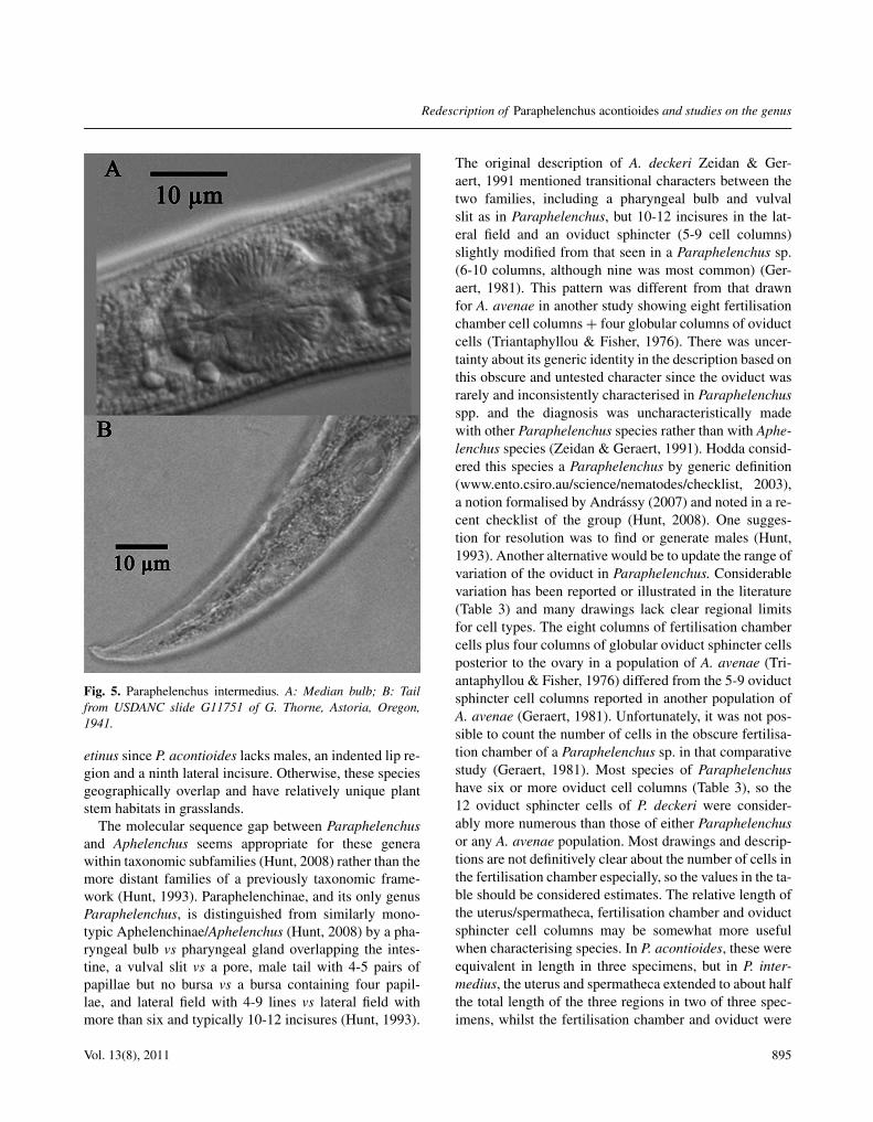
See Tables 1, 2, Figure 5A, B. Morphometrics for thisColorado population of P. acontioides are similar to P. am-blyurus and P. myceliophthorus, except for the lower b ra-tio and longer stylet. In a second paper by the authors ofP. acontioides, the b ratio was considerably lower whennematodes were grown on several different fungi (Pillai &Taylor, 1967a). The post-vulval uterine sac length/vulvalanal distance (Pvs/VAD) was 39-47 vs 50% for P. am-blyurus and 66% for P. myceliophagus. The Pvs lengthis similar for P. acontioides (55 ± 10, 23-65) and P. inter-medius (58 ± 11, 37-74 μm). The Pvs/vulval body diam.(Vbd) was also somewhat shorter in P. acontioides com-pared to P. intermedius (2 ± 0.4, 0.9-2.5 vs 2.5 ± 1.1, 1.1-4.3) (Table 1), and P. pseudoparietinus (Pvs/Vbd = 2-3, Andrássy, 2007). The P. intermedius oviduct had 8-12 cell columns, and the fertilisation chamber had eightcolumns of cells (Fig. 2B), whilst 8-12 cells were ob-served in the fertilisation chamber of P. deckeri. AlthoughP. acontioides has eight lateral incisures, it is otherwisesimilar to species with only six lines, such as P. inter-
892 Nematology

Redescription of Paraphelenchus acontioides and studies on the genus
Fig. 4. 28S Maximum Likelihood Tree of Aphelenchidae implemented in RAxML. Proportion of gaps and completely undeterminedcharacters in alignment: 0.288448, Model parameter alpha: 0.428791, Tree Length: 1.791170, Final ML Optimisation Likelihood:−3800.974955. Bootstrap values are listed for 100 ML replicates.
medius and P. amblyurus, for which males are described.Unfortunately, the lateral field and other features were es-pecially difficult to see in putative P. pseudoparietinusfrom old specimens in the USDANC. Paraphelenchus am-blyurus was described with what appeared at the time tobe a unique differentiation of the anterior median bulb(Steiner, 1934), similar to the bulb in the Colorado popu-lation of P. acontioides. This median bulb differentiationof less striated tissue was also visible in P. intermediusslides (Fig. 5A). The tail of P. intermedius was more at-tenuated toward the distal end (Fig. 5B) than in P. acon-tioides (Fig. 1C).
Discussion
TAXONOMY
This population of P. acontioides extended the reportedsize range with a longer body and stylet, lower V andslightly lower b value. The b values from the first descrip-tion were reduced substantially in a second paper describ-ing basic morphometrics on various fungal cultures (Pillai& Taylor, 1967a). The b values of this Colorado popula-tion are more similar to those in the second paper vs theoriginal. Therefore, relying primarily on morphometricscould lead an identifier astray, especially with species hav-ing few described populations or few replicates of a single
population. As with any identification of a nematode out-side the type locality, definitive assignment of this isolateto P. acontioides is a hypothesis. If molecular sequencesfrom this population are shown to be somewhat differentthan specimens from the type locality in the future, this re-description could serve as a framework for the proposal ofa possible new cryptic or complementary species.
Egg measurements extended the original range forlength and diam. of P. acontioides eggs ((64-74) × (28-32) μm) (Taylor & Pillai, 1967) at either end of the range((51-78) × (24-52) μm). Paraphelenchus intermedius hada similar sized egg (69×27 μm) to P. acontioides, whereasP. myceliophthorus had larger (78×32 μm) and A. avenaesomewhat smaller ((60-88) × (20-30) μm) eggs.
Of the 23 Paraphelenchus species, only eight lacka mucro and nine lack males. Absence of males is some-times a result of the small number of specimens available,but may also be related to temperature threshold. Among39 populations of A. avenae, most were parthenogeneticand males were inducible in many populations only above30◦C (Ali et al., 1999a). Both A. avenae and P. acon-tioides had minimum generation times of 5 and 6 days at35◦C (Pillai & Taylor, 1967b). Genetic diversity of A. ave-nae populations did not correlate with geography, fun-gal host, thermal preference or presence/absence of males(Ali et al., 1999b). Paraphelenchus acontioides may bea complementary species (Osche, 1954) of P. pseudopari-
Vol. 13(8), 2011 893

L.K. Carta et al.
Tabl
e2.
Para
phel
ench
usfe
mal
em
orph
omet
rics
.Sp
ecie
sL
ab
cV
Styl
et>
1m
ucro
c′M
ales
Li
Pvs/
Vbd
Tail
muc
roP.
acon
tioi
des
Ia71
0-88
025
-30
4.4-
5.3
20-3
073
-77
14-1
6–
2.4
–8
2P.
acon
tioi
des
IIb
510-
960
15.9
-35.
63.
5-6.
214
.5-3
071
-80
––
–+
––
P.ac
onti
oide
sC
O54
0-10
5923
-37
3.4-
5.8
16-3
3.7
69-7
8.3
13-2
3–
1.7-
2.4
(2.1
)+
81.
7-2.
3P.
alii
790-
980
23.5
-29.
65.
2-6.
514
.7-2
2.2
73-7
613
.6-1
5.2
–3.
1+
61.
8P.
ambl
yuru
s54
0-93
023
-33
3.7-
5.9
17-2
476
17-1
9–
2.5
+6
2.1
P.ba
sili
550-
590
23.4
-23.
95-
5.3
15.6
-18
70-7
416
–3.
2+
42.
1P.
cren
atus
680-
740
27.6
-30.
84.
8-5.
119
.2-2
3.7
74.6
-74.
713
.8-1
5–
3.4
+8
2.3
P.fid
icau
datu
s67
6-75
938
-39
3.1-
5.3
24.6
-27.
475
-79.
813
22.
1–
82.
8P.
good
eyi
715-
766
209-
1018
-20
7412
–2.
1–
63.
2P.
hete
roli
neat
us61
5-80
319
.6-2
1.6
4.0-
4.8
17.1
-29.
275
.1-7
7.9
15.1
–2
–8
1P.
inte
rmed
ius
700-
1000
24-2
84.
3-5.
623
-26
7615
-19
–2.
4–
61.
1-4.
3P.
myc
elio
phth
orus
580-
820
22-3
44.
1-6.
613
-24
71-7
86-
19–
1.4-
2.7
+6
3.2
P.pa
ram
onov
i58
7-93
718
.9-2
64.
4-5.
518
.1-2
5.2
75.6
-77.
812
.8-1
3.9
–1.
5+
62.
7P.
porr
ectu
s70
4-77
030
.8-3
1.2
4.8
20.7
-23.
376
.4-7
8.9
13–
1.8
+8
–P.
sacc
hari
590-
880
30-3
95.
1-6.
620
-21
68-7
711
-16
–2.
9+
43
P.ps
eudo
pari
etin
us38
0-91
025
-36
(30)
4.1-
6.3
(5)
14-2
4(2
0)68
-78
(74)
13-1
4–
2.7
+9
2-3
P.zi
csii
970
365.
714
7214
-15
24.
3+
4+
23.
2
No
tail
muc
roP.
bata
vicu
s75
032
-37
7-8
2775
12-1
3–
2.2
+12
4.3
P.de
cker
i69
5-85
537
-39
5.2-
622
-25
75-7
814
.5-1
7.5
–2.
3-3.
2–
10-1
22-
3P.
obsc
urus
560-
630
26-4
24.
2-5.
220
-24
72-7
412
-13
–2
+5
3.6
P.oc
toli
neat
us51
2-59
727
.4-3
4.2
3.7-
4.6
17.1
-21.
675
-78.
913
-14
–2.
9+
83.
6P.
orie
ntal
is66
6-71
225
-37
5-6
29-3
773
-77
12-1
3–
1–
83.
5P.
trit
ici
442-
851
26.8
-32
3.4-
5.9
16.7
-21.
277
-78
12-1
3–
2.0
+–
2P.
ussu
rien
sis
830-
841
30.2
-37.
44.
8-5.
620
.2-2
873
.8-7
6.1
15–
2.4
–6
–P.
zeae
658-
700
24.4
8.15
31.3
76.5
16-1
7–
2.0
––
4.4
Tail
muc
ropr
esen
tin
first
15sp
ecie
s.–/
+,ab
senc
e/pr
esen
ceof
mal
es;L
i,la
tera
linc
isur
es;P
vs,p
ost-
vulv
alut
erin
esa
c;V
bd,v
ulva
lbod
ydi
am.M
easu
rem
ents
inth
efo
rm:r
ange
and/
or(m
ean)
.a
From
Tayl
or&
Pilla
i(19
67).
bFr
omPi
llai&
Tayl
or(1
967a
).
894 Nematology
Redescription of Paraphelenchus acontioides and studies on the genus
Fig. 5. Paraphelenchus intermedius. A: Median bulb; B: Tailfrom USDANC slide G11751 of G. Thorne, Astoria, Oregon,1941.
etinus since P. acontioides lacks males, an indented lip re-gion and a ninth lateral incisure. Otherwise, these speciesgeographically overlap and have relatively unique plantstem habitats in grasslands.
The molecular sequence gap between Paraphelenchusand Aphelenchus seems appropriate for these generawithin taxonomic subfamilies (Hunt, 2008) rather than themore distant families of a previously taxonomic frame-work (Hunt, 1993). Paraphelenchinae, and its only genusParaphelenchus, is distinguished from similarly mono-typic Aphelenchinae/Aphelenchus (Hunt, 2008) by a pha-ryngeal bulb vs pharyngeal gland overlapping the intes-tine, a vulval slit vs a pore, male tail with 4-5 pairs ofpapillae but no bursa vs a bursa containing four papil-lae, and lateral field with 4-9 lines vs lateral field withmore than six and typically 10-12 incisures (Hunt, 1993).
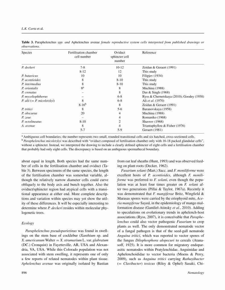
The original description of A. deckeri Zeidan & Ger-aert, 1991 mentioned transitional characters between thetwo families, including a pharyngeal bulb and vulvalslit as in Paraphelenchus, but 10-12 incisures in the lat-eral field and an oviduct sphincter (5-9 cell columns)slightly modified from that seen in a Paraphelenchus sp.(6-10 columns, although nine was most common) (Ger-aert, 1981). This pattern was different from that drawnfor A. avenae in another study showing eight fertilisationchamber cell columns + four globular columns of oviductcells (Triantaphyllou & Fisher, 1976). There was uncer-tainty about its generic identity in the description based onthis obscure and untested character since the oviduct wasrarely and inconsistently characterised in Paraphelenchusspp. and the diagnosis was uncharacteristically madewith other Paraphelenchus species rather than with Aphe-lenchus species (Zeidan & Geraert, 1991). Hodda consid-ered this species a Paraphelenchus by generic definition(www.ento.csiro.au/science/nematodes/checklist, 2003),a notion formalised by Andrássy (2007) and noted in a re-cent checklist of the group (Hunt, 2008). One sugges-tion for resolution was to find or generate males (Hunt,1993). Another alternative would be to update the range ofvariation of the oviduct in Paraphelenchus. Considerablevariation has been reported or illustrated in the literature(Table 3) and many drawings lack clear regional limitsfor cell types. The eight columns of fertilisation chambercells plus four columns of globular oviduct sphincter cellsposterior to the ovary in a population of A. avenae (Tri-antaphyllou & Fisher, 1976) differed from the 5-9 oviductsphincter cell columns reported in another population ofA. avenae (Geraert, 1981). Unfortunately, it was not pos-sible to count the number of cells in the obscure fertilisa-tion chamber of a Paraphelenchus sp. in that comparativestudy (Geraert, 1981). Most species of Paraphelenchushave six or more oviduct cell columns (Table 3), so the12 oviduct sphincter cells of P. deckeri were consider-ably more numerous than those of either Paraphelenchusor any A. avenae population. Most drawings and descrip-tions are not definitively clear about the number of cells inthe fertilisation chamber especially, so the values in the ta-ble should be considered estimates. The relative length ofthe uterus/spermatheca, fertilisation chamber and oviductsphincter cell columns may be somewhat more usefulwhen characterising species. In P. acontioides, these wereequivalent in length in three specimens, but in P. inter-medius, the uterus and spermatheca extended to about halfthe total length of the three regions in two of three spec-imens, whilst the fertilisation chamber and oviduct were
Vol. 13(8), 2011 895
L.K. Carta et al.
Table 3. Paraphelenchus spp. and Aphelenchus avenae female reproductive system cells interpreted from published drawings orobservations.
Species Fertilisation chamber Oviduct Referencecell number sphincter cell
number
P. deckeri 7-8 10-12 Zeidan & Geraert (1991)8-12 12 This study
P. batavicus 10 10 Filipjev (1934)P. acontioides 8 8-10 This studyP. intermedius 8 8-10 This studyP. orientalis 8a 8 Muchina (1988)P. crenatus – 8 Das & Singh (1968)P. myceliophthorus – 6-8 Ryss & Chernetskaya (2010); Goodey (1958)P. alii (= P. micoletzkyi) 8 6-8 Ali et al. (1970)
8-16b 8 Zeidan & Geraert (1991)P. tritici 8 5-6 Baranovskaya (1958)P. obscurus 20 4 Muchina (1988)P. zeae – 4 Romaniko (1968)P. octolineatus 8-10 2 Shavrov (1968)A. avenae 8 4 Triantaphyllou & Fisher (1976)
5-7 5-9 Geraert (1981)
a Ambiguous cell boundaries; the number represents two small, rounded transitional cells and six hatched, cross-sectioned cells.b Paraphelenchus micoletzkyi was described with “oviduct composed of fertilisation chamber only with 16-18 packed glandular cells”,without a sphincter. Instead, we interpreted the drawing to include a clearly defined sphincter of eight cells and a fertilisation chamberthat probably had only eight cells. The discrepancy is based on an ambiguous spermathecal boundary.
about equal in length. Both species had the same num-ber of cells in the fertilisation chamber and oviduct (Ta-ble 3). Between specimens of the same species, the lengthof the fertilisation chamber was somewhat variable, al-though the relatively narrow diameter cells could curveobliquely to the body axis and bunch together. Also theoviduct/sphincter region had atypical cells with a transi-tional appearance at either end. More complete descrip-tions and variation within species may yet show the util-ity of these differences. It will be especially interesting todetermine where P. deckeri resides within molecular phy-logenetic trees.
Ecology
Paraphelenchus pseudoparietinus was found in swell-ings on the stem base of cocklebur (Xanthium sp. andX. americanum Walter = X. strumarium L. var. glabratum(DC.) Cronquist) in Fayetteville, AR, USA and Alexan-dria, VA, USA. While this Colorado population was notassociated with stem swelling, it represents one of onlya few reports of related nematodes within plant tissue.Aphelenchus avenae was originally isolated by Bastian
from oat leaf sheaths (Hunt, 1993) and was observed feed-ing on plant roots (Decker, 1962).
Fusarium solani (Mart.) Sacc. and F. moniliforme wereexcellent hosts of P. acontioides, although F. monili-forme was preferred to F. solani, even though the popu-lation was at least four times greater on F. solani af-ter two generations (Pillai & Taylor, 1967a). Recently itwas demonstrated that F. mangiferae Britz, Wingfield &Marasas spores were carried by the eriophyoid mite, Ace-ria mangiferae Sayed, in the epidemiology of mango mal-formation disease (Gamliel-Atinsky et al., 2010). Addingto speculations on evolutionary trends in aphelench-hostassociations (Ryss, 2007), it is conceivable that Paraphe-lenchus could also vector pathogenic Fusarium to cropplants as well. The only demonstrated nematode vectorof a fungal pathogen is that of the seed-gall nematodeAnguina tritici, which was reported to vector spores ofthe fungus Dilophosphora alopecuri to cereals (Atana-soff, 1925). It is more common for migratory endopar-asitic nematodes within Pratylenchidae, Anguinidae andAphelenchoididae to vector bacteria (Moens & Perry,2009), such as Anguina tritici carrying Rathayibacter(= Clavibacter) toxicus (Riley & Ophel) Sasaki, Chi-
896 Nematology

Redescription of Paraphelenchus acontioides and studies on the genus
jimatsu & Suzuki to wheat (Riley, 1992), and othernematode-bacteria combinations (Dorofeeva et al., 2002).However, most nematode-microbial associations involvewounding or host predisposition rather than a strict vectorrelationship (Powell, 1963).
Cheatgrass is an invasive species that is especiallycommon in western crops such as winter wheat and alfalfa(Young, 2000) and, like many weeds, may be capableof harbouring various plant diseases. Alternatively, likeA. avenae (Rhoades & Linford, 1959), Paraphelenchusmay be an opportunist fungivore scavenging fungi undera variety of conditions.
We look forward to future research involving moredetailed SEM images of various Paraphelenchus speciesso as to reveal clearer profiles and variability of thelateral field in various species, and of the presence ofother female tail papillae homologous to those in malesas reported in P. myceliophthorus (Ryss & Chernetskaya,2010). More molecular sequences and descriptions of theoviduct in more species will undoubtedly improve thesystematic knowledge of this group.
Acknowledgements
We thank Maria Hult, Jennifer Kramer and David Mar-tel of the Nematology Laboratory for technical assistance.We are grateful to David Chitwood of the NematologyLaboratory for bringing to our attention the history of thegeneric status and authorities for Paraphelenchus deckeri.Mention of trade names or commercial products in thispublication is solely for the purpose of providing specificinformation and does not imply recommendation or en-dorsement by the US Department of Agriculture.
References
ALI, M.R., AMIN, B., ADACHI, T. & ISHIBASHI, N. (1999a).Host and temperature preference, male occurrence and mor-phometrics of fungivorous nematode, Aphelenchus avenaeisolates from Japan. Japanese Journal of Nematology 29, 7-17.
ALI, M.R., YAMAGUCHI, Y. & ISHIBASHI, N. (1999b). RAPDand PCR-RFLP analysis on genetic diversity of Aphelenchusavenae isolates collected from Kyushu and some otherdistricts of Japan. Japanese Journal of Nematology 29, 24-34.
ALI, M.S., FAROOQUI, N.M. & SURYAWANSHI, M.V. (1970).On a new species of Paraphelenchus (Micoletzky, 1922) Mi-coletzky, 1925 (Nematoda: Paraphelenchidae) from Marath-wada, India. Rivista di Parassitologia 31, 139-142.
ANDRÁSSY, I. (1989). Six new nematode species from SouthAmerica. Acta Zoologica Hungarica 35, 1-16.
ANDRÁSSY, I. (2007). Free-living nematodes of Hungary,III (Nematoda errantia). Pedozoologica Hungarica No. 4.Budapest, Hungary, Hungarian Natural History Museum& Systematic Zoology Research Group of the HungarianAcademy of Sciences, 496 pp.
ATANASOFF, D. (1925). The Dilophospora disease of cereals.Phytopathology 15, 1-40.
BARANOVSKAYA, I.A. (1958). [Contribution to the knowledgeof the genus Paraphelenchus (Micoletzky, 1922) Micoletzky,1925 (Nematoda: Aphelenchidae).] Zoologichesky Zhurnal37, 13-19.
BARANOVSKAYA, I.A. (1984). [Nematodes of the genus Para-phelenchus (Micoletzky, 1922) Micoletzky, 1925.] In: Turly-gina, E.S. (Ed.). Taksonomiya i biologiya fitogel’mintov.Moscow, USSR, Nauka, pp. 5-35.
DAS, V.M. (1960). Studies on the nematode parasites ofplants in Hyderabad (Andhra Pradesh, India). Zeitschrift fürParasitenkunde 19, 553-605.
DE LEY, P., TANDINGAN DE LEY, I., MORRIS, K., ABEBE,E., MUNDO-OCAMPO, M., YODER, M., HERAS, J., WAU-MANN, D., ROCHA-OLIVARES, A., BURR, A.H.J. et al.(2005). An integrated approach to fast and informativemorphological vouchering of nematodes for applications inmolecular barcoding. Philosophical Transactions of the RoyalSociety B 360, 1945-1958.
DECKER, H. (1962). Zur Biologie und Ökologie von Aphe-lenchus avenae Bastian. Nematologica 7, 9. [Abstr.]
DOROFEEVA, L.V., EVTUSHENKO, L.I., KRAUSOVA, V.I.,KARPOV, A.V., SUBBOTIN, S.A. & TIEDJE, J.M. (2002).Rathayibacter caricis sp. nov. and Rathayibacter festucae sp.nov., isolated from the phyllosphere of Carex sp. and the leafgall induced by the nematode Anguina graminis on Festucarubra L., respectively. International Journal of Systematicand Evolutionary Microbiology 52, 1917-1923.
DRUMMOND, A.J., ASHTON, B., CHEUNG, M., HELED, J.,KEARSE, M., MOIR, R., STONES-HAVAS, S., THIERER,T. & WILSON, A. (2009). Geneious v 5.0. Available from:http://www.geneious.com/
EROSHENKO, A.S. (1966). [Three new species of Paraphe-lenchus (Micoletzky, 1922) Micoletzky, 1925 (Nematoda:Aphelenchidae).] Zoologischesky Zhurnal 45, 1873-1876.
FILIPJEV, I.N. (1934). [Harmful and useful nematodes in ruraleconomy.] Moscow & Leningrad, USSR, Ogiz, 440 pp.
FORTUNER, R. (1985). Notes on nomenclature of plant nema-todes. Revue de Nématologie 8, 77-83.
GAMLIEL-ATINSKY, E., FREEMAN, S., MAYMON, M.,OCHOA, R., BAUCHAN, G.R., SKORACKA, A., PENA, J.& PALEVSKY, E. (2010). The role of eriophyoids in fungalpathogen epidemiology, mere association or true interaction?Experimental and Applied Acarology 51, 191-204.
Vol. 13(8), 2011 897

L.K. Carta et al.
GERAERT, E. (1981). The female reproductive system in nema-tode systematics. Annales de la Société Royale Zoologique deBelgique 110, 73-86.
GOODEY, J.B. (1958). Paraphelenchus myceliophthorus n. sp.Nematologica 3, 1-5.
HAQUE, M.M. (1967). [Description of the new species belong-ing to the genus Paraphelenchus, Micol., 1922 (Nematoda,Paraphelenchidae).] Zoologichesky Zhurnal 46, 1842-1846.
HOLTERMAN, M., KARSSEN, G., VAN DEN ELSEN, S., VAN
MEGEN, H., BAKKER, J. & HELDER, J. (2009). Smallsubunit rDNA-based phylogeny of the Tylenchida sheds lighton relationships among some high-impact plant-parasiticnematodes and the evolution of plant feeding. Phytopathology99, 227-235.
HUNT, D.J. (1993). Aphelenchida, Longidoridae and Tricho-doridae: their systematics and bionomics. Wallingford, UK,CABI Publishing, 352 pp.
HUNT, D.J. (2008). A checklist of the Aphelenchoidea (Nema-toda: Tylenchida). Journal of Nematode Morphology and Sys-tematics 10 (2007), 99-135.
HUSAIN, S.I. & KHAN, A.M. (1967). On the status of thegenera of the superfamily Aphelenchoidea (Fuchs, 1937)Thorne, 1949, with descriptions of six new species from In-dia. Proceedings of the Helminthological Society of Washing-ton 34, 167-174.
LARKIN, M.A., BLACKSHIELDS, G., BROWN, N.P.,CHENNA, R., MCGETTIGAN, P.A., MCWILLIAM, H.,VALENTIN, F., WALLACE, I.M., WILM, A., LOPEZ,R. et al. (2007). ClustalW and ClustalX version 2.0.Bioinformatics 23, 2947-2948.
LUGINBUHL, M. & MULLER, E. (1980). [Endophytic fungusin the above-ground organs of four plants growing together atthe same locations (Buxus, Hedera, Ilex, Ruscus).] Sydowia33, 185-209.
MICOLETZKY, H. (1922). Die freilebenden Erd-Nematodenmit besonderer Berucksichtigung der Steiermark und derBukowina, zugleich mit einer Revision samtlicher nichtmariner, freilebender Nematoden in Form von Genus– Beschreibunger un Bestimmungsschlusseln. Archiv fürNaturgeschlichte 87, 1-650.
MICOLETZKY, H. (1925). Die Freilebenden Susswasser- undMoornematoden Danemarks. Nebst Anhang eber Amo-bosporidien und andere Parasiten bei freilebenden Nemato-den. Kongelige Danske Videnskabernes Selskab Skrifter (8)10, 57-310.
MOENS, M. & PERRY, R.N. (2009). Migratory plant endopar-asitic nematodes: a group rich in contrasts and divergence.Annual Review of Phytopathology 47, 313-332.
MUCHINA, T.I. (1988). [Two new species of nematodes (Para-phelenchidae) with a description of anomalies in their repro-ductive system.] Zoologischesky Zhurnal 67, 1240-1245.
NUNN, G. (1992). Nematode molecular evolution. An investiga-tion of evolutionary patterns among nematodes based upon
DNA sequences. Ph.D. Dissertation, University of Notting-ham, Nottingham, UK.
OSCHE, G. (1954). Über dei gegenwärtig ablaufende Entste-hung von Zwillings- und Komplementärarten bei Rhabditi-den (Nematodes). (Fötalisation und Artbildung.) ZoologischeJahrbücher. Abteilung für Systematik, Ökologie und Geogra-phie der Tiere 82, 618-654.
PILLAI, J.K. & TAYLOR, D.P. (1967a). Influence of fungi onhost preference, host suitability, and morphometrics of fivemycophagous nematodes. Nematologica 13, 529-540.
PILLAI, J.K. & TAYLOR, D.P. (1967b). Effect of temperatureon the time required for hatching and duration of life cycle offive mycophagous nematodes. Nematologica 13, 512-516.
POWELL, N.T. (1963). The role of plant-parasitic nematodes infungus diseases. Phytopathology 53, 28-35.
RHOADES, H.L. & LINFORD, M.B. (1959). Control of Pythiumroot rot by the nematode Aphelenchus avenae. Plant DiseaseReporter 43, 323-328.
RILEY, I.T. (1992). Anguina tritici is a potential vector ofClavibacter toxicus. Australasian Plant Pathology 21, 147-149.
ROMANIKO, V.I. (1968). [A new nematode species of the genusParaphelenchus (Micoletzky, 1922) Micoletzky, 1925.] TrudyBiologicheskii Chelyabinskaia Gosudarstvennogo Pedagog-ickej Institut 23, 40-42.
RYSS, A.YU. (2007). [Main evolution lines of plant parasiticnematodes of the order Aphelenchida Siddiqi, 1980.] Parazi-tologiya 41, 484-511.
RYSS, A.YU. & CHERNETSKAYA, A.YU. (2010). [Life cycleof Paraphelenchus myceliophthorus Goodey, 1958 (Nema-toda: Aphelenchida).] Parazitologiya 44, 105-127.
SCHULZ, B., WANKE, U., DRAEGER, S. & AUST, H.J. (1993).Endophytes from herbaceous plants and shrubs: effectivenessof surface sterilization methods. Mycological Research 97,1447-1450.
SHAVROV, G.N. (1968). [New species of Paraphelenchus(Micoletzky, 1922) Micoletzky, 1925 (Nematoda: Aphe-lenchinae).] Soobshcheniya dal’nevost Filial V.L. KomarovaSibirskoe Otdelenie Akademiya 26, 135-136.
STAMATAKIS, A., HOOVER, P. & ROUGEMONT, J. (2008).A rapid bootstrap algorithm for the RAxML web-servers.Systematic Biology 75, 758-771.
STEINER, G. (1934). Observations on nematodes parasitic in tu-bers of the cinnamon-vine. Proceedings of the Helmintholog-ical Society of Washington 1, 15-17.
STEINER, G. (1941). Nematodes parasitic on and associatedwith roots of marigolds Tagetes hybrids. Proceedings of theBiological Society of Washington 54, 31-34.
SWOFFORD, D.L. (2002). PAUP*: phylogenetic analysis usingparsimony (* and other methods), Version 4. Sunderland,MA, USA, Sinauer Associates.
SWOFFORD, D.L., OLSEN, G.J., WADDELL, P.J. &HILLIS, D.M. (1996). Phylogenetic inference. In: Hillis,D.M., Moritz, C. & Mable, B.K. (Eds). Molecular systemat-
898 Nematology

Redescription of Paraphelenchus acontioides and studies on the genus
ics, 2nd edition. Sunderland, MA, USA, Sinauer Associates,pp. 407-513.
TANDON, R.S. & SINGH, S.P. (1970). On two new nematodes(Aphelenchoidea) from tobacco roots in India. Journal ofHelminthology 44, 323-328.
TAYLOR, D.P. & PILLAI, J.K. (1967). Paraphelenchus acon-tioides n. sp. (Nematoda: Paraphelenchidae), a mycophagousnematode from Illinois, with observations on its feedinghabits and a key to the species of Paraphelenchus. Proceed-ings of the Helminthological Society of Washington 34, 51-54.
THOMAS, W.K., VIDA, J.T., FRISSE, L.M., MUNDO, M.& BALDWIN, J.G. (1997). DNA sequences from formalinfixed nematodes: integrating molecular and morphologicalapproaches to taxonomy. Journal of Nematology 29, 250-254.
THORNE, G. & MALEK, R.B. (1968). Nematodes of the North-ern Great Plains, Part I, Tylenchida (Nemata: Secernen-tea). Technical Bulletin 31, Agricultural Experiment Station,South Dakota State University, 111 pp.
TRIANTAPHYLLOU, A.C. & FISHER, J.M. (1976). Gemeto-genesis in amphimictic and parthenogenetic populations ofAphelenchus avenae. Journal of Nematology 8, 168-177.
YE, W., GIBLIN-DAVIS, R.M., DAVIES, K.A., PURCELL,M.F., SCHEFFER, S.J., TAYLOR, G.S., CENTER, T.D.,MORRIS, K. & THOMAS, W.K. (2007). Molecular phy-logenetics and the evolution of host plant associations inthe nematode genus Fergusobia (Tylenchida: Fergusobiinae).Molecular Phylogenetics and Evolution 45, 123-141.
YOUNG, J. (2000). Bromus tectorum L. In: Bossard, C.C.,Randall, J.M. & Hoshovsky, M.C. (Eds). Invasive plants ofCalifornia’s wildlands. Berkeley, CA, USA, University ofCalifornia Press, pp. 76-80.
ZEIDAN, A.B. & GERAERT, E. (1991). Aphelenchoides, Aphe-lenchus and Paraphelenchus from Sudan with the descriptionof two new species. Nematologica 37, 420-438.
Vol. 13(8), 2011 899