summary of the iadr ariology research, raniofacial iology
TRANSCRIPT

Vol 1, No 1 (2013) ISSN 2167-8677 (online)
DOI 10.5195/d3000.2013.16
http://dentistry3000.pitt.edu
This work is licensed under a Creative Commons Attribution 3.0 United States License.
This site is published by the University Library System, University of Pittsburgh as part of its D-Scribe Digital Publishing Program and is cosponsored by the University of Pittsburgh Press.
Summary of the IADR Cariology Research, Craniofacial Biology, and Mineralized Tissue Groups Symposium, Iguaçu Falls, Brazil, June 2012
Adriana Modesto1, Ophir Klein
2, Livia M.A. Tenuta
3, Raquel F. Gerlach
4, Alexandre R. Vieira
1
1University of Pittsburgh, School of Dental Medicine, Pittsburgh, PA, USA 2University of California, School of Dentistry, San Francisco, CA, USA 3University of Campinas, Piracicaba Dental School, Piracicaba, SP, Brazil 4University of São Paulo, Ribeirão Preto, SP, Brazil
Abstract
Characteristics of enamel may influence or modulate individual susceptibility to caries and erosion. These characteristics are defined during development, which is under strict genetic control, but can easily be modified in many ways by environmental factors. In the symposi-um, translational aspects of embryology, biochemistry, and genetics of amelogenesis were presented. The symposium provided unique insight into how basic sciences integrate with clinically relevant problems. The need for improved understanding of risks at the individual level, taking into consideration both environmental exposures and genetic background, was presented. The symposium was divided into four stepwise and interconnected topics as fol-lows: 1) The Many Faces of Enamel Development; 2) Enamel Pathogenesis: Biochemistry Lessons; 3) Environmental Factors on Enamel Formation; and, 4) Genetic Variation in Enamel Formation Genes.
Citation: Modesto A, Klein O, Tenuta LMA, Gerlach RF, Vieira AR. (2013) Summary of the IADR Cariology Re-search, Craniofacial Biology and Mineralized Tissue Groups Symposium, Iguaçu Falls, Brazil, June 2012. Dentistry 3000 1:a005 doi: 10.5195/d3000.2013.16 Received: November 18, 2013 Accepted: November 25, 2013 Published: December 12, 2013 Copyright: ©2013 Modesto et all. This is an open-access article licensed under a Creative Commons Attribution Work 3.0 United States License. Email: [email protected]
Introduction
Oral diseases are progressive and cumula-tive, and they affect people throughout their life span. The etiology and pathogenesis of diseases and disorders affecting the cranio-facial structures are multifactorial and complex, involving interplay among genetic, environmental, and behavioral factors. Nearly every American has experienced dental caries, which is the single most common chronic childhood disease - five times more common than asthma and seven times more common than hay fever. Over 50 percent of 5- to 9-year-old children have at least one carious lesion or restoration; that proportion increases to 78 percent among 17-year-olds [1]. For the rest of the world, caries is also a major public health problem with an estimated 60-90% of
school children and a vast number of adults affected [2].
According to the Report on the NIH Consen-sus Development Conference on Diagnosis and Management of Dental Caries Through-out Life, there is a need to improve methods of assessing risk and diagnosing dental car-ies to make progress in eliminating this disease. Additionally, it was stated that there is a gap between research findings and oral disease prevention, health promo-tion practices, and education of the public and the health professions. Research is needed to develop better measures of dis-ease and health, to explain the differences among population groups, and to develop interventions targeted at eliminating dis-parities [3].
Tooth enamel is unique because it has the
highest mineral content of all mineralized tissues. It is also an active chemical system that participates in a variety of chemical reactions, including solute and ion transport from saliva to dentin and back, ion-exchange reactions with saliva, and demin-eralization-remineralization processes. However, despite its high-level organization and outstanding physical properties, which make it the hardest tissue in the vertebrate body, tooth enamel can be destroyed fairly rapidly by dental caries [4-5]. Current evi-dence supports the linkage of altered dental enamel development with increased suscep-tibility to dental caries. Increased enamel porosity, decreased mineral content, and the presence of enamel crystal inhibitory proteins all are directly linked to dental caries risk [6].
Gene-environment Interactions and Epigenetics in Oral Diseases: Enamel For-mation and its Clinical Impact on Tooth Defects, Caries, and Erosion
Summary of the IADR Cariology Research, Craniofacial Biology, and Mineralized Tissue Groups Symposium, Iguaçu Falls, Brazil, June 2012
Vol 1, No 1 (2013) DOI 10.5195/d3000.2013.16
http://dentistry3000.pitt.edu 2
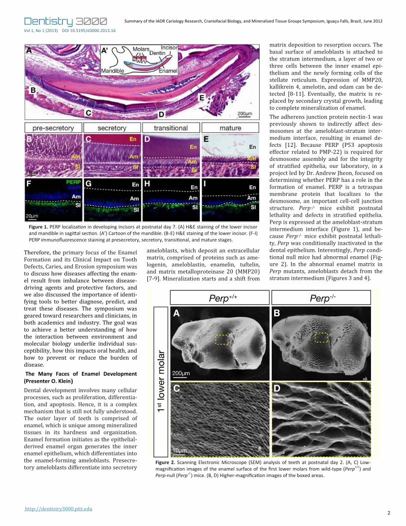
Figure 1. PERP localization in developing incisors at postnatal day 7. (A) H&E staining of the lower incisor and mandible in sagittal section. (A’) Cartoon of the mandible. (B-E) H&E staining of the lower incisor. (F-I) PERP immunofluorescence staining at presecretory, secretory, transitional, and mature stages.
Figure 2. Scanning Electronic Microscope (SEM) analysis of teeth at postnatal day 2. (A, C) Low-magnification images of the enamel surface of the first lower molars from wild-type (Perp+/+) and Perp-null (Perp-/-) mice. (B, D) Higher-magnification images of the boxed areas.
Therefore, the primary focus of the Enamel Formation and its Clinical Impact on Tooth Defects, Caries, and Erosion symposium was to discuss how diseases affecting the enam-el result from imbalance between disease-driving agents and protective factors, and we also discussed the importance of identi-fying tools to better diagnose, predict, and treat these diseases. The symposium was geared toward researchers and clinicians, in both academics and industry. The goal was to achieve a better understanding of how the interaction between environment and molecular biology underlie individual sus-ceptibility, how this impacts oral health, and how to prevent or reduce the burden of disease.
The Many Faces of Enamel Development (Presenter O. Klein) Dental development involves many cellular processes, such as proliferation, differentia-tion, and apoptosis. Hence, it is a complex mechanism that is still not fully understood. The outer layer of teeth is comprised of enamel, which is unique among mineralized tissues in its hardness and organization. Enamel formation initiates as the epithelial-derived enamel organ generates the inner enamel epithelium, which differentiates into the enamel-forming ameloblasts. Presecre-tory ameloblasts differentiate into secretory
ameloblasts, which deposit an extracellular matrix, comprised of proteins such as ame-logenin, ameloblastin, enamelin, tuftelin, and matrix metalloproteinase 20 (MMP20) [7-9]. Mineralization starts and a shift from
matrix deposition to resorption occurs. The basal surface of ameloblasts is attached to the stratum intermedium, a layer of two or three cells between the inner enamel epi-thelium and the newly forming cells of the stellate reticulum. Expression of MMP20, kallikrein 4, amelotin, and odam can be de-tected [8-11]. Eventually, the matrix is re-placed by secondary crystal growth, leading to complete mineralization of enamel.
The adherens junction protein nectin-1 was previously shown to indirectly affect des-mosomes at the ameloblast-stratum inter-medium interface, resulting in enamel de-fects [12]. Because PERP (P53 apoptosis effector related to PMP-22) is required for desmosome assembly and for the integrity of stratified epithelia, our laboratory, in a project led by Dr. Andrew Jheon, focused on determining whether PERP has a role in the formation of enamel. PERP is a tetraspan membrane protein that localizes to the desmosome, an important cell-cell junction structure. Perp-/- mice exhibit postnatal lethality and defects in stratified epithelia. Perp is expressed at the ameloblast-stratum intermedium interface (Figure 1), and be-cause Perp-/- mice exhibit postnatal lethali-ty, Perp was conditionally inactivated in the dental epithelium. Interestingly, Perp condi-tional null mice had abnormal enamel (Fig-ure 2). In the abnormal enamel matrix in Perp mutants, ameloblasts detach from the stratum intermedium (Figures 3 and 4).

Summary of the IADR Cariology Research, Craniofacial Biology, and Mineralized Tissue Groups Symposium, Iguaçu Falls, Brazil, June 2012
Vol 1, No 1 (2013) DOI 10.5195/d3000.2013.16
http://dentistry3000.pitt.edu 3
Perp-mutant incisors had fewer and smaller desmosomes. Several genes involved in amelogenesis (Ambn, Enam, Mmp20, and Klk4) were downregulated in Perp-mutants. These experiments point to a model in which PERP is essential for amelogenesis involving cell-cell adhesion, through desmo-some-mediated interactions between the ameloblasts and the stratum intermedium. These data and additional findings have been published [13].
Enamel Pathogenesis: Biochemistry Lessons (Presenter L.M.A. Tenuta)
Dental enamel is the hardest structure of the human body. It has a repeated structure of prisms in which hydroxyapatite crystals are tightly packed. At the ultrastructural level, the crystalline structure of enamel can be significantly altered, either during its formation, such as in fluorosis, or as a result of the interaction with the oral environ-ment, such as in caries and erosion.
The common fate of dental enamel and teeth has changed during human history. Although in the past the enamel was sub-mitted to abrasive forces due to the rough and hard nature of foods, in the last few centuries, it became more common to see dental enamel completely dissolving due to the action of acids. The big change in the pattern of enamel loss was the result of sugar, which was made highly accessible by man in the last centuries [14]. The problem with sugar is its metabolization by dental biofilm [15], which naturally forms on enamel surface exposed to the oral envi-ronment [16]. Although saliva, and even the fluid phase of dental biofilm, are highly su-
persaturated with respect to dental miner-als [17], upon exposure to fermentable sug-ars, fast and intense pH drop occurs [18]. The pH drop in the biofilm has a direct ef-fect on the stability of hydroxyapatite in enamel. As hydroxyapatite is a phosphate-containing mineral, its solubility increases as the pH decreases [19]. The reduction of the activity product of ions, which compose hydroxyapatite in the biofilm fluid during the low pH, results in the dissolution of sol-id minerals in order to maintain the solubili-ty equilibrium [20]. The demineralization process is followed by a remineralizing pe-riod in which part of minerals dissolved can be replenished [21]. If this cycle frequently continues favoring demineralization, the resulting enamel crystals will gradually dissolve [22,23]. Moreover, when the epi-sodes of exposure to sugar are frequent during the day, acidogenic and aciduric bac-teria will prevail in the biofilm [24]. If the sugar is sucrose, the resulting biofilm is even more cariogenic due to the presence of extracellular polysaccharides that will change the biofilm matrix to a more porous and cariogenic one [25,26].
In the dental caries process, this dissolution is controlled by pores which diffuse acid into enamel, resulting in more severe disso-lution in the subsurface area of enamel [27]. This phenomenon has been attributed to the coupled diffusion of protons inward and of calcium and phosphate outward in enam-el (faster for protons than for calcium and phosphate), to the reprecipitation of miner-als dissolving from the subsurface at the surface, as well as the result of some pro-tecting effects of fluoride on the surface
[28]. The surface of a carious lesion, howev-er, is not intact, but full of dissolution spac-es, which gives the carious enamel a rough and opaque clinical appearance [29].
The great increase in caries rates in parallel to the availability of sucrose in occidental, modern diets culminated with the astonish-ing dental caries rates observed worldwide in the late 60’s. The recently-discovered physicochemical effect of fluoride on miner-al loss, reducing enamel demineralization during a pH drop, and increasing remineral-ization rates by the precipitation of less soluble mineral phases [30] was responsi-ble at that time for the reduction of caries progression even when the cariogenic chal-lenge was maintained [31]. By using fluo-ride in various forms, the worldwide dental caries prevalence considerably declined [2]. Fluoride use in water, as well as in tooth-pastes, has been systematically correlated with decreased dental caries prevalence [32,33]. However, the widespread use of fluoride to control caries has been followed by an increase in prevalence of dental fluo-rosis. Dental fluorosis is a function of the overall chronic exposure to fluoride, and thus, fluoride in water has been related to fluorosis [32]. In regards to fluoride tooth-paste use, the effect on fluorosis is still to be clearly determined [34,35]. Most of all, fluo-rosis as a result of diet, and toothpaste ex-posure is mainly in the mild and very mild degrees, which were shown not to affect the oral health-related quality of life of those affected [36]. For this reason, fluoride-based strategies continue to help us to maintain caries at reduced levels consider-ing our cariogenic diet.
Figure 3. H&E staining of developing mandibular incisors and maxillary molars in wild-type (Perp+/+) and Perp-null (Perp-/-) mice at postnatal day 7 is sagittal sections. (A-D) Low-magnification images of the lower incisor and first upper molar. (A’-D’) Higher magnification images of the boxed areas. Dentin (Dn) and enamel (En) are denoted by yellow and red arrowheads, respectively. Areas of defective enamel are denoted by red asterisks in the incisors and molars of Perp-null mice. P, proximal; D, distal.

Summary of the IADR Cariology Research, Craniofacial Biology, and Mineralized Tissue Groups Symposium, Iguaçu Falls, Brazil, June 2012
Vol 1, No 1 (2013) DOI 10.5195/d3000.2013.16
http://dentistry3000.pitt.edu 4
Figure 4. Displacement of ameloblasts from the stratum intermedium (SI). (A, B) H&E staining of the transitional stage in sagittal sections of incisors at postnatal day 7. Detached cells between ameloblasts and the enamel matrix in Perp-null mice are indicated (red arrowheads). (C, D) DAPI staining of adjacent sections shows the presence of the detached cells (red arrowheads). (E-H) Detached cells express ame-loblast-specific proteins such as ameloblastin (AMBN, red arrowheads) and amelogenin (AMEL; red arrowheads). Matrix vesicles present in both wild-type and Perp-null teeth are indicated (yellow arrow-heads). En, enamel; Am, ameloblasts.
Although fluoride has a history of great suc-cess on caries control, its effect on erosion is still to be proved. The erosive wear, as op-posed to caries, occurs when teeth are ex-posed to very acidic solutions such as soft drinks, juices, or the acid juice from the stomach, which are highly undersaturated in respect to hydroxyapatite. Therefore, minerals would be washed away from the surface of enamel, resulting in a softened etched surface [37]. The etched surface is susceptible to abrasive forces, and enamel loss happens from the surface, layer by lay-er. Fluoride has little effect on preventing mineral loss from erosion, since fluoride-containing minerals are also soluble at the pH levels at which erosion occurs. Although fluoride might have an effect of enhancing remineralization of the eroded surface, the clinical significance is yet to be properly determined.
In an overview of these processes, dental caries is considered a disease dependent of biofilm accumulation with a strong influ-ence of the diet [38]. Saliva and its reminer-alizing capacity and fluoride, which increase enamel remineralization, have positive ef-fects in the reduction of the disease out-come. Other factors, such as virulent bacte-rial species or the composition of the tooth structure, might also affect dental caries progression rates. For instance, it is well known that in the enamel of primary teeth, caries progresses faster as a result of the differential composition of both [39]. In the near future, perhaps we could identify other subtle differences in tooth composition that might also affect the disease.
Dental caries is also influenced by social modifying factors, which today make it a disease of the poor, who have an increasing vulnerability to diseases and less access to prevention policies. It is possible that be-sides the environmental influence on this subject, genetics also play a role on influenc-ing the biological aspects of this disease. Additionally, dental erosion has also been described by interplay of biological, chemi-cal and behavioral factors, and some of these might be influenced by genetics as well.
In summary, for thousands of years the car-iogenic diet has been restricted to starchy products with low fermentable capacity and mineral loss was usually the result of abra-sive forces. With the common availability of sugar since the colonial time, caries flour-ished as a widespread disease in our mod-ern societies. Our diet pattern (processed foods, highly available soft drinks) still sup-ports diseases such as dental caries and erosion. Hopefully, in the future, as long as we understand our intrinsic influences to
these diseases, we can develop technologies to help us better modulate them.
Environmental Factors on Enamel For-mation (Presenter R. Gerlach)
Dental enamel is affected by many insults during its formation (called amelogenesis). The environmental factors most widely known to negatively interfere with amelo-genesis are fevers, hypoxia, undernutrition, and exposure to certain substances that are toxic to enamel cells during enamel devel-opment. Other factors that also influence amelogenesis are antibiotics, environmental pollutants, and socioeconomic status. Such external factors may affect enamel for-mation when cells are secreting the enamel
matrix and/or during the mineralizing pro-cess.
Molar-incisor hypomineralization is a clini-cal entity that exemplifies the possible ef-fects of environmental factors on enamel formation. Whereas a genetic component exists [40], medical issues during prenatal, perinatal, and postnatal stages also lead to enamel hypomineralization, as well as use of medications during the first year of life, and early life exposure to fluorides or envi-ronmental pollutants (dioxins and polychlo-rinated biphenyls or PCBs) [41].
In the continuously growing incisor of rats, fluorotic enamel clinically displays white discoloration that microscopically shows a

Summary of the IADR Cariology Research, Craniofacial Biology, and Mineralized Tissue Groups Symposium, Iguaçu Falls, Brazil, June 2012
Vol 1, No 1 (2013) DOI 10.5195/d3000.2013.16
http://dentistry3000.pitt.edu 5
pattern of repeated white and pigmented bands. These white bands represent hypo-mineralized superficial enamel, but no sub-surface lesions exist [42]. In the presence of lead, clinical alterations due to the chronic exposure to higher levels of fluoride are exacerbated [43]. Like other agents, if the attack on the enamel lasts for a short period of time, there will probably be no visible defects in the enamel. Enamel maturation of permanent teeth lasts up to four years; hence, chronic exposures may lead to clini-cal alterations of enamel. This long matura-tion period probably explains why teeth harbor metal traces to which they were exposed (sodium, chloride, lead). This char-acteristic makes teeth useful for detecting metals in studies of samples from pre-industrial ages, of hypoplasic and hypo-mineralized enamel, caries, and to test hy-pothesis involving the effect of the syner-gism of multiple environmental contami-nants.
Genetic Variation in Enamel Formation Genes (Presenter A.R. Vieira)
Genes responsible for enamel formation have been proposed as potentially involved in caries susceptibility, and positive associa-tions between genetic variation in amelogenin, tuftelin, and enamelin and higher caries experience have been report-ed by our group and others [44-46].
These results, however, are not consistent in all populations. In fact, the only con-sistent result is the lack of association be-tween caries experience and variation in tuftelin interacting protein 11 (Table 1). These latest data are published [47].
One big limitation of these studies is the definition of the phenotype of caries. DMFT/DMFS scores provide a picture of the burden of the disease but little insight into the disease initiation and process.
One approach we proposed was defining caries based on enamel microhardness. We tested enamel microhardness at baseline, after creation of artificial carious lesions, and after fluoride application. As expected, enamel microhardness decreased after cre-ation of artificial caries lesions and then increased to levels similar to baseline after the one-time fluoride treatment and the pH-cycling protocol for 14 days. Lower baseline microhardness was significantly associated with amelogenin (p=0.03 for buccal sur-face), tuftelin (p=0.03 for mesial and p=0.02 for buccal + lingual surfaces), and ameloblastin (p=0.04 for distal surface). After artificial caries creation, lower micro-hardness was significantly associated with tuftelin (p=0.02 for buccal + lingual surfaces and p=0.006 for distal sur-face), enamelin (p=0.02 for distal surface),
and tuftelin interacting protein 11 (p=0.0006 for buccal + lingual and p=0.009 for occlusal surfaces). After fluo-ride treatment, microhardness was signifi-cantly associated with tuftelin (p=0.03 for occlusal surface). The ratio of change of microhardness after pH-cycling treatment was significantly associated with amelogenin (p=0.03 for buccal + lin-gual and p=0.03 for mesial surfac-es), tuftelin (p=0.02 for occlusal sur-face), enamelin (p=0.01 for occlusal sur-face), and tuftelin interacting protein 11 (p=0.04 for buccal + lingual and p=0.009 for occlusal surfaces).
To help overcome the limitations of using DMFT/DMFS scores, we decided to design a series of functional assays to evaluate the response of enamel samples with known genotypes of the genes involved in enamel formation to simulated cariogenic challenges.
Despite the limitation of having samples from several different types of teeth (first, second, and third molars, premolars, canines, and incisors), the results of these experiments suggest that there may be some truth to the popular belief that some individuals may have “weaker” teeth, and hence, are more prone to caries development.
Another observation in our study is that enamel microhardness varies from individ-ual to individual, sometimes substantially. Traditional protocols avoid these variations by eliminating samples that are outside a specific range (i.e., limiting the study to specimens with knoop microhardness val-ues between 350 and 380). Although this methodological approach reduces inter-specimen variation, it also eliminates the chance of interpreting the results in light of individual variation. Our results suggest that the influence of genetic variation of enamel formation genes may influence the dynamic interactions between the enamel
surface and the oral cavity. Components not studied here include biofilm formation (both adhesion to the enamel and matura-tion), and the influence of salivary compo-nents. Despite these limitations, the deter-mination of the presence of specific genetic variants in patients holds the promise for allowing customized treatments that may better impact individual risks for caries.
Acknowledgements The authors are grateful to the IADR Cariol-ogy Research, Craniofacial Biology and Min-eralized Tissue research groups. Jaime A. Cury helped plan the symposium. Colgate provided financial support for the symposi-um. A.R. Vieira is supported by the NIH Grant R01-DE018914.
References 1. Genetic determinants of cranio-facial morphol-
ogy: a twin study. Nakata M, Yu PL, Davis B, Nance WE. Ann Hum Genet 1974;37:431-43.
2. U.S. Department of Health and Human Services. Oral Health in America: A Report of the Surgeon General—Executive Summary. Rockville, MD: U.S. Department of Health and Human Services, National Institute of Dental and Craniofacial Re-search, National Institutes of Health, 2000.
3. World Health Organisation. The World Oral Health Report 2003. Geneva, Switzerland: World Health Organisation, 2003. (Accessed September 15, 2013, at http://www.who.int/oral_health/publications/report03/en/)
4. A report on the NIH Consensus Development Conference on Diagnosis and Management of Dental Caries Throughout Life. Horowitz AM.J Dent Res. 2004;83 Spec No C:C15-7.PMID:15286115
5. Dental enamel formation and its impact on clinical dentistry. Simmer JP, Hu JC. J Dent Educ. 2001 Sep;65(9):896-905. Review. PMID:11569606
6. Molecular mechanisms of dental enamel for-mation. Simmer JP, Fincham AG. Crit Rev Oral Biol Med. 1995;6(2):84-108. Review. PMID:7548623
Table 1. Summary of association results between enamel formation gene variants and caries experience. The only consistent result is the lack of association between caries and tuftelin interacting protein 11.
Gene
Iowa, USA [44]
Tiquisate, Guatemala
[45]
Istanbul, Turkey
[46]
Cebu, Philippines
[47]
Patagonia, Argentina
[47]
Curitiba, Brazil [47]
Rio de Janeiro,
Brazil [47]
ameloblastin - - + + - - -
amelogenin - + + + - - -
enamelin - - +* - - + -
tuftelin +* +# + - + - +
tuftelin interacting protein 11
- - - - - - -
* In the presence of Streptococcus mutans; # Only in less severely affected cases

Summary of the IADR Cariology Research, Craniofacial Biology, and Mineralized Tissue Groups Symposium, Iguaçu Falls, Brazil, June 2012
Vol 1, No 1 (2013) DOI 10.5195/d3000.2013.16
http://dentistry3000.pitt.edu 6
7. Inherited risks for susceptibility to dental car-ies. Shuler CF. J Dent Educ. 2001 Oct;65(10):1038-45. PMID:11699975
8. Cloning, characterization, and tissue expression pattern of mouse tuftelin cDNA. MacDougall M, Simmons D, Dodds A, Knight C, Luan X, Zeich-ner-David M, Zhang C, Ryu OH, Qian Q, Simmer JP, Hu CC. J Dent Res. 1998 Dec;77(12):1970-8. PMID: 9839784
9. Genes and related proteins involved in amelo-genesis imperfecta. Stephanopoulos G, Gare-falaki ME, Lyroudia K. J Dent Res. 2005 Dec;84(12):1117-26. Review. PMID:16304440
10. Enamel formation and amelogenesis imperfec-ta. Hu JC, Chun YH, Al Hazzazzi T, Simmer JP. Cells Tissues Organs. 2007;186(1):78-85. Re-view. PMID:17627121
11. Amelotin--a Novel Secreted, Ameloblast-specific Protein. Iwasaki K, Bajenova E, So-mogyi-Ganss E, Miller M, Nguyen V, Nourkeyhani H, Gao Y, Wendel M, Ganss B. J Dent Res. 2005 Dec;84(12):1127-32. PMID:16304441
12. Characterization of Apin, a secreted protein highly expressed in tooth-associated epithelia. Moffatt P, Smith CE, St-Arnaud R, Nanci A. J Cell Biochem. 2008 Feb 15;103(3):941-56. PMID:17647262
13. The cell adhesion molecule nectin-1 is critical for normal enamel formation in mice. Barron MJ, Brookes SJ, Draper CE, Garrod D, Kirkham J, Shore RC, Dixon MJ. Hum Mol Genet. 2008 Nov 15;17(22):3509-20. doi: 10.1093/hmg/ddn243. Epub 2008 Aug 14. PMID:18703497
14. PERP regulates enamel formation via effects on cell-cell adhesion and gene expression. Jheon AH, Mostowfi P, Snead ML, Ihrie RA, Sone E, Pramparo T, Attardi LD, Klein OD. J Cell Sci. 2011 Mar 1;124(Pt 5):745-54. doi: 10.1242/jcs.078071. Epub 2011 Feb 1. PMID:21285247
15. Public health: The toxic truth about sugar. Lustig RH, Schmidt LA, Brindis CD. Nature. 2012 Feb 1;482(7383):27-9. doi: 10.1038/482027a. PMID:22297952
16. Microbial ecology of dental plaque and its sig-nificance in health and disease. Marsh PD. Adv Dent Res. 1994;8:263-71. PMID:7865085
17. Scanning electron microscopy of early microbial colonization of human enamel and root surfac-es in vivo. Nyvad B, Fejerskov O. Scand J Dent Res. 1987;95:287-96. PMID:3476984
18. Degrees of saturation with respect to apatites in parotid saliva at various ph values. Larsen MJ. Scand J Dent Res. 1975 Jan;83(1):7-12. PMID:237316
19. Intra-oral hydrogen-ion concentrations associ-ated with dental caries activity. Stephan RM. J Dent Res. 1944;23:257-66.
20. Influence of fluoride in solution on tooth de-mineralization. I. Chemical data. ten Cate JM, Duijsters PP. Caries Res. 1983;17:193-9. PMID:6573960
21. Thermodynamic solubility product of human tooth enamel: powdered sample. Patel PR, Brown WE. J Dent Res. 1975 Jul-Aug;54(4):728-36. PMID:239974
22. Alternating demineralization and remineraliza-tion of artificial enamel lesions. ten Cate JM,
Duijsters PP. Caries Res. 1982;16(3):201-10. PMID:6953998
23. Concepts of dental caries and their conse-quences for understanding the disease. Fejer-skov O. Community Dent Oral Epidemiol. 1997 Feb;25(1):5-12. PMID:9088687
24. Changing concepts in cariology: forty years on. Kidd E, Fejerskov O. Dent Update. 2013 May;40(4):277-8, 280-2, 285-6. PMID:23829008
25. Effects of carbohydrate pulses and pH on popu-lation shifts within oral microbial communities in vitro. Bradshaw DJ, McKee AS, Marsh PD. J Dent Res. 1989 Sep;68(9):1298-302. PMID:2674233
26. Physical and biochemical studies of Streptococ-cus mutans sediments suggest new factors link-ing the cariogenicity of plaque with its extracel-lular polysaccharide content. Dibdin GH, Shellis RP. J Dent Res. 1988 Jun;67(6):890-5. PMID:3170900
27. Biochemical composition and cariogenicity of dental plaque formed in the presence of su-crose or glucose and fructose. Cury JA, Rebelo MA, Del Bel Cury AA, Derbyshire MT, Tabchoury CP. Caries Res 2000;34:491-7. PMID:11093024
28. Electron microscopy of fissure caries in man. Frank RM. Arch Oral Biol. 1973 Jan;18(1):9-25. PMID:4513117
29. Assimilation of fluoride by enamel throughout the life of the tooth. Weatherell JA, Deutsch D, Robinson C, Hallsworth AS. Caries Res. 1977;11(Suppl 1):85-115. PMID:318575
30. Thylstrup A, Featherstone JDB, Fredebo L. Sur-face morphology and dynamics of early enamel caries development. In: Leach SA, Edgar WM. Demineralization and remineralization of the teeth. London: IRL Press, 1983:165-84.
31. Current concepts on the theories of the mech-anism of action of fluoride. ten Cate JM. Acta Odontol Scand. 1999 Dec;57(6):325-9. PMID:10777135
32. Fluoride: its role in dentistry. Tenuta LM, Cury JA. Braz Oral Res 2010; 24:Suppl 1: 9-17. PMID:20857070
33. Systematic review of water fluoridation. McDonagh MS, Whiting PF, Wilson PM, Sutton AJ, Chestnutt I, Cooper J, Misso K, Bradley M, Treasure E, Kleijnen J. Brit Med J. 2000;321(7265):855-9. PMID:11021861
34. Fluoride toothpastes for preventing dental caries in children and adolescents. Marinho VCC, Higgins JPT, Logan S, Sheiham A. Cochrane Database Syst Rev 2003, Issue 1. Art. No.: CD002278. PMID:12535435
35. Topical fluoride as a cause of dental fluorosis in children. Wong MC, Glenny AM, Tsang BW, Lo EC, Worthington HV, Marinho VC. Cochrane Da-tabase Syst Rev. 2010 Jan 20;(1):CD007693. doi: 10.1002/14651858.CD007693.pub2. PMID:20091645
36. Effects of low and standard fluoride tooth-pastes on caries and fluorosis: systematic re-view and meta-analysis. Santos AP, Oliveira BH, Nadanovsky P. Caries Res. 2013;47(5):382-90. doi: 10.1159/000348492. PMID:23572031
37. A literature review of aesthetic perceptions of dental fluorosis and relationships with psycho-social aspects/oral health-related quality of life. Chankanka O, Levy SM, Warren JJ, Chalmers JM. Community Dent Oral Epidemiol.
2010 Apr;38(2):97-109. doi: 10.1111/j.1600-0528.2009.00507.x. PMID:20002631
38. Initial erosion models. Young A, Tenuta LM. Caries Res. 2011;45(suppl 1):33-42. PMID:21625131
39. Fejerskov O, Manji F. Risk assessment in dental caries. In: Batler JD. ed. Risk assesstnent in dentistry. Chapel Hill: University of North Caro-lina Dental Ecology, 1990; 215-7.
40. Quantitative determination of type A and type B carbonate in human deciduous and perma-nent enamel by means of Fourier transform in-frared spectrometry. Sønju Clasen AB, Ruyter IE. Adv Dent Res. 1997 Nov;11(4):523-7. PMID:9470513
41. Genes expressed in dental enamel develop-ment are associated with molar-incisor hypo-mineralization. Jeremias F, Koruyucu M, Küchler EC, Bayram M, Tuna EB, Deeley K, Pierri RA, Souza JF, Fragelli CM, Paschoal MA, Gencay K, Seymen F, Caminaga RM, dos Santos-Pinto L, Vieira AR. Arch Oral Biol. 2013 Oct;58(10):1434-42. doi: 10.1016/j.archoralbio.2013.05.005. Epub 2013 Jun 19. PMID:23790503
42. Aetiology of Molar-Incisor Hypomineralisation: A systematic review. Alaluusua S. Eur Arch Pae-diatr Dent. 2010 Apr;11(2):53-8. Review. PMID:20403298
43. Morphological characterization of rat inci-sor fluorotic lesions. Saiani RA, Porto IM, Mar-cantonio Junior E, Cury JA, de Sousa FB, Gerlach RF. Arch Oral Biol. 2009 Nov;54(11):1008-15. doi: 10.1016/j.archoralbio.2009.08.009. Epub 2009 Sep 24. PMID:19781688
44. Exposure to lead exacerbates dental fluorosis. Leite GA, Sawan RM, Teófilo JM, Porto IM, Sou-sa FB, Gerlach RF. Arch Oral Biol. 2011 Jul;56(7):695-702. doi: 10.1016/j.archoralbio.2010.12.011. Epub 2011 Jan 26. PMID:21269604
45. Tuftelin, mutans streptococci, and dental caries susceptibility. Slayton RL, Cooper ME, Marazita ML. J Dent Res. 2005 Aug;84(8):711-4. PMID:16040727
46. Possible association of amelogenin to high caries experience in a Guatemalan-Mayan pop-ulation. Deeley K, Letra A, Rose EK, Brandon CA, Resick JM, Marazita ML, Vieira AR. Caries Res. 2008;42(1):8-13. Epub 2007 Nov 27. PMID:18042988
47. Enamel formation genes are associated with high caries experience in Turkish children. Patir A, Seymen F, Yildirim M, Deeley K, Cooper ME, Marazita ML, Vieira AR. Caries Res. 2008;42(5):394-400. doi: 10.1159/000154785. Epub 2008 Sep 10. PMID:18781068
48. Enamel formation genes influence enamel microhardness before and after cariogenic chal-lenge. Shimizu T, Ho B, Deeley K, Briseño-Ruiz J, Faraco IM Jr, Schupack BI, Brancher JA, Pecharki GD, Küchler EC, Tannure PN, Lips A, Vieira TC, Patir A, Yildirim M, Poletta FA, Mereb JC, Resick JM, Brandon CA, Orioli IM, Castilla EE, Marazita ML, Seymen F, Costa MC, Granjeiro JM, Tre-vilatto PC,Vieira AR. PLoS One. 2012;7(9):e45022. doi: 10.1371/journal.pone.0045022. Epub 2012 Sep 24. PMID:23028741