summary of clinical studies in saphenous …...summary of clinical study in the carotid vasculature...
TRANSCRIPT

50606897-01
TABLE OF CONTENTS
WARNING .......................................................................................................................................1
DEVICE DESCRIPTION...................................................................................................................1Figure 1. Protection Wire..............................................................................................1Table 1. Contents ............................................................................................................1Table 2. Available Sizes.................................................................................................2
INTENDED USE/INDICATIONS FOR USE....................................................................................2
CONTRAINDICATIONS ..................................................................................................................2
WARNINGS .....................................................................................................................................2
PRECAUTIONS................................................................................................................................2
ADVERSE EVENTS .........................................................................................................................2
HOW SUPPLIED..............................................................................................................................2
Handling and Storage ...........................................................................................................2
INSTRUCTIONS FOR USE .............................................................................................................2
Preparation of the Protection Wire and EZ Delivery Sheath: .........................................2
Insertion and Positioning of the FilterWire EZ™ System: ...............................................3Table 3. Minimum Landing Zone Requirements ........................................................3Figure 2. Minimum Landing Zone ................................................................................3
During the Procedure: ...........................................................................................................3
Preparation of the EZ Retrieval Sheath and Filter Capture: ............................................3Figure 3. Retrieved Filter Position inside the EZ Retrieval Sheath.........................3
Removal of the Protection Wire Using the EZ Retrieval Sheath: ...................................3
Repositioning the Filter Using the EZ Retrieval Sheath: ..................................................3
CLINICAL STUDIES FOR THE FILTERWIRE EZ SYSTEM OVERVIEW OF CLINICAL STUDIES IN THE CAROTID VASCULATURE (BEACH AND CABERNET) ................................4
Table 4. Overview of BEACH Trial Study Design .......................................................4
Adverse Events ......................................................................................................................4Table 5. BEACH Trial Major Adverse Events .............................................................4Table 6. BEACH Trial Serious Adverse Events ..........................................................5Table 7. Causes of Death ..............................................................................................5
SUMMARY OF CLINICAL STUDIES IN THE CAROTID VASCULATURE (BEACH) .................5
Eligibility Criteria Summary ..................................................................................................5
Description of Patients Evaluated .......................................................................................5Table 8. BEACH Patient Follow-Up ..............................................................................6Table 9. Baseline Patient Demographics, Lesion Characteristics, and High-Risk Inclusion Criteria .........................................................................................6
Results .....................................................................................................................................6
Table 10. Clinical Results Through 360 Days Follow-Up ..........................................6Figure 4. All Pivotal Patients, Freedom from Morbidity and Mortalitythrough 360 Days ...........................................................................................................7Figure 5. Sympotmatic Patients, Freedom from Morbidity and Mortality through 360 Days ...........................................................................................................7Figure 6. Asymptomatic Patients, Freedom from Morbidity and Mortality through 360 Days ...........................................................................................................7
OVERVIEW OF CLINICAL STUDY IN THE CAROTID VASCULATURE (CABERNET) ..............7Table 11. Overview of CABERNET Trial Study Design..............................................7
Adverse Events ......................................................................................................................8Table 12. CABERNET Major Adverse Events .............................................................8Table 13. CABERNET Trial Serious Adverse Events .................................................8Table 14. Causes of Death ............................................................................................8
SUMMARY OF CLINICAL STUDY IN THE CAROTID VASCULATURE (CABERNET) ........................................................................................................8
Inclusion Criteria ....................................................................................................................8
Exclusion Criteria ...................................................................................................................9
Description of Patients Evaluated .......................................................................................9Table 15. Patient Enrollment and Disposition ............................................................9Table 16. Baseline Patient Demographics .................................................................9Table 17. Patient High-Risk Inclusion Criteria .........................................................10Table 18. Baseline Lesion Characteristics (Pre-Procedure) ................................10
Trial Results...........................................................................................................................10Table 19. Primary Endpoints (Major Adverse Events) ............................................10Table 20. Secondary Endpoints .................................................................................10
Additional Analysis ..............................................................................................................10
Endpoint Analysis ................................................................................................................10Figure 7. Survival from Major Adverse Events (All Deaths, Strokes and MIs) through 1 Year. ..............................................................................................................11Figure 8. Survival from Major Adverse Events (All Deaths, Strokes and MIs) at 30 Days; Plus Ipsilateral Stroke or Death Related to Ipsilateral Stroke through 1 Year. ..............................................................................................................11Figure 9. Survival from Major Adverse Events (All Deaths, Strokes and MIs)at 1 Year Symptomatic Patient Group .......................................................................11Figure 10. Survival from Major Adverse Events (All Deaths, Strokes and MIs) at 1 Year Asymptomatic Patient Group ....................................................................11Figure 11. Survival from Major Adverse Events (All Deaths, Strokes and MIs) at 30 Days Plus Ipsilateral Stroke or Death Related to Ipsilateral Stroke through 1 Year Symptomatic Patient Group ............................................................11Figure 12. Survival from Major Adverse Events (All Deaths, Strokes and MIs) at 30 Days Plus Ipsilateral Stroke or Death Related to Ipsilateral Stroke through 1 Year Asymptomatic Patient Group ..........................................................11
Study Conclusions ..............................................................................................................11
OVERVIEW OF CLINICAL STUDIES IN SAPHENOUS VEIN BYPASS GRAFTS (BLAZE II, BLAZE, AND FIRE) ........................................................................................................................12
Clinical Trial Comparison ............................................................................................12
ADVERSE EFFECTS ......................................................................................................................12Table 21. Major Adverse Events– In–and Out–of–Hospital (To 30 days) – FilterWire EZ System (2.25 mm - 3.5 mm) in the BLAZE II Study Compared to Subset of GuardWire Plus® Device patients from FIRE Trial (BLAZE II Study and FIRE Trial Data are listed as per Patient) ............................13Table 22. Major Adverse Events – In-and-Out-of-Hospital (To 30 days) – FilterWire EZ System (3.5 mm - 5.5 mm) in the BLAZE Study and FilterWire EX®
System in the FIRE Trial Comparison (Patients Treated in BLAZE Study and FIRE Trial Data listed as per Patient) ........................................................................14
SUMMARY OF CLINICAL STUDIES IN SAPHENOUS VEIN BYPASS GRAFTS (BLAZE II, BLAZE, AND FIRE) ........................................................................................................................14
FilterWire EZ System (2.25 mm - 3.5 mm) .........................................................................14
Inclusion/Exclusion Criteria ...............................................................................................14
Exclusion Criteria .................................................................................................................14Table 23. Revised analysis imputing 1 additional MACE. ......................................15Table 24. Principal Effectiveness and Safety Results - Vessel Diameter per QCA. ...............................................................................................................................15
SUMMARY OF CLINICAL STUDIES IN SAPHENOUS VEIN BYPASS GRAFTS (BLAZE II, BLAZE, AND FIRE) ........................................................................................................................15
FilterWire EZ System (2.25 mm - 3.5 mm) .........................................................................15
Inclusion/Exclusion Criteria ...............................................................................................15
Exclusion Criteria .................................................................................................................15Table 23. Revised analysis imputing 1 additional MACE. ......................................15Table 24. Principal Effectiveness and Safety Results - Vessel Diameter per QCA. ...............................................................................................................................16Table 25. Results: FilterWire EZ System (2.25 mm – 3.5 mm) in the BLAZE II Study compared with GuardWire Plus Device (2.25 mm – 3.5 mm) subset from the FIRE Trial ........................................................................................................16Table 26. Myocardial Infarction Classification – FilterWire EZ System (2.25 mm – 3.5 mm) in the BLAZE II Study compared with the GuardWire Plus Device (2.25 mm – 3.5 mm) subset from the FIRE Trial .............16Table 27. FilterWire EZ System (2.25 mm – 3.5 mm) in the BLAZE II Study compared to the GuardWire Plus Device (2.25 mm – 3.5 mm) System subset from the FIRE Trial With IIb/IIIa Inhibitors ...............................................................16Table 28. FilterWire EZ System (2.25 mm – 3.5 mm) in the BLAZE II Study compared to the GuardWire Plus Device (2.25 mm – 3.5 mm) subset fromthe FIRE Trial Without IIb/IIIa Inhibitors ...................................................................17Table 29. Quantitative Coronary Angiography – FilterWire EZ System (2.25 mm – 3.5 mm) in the BLAZE II Study compared to the GuardWire Plus Device (2.25 mm – 3.5 mm) subset from the FIRE Trial ..........................................17BLAZE II - Cumulative Distribution of CK-MB Elevations......................................17
FilterWire EZ System (3.5 mm - 5.5 mm) ...........................................................................17
Inclusion/Exclusion Criteria ...............................................................................................17Table 30. Results: FilterWire EZ System (3.5 mm - 5.5 mm) in the BLAZE Study Compared with FilterWire EX System in the FIRE Trial .................18Table 31. Myocardial Infarction Classification – FilterWire EZ System (3.5 mm - 5.5mm) in the BLAZE Study Compared with FilterWire EX System in the FIRE Trial .............................................................................................................19Table 32. Major Adverse Cardiac Events (By IIB/IIIA Antagonist Use) – FilterWire EZ System (3.5 mm - 5.5 mm) in the BLAZE Study Compared with FilterWire EX System in the FIRE Trial ......................................................................19Table 33. Quantitative Coronary Angiography – FilterWire EZ System (3.5 mm - 5.5 mm) in the BLAZE Study Compared with FilterWire EX System in the FI .......................................................................19BLAZE - Cumulative Distribution of CK-MB Elevations .........................................19
FilterWire EX System ...........................................................................................................19Figure 13. FIRE Trial Design ........................................................................................19
Inclusion/Exclusion Criteria ...............................................................................................20FIRE - Cumulative Distribution of CK-MB Elevations .............................................20
WARRANTY ..................................................................................................................................20
Bo
sto
n S
cien
tifi
c (M
aste
r B
ran
d D
FU Te
mp
late
8.2
677i
n x
11.
6929
in A
4, 9
0105
918A
W),
eD
FU, M
B, F
ilter
Wir
e E
Z, E
N, 5
0606
897-
01A
Black (K) ∆E ≤5.0
ONLYCaution: Federal Law (USA) restricts this device to sale by or on the order of a physician.
WARNING
Contents supplied STERILE using Radiation process. Do not use if sterile barrier is damaged. If damage is found, call your Boston Scientific representative.
For single use only. Do not reuse, reprocess or resterilize. Reuse, reprocessing or resterilization may compromise the structural integrity of the device and/or lead to device failure which, in turn, may result in patient injury, illness or death. Reuse, reprocessing or resterilization may also create a risk of contamination of the device and/or cause patient infection or cross-infection, including, but not limited to, the transmission of infectious disease(s) from one patient to another. Contamination of the device may lead to injury, illness or death of the patient.
After use, dispose of product and packaging in accordance with hospital, administrative and/or local government policy.
DEVICE DESCRIPTION
The Boston Scientific FilterWire EZ Embolic Protection System is a temporary intravascular 0.014 in (0.36 mm) guidewire filtration system that is placed distal to the target lesion to be treated by interventional procedures. The system consists of a protection wire, an EZ Delivery Sheath, an EZ Retrieval Sheath and accessories. When deployed, the protection wire’s filter bag is designed to contain and remove embolic material that may be liberated during the interventional procedure. The protection wire is used as a standard 0.014 in (0.36 mm) steerable guidewire. The spring coil tip of the protection wire and the filter loop are radiopaque to enable visual guidance during placement. At the completion of the procedure, the filter is captured using the EZ Retrieval Sheath and then removed from the patient.
Protection Wireexiting DeliverySheath
Delivery Sheath
Catheter Stop
Spinner Tube
Filter Loop
Filter Bag NoseconeSpringCoil Tip
Figure 1. Protection Wire
Table 1. Contents
Quantity Description1 each Protection Wire1 each EZ Delivery Sheath1 each EZ Retrieval Sheath1 each Accessory Tool Kit (One Wire Torquer, One Peel-away
Introducer, One Valve Dilator Tool)
FilterWire EZ™
Embolic Protection System
2017-11< EN >

2
Bo
sto
n S
cien
tifi
c (M
aste
r B
ran
d D
FU Te
mp
late
8.2
677i
n x
11.
6929
in A
4, 9
0105
918A
W),
eD
FU, M
B, F
ilter
Wir
e E
Z, E
N, 5
0606
897-
01A
Black (K) ∆E ≤5.0
Table 2. Available Sizes
ProtectionWire Length
Vessel Diameter
Clinical Applications
Clinical Study Evaluation
(see Clinical Studies Section)
190 cm or
300 cm
2.25 mm - 3.5 mm
Saphenous Vein Bypass Grafts BLAZE II Study
3.5 mm - 5.5 mm
Saphenous Vein Bypass Grafts;
Carotid Arteries
BLAZE Study; BEACH Study
Caution: To prevent overstretching and potential dissection of the artery, ensure the device selected matches the reference vessel diameter.
Note: The 190 cm protection wire is compatible with the Boston Scientific AddWire® Extension Wire (catalog number 22150-01). The EZ Bent Tip Retrieval Sheath (catalog number 50100-150), an alternate retrieval sheath, is available separately.
INTENDED USE/INDICATIONS FOR USE
The Boston Scientific FilterWire EZ™ Embolic Protection System is indicated for use as a guidewire and embolic protection system to contain and remove embolic material (thrombus/debris) while performing angioplasty and stenting procedures in coronary saphenous vein bypass grafts and carotid arteries. The diameter of the vessel at the site of filter loop placement should be between 2.25 mm and 5.5 mm for coronary saphenous vein bypass graft procedures and between 3.5 mm and 5.5 mm for carotid procedures.
The safety and effectiveness of this device as an embolic protection system have not been established in the cerebral vasculature, peripheral vessels other than carotid arteries, or in treating native coronaries, including acute myocardial infarction.
CONTRAINDICATIONS
• Patients with severe allergy to heparin
• Patients with bleeding diathesis or other disorders that limit the use of anticoagulant therapy
WARNINGS
• Only physicians experienced in percutaneous intravascular techniques and procedures should use the FilterWire EZ Embolic Protection System.
• The safety and effectiveness of coronary drug-eluting stents (DES) when used with embolic protection devices have not been established.
• The safety and effectiveness of the FilterWire EZ Embolic Protection System have not been demonstrated with carotid stents other than the Carotid WALLSTENT® Monorail® Endoprosthesis and Delivery System (Carotid WALLSTENT Endoprosthesis) and the NexStent® Carotid Stent and Monorail Delivery System (NexStent Carotid Stent).
• The appropriate antiplatelet and anticoagulation therapy should be administered pre- and post-procedure to minimize the risk of embolism and thrombosis.
• Avoid using power injection in the cerebral circulation.
• Filter safety and effectiveness has not been tested with power injection.
• Failure to follow recommended device preparation and delivery instructions may result in air embolism or other adverse reaction.
• Introduce and advance devices slowly to prevent air embolism or trauma to the vasculature.
• Do not attempt to move the protection wire without observing the resultant tip response.
• All distal wire tips have the potential to cause vessel injury. Confirm that the wire tip is free within the vessel.
• Do not use excessive force when attempting to cross the lesion with the FilterWire EZ System.
• Observe all protection wire movement in the vessels under fluoroscopic imaging.
• Always keep the open filter loop distal to a deployed stent. Pulling the filter loop into the stent area may lead to entanglement with the stent and possible filter loop detachment.
• Ensure that the protection wire is stabilized throughout the procedure. Failure to stabilize the protection wire could lead to inadvertent movement of the filter, resulting in protection wire entanglement and/or delay in the procedure.
• Do not pull excessively on the protection wire or the EZ Retrieval Sheath to avoid filter bag tears, filter loop detachment or other protection wire damage.
PRECAUTIONS
• Do not autoclave or expose the FilterWire EZ Embolic Protection System to organic solvents.
• Confirm that the interventional devices to be used are compatible with the 0.014 in (0.36 mm) diameter protection wire before actual use.
• For SVG applications, prior to use, heparinize the patient to achieve an Activated Clotting Time (ACT) of >300 seconds (>200 seconds if using
GP IIb/IIIa inhibitors) or in absence of ACT measurement, administer an appropriate weight based bolus of heparin (approximately 125 units/kg).
• For carotid procedures, administer heparin and monitor ACT to maintain >275 seconds throughout the procedure.
• Venous access should be available in order to manage bradycardia and/or hypotension by either pharmaceutical intervention or placement of a temporary pacemaker, if needed.
• Prior to use, inspect the protection wire and sheaths for damage (e.g. bends, kinks, filter tears). Do not use the FilterWire EZ System if damaged. Guidewires are delicate instruments and should be handled carefully.
• Maintain the sterile contents of the pouch (including the coils and accessory tool kit) in the sterile field during the procedure. Do not store the accessory tool kit for future use.
• Confirm angiographically that the vessel diameter is appropriate for treatment with the FilterWire EZ System and ensure that the correct device size is selected. The reference vessel diameter at the filter loop deployment site must be at least 2.25 mm but no greater than 3.5 mm for the FilterWire EZ System (2.25 mm - 3.5 mm) and at least 3.5 mm but no greater than 5.5 mm for the FilterWire EZ System (3.5 mm - 5.5 mm).
• To prevent overstretching and potential dissection of the artery, ensure the device selected matches the reference vessel diameter.
• Confirm angiographically that there is sufficient vessel length to place the filter to allow for and maintain adequate distance between the protection wire’s catheter stop and the stent delivery system or other compatible interventional devices (see Table 3 and Figure 2).
• Minimum inner diameter of a guide catheter or guide sheath must be 0.066 in (1.68 mm).
• Minimum inner diameter bore of a rotating hemostasis valve must be 0.075 in (1.91 mm) to accommodate use of the peel-away introducer supplied with the FilterWire EZ Embolic Protection System.
• Use of a guide catheter with a rotating hemostasis valve is recommended when performing a procedure with the FilterWire EZ Embolic Protection System.
• When using a rotating hemostasis valve, ensure that the hemostasis valve is sufficiently opened prior to insertion of any device.
• A stent that is not well apposed to the vessel wall can lead to difficulty crossing with the retrieval sheath and may increase the risk of filter entanglement with the stent.
• Ensure the stent selected matches the diameter of the vessel being treated to maximize the potential for adequate stent apposition to the vessel wall.
• Ensure adequate guide catheter or guide sheath support and position during the interventional procedure. Guide catheter or guide sheath movement can cause the filter to be inadvertently pulled proximally in the vessel.
• During the procedure, always attempt to keep the filter loop, lesion site, and radiopaque tip of the guide catheter or guide sheath in the field of view, minimizing the likelihood of the guide catheter or guide sheath backing out or prolapsing into the aortic arch.
• Prolapse of the guide catheter or guide sheath can result in movement of an open filter through an untreated lesion, filter/stent entanglement, filter basket detachment, proximal movement of the stent, or protection wire damage.
• Always advance or retract the FilterWire EZ Embolic Protection System slowly under fluoroscopic imaging. If resistance is felt and/or observed, determine the cause and take any necessary remedial action.
• Advancing or torquing the FilterWire EZ System against resistance could result in vessel trauma.
• Always tighten the wire torquer appropriately to the protection wire. Over-tightening of the wire torquer may cause difficulty/inability to remove the wire torquer from the protection wire. Under-tightening of the wire torquer may lead to improper filter deployment.
• Always advance/retract the wire torquer carefully along the protection wire to prevent kinking.
• Keep the wire position stable by using the wire torquer while the filter is deployed.
• In vitro testing for compatibility of the FilterWire EZ System with coronary drug-eluting stents (DES) indicates that the DES coating should not be compromised upon advancing or retracting the FilterWire EZ System; however, care should still be taken not to disrupt the drug coating if a coronary DES has been recently placed.
• To address lesions in subsequent vessels, use a new FilterWire EZ Embolic Protection System for each vessel.
• Do not use the FilterWire EZ System if the yellow protective housing is not clipped to the coil and the filter and spring coil tip are exposed.
• Do not use the FilterWire EZ System if the retention clip is not clipped onto the coil with the delivery sheath secured to it.
• If a buddy wire is used to facilitate crossing the lesion, ensure that the buddy wire does not wrap around the protection wire.
• If a buddy wire is used, leaving the buddy wire in place during lesion treatment can lead to vessel trauma or accidental entrapment of the protection wire.
• To avoid device damage, do not pre-dilate the lesion with the sheathed FilterWire EZ System in the vicinity of the lesion.
• During deployment, failure to position the wire torquer against the hemostasis valve and secure it to the protection wire may lead to wire kinking and/or filter movement during deployment.
• If blood flow is slowed during the procedure, the protection wire may be removed and replaced, if necessary. However, at the operator’s discretion, the procedure may be completed before removing the protection wire.
• If there is no blood flow, the protection wire should be removed and replaced (unless the procedure is complete).
• During FilterWire EZ System retrieval, do not rotate the EZ Retrieval Sheath more than 90 degrees in either direction; this can cause the protection wire to wrap around the EZ Retrieval Sheath shaft and prevent capture of the filter.
• Always use a FilterWire EZ System compatible retrieval sheath to remove the protection wire. Pulling an open filter through a stent should be a last resort for retrieval as it may lead to entanglement with the stent and possible filter loop detachment.
• Use caution when advancing or retracting the FilterWire EZ System through a deployed stent as this may cause stent/filter entanglement or stent dislocation.
• Use of a guide sheath with a fixed hemostasis valve requires use of a valve dilator tool. The use of a guide sheath with a fixed hemostasis valve may cause the filter bag to tear at the hemostasis valve upon removal.
ADVERSE EVENTS
Possible adverse events associated with FilterWire EZ Embolic Protection System use and application procedure include, but are not limited to, the following:
• Angina
• Bleeding complications
• Bradycardia or arrhythmias, including ventricular fibrillation or tachycardia
• Congestive heart failure
• Damage to or dislocation of the implanted stent(s)
• Death
• Detachment and/or implantation of a component of the system
• Drug reaction, allergic reaction to contrast media, medications or device materials
• Embolization of air, tissue, thrombus or other embolic debris
• Emergent surgery
• End organ ischemia/infarction
• Headache
• Hypotension/hypertension
• Infection (local or systemic)
• Myocardial infarction
• No-reflow resulting from reduced blood flow through the FilterWire EZ System filter
• Pain
• Puncture site complications (i.e., vessel occlusion, hemorrhage, hematoma, pseudoaneurysm or arteriovenous fistula)
• Renal insufficiency, kidney failure, hematuria
• Stroke/cerebrovascular accident (CVA), transient ischemic attack (TIA) or seizure
• Vessel damage, dissection, occlusion, aneurysm, perforation, rupture, injury, thrombosis, or spasm
Adverse events experienced during clinical studies are presented in the Clinical Study Overview sections.
HOW SUPPLIED
• Do not use if package is opened or damaged.
• Do not use if labeling is incomplete or illegible.
Handling and Storage
• Store in a cool, dry, dark place.
• Use the device prior to the “Use By” date noted on the box and pouch
INSTRUCTIONS FOR USE
Preparation of the Protection Wire and EZ Delivery Sheath:
1. Open the pouch using standard sterile handling procedures and place the packaging coils and accessory tool kit into the sterile field.
2. Unclip and carefully remove the yellow protective housing from the filter.
Note: The protection wire is preloaded into the EZ Delivery Sheath.
Caution: Do not use the FilterWire EZ System if the yellow protective housing is not clipped to the coil and the filter and spring coil tip are exposed.
3. Remove the preloaded protection wire from the retaining clip; advance the EZ Delivery Sheath until the clear section is exposed; then, grasp the clear section of the sheath, and remove the preloaded protection wire from the packaging coil.
Caution: Do not use the FilterWire EZ System if the retention clip is not clipped onto the coil with the delivery sheath secured to it.
4. Retain the packaging coil on the sterile field to ensure availability of the EZ Retrieval Sheath when needed.
5. Attach the wire torquer to the protection wire near the exit port. This will facilitate retraction of the filter into the EZ Delivery Sheath during preparation.
6. Grasp the EZ Delivery Sheath towards the very distal end of the clear section and submerge the filter and sheath in heparinized saline.
7. While the filter and delivery sheath are submerged in the heparinized saline, sheath the protection wire by slowly retracting it into the EZ Delivery Sheath until the nose cone is partially retracted into the sheath.

3
Bo
sto
n S
cien
tifi
c (M
aste
r B
ran
d D
FU Te
mp
late
8.2
677i
n x
11.
6929
in A
4, 9
0105
918A
W),
eD
FU, M
B, F
ilter
Wir
e E
Z, E
N, 5
0606
897-
01A
Black (K) ∆E ≤5.0
Note: Do not over-retract the filter.
Note: Do not sheath the filter until immediately prior to use. Make certain that the filter is thoroughly saturated with saline prior to retracting the filter into the EZ Delivery Sheath.
Insertion and Positioning of the FilterWire EZ™ System:
Warning: Introduce and advance devices slowly to prevent air embolism or trauma to the vasculature.
1. Carefully insert the sheathed protection wire tip into the peel-away introducer. If desired, shape the spring coil tip of the protection wire. In order to prevent spring coil tip damage, ensure the tip is retracted inside the peel-away introducer.
2. Advance the FilterWire EZ System and peel-away introducer assembly into the hemostasis valve attached to a guide catheter or guide sheath.
3. Carefully remove the peel-away introducer.
4. Advance the FilterWire EZ System through the guide catheter or guide sheath.
Note: Carefully advance both the EZ Delivery Sheath and protection wire together to prevent inadvertent deployment of the filter.
Warning: Do not attempt to move the protection wire without observing the resultant tip response.
Warning: All distal wire tips have the potential to cause vessel injury. Confirm that the wire tip is free within the vessel.
5. Under fluoroscopic imaging, steer the FilterWire EZ System into the target vessel. Using a two-handed technique, torque the protection wire with one hand and advance the EZ Delivery Sheath with the other hand.
Note: Always advance the protection wire and EZ Delivery Sheath together to prevent inadvertent deployment.
Note: Do not allow the protection wire to wrap around the EZ Delivery Sheath shaft to ensure the filter can be deployed smoothly.
Note: Consider using a “buddy wire” or pre-dilate the lesion if it is tight and difficult to pass.
Caution: If a buddy wire is used, ensure that the buddy wire does not wrap around the protection wire.
Caution: If a buddy wire is used, leaving the buddy wire in place during lesion treatment can lead to vessel trauma or accidental entrapment of the protection wire.
Caution: To avoid device damage, do not pre-dilate the lesion with the sheathed FilterWire EZ System in the vicinity of the lesion.
6. Advance the FilterWire EZ System across the lesion until the apex of the filter loop can be deployed in the recommended minimum landing zone (see Table 3 and Figure 2). This distance should be maintained throughout the procedure.
Warning: Do not use excessive force when attempting to cross the lesion with the FilterWire EZ System.
Note: If extra landing zone length is available, placing the filter loop beyond the minimum distance is desirable.
Table 3. Minimum Landing Zone Requirements
Minimum Landing Zone should be:FilterWire EZ System (2.25 mm - 3.5 mm) ≥2.5 cm
FilterWire EZ System (3.5 mm - 5.5 mm) ≥3.0 cm
Note: Either the FilterWire EZ System (2.25 mm – 3.5 mm) or the FilterWire EZ System (3.5 mm – 5.5 mm) may be used if the vessel diameter is 3.5 mm at the deployment site. If less than 3.0 cm of vessel is available for the landing zone, consider the FilterWire EZ System (2.25 mm – 3.5 mm). If there is a possibility that the vessel diameter is underestimated due to slow flow or if there is a possibility of vessel expansion during the procedure, consider the FilterWire EZ System (3.5 mm – 5.5 mm).
LesionSee Table 3 for Minimum Landing Zone Requirements
Guide wire / bare wireCatheter Stop
Figure 2. Minimum Landing Zone
Note: If an interventional device with a tip-to-shoulder length greater than 10 mm is to be used, a longer vessel length and landing zone may be required to place the protection wire in order to avoid contact between the protection wire’s catheter stop and the tip of the interventional device.
7. Once the protection wire is advanced past the lesion, slide the wire torquer along the protection wire and secure it against the hemostasis valve.
8. Deploy the filter by holding the protection wire in place with the wire torquer pressed against the hemostasis valve while simultaneously retracting the EZ Delivery Sheath.
Caution: Failure to position the wire torquer against the hemostasis valve and secure it to the protection wire may lead to wire kinking and/or filter movement during deployment.
9. Continue retracting the EZ Delivery Sheath, peeling the sheath away until the clear section of the EZ Delivery Sheath is encountered. Remove the wire torquer and carefully remove the remaining distal segment of the EZ Delivery Sheath from the wire while maintaining protection wire stability.
10. Inject contrast media to verify the protection wire is in the proper position and that there is adequate flow.
Note: If repositioning is required, refer to “Repositioning the Filter Using the EZ Retrieval Sheath” section.
During the Procedure:
1. Use the protection wire like a traditional guidewire to track catheters and stent delivery systems to the target treatment site. Carefully maintain the position of the filter, particularly during device exchanges.
Warning: Observe all protection wire movement in the vessels under fluoroscopic imaging.
Warning: Always keep the open filter loop distal to the deployed stent. Pulling the filter loop into the stent area may lead to entanglement with the stent and possible filter loop detachment.
Warning: Ensure that the protection wire is stabilized throughout the procedure. Failure to stabilize the protection wire could lead to inadvertent movement of the filter, resulting in protection wire entanglement and/or delay in the procedure.
Note: Inject contrast after any intervention or exchange. Visually verify that blood flow is not obstructed and that filter loop/vessel wall apposition is maintained. Reposition if necessary.
Caution: If blood flow is slowed during the procedure, the protection wire may be removed and replaced. However, at the operator’s discretion, the procedure may be completed before removing the protection wire.
Caution: If there is no blood flow, the protection wire should be removed and replaced if further intervention is required.
2. Carefully remove all interventional devices, ensuring that the protection wire is not kinked and its position is maintained during removal.
Preparation of the EZ Retrieval Sheath and Filter Capture:
1. Remove the EZ Retrieval Sheath from its packaging coil by grasping it at the white proximal handle and pulling it carefully out of the packaging coil, taking care not to damage the EZ Retrieval Sheath.
2. Flush the EZ Retrieval Sheath with heparinized saline. Be careful not to damage the EZ Retrieval Sheath tip with the syringe.
Note: If the EZ Retrieval Sheath was used for repositioning, flush it again with heparinized saline and clean the outer surface before using it for filter retrieval.
Warning: Observe all protection wire movement in the vessels under fluoroscopic imaging.
3. While maintaining the protection wire position distal to the lesion, advance the EZ Retrieval Sheath over the protection wire past any deployed stent(s) until the tip of the EZ Retrieval Sheath reaches the protection wire’s catheter stop.
Caution: Do not rotate the EZ Retrieval Sheath more than 90 degrees in either direction; this can cause the protection wire to wrap around the EZ Retrieval Sheath shaft and prevent capture of the filter.
Caution: Always use a FilterWire EZ System compatible retrieval sheath to remove the protection wire. Pulling an open filter through a stent should be a last resort for retrieval as it may lead to entanglement with the stent and possible filter loop detachment.
Note: If it is difficult to cross a deployed stent or tortuous vessel anatomy with the EZ Retrieval Sheath, the EZ Bent Tip Retrieval Sheath (available separately) may be used.
4. Slide the wire torquer along the protection wire and secure it against the hemostasis valve. Gently and slowly retract the protection wire and filter loop back into the EZ Retrieval Sheath until resistance is felt.
Warning: Do not pull excessively on the protection wire to avoid filter bag tears, filter loop detachment, or other protection wire damage.
Note: The distal edge of the collapsed filter loop should align with, or be proximal to, the EZ Retrieval Sheath marker band (see Figure 3).
Internal StopFilter Loop
Retrieval Sheath Marker Band
Figure 3. Retrieved Filter Position inside the EZ Retrieval Sheath
Note: If the distal edge of the collapsed filter loop is not aligned with, or proximal to, the EZ Retrieval Sheath marker band, it is possible that the filter is overfilled. Carefully retract the protection wire and EZ Retrieval Sheath together as a system.
Note: If repositioning is required, use the EZ Retrieval Sheath. Refer to “Repositioning the Filter Using the EZ Retrieval Sheath” section for instructions.
Removal of the Protection Wire Using the EZ Retrieval Sheath:
1. Slowly and carefully retract the entire system as a unit until the tip of the EZ Retrieval Sheath is adjacent to the tip of the guide catheter or guide sheath.
Caution: Use caution when advancing or retracting the FilterWire EZ System through a deployed stent as this may cause filter/stent entanglement or stent dislocation.
Warning: Do not pull excessively on the protection wire or the EZ Retrieval Sheath to avoid filter bag tears, filter loop detachment, or other protection wire damage.
Note: If any resistance is met during retraction of the sheathed protection wire and filter, slightly advance the sheathed protection wire and rotate the EZ Retrieval Sheath before continuing to retract.
2. Carefully retract the FilterWire EZ System across the guide catheter or guide sheath up to the hemostasis valve.
Note: If there is any resistance at the guide catheter or guide sheath, retract the guide catheter or guide sheath and FilterWire EZ System together.
3. Remove the FilterWire EZ System from the patient, ensuring that the hemostasis valve is fully opened prior to FilterWire EZ System removal. If a fixed hemostasis valve is used, place the valve dilator tool on the shaft of the retrieval sheath and slide the tool into the valve. Then, remove the FilterWire EZ System from the patient through the valve dilator tool. Remove the valve dilator tool.
Caution: Use of a guide sheath with a fixed hemostasis valve requires use of a valve dilator tool. The use of a guide sheath with a fixed hemostasis valve may cause the filter bag to tear at the hemostasis valve upon removal.
Repositioning the Filter Using the EZ Retrieval Sheath:
1. Prepare the EZ Retrieval Sheath and capture the filter according to the instructions outlined in the “Preparation of the EZ Retrieval Sheath and Filter Capture” section.
Note: Observe all protection wire movement in the vessels under fluoroscopic imaging.
2. Guide the EZ Retrieval Sheath and protection wire assembly to the desired location. Make sure to advance both the EZ Retrieval Sheath and the protection wire as a system to ensure that the protection wire does not prematurely deploy.
3. If needed, the spring coil tip of the protection wire can be steered by turning the wire torquer previously installed on the protection wire.
Note: Do not allow the protection wire to wrap around the EZ Retrieval Sheath shaft to ensure that the filter can be deployed smoothly.
Note: If it is difficult to advance or reposition the protection wire, this may be the result of a full filter. Care must be taken not to damage the filter.
4. Advance the FilterWire EZ System until the apex of the filter loop can be deployed in the recommended minimum landing zone (see Table 3 and Figure 2). This distance should be maintained throughout the procedure.
Note: Deploy the filter as distal as possible to the lesion.
5. Once the protection wire is in the desired location, slide the wire torquer along the protection wire and secure it against the hemostasis valve.
6. Deploy the filter by holding the protection wire in place with the wire torquer pressed against the hemostasis valve while simultaneously retracting the EZ Retrieval Sheath.
7. Ensure that the filter has been deployed in the desired location. Remove the wire torquer and the EZ Retrieval Sheath from the patient while holding the protection wire in place.
8. Inspect the EZ Retrieval Sheath for damage. If no damage is noted, set it aside for eventual retrieval and removal of the protection wire.

4
Bo
sto
n S
cien
tifi
c (M
aste
r B
ran
d D
FU Te
mp
late
8.2
677i
n x
11.
6929
in A
4, 9
0105
918A
W),
eD
FU, M
B, F
ilter
Wir
e E
Z, E
N, 5
0606
897-
01A
Black (K) ∆E ≤5.0
CLINICAL STUDIES FOR THE FILTERWIRE EZ SYSTEM OVERVIEW OF CLINICAL STUDIES IN THE CAROTID VASCULATURE (BEACH AND CABERNET)
BEACH (Boston Scientific EPI: A Carotid Stenting Trial for High-Risk Surgical Patients) was a prospective, single-arm, multi-center trial to evaluate the safety and efficacy of the Carotid WALLSTENT® Monorail® Endoprosthesis in conjunction with the FilterWire EX®/FilterWire EZ™ Embolic Protection System (FilterWire EX/EZ System) to treat high-surgical-risk, symptomatic (≥50% stenosis) and asymptomatic (≥80% stenosis) patients with disease in the carotid artery. The primary objective of the trial was to show non-inferiority between carotid stenting and a historical control representative of outcomes with carotid endarterectomy, based upon the 1-year morbidity and mortality rate including non Q-wave MI through 24 hours; death, stroke, and Q-wave MI through 30 days; and ipsilateral stroke and neurologic death from 31 to 360 days. A total of 747 patients were enrolled at 47 centers in the United States, including 189 roll-in patients, 480 pivotal patients and 78 bilateral registry patients. This trial is summarized in Table 4.
Table 4. Overview of BEACH Trial Study Design
Product Evaluated:Carotid WALLSTENT Endoprosthesis and FilterWire EX System/FilterWire EZ SystemSample Size for Pivotal Patients: 480Number of Centers: 47Primary Endpoint: 1-year morbidity and mortality:Non Q-wave MI through 24 hoursDeath, Stroke, Q-wave MI through 30 daysIpsilateral Stroke, Neurologic Death 31-360 days
Secondary Endpoints:
- FilterWire EX System/FilterWire EZ System Technical Success1
- Carotid WALLSTENT Endoprosthesis Technical Success2
- System Technical Success3
- Angiographic Success4
- Procedure Success5
- 30-Day Clinical Success6
- Peri-Procedural Morbidity and Mortality7
- Peri-Procedural Overall Morbidity8
- 1-Year Clinical Success9
- Late Stroke, TIA and Death10
Study Hypothesis: Non-inferiority to historical controlPatient Follow-up:
- Neurological assessment by independent neurologist
- CK/CKMB to 24 hours
- ECG: discharge and 30 days
- Carotid ultrasound: discharge, 30 days, 6 months and 1 year to 3 years
- AEs: discharge, 30 days, 6 months, 1 year to 3 years
1 FilterWire EX System/FilterWire EZ System successfully delivered and deployed beyond the target lesion and successfully retrieved after completion of the stent placement. Calculated based on the number of FilterWire System uses attempted.
2 Deployment of the Carotid WALLSTENT Endoprosthesis at the intended location and successful retrieval of the delivery catheter after stent placement. Calculated based on the number of stent implantations attempted.
3 Includes FilterWire System Technical Success combined with Carotid WALLSTENT Endoprosthesis Technical Success. Calculated based on the number of system placement attempts.
4 System Technical Success with a residual diameter stenosis ≤30% immediately after post-dilatation as determined by angiographic core lab. Calculated based on number of patients on whom a procedure is attempted.
5 Includes System Technical Success and Angiographic Success without death, stroke and MI (Q-wave and non Q-wave) immediately following the index procedure. Based on number of patients attempted to be treated.
6 Procedure Success without any death, stroke or MI (Q-wave) up to and including 30 days post procedure. Calculated based on number of patients on whom a procedure is attempted.
7 Non Q-wave MI through 24 hours post procedure and death, stroke and Q-wave MI through 30 days post procedure.
8 Morbidity occurring up to and including 30 days after the index procedure, including complications associated with routine catheterization, e.g., infection, hematoma, etc.
9 Defined as a patent vessel by Duplex Ultrasound (as assessed by core laboratory to be <50% stenosis and confirmed by angiogram in patients that develop symptoms post procedurally) combined with freedom from stroke and death through 30 days, ipsilateral stroke and neurologic death 31-360 days and interim target vessel revascularization through 360 days. One-year clinical success was based on the number of patients treated.
10 Defined as the incidence of any stroke (major or minor), TIA or death occurring after 30 days and up to and including 1-year post procedure. Major stroke: a new focal ischemic neurological deficit of abrupt onset, which is present after 7 days and increases the NIH Stroke Scale by ≥4. Minor stroke: a new focal ischemic neurological deficit of abrupt onset, lasting >24 hours and increases the NIH Stroke Scale by ≤3. TIA: a focal ischemic neurological deficit of abrupt onset and of presumed vascular etiology that resolves completely within 24 hours of onset.
Adverse Events
Because the FilterWire EX/FilterWire EZ System is used in an acute manner, peri-procedural adverse events are most relevant to an evaluation of device safety. Tables 5 and 6 represents the major adverse events (MAE) and the serious adverse events (SAE) reported in the BEACH pivotal trial patients up to and including 30 days. An SAE may or may not be considered related to the device and may be described as follows:
• Death due to any cause
• Life-threatening condition (e.g., stroke)
• Persistent or significant disability/incapacity
• Any event resulting in an unscheduled in-patient hospitalization or prolongation of existing hospitalization >72 hours post index procedure
• Any event requiring intervention, except for comorbid scheduled events, which are scheduled and planned during the follow-up period
• Congenital abnormality or birth defect
The SAEs have been coded using the Medical Dictionary for Regulatory Activities (MedDRA™) version 5.0 and are presented by System Organ Class and Preferred Term as follows:
• BLOOD AND LYMPHATIC SYSTEM DISORDERS include events such as anemia.
• CARDIAC DISORDERS include events such as angina, arrhythmias, cardiac failure congestive and myocardial infarction.
• EYE DISORDERS include events such as retinal infarction.
• GASTROINTESTINAL DISORDERS include events such as gastrointestinal hemorrhage and retroperitoneal hemorrhage.
• GENERAL DISORDERS AND ADMINISTRATION SITE CONDITIONS include events such as death, multi-organ failure, and pyrexia.
• HEPATOBILIARY DISORDERS include events such as cholelithiasis.
• INFECTIONS AND INFESTATIONS include events such as pneumonia, sepsis and urinary tract infections.
• INJURY, POISONING AND PROCEDURAL COMPLICATIONS include events such as hip fracture and stent occlusion.
• INVESTIGATIONS include events such as blood creatinine increased and neurological examination abnormal.
• METABOLISM AND NUTRITION DISORDERS include events such as dehydration and hyperglycemia.
• MUSCULOSKELETAL AND CONNECTIVE TISSUE DISORDERS include events such as arthritis and pain.
• NEOPLASMS BENIGN, MALIGNANT AND UNSPECIFIED (INCLUDING CYSTS AND POLYPS) include events such as carcinomas, lung cancer, and neoplasms.
• NERVOUS SYSTEM DISORDERS include events such as cerebral hemorrhage, cerebrovascular accident, convulsions, dizziness, syncope and transient ischemic attack.
• PSYCHIATRIC DISORDERS include events such as confusion, depression and mental status changes.
• RENAL AND URINARY DISORDERS include events such as renal failure and impairment.
• REPRODUCTIVE SYSTEM AND BREAST DISORDERS include events such as vaginal hemorrhage.
• RESPIRATORY, THORACIC AND MEDIASTINAL DISORDERS include events such as chronic obstructive airway disease, dyspnea, pulmonary fibrosis and respiratory failure.
• SKIN AND SUBCUTANEOUS TISSUE DISORDERS include events such as skin ulcer.
• SURGICAL AND MEDICAL PROCEDURES include events such as aortic valve replacement, arterial stent insertion, carotid endarterectomy, coronary artery surgery and revascularization, and hip arthroplasty.
• VASCULAR DISORDERS include events such as hematoma, hemorrhage, hypertension, hypotension, peripheral revascularization and vascular pseudoaneurysm.
Table 5. BEACH Trial Major Adverse Events
Adverse Events ≤30 Days
Major Adverse Events1 # of Events # of Patients % Patients (N=478)
Death 7 7 1.5%
Neurologic 2 2 0.4%
Non-neurologic 5 5 1.0%
Stroke 20 20 4.2%
Ipsilateral Stroke 15 15 3.1%
Major 5 5 1.0%
Minor 9 9 1.9%
Contralateral 5 5 1.0%
Major 0 0 0.0%
Minor 3 3 0.6%
Myocardial Infarction (MI) 5 5 1.0%
Non-Q-wave MI 4 4 0.8%
Q-wave MI 1 1 0.2%
1Major adverse events are defined as any death, stoke, or myocardial infarction.

5
Bo
sto
n S
cien
tifi
c (M
aste
r B
ran
d D
FU Te
mp
late
8.2
677i
n x
11.
6929
in A
4, 9
0105
918A
W),
eD
FU, M
B, F
ilter
Wir
e E
Z, E
N, 5
0606
897-
01A
Black (K) ∆E ≤5.0
Table 6. BEACH Trial Serious Adverse Events
System Organ Class/Preferred Term≤30 Days
Events Pivotal (N=480)
Blood And Lymphatic System Disorders 9 9 (1.9%)
Anemia Not Otherwise Specified 9 9 (1.9%)
Cardiac Disorders 26 21 (4.4%)
Angina Pectoris 2 2 (0.4%)
Angina Unstable 0 0 (0.0%)
Bradycardia Not Otherwise Specified 3 3 (0.6%)
Cardiac Arrest 2 2 (0.4%)
Cardiac Failure Congestive 2 2 (0.4%)
Coronary Artery Disease Not Otherwise Specified 1 1 (0.2%)
Myocardial Infarction 6 6 (1.3%)
Other Cardiac Disorders 10 8 (1.7%)
Eye Disorders 1 1 (0.2%)
Gastrointestinal Disorders 15 12 (2.5%)General Disorders And Administration Site Conditions 6 5 (1.0%)
Death Not Otherwise Specified 0 0 (0.0%)Other General Disorders and Administration Site Conditions 6 5 (1.0%)
Hepatobiliary Disorders 0 0 (0.0%)Infections And Infestations 6 6 (1.3%)
Injury, Poisoning And Procedural Complications 1 1 (0.2%)
Stent Occlusion 0 0 (0.0%)
Other Injury, Poisoning and Procedural Complications 1 1 (0.2%)
Investigations 5 4 (0.8%)
Metabolism And Nutrition Disorders 2 2 (0.4%)
Musculoskeletal And Connective Tissue Disorders 1 1 (0.2%)
Neoplasms Benign, Malignant And Unspecified (Including Cysts and Polyps)
0 0 (0.0%)
Nervous System Disorders 53 43 (9.0%)Carotid Artery Dissection 3 3 (0.6%)Carotid Artery Occlusion 3 3 (0.6%)Carotid Artery Stenosis 0 0 (0.0%)Cerebral Hemorrhage 2 2 (0.4%)Cerebrovascular Accident 14 14 (2.9%)Transient Ischemic Attack 17 17 (3.5%)Vasovagal Attack 1 1 (0.2%)Other Nervous System Disorders 13 11 (2.3%)
Psychiatric Disorders 2 1 (0.2%)
Renal And Urinary Disorders 10 10 (2.1%)
Reproductive System And Breast Disorders 1 1 (0.2%)Respiratory, Thoracic And Mediastinal Disorders 8 7 (1.5%)
Skin And Subcutaneous Tissue Disorders 0 0 (0.0%)
Surgical And Medical Procedures 16 15 (3.1%)Carotid Endarterectomy 0 0 (0.0%)Other Surgical and Medical Procedures 16 15 (3.1%)
Vascular Disorders 34 28 (5.8%)Hematoma Not Otherwise Specified 8 8 (1.7%)Hemorrhage Not OtherwiseSpecified 2 2 (0.4%)
Hypotension Aggravated 1 1 (0.2%)Hypotension Not Otherwise Specified 10 10 (2.1%)Vascular Pseudoaneurysm 3 3 (0.6%)Other Vascular Disorders 10 10 (2.1%)
Table 7 presents all deaths occurring up to and including 30 days regardless of devices or procedure relatedness.
Table 7. Causes of Death
Death (by type)0-30 Days(N=480)
n %
Neurologic 2 0.4Cardiac 3 0.6General 2 0.4Respiratory/Pulmonary 0 0.0Infectious/Inflammatory 0 0.0
SUMMARY OF CLINICAL STUDIES IN THE CAROTID VASCULATURE (BEACH)
BEACH (Boston Scientific EPI: A Carotid Stenting Trial for High-Risk Surgical Patients) was a prospective, single-arm, multi-center trial to evaluate the safety and efficacy of the Carotid WALLSTENT® Monorail® Endoprosthesis in conjunction with the FilterWire EX® System/ FilterWire EZ™ System to treat high-surgical-risk, symptomatic (≥50% stenosis) and asymptomatic (≥80% stenosis) patients with disease in the carotid artery. A trial design utilizing a roll-in phase for initial clinical experience was employed in the study. In addition, a bilateral registry was included for patients presenting with bilateral carotid artery disease requiring treatment. A total of 747 patients were enrolled at 47 US clinical study centers, including 189 roll-in patients, 480 pivotal patients and 78 bilateral registry patients.
The BEACH trial was designed to show non-inferiority between carotid stenting and a historical control, based on standard of care. The historical control was established based on a review of the current literature on carotid endarterectomy and was defined as a weighted Objective Performance Criterion (OPC). A criterion of 15% for patients who had comorbidity risk factors and a criterion of 11% for patients who had anatomic risk factors were selected. A spread of 4% for the “delta” definition of equivalency was selected.
Weighted OPC = (% Comorbid x 15%) + (% Anatomic x 11%)
Two patients did not meet either the comorbid or anatomic high-risk criteria. Of the remaining 478 patients, 41.2% (197/478) were in the comorbid group and 58.8% (281/478) were in the anatomic group; therefore, the weighted OPC for BEACH was 12.6%. Note that 59 patients included in the comorbid group presented with both comorbid and anatomic risk factors.
12.6 % = (41.2% x 15%) + (58.8% x 11%)
Based on the weighted OPC of 12.6% and the pre-specified delta of 4%, the threshold for claiming non-inferiority to CEA is 16.6%, i.e., the one-sided upper 95% confidence limit of the primary endpoint must be <16.6% to conclude non-inferiority.
The protocol required regular patient follow-up by the treating physician and follow-up neurological assessments by an independent neurologist. Core laboratories provided independent assessments for angiographic, ultrasound, ECG and CT/MRI testing. Monitors reviewed all safety data to ensure appropriate reporting of adverse events. A Clinical Events Committee adjudicated suspected primary endpoint events. A Data Safety Monitoring Board reviewed adverse events to ensure patient safety.
Eligibility Criteria Summary
The study population consisted of male and female patients, at least 18 years of age, with discrete lesions in the common carotid artery (CCA), internal carotid artery (ICA) or carotid bifurcation. Patients had to be at high-risk for surgical intervention; both symptomatic (≥50% stenosis) and asymptomatic (≥80% stenosis) patients were eligible.
The key inclusion criteria included the following:
• Symptomatic: Carotid stenosis of ≥50% via angiography with cerebral or retinal TIA or ischemic stroke symptoms determined to have occurred ipsilateral to the target lesion and to be reasonably attributable to the lesion within 180 days of the stenting procedure
• Asymptomatic: Carotid stenosis of ≥80% via angiography without cerebral or retinal TIA or ischemic stroke symptoms within 180 days of the stenting procedure
• Patient had to have an anatomic or comorbid high-risk condition as follows:
Anatomic High-Risk Conditions:
ONE (1) criterion qualifies
1. Surgically inaccessible lesions at or above C2 or below the clavicle
2. Previous neck or head radiation therapy or surgery that included the area of stenosis/repair or ipsilateral radical neck dissection for cancer
3. Spinal immobility of the neck due to cervical arthritis or other cervical disorders
4. Restenosis after a previous or unsuccessful attempt of CEA (≥50% symptomatic, ≥80% asymptomatic) at least 31 days prior to enrollment if arteriotomy was performed
5. Presence of laryngeal palsy or laryngectomy
6. Presence of a tracheostoma
7. Contralateral total occlusion with a qualifying lesion on the ipsilateral side (Note: Applied to Roll-In and Pivotal groups only)
8. Bilateral carotid artery disease (Note: Patients with bilateral disease were placed in the Bilateral Registry provided that both ipsilateral and contralateral arteries required treatment at the time of enrollment)
Comorbid High-Risk Conditions:
CLASS I [ONE (1) criterion qualifies]
1. Congestive heart failure (NYHA Class III/IV)
2. Unstable angina (CCS Class III/IV)
3. Requirement for staged and scheduled Coronary Artery Bypass Graft (CABG) or valve replacement post carotid index procedure (Note: The staged procedure had to occur >30 days post index procedure.)
4. Chronic Obstructive Pulmonary Disease (COPD) manifested with a forced expiratory volume (FEV) ≤30%
5. Known severe left ventricular ejection fraction (LVEF) ≤30%
CLASS II [TWO (2) criteria qualify]
1. Age ≥75 years
2. Recent MI (Q-wave and/or non Q-wave) >72 hours and ≤30 days, with any elevation in CK-MB greater than the local laboratory upper limit of normal values
3. Two or more major diseased coronary arteries with ≥70% stenosis at the time of index procedure in patients with a history of angina
4. Requirement for staged and scheduled peripheral vascular surgery or other major surgeries [e.g., abdominal aortic aneurysm (AAA)] post carotid index procedure
Specific Inclusion Criteria for the Carotid WALLSTENT Monorail Endoprosthesis and the FilterWire EZ System
1. Target lesion in the common carotid artery (CCA), internal carotid artery (ICA) or carotid bifurcation
2. Diameter of the target arterial segment to be stented ≥4.0 mm and ≤9.0 mm
3. Vessel diameter distal to the target lesion ≥3.5 mm and ≤5.5 mm as an optimal “landing zone” for placement of the FilterWire EZ System with visual angiographic recommendations
Description of Patients Evaluated
Table 8 summarizes patient follow-up at the endpoint evaluation time points of 30 days, 6 months, and 12 months. Patients were considered to have been evaluated if they had physician contact evidenced by at least one of the following at the given time point: office visit, neurologic evaluation, AE log, stroke scales, event forms such as Repeat Carotid Angiography Form, SAE Notification Form, Subsequent Hospitalization Form, Vascular Event Form, Neurological Event Form, etc.

6
Bo
sto
n S
cien
tifi
c (M
aste
r B
ran
d D
FU Te
mp
late
8.2
677i
n x
11.
6929
in A
4, 9
0105
918A
W),
eD
FU, M
B, F
ilter
Wir
e E
Z, E
N, 5
0606
897-
01A
Black (K) ∆E ≤5.0
Table 8. BEACH Patient Follow-Up
Pivotal (N=480)
Primary Analysis Sample (ITT1) 480
30-day Follow-up Evaluation Completed 4666-month Follow-up Evaluation Completed 43512-month Follow-up Evaluation Completed 41812-month Follow-up Evaluation not Completed 62
Death 36Lost to Follow-up 10Missed Visit 16
Patients with Ultrasound Data Pre-Procedure 455Patients with Ultrasound Data at 30 Days 446Patients with Ultrasound Data at 6 Months 418Patients with Ultrasound Data at 12 Months 377
1ITT is Intent to Treat
Baseline demographics, lesion characteristics, and High-Risk Inclusion Criteria for the study are presented in Table 9. All reported angiographic data on the treated lesions are based on measurements obtained by the centralized angiographic core laboratory.
Table 9. Baseline Patient Demographics, Lesion Characteristics, and High-Risk Inclusion Criteria
Demographic and Medical History Value 95% CI
Age (years)
Mean ± SD (N) 70.9±9.3 (480) [70.0, 71.7]
Range (min, max) (41.0, 92.0)Gender %
Male 65.2% (313/480) [60.8%, 69.5%]History %History of Diabetes mellitus 33.8% (162/480) [29.5%, 38.2%]History of Hypertension 89.4% (429/480) [86.3%, 92.0%]History of Hyperlipidemia 86.5% (415/480) [83.1%, 89.4%]Current or history of smoking 74.6% (358/480) [70.4%, 78.4%]Number of Symptomatic Patients 23.3% (112/480) [19.6%, 27.4%]
Baseline Lesion Characteristics %Calcification 48.8% (234/480) [44.2%, 53.3%]Lesion Length (mm)
Mean ± SD (N) 15.13±7.25 (480) [14.48, 15.78]Range (min, max) (2.46, 57.60)
Minimal Lumen Diameter (mm)Mean ± SD (N) 1.33±0.58 (480) [1.27, 1.38]
Range (min, max) (0.12, 3.51)Percent Diameter Stenosis (%)
Mean ± SD (N) 71.61%±10.71% (480) [70.65, 72.58]
Range (min, max) (36.75%, 96.52%)High-Risk Inclusion Criteria ValueAnatomic High-Risk Conditions (One Criterion Qualifies)Surgically inaccessible lesions 9.2% (44/480)Previous head/neck radiation therapy or radical neck surgery 10.8% (52/480)
Spinal immobility 7.3% (35/480)Restenosis after previous, or unsuccessful attempt, of CEA 34.2% (164/480)
Presence of laryngeal palsy or laryngectomy 1.0% (5/480)Presence of tracheostoma 2.1% (10/480)Contralateral total occlusion 18.1% (87/480)Comorbid High-Risk Conditions - Class I(One Criterion Qualifies)Congestive heart failure(NYHA Class III/IV) 11.7% (56/480)
Unstable angina (CCS Class III/IV) 12.5% (60/480)Requirement for CABG or valve replacement 6.5% (31/480)COPD manifested with a forced expired volume (FEV) ≤30% 2.3% (11/480)
Known severe left ventricular ejection fraction (LVEF) ≤30% 12.1% (58/480)
Comorbid High-Risk Conditions - Class II (Two Criteria Qualify)Age ≥75 years old 39.0% (187/480)Recent MI (Q-wave and/or non Q-wave) >72 hours and ≤30 days 1.3% (6/480)
Two or more major diseased coronary arteries with ≥70% stenosis 21.7% (104/480)
Requirement for peripheral vascular or other major surgery 2.9% (14/480)
Results
The primary endpoint for the BEACH trial was 1-year morbidity and mortality defined as the cumulative incidence of any non Q-wave myocardial infarction within the 24 hours following carotid stenting, peri-procedural (≤30 days) death, stroke, Q-wave myocardial infarction, and late ipsilateral stroke or death due to neurologic events from 31 to 360 days. The 1-year morbidity and mortality rate was 8.9%. Rates for each contributor to the composite primary endpoint rate are presented along with the secondary endpoints in Table 10.
The trial utilized the FilterWire EX® System and the FilterWire EZ™ System devices. A total of 195 patients were enrolled using the FilterWire EX System and 285 patients were enrolled using the FilterWire EZ System. Poolability analysis was conducted to determine baseline homogeneity. No significant differences between the groups were found. In addition, a group difference on peri-procedural outcome analysis was performed. There was no evidence found against pooling the FilterWire EX System and FilterWire EZ System groups for purposes of estimating the treatment effect on 1-year morbidity and mortality.
The primary objective of the BEACH trial was met. The observed 1-year morbidity and mortality rate of 8.9% with an upper confidence limit of 11.5% fell well below 16.6%, the predefined weighted OPC + delta, demonstrating that carotid stenting with the Carotid WALLSTENT Endoprosthesis and the FilterWire EX System/FilterWire EZ System is non-inferior to surgical treatment for carotid artery disease in patients who were at high risk for CEA.
Table 10. Clinical Results Through 360 Days Follow-Up
Primary Endpoint Measures Pivotal (N=480) 95% CI1
1-Year Morbidity and Mortality 8.9% (40/448) [11.5%]Non Q-wave MI (Through 24 hours) 0.9% (4/448) [0.2%, 2.3%]Death, Stroke, Q-wave MI (Through 30 days) 5.4% (24/448) [3.5%, 7.9%]
Death 1.6% (7/448) [0.6%, 3.2%] Neurologic 0.4% (2/448) [0.1%, 1.6%] Cardiac 0.7% (3/448) [0.1%, 1.9%] General 0.4% (2/448) [0.1%, 1.6%]Stroke 4.5% (20/448) [2.8%, 6.8%] Ipsilateral2 3.3% (15/448) [1.9%, 5.5%] Major Ischemic 1.1% (5/448) [0.4%, 2.6%] Minor Ischemic 2.0% (9/448) [0.9%, 3.8%] Hemorrhagic (excludes Subarachnoid Hemorrhages) 0.2% (1/448) [0.0%, 1.2%] Contralateral 1.1% (5/448) [0.4%, 2.6%] Major Ischemic 0.0% (0/448) [0.0%, 0.8%] Minor Ischemic 0.7% (3/448) [0.1%, 1.9%] Hemorrhagic (excludes Subarachnoid Hemorrhages) 0.4% (2/448) [0.1%, 1.6%] Subarachnoid Hemorrhagic 0.0% (0/448) [0.0%, 0.8%]Q-wave MI 0.2% (1/448) [0.0%, 1.2%]
Neurologic Death, Ipsilateral Stroke (31-360 days) 3.1% (14/448) [1.7%, 5.2%]Neurologic Death 1.6% (7/448) [0.6%, 3.2%]Ipsilateral Stroke 2.5% (11/448) [1.2%, 4.4%] Major Ischemic 1.3% (6/448) [0.5%, 2.9%] Minor Ischemic 0.4% (2/448) [0.1%, 1.6%] Hemorrhagic (excludes Subarachnoid Hemorrhages) 0.7% (3/448) [0.1%, 1.9%]
Freedom from 1-Year Morbidity and Mortality – KM Estimate 91.6% [89.0%, 94.2%]
Secondary Endpoint Measures Pivotal (N=480) 95% CI
FilterWire EX and FilterWire EZ System Technical Success3 97.1% (475/489) [95.2%,98.4%]Carotid WALLSTENT® Monorail® Endoprosthesis Technical Success4 94.1% (475/505) [91.6%,96.0%]System Technical Success5 98.3% (469/477) [96.7%,99.3%]Angiographic Success6 90.8% (433/477) [87.8%,93.2%]Procedure Success7 87.6% (418/477) [84.3%,90.5%]30-Day Clinical Success8 85.3% (405/475) [81.8%,88.3%]Peri-Procedural Morbidity and Mortality9 5.6% (27/478) [3.8%,8.1%]Peri-Procedural Overall Morbidity10 68.5% (328/479) [64.1%,72.6%]1-Year Clinical Success11 69.9% (297/425) [65.3%,74.2%]Late Stroke, TIA and Death (31-360 days)12 10.6% (49/462) [7.9%,13.8%]Post-procedure In-lesion Minimal Lumen Diameter (mm):
Mean + SD (N)
Range (min, max)4.2±0.8 (478)
(2.3, 7.9)
[4.1, 4.2]
Post-procedure In-lesion Percent Diameter Stenosis:
Mean + SD (N)
Range (min, max)10.6%±14.4% (478)
(-73.3%,51.9%)
[9.4%, 11.9%]
Target Vessel Revascularization (TVR) Rate (Through 360 days)13 4.7% (20/425) [2.9%, 7.2%]1-Year Restenosis Rate (≥50% Stenosis via Duplex U/S) 18.7% (72/385) [14.9%, 23.0%]
Carotid Duplex Ultrasound ICA/CCA Ratio:
Pre-Procedure
Post-Procedure
At 1 month
At 6 months
At 12 months
5.3±3.1 (420)
1.4±0.5 (438)
1.4±0.5 (434)
1.9±1.2 (399)
1.9±1.1 (362)
[5.0, 5.6]
[1.4, 1.5]
[1.4, 1.5]
[1.8, 2.1]
[1.8, 2.0]
Numbers are % (count/sample size) or %.
1 1-sided 95% upper confidence limit is presented for 1-year morbidity and mortality.
2 Patient 42-014 was originally denoted to have suffered a minor ipsilateral stroke 27 days post-procedure. This event was sent back to the CEC for additional review after the CT/MRI core lab provided a review of films made available to them. Based upon the core lab report, the CEC adjudicated the event as a TIA.
3 FilterWire EX/FilterWire EZ System successfully delivered and deployed beyond the target lesion and successfully retrieved after completion of the stent placement. Calculated based on the number of FilterWire® uses attempted.
4 Deployment of the Carotid WALLSTENT Monorail Endoprosthesis (Carotid WALLSTENT Endoprosthesis) at the intended location and successful retrieval of the delivery catheter after stent placement. Calculated based on the number of stent implantations attempted. Three patients did not have a Carotid WALLSTENT Endoprosthesis implantation attempted.
5 Includes FilterWire System Technical Success combined with Carotid WALLSTENT Endoprosthesis Technical Success. Calculated based on the number of system placement attempts.
6 System Technical Success with a residual diameter stenosis ≤30% immediately after post-dilatation as determined by angiographic core lab. Based on number of patients on whom a procedure is attempted.
7 Includes System Technical Success and Angiographic Success without death, stroke and MI (Q-wave and non Q-wave) immediately following the index procedure. Based on number of patients attempted to be treated.
7
Bo
sto
n S
cien
tifi
c (M
aste
r B
ran
d D
FU Te
mp
late
8.2
677i
n x
11.
6929
in A
4, 9
0105
918A
W),
eD
FU, M
B, F
ilter
Wir
e E
Z, E
N, 5
0606
897-
01A
Black (K) ∆E ≤5.0
8 Procedure Success without any death, stroke or MI (Q-wave) up to and including 30 days post procedure. Based on number of patients on whom a procedure is attempted.
9 Non Q-wave MI through 24 hours post procedure and death, stroke and Q-wave MI through 30 days post procedure.
10 Morbidity occurring up to and including 30 days after the index procedure, including complications associated with routine catheterization, e.g., infection, hematoma, etc.
11 Defined as a patent vessel by Duplex Ultrasound (as assessed by core laboratory to be <50% stenosis and confirmed by angiogram in patients that develop symptoms post procedurally) combined with freedom from stroke and death through 30 days, ipsilateral stroke and neurologic death 31-360 days and interim target vessel revascularization through 360 days. One-year clinical success was based on the number of patients treated.
12 Defined as the incidence of any stroke (major or minor), TIA or death occurring after 30 days and up to and including 1-year post procedure. Major stroke: a new focal ischemic neurological deficit of abrupt onset, which is present after 7 days and increases the NIH Stroke Scale by ≥4. Minor stroke: a new focal ischemic neurological deficit of abrupt onset, lasting >24 hours and increases the NIH Stroke Scale by ≤3. TIA: a focal ischemic neurological deficit of abrupt onset and of presumed vascular etiology that resolves completely within 24 hours of onset.
13 Defined as any surgical or percutaneous attempt to revascularize the target lesion after the initial treatment. The target lesion is defined as the stented segment including 0.5 cm at the proximal and distal margins of the stented segment.
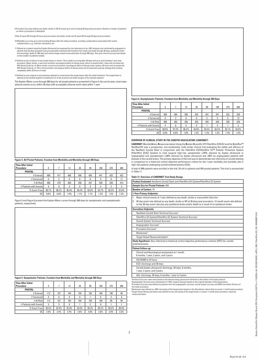
The Kaplan-Meier curve through 360 days for all pivotal patients is presented in Figure 4. As can be seen, most major adverse events occur within 30 days with acceptable adverse event rates within 1 year.
Figure 4. All Pivotal Patients, Freedom from Morbidity and Mortality through 360 Days
Time After Initial Procedure 0 7 14 30 90 180 270 360
PIVOTAL# Entered 480 471 460 456 450 441 432 422
# Censored 0 2 0 0 6 5 8 17# At Risk 480 470 460 456 447 439 428 414
# Patients with Events 9 9 4 6 3 4 2 3% Event-Free 98.1% 96.2% 95.4% 94.2% 93.5% 92.7% 92.2% 91.6%
SE 0.6% 0.9% 1.0% 1.1% 1.1% 1.2% 1.3% 1.3%
Figure 5 and Figure 6 present the Kaplan-Meier curves through 360 days for symptomatic and asymptomatic patients, respectively.
Figure 5. Sympotmatic Patients, Freedom from Morbidity and Mortality through 360 Days
Time After Initial Procedure 0 7 14 30 90 180 270 360
PIVOTAL# Entered 112 107 104 104 103 100 100 96
# Censored 0 0 0 0 1 0 3 5# At Risk 112 107 104 104 103 100 99 94
# Patients with Events 5 3 0 1 2 0 1 1% Event-Free 95.5% 92.9% 92.9% 92.0% 90.2% 90.2% 89.3% 88.3%
SE 2.0% 2.4% 2.4% 2.6% 2.8% 2.8% 3.0% 3.2%
Figure 6. Asymptomatic Patients, Freedom from Morbidity and Mortality through 360 Days
Time After Initial Procedure 0 7 14 30 90 180 270 360
PIVOTAL# Entered 368 364 356 352 347 341 332 326
# Censored 0 2 0 0 5 5 5 12# At Risk 368 363 356 352 345 339 330 320
# Patients with Events 4 6 4 5 1 4 1 2% Event-Free 98.9% 97.3% 96.2% 94.8% 94.5% 93.4% 93.1% 92.6%
SE 0.5% 0.9% 1.0% 1.2% 1.2% 1.3% 1.4% 1.4%
OVERVIEW OF CLINICAL STUDY IN THE CAROTID VASCULATURE (CABERNET)
CABERNET (Carotid Artery Revascularization Using the Boston Scientific EPI FilterWire EX®/EZ and the EndoTex™ NexStent®) was a prospective, non-randomized, multi-center clinical trial evaluating the safety and efficacy of the NexStent Carotid Stent in conjunction with the FilterWire EX/FilterWire EZ™ Embolic Protection System (FilterWire EX/EZ System) to treat surgical high-risk, symptomatic (≥50% stenosis by duplex ultrasound or angiography) and asymptomatic (≥80% stenosis by duplex ultrasound and ≥60% by angiography) patients with disease in the carotid artery. The primary objective of the trial was to demonstrate non-inferiority of carotid stenting in comparison to a historical control objective performance criteria for the 1-year morbidity and mortality rate in high-risk patients undergoing carotid endarterectomy (CEA).
A total of 488 patients were enrolled in the trial: 34 roll-in patients and 454 pivotal patients. This trial is summarized in Table 11.
Table 11. Overview of CABERNET Trial Study Design
Product Evaluated: NexStent Carotid Stent and FilterWire EX System/FilterWire EZ System
Sample Size for Pivotal Patients: 454
Number of Centers: 19
1-Year Primary Endpoints:
1. Major clinical events at 1 year defined as any death, stroke or myocardial infarction
2. 30-day event rate defined as any death, stroke or MI at 30 days post procedure. 12-month event rate defined as the 30-day event rate plus any ipsilateral stroke and/or death as a result of an ipsilateral stroke
Secondary Endpoints:
- NexStent Carotid Stent Technical Success1
- FilterWire EX System/FilterWire EZ System Technical Success1
- Overall System Technical Success1
- Angiographic Success2
- Procedure Success3
- Restenosis4
- Target Vessel Revascularization5
Study Hypothesis: Non-inferiority to historical control objective performance criterion (OPC) for carotid endarterectomy
Patient Follow-up:
- Clinical and Neurological assessment at 1 month, 6 months, 1 year, 2 years, and 3 years
- CK/CKMB to 24 hours
- ECG: discharge and 30 days
- Carotid duplex ultrasound: discharge, 30 days, 6 months, 1 year, 2 years, and 3 years
- AEs: discharge, 30 days, 6 months, 1 year to 3 years1 Technical Success rates were based on the devices being placed and retrieved as described in the study protocol.2 Angiographic Success was achievement of ≤50% residual stenosis based on the original diameter of the target lesion.3 Procedure Success was defined as patients who had angiographic success, overall system success and MAE free within 24 hours of the index procedure.
4 Restenosis was defined as >80% narrowing of the target lesion based on the Strandness criteria that occurred >1 month post procedure.5 Target Vessel Revascularization was defined as any narrowing of the target lesion or vessel >1 month post procedure, requiring revascularization.

8
Bo
sto
n S
cien
tifi
c (M
aste
r B
ran
d D
FU Te
mp
late
8.2
677i
n x
11.
6929
in A
4, 9
0105
918A
W),
eD
FU, M
B, F
ilter
Wir
e E
Z, E
N, 5
0606
897-
01A
Black (K) ∆E ≤5.0
≤30 daysEvent Categories1 # Patients % Patients
Procedure RelatedAngina 3 0.7
Bleeding/Anemia 17 3.7
Cardiac Dysrrhythmia 13 2.9
Cardiogenic Shock 1 0.2
Ischemia/Increased Enzymes 4 0.9
Syncope 4 0.9
Cerebrovascular 3 0.7
Emergent CEA 1 0.2
Genitourinary 1 0.2
Hypotension 16 3.5
Hypertension 1 0.2
Infection 3 0.7
Metabolic 5 1.1
Musculoskeletal 2 0.4
Neurological 9 2.0
Prolonged Hospitalization 1 0.2
Respiratory 2 0.4
Vascular 8 1.8
Other 2 0.4
Access Site ComplicationsBleeding and Hematoma 9 9
Ecchymosis 1 0.2
Pseudoaneurysm 3 0.7
Poss, Femoral Artery Thrombosis 1 0.2
Wound Infection 2 0.4
Total Procedure Related 78 17.2Non-Procedure RelatedCardiacAngina 2 0.4Congestive Heart Failure (CHF) 3 0.7Coronary Artery Disease 1 0.2Dysrhythmia 2 0.4Ischemia/Increased Enzymes 3 0.7Syncope 1 0.2NeurologicalAltered Mental State 1 0.2Seizure 1 0.2Syncope/Dizziness 1 0.2Visual Disturbance 1 0.2Other (Glazed Look, Neuropathies) 1 0.2Other SystemsBleeding 2 0.4Blood Dyscrasia 8 1.8Carcinoma 1 0.2Gastrointestinal 6 1.3Genitourinary 3 0.7Hemodynamic 5 1.1Infection 3 0.7Metabolic 2 0.4Musculoskeletal 2 0.4Respiratory 2 0.4Vascular 4 0.9Other 7 1.5Total Non-Procedure Related 53 11.7
1Patients may have had multiple events and therefore can be counted in more than one category/subcategory of event. Counts represent the number of patients who have experienced one or more events.
Table 14. Causes of Death
Death (by type) 0-30 Days
(n=454)
n %
Cardiac 1 0.2
Sepsis 1 0.2
SUMMARY OF CLINICAL STUDY IN THE CAROTID VASCULATURE (CABERNET)
CABERNET (Carotid Artery Revascularization Using the Boston Scientific EPI FilterWire EX/EZ and the EndoTex™ NexStent®) was a prospective, single-arm, multi-center trial evaluating the safety and efficacy of the NexStent Carotid Stent in conjunction with the FilterWire EX System/FilterWire EZ System to treat surgical high-risk, symptomatic (≥50% stenosis by duplex ultrasound or angiography) and asymptomatic (≥80% stenosis by duplex ultrasound or ≥60% by angiography) patients with disease in the carotid artery. A trial design utilizing a roll-in phase for initial clinical experience was employed in the study. A total of 488 patients were enrolled at 19 centers involving 21 clinical sites (15 centers in the United States and 4 centers outside the United States), including 34 roll-in patients and 454 pivotal patients.
The CABERNET trial was designed to show non-inferiority between carotid stenting and a historical control, based on standard of care. The historical control was established based on a review of the current literature on carotid endarterectomy and was defined as a weighted OPC. A criterion of 14% for patients who had comorbidity risk factors and a criterion of 11% for patients who had anatomical risk factors were selected. A spread of 4% for the “delta” definition of equivalency was selected.
πOPC-Anatomic = OPC 1 year complication rate for anatomical high risk factors = 11%
πOPC-Comorbid = OPC 1 year rate for comorbid high risk factors = 14%
πOPC-weighted = (wC*[πOPC-Comorbid]) + (wA*[πOPC-Anatomic])
Enrollment percentages in each category were 36.3% (165/454) in the comorbid group and 63.7% (288/454) in the anatomic group; therefore, the weighted OPC for CABERNET was 12.1%. Note that 16.7% (76/454) of the patients presented with both comorbid and anatomic risk factors and were included in the comorbid group for the calculation of the OPC (89 comorbid only +76 comorbid/anatomic = 165 comorbid patients or 36.3%).
12.1% = (36.3% x 0.14) + (63.7% x 0.11)
Based on the weighted OPC of 12.1% and the pre-specified delta of 4%, the threshold for claiming non-inferiority to CEA is 16.1%.
Patients were followed at hospital discharge, 1 month, 6 months, and 1 year. Additionally, registry patients were followed annually through 3 years from the date of the index procedure. Patients were seen in follow-up by the treating physician as well as independent neurologist (neurological assessments). Core laboratories were utilized for the analysis of angiographic, ultrasound, ECG and CT/MRI (only for evaluation of neurological events or symptomology) data. To ensure patient safety, medical monitors reviewed safety data to ensure appropriate reporting of adverse events. A Clinical Events Committee (CEC) reviewed all reported major adverse events for the primary endpoint and a Data Safety Monitoring Board (DSMB) reviewed the summary of these events to ensure patient safety.
Inclusion Criteria
The study population consisted of male and female patients, at least 18 years of age, with discrete lesions in the common carotid artery (CCA), internal carotid artery (ICA) or carotid bifurcation. Patients had to be at high-risk for surgical intervention; both symptomatic (≥50% stenosis by duplex ultrasound or angiography) and asymptomatic (≥80% stenosis by duplex ultrasound and ≥60% by angiography) patients were eligible.
The key inclusion criteria included the following:
1. Target lesion is located in the common carotid artery (CCA) and/or the internal carotid artery (ICA) or the carotid bifurcation
2. Target vessel is the only vessel being treated at this intervention and the lesion is < 30 mm and can be treated with a single stent
3. Vessel to be treated is between 4 mm and 9 mm in diameter
4. Distal vessel “landing zone” for placement of the FilterWire EX System/FilterWire EZ System must be between 3.5 mm and 5.5 mm in diameter and have available the following artery lengths:
FilterWire EX System:
• A minimum of 2.0 cm distal to the target lesion
• A minimum of 2.0 cm straight vessel segment for placement of the filter
• A minimum of 1.0 cm from a major vessel curve FilterWire EZ System:
• Visual angiographic assessment recommendations as described in the DFU
Patients were required to meet one of the following criteria:
• Symptomatic: Carotid stenosis of ≥50% as determined by duplex ultrasound and angiogram with a history of stroke, TIA and/or amaurosis fugax in the hemisphere supplied by the target vessel within 180 days of the procedure.
• Asymptomatic: Carotid stenosis of ≥80% as determined by duplex ultrasound and ≥60% as determined by angiogram without any neurological symptoms.
• Patient had to meet at least ONE High-Risk Category as follows:
Anatomic High-Risk Conditions:
ONE (1) criterion qualifies
1. Previous carotid endarterectomy with significant restenosis (as defined above for symptomatic or asymptomatic patients)
2. Total occlusion of the contralateral carotid artery
3. Previous radiation treatment to the neck or radical neck dissection
4. Target lesion is at or above the second vertebral body C2 or below the clavicle
5. Inability to extend the head due to cervical arthritis or other cervical disorders
6. Tracheostomy or tracheal stoma
7. Presence of laryngeal nerve palsy
Adverse Events
Because the FilterWire EX® System/FilterWire EZ™ System is used in an acute manner, peri-procedural adverse events are most relevant to an evaluation of device safety. Tables 12 and 13 present the major adverse events (MAE) and the serious adverse events (SAE) reported for the enrolled patients through 30-days. Table 14 presents the patient deaths by causation. The MAE table summarizes all deaths, strokes and MIs. A SAE may or may not be considered related to the device and may be described as follows:
• Death due to any cause
• Life-threatening condition (e.g., stroke)
• Persistent or significant disability/incapacity
• Any event resulting in an unscheduled in-patient hospitalization (admission to hospital for >24 hours) or prolongation of existing hospitalization (>72 hours)
• Any event requiring intervention (except for comorbid scheduled events which are scheduled and planned during the follow-up period)
• New cancer/malignancy not present at the time eligibility was established
• Congenital abnormality or birth defect
Events are categorized by body system and are defined as follows:
• Access site includes such events as aneurysm, bleeding, bruising or ecchymosis, hematoma, pseudo-aneurysm, pain and arterial thrombosis.
• Bleeding includes such non access-site bleeding, nose bleed, surgical or incisional bleeding and retroperitoneal bleed.
• Blood Dyscrasia includes events such as anemia, thrombocytopenia, and leucopenia.
• Carcinoma includes such events as lung cancer, breast cancer, leukemia, brain tumor, and rectal cancer.
• Cardiac includes such events as angina, coronary artery disease, cardiac dysrhythmia, congestive heart failure, cardiac-related syncope and valvular disease (aortic and mitral).
• Cerebrovascular includes such events as headache and brain hemorrhage.
• Gastrointestinal includes events such as dysphagia, indigestion, nausea, vomiting, esophageal stenosis or varicies, ulcer, bowel obstruction, GI Bleed, colitis, cholecystitis, pancreatitis, hepatic disorders, diverticulitis, melena and rectal prolapse.
• Genitourinary includes events such as urinary retention, hematuria, nocturia related to prostatic hyperplasia and lower abdominal pain related to the bladder or prostate.
• Hemodynamic includes events such as hypotension and hypertension.
• Metabolic includes events such as diabetes, dehydration, electrolyte imbalance and renal failure.
• Musculoskeletal includes events such as bone, muscle or joint pain, fractures and arthritis injury, and inguinal hernia.
• Infection includes events such as conjunctivitis, cellulitis, parotitis, abscess, system infection, sepsis, fungal infection, urinary tract infection, wound infection and non-specified infection.
• Neurological includes all non-stroke related events such as altered mental status/confusion/dementia/organic brain syndrome, seizure, sensory deficits (peripheral numbness or weakness), visual/speech disturbances, neurologic- related syncope or dizziness, and TIA.
• Respiratory includes events such as pneumonia, hemoptysis, respiratory failure and chronic obstructive lung disease.
• Vascular includes events such as carotid restenosis (target and non-target lesion), peripheral arterial disease, and peripheral arterial or venous thrombosis.
• Other Hospitalizations include hospitalizations for other medical/surgical treatment.
• Other is a miscellaneous category that includes such events as agitation, drug hypersensitivity, patient fall (non-neurologic), rash, general weakness, gout, peripheral edema, fatigue, etc.
Table 12. CABERNET Major Adverse Events
Major Adverse Events ≤30 days(n=439)
Primary Endpoints # Patients %All Death Stroke and MI at 30 Days 17 3.9
Major Adverse Events by Classification ≤30 days(n=439)
# Patients %Death 2 0.5Stroke 15 3.4
Ipsilateral Stroke 12 2.7Major 5 1.1
Minor 7 1.6Non-Ipsilateral Stroke 3 0.7
Major 1 0.2Minor 2 0.5
Myocardial Infarction (MI) 1 0.2
Table 13. CABERNET Trial Serious Adverse Events

9
Bo
sto
n S
cien
tifi
c (M
aste
r B
ran
d D
FU Te
mp
late
8.2
677i
n x
11.
6929
in A
4, 9
0105
918A
W),
eD
FU, M
B, F
ilter
Wir
e E
Z, E
N, 5
0606
897-
01A
Black (K) ∆E ≤5.0
8. Bilateral carotid artery stenosis as determined by angiography in which both carotid arteries require treatment, as defined as:
• Bilateral asymptomatic stenosis ≥60% or
• Bilateral symptomatic stenosis ≥50% or
• Bilateral stenoses, one side with a symptomatic stenosis ≥50% and the other side asymptomatic with a stenosis ≥60%
A second NexStent Carotid Stent and the FilterWire EX System/FilterWire EZ System procedure may be conducted and included in study evaluation, providing the second procedure is staged >30 days following the first NexStent Carotid Stent and the FilterWire EX System/FilterWire EZ System procedure. A total of 41 patients went on to undergo treatment of the second artery. These patients were followed for 3 years from the date of their index procedure.
Comorbid High-Risk Conditions:
CLASS I [ONE (1) criterion qualifies]
1. Unstable angina (chest pain with ECG changes)
2. Known severe left ventricular dysfunction, LVEF <30%
3. Congestive heart failure (CHF) - New York Heart Association Functional Class III or IV
4. Dialysis dependent renal failure
5. Chronic obstructive pulmonary disease (COPD) with either
• Forced Expiratory Volume in 1 Second (FEV1) <50% predicted or
• chronic oxygen therapy or
• resting PO2 of ≤60 mmHg or
• baseline hematocrit ≥50%
6. Requirement for staged CABG or valve replacement post carotid index procedure
CLASS II [TWO (2) criteria qualify]
1. Patient is ≥75 years of age
2. Myocardial infarction within previous 6 weeks
3. Requires staged peripheral vascular surgery (i.e., abdominal aortic aneurysm repair) or other major surgery post carotid index procedure
4. Two or more proximal or major diseased coronary arteries with ≥70% stenosis that have not or cannot be revascularized
Exclusion Criteria
A patient with any of the following criteria was not considered eligible for treatment in the trial.
1. Previously placed stent in target vessel
2. Total occlusion of target vessel (ICA or CCA)
3. Angiographically visible thrombus
4. Carotid string sign (a tiny, long segment of contrast in the true lumen of the artery, aneurysmal pouch formation, and the distal location of the arteriopathy) with poor visualization of the distal vessel
5. Vertebrobasilar insufficiency symptoms only, without clearly identifiable symptoms referable to the targeted carotid artery
6. Vessel anatomy precluding use of stent system or distal protection system.
7. Presence of carotid artery dissection
8. Requirement for staged CABG, valve replacement or abdominal aortic aneurysm procedure within 30 days of the index procedure
9. Evidence of a major disabling stroke within the previous 30 days
10. Patient has an evolving stroke or has experienced a major stroke (NIHSS score >15) within 3 months
11. History of intracranial hemorrhage within the past 12 months
12. Any condition that precludes proper angiographic assessment or makes percutaneous arterial access unsafe, e.g., morbid obesity, history of chronic hypertension that is not controlled by medical therapy
13. Contraindication to heparin, aspirin, clopidogrel (Plavix® Clopidogrel), X-ray contrast or ticlopidine (Ticlid® Ticlopidine HCL) in cases of intolerance to clopidogrel
14. History of liver failure with elevated prothrombin time
15. History or current indication of bleeding diathesis or coagulopathy
16. Hgb <8gm/dl (unless on dialysis), platelet count <50,000, WBC >15,000, INR >l.5 (irreversible) or heparin-associated thrombocytopenia
17. Known cardiac sources of emboli not under treatment with anticoagulant therapy
18. Atherosclerotic disease involving adjoining vessels precluding safe placement of the guiding catheter or sheath
19. Planned treatment of non-target lesion within 30 days
20. Other abnormal angiographic findings that indicate the patient is at risk of a stroke due to a problem other than the target lesion, such as: ipsilateral arterial stenosis greater in severity than the target lesion, cerebral aneurysm, or arteriovenous malformation (AVM) of the cerebral vasculature
21. Dementia or confusion
22. The patient is enrolled in another study protocol
23. Patient may not participate in another investigational trial up to 12 months post- index procedure
Description of Patients Evaluated
Table 15 summarizes patient follow-up at the endpoint evaluation time points of 30 days and 1 year. Patients were considered to have been evaluated if they had physician contact as evidenced by a physician visit that included at least one of the following at the specified study interval: clinical evaluation, neurologic evaluation, AE log, SAE Notification Form, Subsequent Hospitalization Form, Vascular Event Form, Neurological Event Form. Phone visits were permitted if patients were unable to attend the visit in person (e.g., unable to attend due to illness, geographic/transportation constraints or hospitalization) and it was required that information regarding occurrence of adverse events or hospitalizations due to adverse events be obtained as well as all available supporting documentation.
Table 15. Patient Enrollment and Disposition
Patient Population Index Procedure 30-Days 1-Year
Total Number of Patients Enrolled 488
Roll-In Patients 34Patients Enrolled in Main Trial Registry 454
Patients Without a NexStent® Carotid Stent Implanted
11
Patients Lost to Follow-Up or Withdrawals (Cumulative) 0 1 13
Deaths (Cumulative) 0 2 19Evaluable Patients Available for Follow-Up 4431 443 434
Total Missed Visits Among Evaluable Patients 4 12
Total Patients Evaluated 436 398Clinical Assessments Performed
428 (97.7%) 377 (93.3%)
Neurologic Assessments Performed
412 (94.1%) 365 (90.3%)
12 Lead ECGs (30-Day FU Only) Performed
335 (76.5%)
Duplex Ultrasound Exams Performed
387 (88.3%) 331 (81.9%)
Stroke Scales Performed 412 (93.6%) 354 (87.6%)
Compliance to Follow-Up 436 (98.4%) 398 (91.7%)
1Evaluated patients only include those that had a NexStent Carotid Stent implanted.
Baseline demographics, high-risk inclusion criteria and lesion characteristics for the study are presented in Tables 16, 17, and 18. All reported angiographic data on the treated lesions are based on measurements obtained by the centralized angiographic core laboratory.
Table 16. Baseline Patient Demographics
Patient Characteristic Patients (N = 454)
% (except as noted)
Age (years) Mean + SD 72.5 + 9.6 yrsMinimum to Maximum 46 to 94 yrs
Age by Decade of Life
40-49 6 1.3
50-59 29 6.4
60-69 124 27.3
70-79 194 42.7
80-89 97 21.4
>90 4 0.9
Gender Male 297 65.4
Female 157 34.6
Ethnicity Caucasian 414 91.2
Black 21 4.6
Hispanic 10 2.2
Asian 5 1.1
Other 4 0.9
Patient Classification
Symptomatic 110 24.2
Asymptomatic 344 75.8
Medical History Diabetes Mellitus 150 33.0Liver Failure 0 0.0Dyslipidemia 314 69.2GI Bleeding/PUD 29 6.4Hypertension 377 83.0Uncontrolled Hypertension 6 1.3
Cigarette Smoker 320 70.5Current Cigarette Smoker 83 18.3
Family Hx Premature ASD 74 16.3
Significant Aortic Arch Atherosclerosis
5 1.1
Cardiac Arrhythmia 81 17.8
Valvular Disease 26 5.7CAD 283 62.3PVD 178 39.2Prior PTCA 119 26.2Prior Valve Replacement 21 4.6
Prior CABG 161 35.5
Prior Carotid PTA 13 2.9
Prior Carotid Stenting 12 2.6
Hx of TIA 124 27.3
Hx of Stroke 96 21.1Family Hx of Stroke 53 11.7
Hx of Seizures 12 2.6
Hx of Other Neuro 31 6.8Prior Vertebrobasilar Intervention
1 0.2
Other 74 16.3

10
Bo
sto
n S
cien
tifi
c (M
aste
r B
ran
d D
FU Te
mp
late
8.2
677i
n x
11.
6929
in A
4, 9
0105
918A
W),
eD
FU, M
B, F
ilter
Wir
e E
Z, E
N, 5
0606
897-
01A
Black (K) ∆E ≤5.0
Table 17. Patient High-Risk Inclusion Criteria
Inclusion Criteria Patients (N = 454) %
High-Risk CategoryAnatomical Only 288 63.4Comorbid Only 89 19.6Both Anatomic and Comorbid 77 17.0Class I - Comorbid 139 30.6Class II - Comorbid 53 11.7Anatomical High-Risk Inclusion Criteria (A)Previous CEA 95 20.9Total Occlusion of Contralateral Carotid Artery 85 18.7
Previous Radial Neck Dissection or Radiation Therapy to Neck Region 30 6.6
Target Lesion at or above C2 or below Clavicle 25 5.5
Spinal Immobility of Neck (cervical arthritis or other) 54 11.9
Tracheostomy or tracheal stoma 3 0.7Presence of laryngeal nerve palsy 8 1.8Bilateral Carotid Artery Stenosis 143 31.5Class I: Comorbid High-Risk Inclusion Criteria (CI)Unstable Angina 26 5.7Severe LV Dysfunction 42 9.3CHF (NYHA Class III/IV) 40 8.8Renal Failure (Dialysis Dependent) 5 1.1Severe COPD 38 8.4Requires Staged CABG or Valve Surgery 21 4.6
Class II: Comorbid High-Risk Inclusion Criteria (CII) Patient is ≥75 years of Age 199 43.8MI within 6 weeks 5 1.1Requires Staged Peripheral Vascular Surgery 34 7.5Major CAD (≥70% Stenosis that have not or cannot be revascularized) 64 14.1
Table 18. Baseline Lesion Characteristics (Pre-Procedure)
Angiographic Data (Core Laboratory Assessment) Results
Parameter Patients (N = 454) %
Lesion Typede novo 360 79.3Restenotic 94 20.7Lesion Morphology1Eccentric 327 72.0Ulceration 169 37.2Calcification 190 41.9Target Lesion Length (mm)Mean + SD n=4412 13.9 + 5.9Minimum Lumen Diameter (mm)Mean + SD n=4432 1.3 + 0.6Baseline Percent Diameter Stenosis (%)Mean + SD n=4432 71.9 +
11.01 Lesions may have had more than one type of morphology.2 Lesion length was not available in 13 patients; MLD and Percent Diameter Stenosis were not available in 11 patients.
Trial Results
The primary and secondary endpoints evaluating the safety and efficacy of the NexStent® Carotid Stent in the CABERNET trial are presented in Tables 19 and 20 respectively. The 30-day primary endpoint MAE rate (all death, stroke and MI within 30 days) was 3.9%. The 1-year endpoint MAE rate for all death, stroke and MI was 11.9%. The rate for the composite 1-year endpoint that includes the 30-day MAEs plus ipsilateral stroke or death from ipsilateral stroke within 31-365 days was 4.7%.
The primary endpoints in this study were further explored through a time-to-event analysis using a Kaplan-Meier (KM) estimator for the survival function. Life table estimates were obtained for:
a. Freedom from major adverse events (All Deaths, Strokes and MIs) at 1 year
b. Freedom from major adverse events (All Deaths, Strokes and MIs) at 30 days; plus ipsilateral stroke or death related to ipsilateral stroke at 1 year.
Angiographic success was defined as less than 50% residual stenosis following completion of the stent implantation and was attained in 97.7% (423/433) of evaluable patients. The mean post-procedure residual stenosis was 20.5% as assessed by the core laboratory and 6.45% as per the visual inspection performed by the treating physician at the completion of the procedure. Acute procedure success, defined as the composite of angiographic success and overall technical success without the occurrence of an MAE within 24 hours, was demonstrated in 395 patients (87.0%).
The primary endpoints of the trial were met. The upper confidence limits for both primary endpoint MAE rates (14.8%, 6.8% respectively) fell below the reference OPC (12.1%) plus an additional delta of 4% established for this study (16.1%), allowing acceptance of the hypothesis that carotid stenting using the NexStent Carotid Stent with the FilterWire EZ™ System meets acceptance of the study hypothesis of comparability to CEA in the high-surgical-risk study population.
Table 19. Primary Endpoints (Major Adverse Events)
Major Adverse Events
≤30 days (n=439)
31-365 days (n=415)²
Cumulative 0-365 Days (n=421)¹
Primary Endpoints # Patients % # Patients % # Patients %All Death Stroke and MI at 1 year 17 3.9 35 8.4 50 11.9
UCL=14.8%
# Patients %Patients (n=438) # Patients %Patients
(n=404)² # Patients %Patients (n=404)²
All Death Stroke and MI ≤ 30 days; plus the 31-day to 12-month event rate defined as any ipsilateral stroke including any death as a result of an ipsilateral stroke
17 3.9 3 0.7 19 4.7 UCL=6.8%
Major Adverse Events by Classification
≤30 days (n=439)
31-365 days (n=415)¹
Cumulative 0-365 Days (n=421)¹
# Patients % # Patients % # Patients %Death 2 0.5 17 4.0 19 4.5Stroke 15 3.4 8 1.9 21 5.0Ipsilateral Stroke 12 2.7 3 0.7 14 3.3Major 5 1.1 1 0.2 5 1.2Minor 7 1.6 2 0.5 9 2.1Non-Ipsilateral Stroke 3 0.7 6 1.4 8 1.9Major 1 0.2 3 0.7 4 1.0Minor 2 0.5 3 0.7 4 1.0Myocardial Infarction (MI) 1 0.2 16 3.9 17 4.0
1n=421 for the 0-365 day time periods. This includes 398 patients evaluated at 1 year, 19 deaths, and 4 patients that did not have a 1-year visit but experienced an adverse event (398+19+4 = 421).2In an effort to present the most conservative analysis for the MAE rate concerning the composite Primary Endpoint n=404 was used. Seventeen patient deaths that occurred during the 31-365 day time period were not due to ipsilateral stroke and were excluded from this analysis. Therefore n = 404 (421-17=404)
Table 20. Secondary Endpoints
Measure Result* 95% Confidence Limit (%)FilterWire EX® System/FilterWire EZ System Technical Success 95.2% (454/477) LCL=93.2
NexStent Carotid Stent Technical Success 94.3% (443/470) LCL=92.2Overall System Technical Success 93.0% (422/454) LCL=90.2Angiographic Success 97.7% (423/433) LCL=96.1Procedure Success (Acute) 87.0% (395/454) LCL=84.1Restenosis 50-79% (Cumulative 0 to 6 months) (Cumulative 0 to 1 year)
16.0% (55/344)1 18.7% (65/347)2
UCL=19.6 UCL=22.5
Restenosis >80% (Cumulative 0 to 6 months) (Cumulative 0 to 1 year)
0.9% (3/341)3 2.7% (9/334)4
UCL=2.3 UCL=4.7
Target Vessel Revascularization (6 months) (1 year)
0.9% (4/443) 2.3% (10/443)
UCL=2.1 UCL=3.8
*Numbers are % (count/sample size).1n=344 includes all patients with duplex ultrasound data available at 6 months plus any patient without a 6-month evaluation that had 50-79% restenosis at an earlier evaluation (340+4=344).2n=347 includes all patients with duplex ultrasound data available at 1 year plus any patient without a 1-year evaluation that had 50-79% restenosis at an earlier evaluation (332+15=347).3n=341 includes all patients with duplex ultrasound data available at 6 months plus any patient without a 6-month evaluation that had >80% restenosis at an earlier evaluation (340+1=341).4n=334 includes all patients with duplex ultrasound data available at 1 year plus any patient without a 1-year evaluation that had >80% restenosis at an earlier evaluation and one patient that had a late 1-year follow-up who had >80% restenosis (332+2=334).
Additional Analysis
The primary endpoints in this study were further explored through a time-to-event analysis using a Kaplan-Meier estimator for the survival function. Life table estimates were obtained for:
a. Survival from major adverse events (All Deaths, Strokes and MIs) through 1 year
b. Survival from major adverse events (All Deaths, Strokes and MIs) at 30 days; plus ipsilateral stroke or death related to ipsilateral stroke through 1 year.
Endpoint Analysis
Patients were included in these analyses if they had a successful NexStent Carotid Stent implant. The survival time for patients who experienced a major adverse event was calculated as the number of days from the index procedure to the date the adverse event was first identified. For patients who withdrew from the study before the time of analysis, the survival time was calculated as the number of days from the index procedure to the date of their withdrawal. For the composite endpoint, for patients who died of causes unrelated to an ipsilateral stroke, the survival time for the composite endpoint analysis was calculated as the number of days from the index procedure to the date of their death. Patients who were terminated early from the study are considered censored in the analyses performed. Figures 7 and 8 present the KM analysis for each endpoint. Figures 9 through 12 present the KM analysis for asymptomatic and symptomatic patients for each endpoint. The event rate estimate of 11.7% obtained for all death, strokes, and MIs at 12 months from the life table is consistent with the estimate of 11.9% obtained according to the analysis performed per protocol. Similarly, the estimate obtained from the life table of 4.3% for the event rate for the composite endpoint of major adverse events (All Deaths, Strokes and MIs) at 30 days; plus ipsilateral stroke or death related to ipsilateral stroke at 1 year is consistent with the obtained estimate of 4.7% according to the analysis performed per protocol.

11
Bo
sto
n S
cien
tifi
c (M
aste
r B
ran
d D
FU Te
mp
late
8.2
677i
n x
11.
6929
in A
4, 9
0105
918A
W),
eD
FU, M
B, F
ilter
Wir
e E
Z, E
N, 5
0606
897-
01A
Black (K) ∆E ≤5.0
1 - ( OPC Rate + 4% ) Threshold
Any MAE:At Risk:441 422 414 409 404 400 398 389 385 383 378 369 239Events: 0 17 20 24 27 31 32 36 39 40 42 43 50
47.11 39.9 86.9 12.9 79.8 62.8 23.7 90.7 61.6 74.5 45.4 68.3 00.0 :% tnevE
KM Estimate Censored
Per
cent
of P
atie
nts
NO
T E
xper
ienc
ing
an E
vent
50
55
60
65
70
75
80
85
90
95
100
Study Month
0 1 2 3 4 5 6 7 8 9 10 11 12
Figure 7. Survival from Major Adverse Events (All Deaths, Strokes and MIs) through 1 Year.
1 - ( OPC Rate + 4% ) Threshold
Ipsilateral MAE:At Risk:441 421 409 408 406 405 399 390 389 387 383 374 244Events: 0 17 17 17 17 18 19 19 19 19 19 19 19
33.4 33.4 33.4 33.4 33.4 33.4 33.4 90.4 68.3 68.3 68.3 68.3 00.0 :% tnevE
KM Estimate Censored
Pe
rce
nt
of
Pa
tien
ts N
OT
Exp
erie
nci
ng
an
Eve
nt
50
55
60
65
70
75
80
85
90
95
100
Study Month
0 1 2 3 4 5 6 7 8 9 10 11 12
Figure 8. Survival from Major Adverse Events (All Deaths, Strokes and MIs) at 30 Days; Plus Ipsilateral Stroke or Death Related to Ipsilateral Stroke through 1 Year.
KM Estimate Censored
Perc
ent o
f Pat
ient
s N
OT
Expe
rienc
ing
an E
vent
50
55
60
65
70
75
80
85
90
95
100
Study Month
0 1 2 3 4 5 6 7 8 9 10 11 12
Figure 9. Survival from Major Adverse Events (All Deaths, Strokes and MIs) at 1 Year Symptomatic Patient Group
KM Estimate Censored
Perc
ent o
f Pat
ient
s N
OT
Expe
rienc
ing
an E
vent
50
55
60
65
70
75
80
85
90
95
100
Study Month
0 1 2 3 4 5 6 7 8 9 10 11 12
Figure 10. Survival from Major Adverse Events (All Deaths, Strokes and MIs) at 1 Year Asymptomatic Patient Group
KM Estimate Censored
Perc
ent o
f Pat
ient
s N
OT
Expe
rienc
ing
an E
vent
50
55
60
65
70
75
80
85
90
95
100
Study Month
0 1 2 3 4 5 6 7 8 9 10 11 12
Figure 11. Survival from Major Adverse Events (All Deaths, Strokes and MIs) at 30 Days Plus Ipsilateral Stroke or Death Related to Ipsilateral Stroke through 1 Year Symptomatic Patient Group
KM Estimate Censored
Perc
ent o
f Pat
ient
s N
OT
Expe
rienc
ing
an E
vent
50
55
60
65
70
75
80
85
90
95
100
Study Month
0 1 2 3 4 5 6 7 8 9 10 11 12
Figure 12. Survival from Major Adverse Events (All Deaths, Strokes and MIs) at 30 Days Plus Ipsilateral Stroke or Death Related to Ipsilateral Stroke through 1 Year Asymptomatic Patient Group
Study Conclusions
The upper confidence limits for both primary endpoint MAE rates fell below the Reference OPC plus the Non-Inferiority Margin of 4% established for this study and both null hypotheses were rejected. The primary endpoints of the trial were met.
The results of the CABERNET trial demonstrate that the NexStent® Carotid Stent used with the FilterWire EZ™ System were found to be safe and effective as treatment for carotid artery disease in the population indicated. Results from the pre-clinical and clinical evaluations provide valid scientific evidence and reasonable assurance that the devices were safe and effective when used in accordance with the instructions for use.

12
Bo
sto
n S
cien
tifi
c (M
aste
r B
ran
d D
FU Te
mp
late
8.2
677i
n x
11.
6929
in A
4, 9
0105
918A
W),
eD
FU, M
B, F
ilter
Wir
e E
Z, E
N, 5
0606
897-
01A
Black (K) ∆E ≤5.0
OVERVIEW OF CLINICAL STUDIES IN SAPHENOUS VEIN BYPASS GRAFTS (BLAZE II, BLAZE, AND FIRE)
BLAZE II - FilterWire EZ™ System (2.25 mm – 3.5 mm): BLAZE II was a prospective, multi-center, non-randomized study. One hundred thirty-one (131) registry patients from 19 U.S. sites were enrolled and treated. The protocol was similar to the BLAZE study and FIRE trial protocols except that the vessel diameter inclusion criteria allowed for protection wire placement in reference vessel diameters (RVDs) between 2.25 mm and 3.5 mm by visual estimate. The study was performed to evaluate the safety and performance of the FilterWire EZ System (2.25 mm – 3.5 mm) in patients being treated for saphenous vein graft stenosis. The primary objective of the study was to assess 30-day MACE rates when compared to a subset of the Medtronic’s GuardWire Plus® Device (GuardWire Plus Device) patients with SVG diameters between 2.5 mm and 3.5 mm * from the Randomized FIRE trial.
BLAZE - FilterWire EZ System (3.5 mm – 5.5 mm): BLAZE was a prospective, multi-center, non-randomized study. Ninety (90) registry patients from 16 U.S. sites and 6 European sites were enrolled and treated. Clinical data from European and US study sites were pooled. There were no statistically significant differences in the important demographic or angiographic variables between patients from the two geographic regions, i.e., age, gender, baseline QCA, and final QCA.
This study was performed to assess the safety and performance of the FilterWire EZ System (3.5 mm - 5.5 mm) during percutaneous treatment of saphenous vein graft stenosis. The primary endpoint of this Study was Major Adverse Cardiac Events (MACE) at 30 days post procedure. Secondary endpoints included MACE during index hospitalization, device success, clinical success, and final TIMI flow. After the procedure there was no standard anticoagulation regimen but patients who were treated with stents were to receive Clopidogrel (75 mg daily) or Ticlopidine (250 mg b.i.d.) for 30 days and Aspirin (325 mg daily) for at least 6 months. Follow-up at 30 days was completed. No further follow-up was required per protocol.
FIRE - FilterWire EX® System: FIRE was a prospective multi-center, randomized, two-arm, controlled trial that utilized a hybrid design combining a superiority trial comparing FilterWire EX System to a conventional guide wire with no protection, and a non-inferiority trial comparing FilterWire EX System to the GuardWire Plus Device. A total of eight hundred sixty-four (864) patients from 62 U.S. sites and 4 Canadian sites were enrolled. The FIRE trial was performed to establish the safety and efficacy of treatment with the Boston Scientific FilterWire EX System during angioplasty/stenting of saphenous vein grafts with vessel diameters between 3.5 mm and 5.5 mm. The FIRE trial was originally designed as a prospective, randomized, controlled trial comparing FilterWire EX System to no protection. During the course of the trial, the GuardWire Plus Device became commercially available. At this point, a “Hybrid Trial” was conceived to create two separate trial arms – the original arm (for randomization to no protection) and an arm for randomization to the GuardWire Plus Device. Two hundred thirteen (213) patients were enrolled and randomized to no protection. A total of six hundred fifty-one (651) patients were randomized to the commercially available GuardWire Plus Device.
The primary endpoint of the FIRE trial was MACE at 30 days post procedure. Secondary safety endpoints were CK and CK-MB post procedure, target vessel failure at 6 months and MACE during index hospitalization. Secondary efficacy endpoints were device success, clinical success and final TIMI flow. After the procedure all patients were to be treated with Aspirin (325 mg daily) indefinitely. There was no other prescribed anticoagulation regimen but patients who were treated with stents were to receive Clopidogrel (75 mg daily) or Ticlopidine (250 mg b.i.d.) for 30 days. Follow-up at 30 days and 6 months was completed.
All patients treated in the FIRE Trial received bare metal stents. Between the time of FIRE and initiation of BLAZE II, drug eluting stents (DES) were introduced to the market. Although DES are not currently approved for SVGs, 95% of patients treated in the BLAZE II study received DES. No conclusions can be drawn from their use in this study.
* The subset of patients from FIRE used in the BLAZE II analysis included all GuardWire Plus Device patients who had reference vessel diameters (RVD) 2.25 mm -3.5 mm as measured by QCA at a validated angiographic core lab. These data were taken from the database received directly from the core lab. The GuardWire Plus Device cohort from the FIRE Trial was chosen for comparison in BLAZE II, rather than the FilterWire EZ System cohort, because at the time of designing the BLAZE II study, GuardWire Plus Device in smaller vessels (2.5 mm – 5.0 mm) had received FDA clearance, whereas the FilterWire EZ System was only indicated for vessel sizes 3.5 mm - 5.5 mm
Clinical Trial Comparison
BLAZE II SVG (registry)
FilterWire EZ System (2.25 mm - 3.5 mm)
BLAZE SVG (registry)
FilterWire EZ System (3.5 mm - 5.5 mm)
FIRE SVG Trial
(non-inferiority arm) FilterWire EX System
(superiority arm) FilterWire EX System
Number of Patients
131 Registry 169 GuardWire Plus Device subset from FIRE SVG Trial
90 Registry 332 FilterWire EX System
110 FilterWire EX System
319 GuardWire Plus Device
103 No Protection
131 Total Patients 169 Total Patients 90 Total Patients 351 Total Patients 213 Total Patients
Study Type • Prospective • Multi-center • Non-randomized
• Historical Control • Prospective • Multi-center • Non-randomized
• Prospective • Multi-center • Randomized • Controlled
Lesion Criteria RVD
> 2.25 mm and < 3.5 mm
> 2.25 mm and < 3.5 mm
> 3.5 mm and < 5.5 mm
> 3.5 mm and < 5.5 mm
Placement Landing Zone
> 2.5 cm distal to lesion; > 2.5 cm from distal anastomosis
2.0 cm > 1.5 cm distal to lesion; > 3.0 cm from distal anastomosis
> 2.0 cm straight vessel segment > 1.5 cm distal to lesion; > 2.5 cm from distal anastomosis
Type SVG SVG SVG SVG
Stenosis > 50% and < 100% > 50% and < 100% > 50% and < 100% > 50% and < 100%
Device Used/Differences
FilterWire EZ System (2.25 mm- 3.5 mm) Guide wire and sheaths the same as FilterWire EZ System (3.5 mm - 5.5 mm) Smaller filter loop and filter
GuardWire Plus Device (2.5 mm - -5.0 mm) Inflatable Balloon Occlusion
FilterWire EZ System (3.5 mm - 5.5 mm)
• Guidewire passes through the filter loop; support arm connects wire to loop.
• Guidewire silicone coated
• Delivery Sheath 3.2 F (peel-away configuration)
• Delivery Sheath silicone coated
FilterWire EX SystemFilter loop attached directly to the guidewire Delivery sheath 4.0 F
Follow-up 30-days: clinical 30-days: clinical 180-days: clinical
30-days: clinical 30-days: clinical 180-days: clinical
ADVERSE EFFECTS
One Hundred thirty one (131) registry patients received the FilterWire EZ System (2.25 mm - 3.5 mm) in the BLAZE II SVG registry. Ninety (90) registry patients received the FilterWire EZ System in the BLAZE Study. Three hundred thirty-two (332) patients received the FilterWire EX System when randomized to the GuardWire Plus Device (non-inferiority arm; FIRE Trial). Adverse events associated with percutaneous intervention of a diseased saphenous vein graft using the FilterWire EZ System (2.25 mm – 3.5 mm) compared with the subset of GuardWire Plus Device patients from the FIRE Trial who received the GuardWire Plus Device in like-diameter vessels occurred at the rates listed in Table 21. Adverse events associated with percutaneous intervention of a diseased saphenous vein graft using the FilterWire EZ System (3.5 mm - 5.5 mm) or FilterWire EX System occurred at the rates listed in Table 22.

13
Bo
sto
n S
cien
tifi
c (M
aste
r B
ran
d D
FU Te
mp
late
8.2
677i
n x
11.
6929
in A
4, 9
0105
918A
W),
eD
FU, M
B, F
ilter
Wir
e E
Z, E
N, 5
0606
897-
01A
Black (K) ∆E ≤5.0
Table 21. Major Adverse Events– In–and Out–of–Hospital (To 30 days) – FilterWire EZ™ System (2.25 mm - 3.5 mm) in the BLAZE II Study Compared to Subset of GuardWire Plus® Device patients from FIRE Trial (BLAZE II Study and FIRE Trial Data are listed as per Patient)
FilterWire EZ System (2.25 mm - 3.5 mm) GuardWire Plus Device
BLAZE II Registry (n =131 Patients, 133 Lesions)FIRE Trial subset (2.25 mm - 3.5 mm)
(n = 169 Patients, 176 Lesions)
Number % Number %MACE (Death, MI, Emergent CABG, TVR)In-HospitalOut-Of-Hospital
550
3.83.80.0
21210
12.411.21.8
DeathIn-HospitalOut-of-Hospital
000
0.00.00.0
110
0.60.60.0
Myocardial Infarction (Q wave or Non-Q wave)In-HospitalOut-of-Hospital
550
3.83.80.0
18181
10.710.10.6
Q waveIn-HospitalOut-of-Hospital
110
0.80.80.0
110
0.60.60.0
Non-Q wave MI (Protocol)In-HospitalOut-of-Hospital
440
3.13.10.0
18171
10.710.10.6
Emergent CABGIn-HospitalOut-of-Hospital
000
0.00.00.0
000
0.00.00.0
Target Lesion RevascularizationIn-HospitalOut-of-Hospital
000
0.00.00.0
220
1.21.20.0
TV/non-TL-CABGIn-HospitalOut-of-Hospital
000
0.00.00.0
101
0.60.00.6
TV/non-TL-PTCAIn-HospitalOut-of-Hospital
000
0.00.00.0
101
0.60.00.6
TransfusionIn-HospitalOut-of-Hospital
220
1.51.50.0
10100
5.95.90.0
Vascular Surgical RepairIn-HospitalOut-of-Hospital
000
0.00.00.0
220
1.21.20.0
Cerebrovascular Accident (CVA)In-HospitalOut-of-Hospital
101
0.80.00.8
110
0.60.60.0
Subacute ClosureIn-HospitalOut-of-Hospital
000
0.00.00.0
110
0.60.60.0
Major Vascular and Bleeding ComplicationsIn-HospitalOut-of-Hospital
330
2.32.30.0
770
4.14.10.0
PerforationIn-HospitalOut-of-Hospital
000
0.00.00.0
220
1.21.20.0
Acute Stent ThrombosisIn-HospitalOut-of-Hospital
000
0.00.00.0
110
0.60.60.0
Subacute Stent ThrombosisIn-HospitalOut-of-Hospital
000
0.00.00.0
000
0.00.00.0
Numbers are % (counts/sample size) or Mean ± 1 SD.Device success is defined as successful delivery and deployment of the temporary arterial filter to the target site and retrieval of the filter.Clinical success is defined as device success with no procedural events.MACE is defined as death, any MI, emergent coronary artery bypass surgery, or target vessel revascularization.Cerebrovascular Accident (CVA) is defined as sudden onset of vertigo, numbness, aphasia, or dysarthria due to vascular lesions of the brain such as hemorrhage, embolism, thrombosis, or rupturing aneurysm, that persisted >24 hours.Major bleeding complication is defined as a procedural related event, which requires a transfusion of blood or blood products.Perforations: Angiographic Perforation. Perforation detected by the clinical site or the core laboratory at any point during the procedure. Clinical Perforation. Perforation requiring additional treatment (including efforts to seal the perforation or pericardial drainage), or resulting in significant pericardial effusion, abrupt closure, myocardial infarction, or death. Pericardial Hemorrhage/Tamponade. Perforation resulting in cardiac tamponade.Stent thrombosis is defined as angiographic thrombus or subacute closure within the stented vessel at the time of the clinically driven angiographic restudy for documented ischemia (chest pain and ECG changes). Any death not attributed to a non-cardiac cause is considered a surrogate for stent thrombosis in the absence of documented angiographic stent patency.TLR = Target Lesion Revascularization/TVR = Target Vessel Revascularization/TVF = Target Vessel Failure/MACE = Major Adverse Cardiac Event

14
Bo
sto
n S
cien
tifi
c (M
aste
r B
ran
d D
FU Te
mp
late
8.2
677i
n x
11.
6929
in A
4, 9
0105
918A
W),
eD
FU, M
B, F
ilter
Wir
e E
Z, E
N, 5
0606
897-
01A
Black (K) ∆E ≤5.0
Table 22. Major Adverse Events – In-and-Out-of-Hospital (To 30 days) – FilterWire EZ™ System (3.5 mm - 5.5 mm) in the BLAZE Study and FilterWire EX® System in the FIRE Trial Comparison (Patients Treated in BLAZE Study and FIRE Trial Data listed as per Patient)
BLAZE FIRE BLAZE FIRE
FilterWire EZRegistry(N = 90
Patients,N = 96
Lesions)
FilterWire EX(N = 332 Patients, N = 348 Lesions)
Medtronic PercuSurge
GuardWire® Control(N = 319 patients, N = 334 Lesions)
FilterWire EZRegistry(N = 90
Patients,N = 96 Lesions)
FilterWire EX(N = 332 Patients, N = 348 Lesions)
Medtronic PercuSurge GuardWire
Control(N = 319 patients, N = 334 Lesions)
No. % No. % No. % No. % No. % No. %MACE (Death, MI, Emergent CABG, TVR)In-HospitalOut-Of-Hospital
6
60
6.7%
6.7%0.0%
33
322
9.9%
9.6%0.6%
37
344
11.6%
10.7%1.3%
Target Vessel Revascularization not involving the Target LesionIn-HospitalOut-Of-Hospital
0
00
0.0%
0.0%0.0%
4
22
1.2%
0.6%0.6%
6
33
1.9%
0.9%0.9%
DeathIn-HospitalOut-Of-Hospital
000
0.0%0.0%0.0%
330
0.9%0.9%0.0%
330
0.9%0.9%0.0%
TV/Non-TL-CABGIn-HospitalOut-Of-Hospital
000
0.0%0.0%0.0%
000
0.0%0.0%0.0%
000
0.0%0.0%0.0%
Myocardial Infarction (Q Wave or Non-Q Wave)In-HospitalOut-Of-Hospital
6
60
6.7%
6.7%0.0%
30
291
9.0%
8.7%0.3%
32
312
10.0%
9.7%0.6%
TV/Non-TL- PTCAIn-HospitalOut-Of-Hospital
000
0.0%0.0%0.0%
222
0.6%0.6%0.6%
303
0.9%0.0%0.9%
Q Wave MIIn-HospitalOut-Of-Hospital
000
0.0%0.0%0.0%
330
0.9%0.9%0.0%
220
0.6%0.6%0.0%
ArrhythmiaIn-HospitalOut-Of-Hospital
550
5.5%5.5%0.0%
15132
4.5%3.9%0.6%
13112
4.1%3.4%0.6%
Non-Q Wave MIIn-HospitalOut-Of-Hospital
660
6.7%6.7%0.0%
27261
8.1%7.8%0.3%
31292
9.7%9.1%0.6%
PerforationIn-HospitalOut-Of-Hospital
000
0.0%0.0%0.0%
330
0.9%0.9%0.0%
660
1.9%1.9%0.0%
Emergent CABGIn-HospitalOut-Of-Hospital
000
0.0%0.0%0.0%
000
0.0%0.0%0.0%
000
0.0%0.0%0.0%
TransfusionIn-HospitalOut-Of-Hospital
431
4.4%3.3%1.1%
18171
5.4%5.1%0.3%
19190
6.0%6.0%0.0%
Target Lesion RevascularizationIn-HospitalOut-Of-Hospital
000
0.0%0.0%0.0%
422
1.2%0.6%0.6%
431
1.3%0.9%0.3%
Vascular Surgical RepairIn-HospitalOut-Of-Hospital
000
0.0%0.0%0.0%
440
1.2%1.2%0.0%
440
1.2%1.2%0.0%
Target Lesion -CABGIn-HospitalOut-Of-Hospital
000
0.0%0.0%0.0%
000
0.0%0.0%0.0%
101
0.3%0.0%0.3%
Cerebrovascular Accident (CVA)In-HospitalOut-Of-Hospital
000
0.0%0.0%0.0%
321
0.9%0.6%0.3%
220
0.6%0.6%0.0%
Target Lesion -PTCAIn-HospitalOut-Of-Hospital
000
0.0%0.0%0.0%
422
1.2%0.6%0.6%
330
0.9%0.9%0.0%
Subacute ClosureIn-HospitalOut-Of-Hospital
000
0.0%0.0%0.0%
211
0.6%0.3%0.3%
5 41
1.6%1.3%0.3%
Vascular ComplicationsIn-HospitalOut-Of-Hospital
211
2.2%1.1%1.1%
21202
6.3%6.0%0.6%
14140
4.4%4.4%0.0%
Reported events may not be additive due to some individual patients having both in-hospital and out-of-hospital events.MACE is defined as death, Q wave and Non-Q wave MI, emergent bypass surgery, or repeat revascularization of the target vessel within 30 days.Q-wave MI is defined as development of new, pathological Q waves in 2 or more contiguous leads.Non Q-wave MI is defined as any elevation of post-procedure CK-MB levels to > 3 times normal in the absence of pathological Q waves.Emergent CABG is defined as coronary artery bypass surgery performed on an urgent or emergent basis for severe vessel dissection or closure, or treatment failure resulting in new ischemia.Target Lesion Revascularization is defined as any clinically driven repeat percutaneous intervention of the target lesion or bypass surgery of the target vessel.Target Vessel Revascularization (TVR) is defined as any clinically driven repeat percutaneous intervention of the target vessel or bypass surgery of the target vessel.Arrhythmia is defined as any cardiac rhythm disturbance not present prior to the index procedure.Perforations were classified as follows: Angiographic perforation. Perforation detected by the clinical site or the core laboratory at any point during the procedure. Clinical perforation. Perforation requiring additional treatment (including efforts to seal the perforation or pericardial drainage), or resulting in significant pericardial effusion, abrupt closure, myocardial infarction, or death. Pericardial hemorrhage/tamponade. Perforation resulting in cardiac tamponade.Transfusion – Any transfusion of blood or blood products related to the access site of the index procedure or related to the index procedure but not the access site.Vascular Surgical Repair – Any surgery for a vascular complication related to the index procedure.Cerebrovascular Accident – Sudden onset of vertigo, numbness, aphasia, or dysarthria due to vascular lesions of the brain such as hemorrhage, embolism, thrombosis, or rupturing aneurysm, that persisted >24 hours.Subacute closure was defined as abrupt closure that occurred after the index procedure is completed (and the patient left the catheterization laboratory) and before the 30 day follow-up.Vascular complications included a complication at the access site (e.g., difficult arterial puncture/sheath insertion, peripheral vessel perforation, hematoma > 5 cm, false aneurysm, AV fistula, retroperitoneal bleed, peripheral ischemia/nerve injury or excessive bleeding/oozing), a transfusion and pericardial hemorrhage and/or tamponade.
SUMMARY OF CLINICAL STUDIES IN SAPHENOUS VEIN BYPASS GRAFTS (BLAZE II, BLAZE, AND FIRE)
BLAZE II (EmBoLic Protection TrAnsluminally with the FilterWire EZ DEvice in Saphenous Vein Grafts)
FilterWire EZ System (2.25 mm - 3.5 mm)
One hundred thirty-one (131) registry patients were enrolled at nineteen (19) clinical sites in this prospective, multi-center, non-randomized study designed to evaluate the safety and performance of the FilterWire EZ System (2.25 mm – 3.5 mm) in patients undergoing percutaneous treatment of saphenous vein graft stenosis. Clinical follow-up was performed 30 days post procedure. The BLAZE II registry protocol was similar to the randomized FIRE Trial protocol except for 1.) the vessel diameter requirements, 2.) tortuosity restrictions and 3.) the randomization scheme. In FIRE, the inclusion criterion was Reference Vessel Diameter (RVD) ≥ 3.5 mm and ≤ 5.5 mm, whereas in the BLAZE II registry the inclusion criterion was RVD ≥ 2.25 mm and ≤ 3.5 mm. In the FIRE trial, placement of the FilterWire EX® System was restricted to straight vessel segments, but in the BLAZE II registry, placement of the FilterWire EZ System (2.25 mm – 3.5 mm) was not limited based on vessel tortuosity at the site of filter loop placement. Since BLAZE II was a registry, no randomization was required whereas it was required in FIRE. The study was completed in January 2005.
The primary objective of this study was to assess 30-day Major Adverse Cardiac Event (MACE) rates when compared to the subset of GuardWire Plus® Device patients that had vessels treated in the 2.25 mm to 3.5 mm diameter range from the Randomized FIRE Trial.
Inclusion/Exclusion Criteria
Candidates for the study met ALL of the following criteria:
Inclusion Criteria
• Patients were > 18 years of age
• The patient had evidence of myocardial ischemia as evidenced by stable or unstable angina pectoris, reversible ECG changes consistent with ischemia, or a positive study.
• The target lesion was located within a coronary artery saphenous vein graft and was >50% and <100% stenosed.
• The vessel had TIMI 1 or better flow.
• The reference vessel was > 2.25 mm and ≤ 3.5 mm in diameter by visual estimation at the proposed site of filter deployment.
• The investigator determined that there were no significant unprotected branch vessels distal to the target lesion and proximal to the proposed site of deployment of the embolic protection device.
• The FilterWire EZ System loop should have been deployed a minimum of 2.5 cm distal to the lesion.
• There was a minimum of 2.5 cm from the distal edge of the lesion to the graft’s distal anastomosis.
• The lesion required treatment with only PTCA prior to stent placement (use of a cutting balloon was allowed, however).
• The patient agreed to the post-procedure follow-up.
• Women with childbearing potential must have had a negative pregnancy test.
• The patient provided written informed consent.
• The patient was eligible for surgery.
Exclusion Criteria
Candidates were excluded from the study if ANY of the following conditions applied:
• The patient had a history of intolerance, allergic reaction or contraindication to nitinol, the stent or the stent material, any of the required study medications, including heparin, Ticlopidine and Clopidogrel, aspirin, or sensitivity to contrast media or anesthesia which could not be adequately pre-medicated.
• The target lesion was a total occlusion.
• The patient had a CK-MB above normal limits at the time of treatment, or patient had experienced an acute myocardial infarction within the past 24 hours (CK-MB >2x upper limit of normal).
• The patient had undergone an angioplasty or stenting procedure within the past 24 hours.
• The patient required immediate angioplasty/stent treatment in more than one vessel, and one of the vessels did not qualify according to the eligibility criteria.
• The patient required treatment in a native coronary vessel at the time of the procedure.
• The lesion required treatment other than PTCA prior to stent placement. (Atherectomy, laser or other debulking devices were not permitted in this study; however, use of a cutting balloon was allowed.)
• In the investigators opinion, the lesion was not suitable for stenting.
• The patient had a Left Ventricular Ejection Fraction (LVEF) <25%.
• The target lesion was in an arterial conduit.
• The target lesion was in a SVG, which was less than six months post placement of the graft.
• The patient suffered a stroke or transient ischemic neurological attack (TIA) within the last 60 days.
• The patient was currently participating in another active investigational drug or device trial.
• The patient demonstrated impaired renal function (Creatinine ≥ 2.5 mg/dl) at the time of treatment AND was not on dialysis.
• There was an expected inability to deliver the FilterWire EZ System (2.25 mm - 3.5 mm) distal to the target lesion due to excessive vessel calcification.
• The patient had a history of bleeding diathesis, peptic ulceration or other illness, which limited the use of anti-platelet or anti-coagulant therapy.
Methods: Baseline clinical and angiographic data were collected on standardized case report forms by clinical coordinators at the clinical sites. Clinical follow-up was required at 30-days post-procedure and safety measures were to be recorded through this time. The primary endpoint of this study was the clinical composite endpoint of MACE which includes death, any myocardial infarction (MI), emergent coronary artery bypass surgery, or target vessel revascularization at 30 days. Patients in the BLAZE II registry receiving stent therapy were pre-treated with aspirin (325 mg) orally within 24 hours pre-procedure; Clopidogrel (75 mg qid) for three days or Ticlopidine (250 mg bid) for three days. Alternatively, a minimum loading dose of Clopidogrel (300 mg) or Ticlopidine (500 mg) was administered pre-procedure or within 4 hours post-procedure. If a IIb/IIIa inhibitor was to be given, it was to be done according to the hospital procedures. Treatment included multiple target lesions within the same or multiple saphenous vein grafts. Patients had a mean age of 68.10 (range 45 to 89), 77.1% were male, 44.3% were diabetic, 54.2% had prior MI and mean LVEF was 52.8% (range 30 to 82%). Lesion characteristics included: calcification (8.3%), significant thrombus (7.6%), and angulation > 45º (6.1%). Mean lesion length was 12.10 mm and mean percent stenosis was 71.9%.
The Primary endpoint of the study was the incidence of major adverse cardiac events (MACE) defined as death, Q-wave or non-Q wave myocardial infarction (MI), emergent coronary artery bypass surgery (CABG), or target vessel revascularization at 30 days post-procedure compared to that of a subset of the GuardWire Plus Device patients from the randomized FIRE trial with vessel diameters from 2.5 mm to 3.5 mm. The secondary endpoints were: device success, defined as successful delivery and deployment of the temporary arterial filter at the target site and retrieval of the filter; clinical procedure success, defined as device success without in-hospital MACE; MACE at index hospitalization discharge; and final TIMI flow.
Conclusions: The 30-day MACE rate observed for the FilterWire EZ System (2.25 mm – 3.5 mm) in the BLAZE II registry based on intent to treat was 3.8%. This 30-day MACE rate compares favorably to the historical control 30-day MACE rate observed for the FIRE Trial GuardWire Plus Device control in the subset of patients with treatment in 2.25 – 3.5 mm vessel diameters which was 12.4% (Difference of -8.6% [95% C.I. -14.6%, -2.7%]).
The FilterWire EZ System (2.25 mm - 3.5 mm) demonstrated device performance with device success of 98.5%, clinical success of 94.7% and final TIMI 3 flow of 100%. Methods from the BLAZE II registry were analogous to the previously completed Randomized FIRE Trial (FilterWire EX System/GuardWire Plus Device) and the BLAZE study

15
Bo
sto
n S
cien
tifi
c (M
aste
r B
ran
d D
FU Te
mp
late
8.2
677i
n x
11.
6929
in A
4, 9
0105
918A
W),
eD
FU, M
B, F
ilter
Wir
e E
Z, E
N, 5
0606
897-
01A
Black (K) ∆E ≤5.0
(FilterWire EZ™ System (3.5 mm - 5.5 mm)) with respect to enrollment criteria and primary endpoint. Due to the non-randomized nature of group membership, adjusted one-sided and two-sided confidence intervals of the treatment difference in 30-day MACE rate were also calculated as follows. For each individual a propensity score for group (FilterWire EZ System (2.25 mm – 3.5 mm), GuardWire Plus® Device) membership was calculated using logistic regression, with “group” as the outcome and FilterWire EZ System baseline and predictor variables as independent variables. Patients were then categorized into quintiles based on this propensity score. An estimate of the overall treatment difference (and its confidence interval) across all propensity score quintiles, adjusted for propensity score quintile, was calculated. By this method we obtain an adjusted difference between FilterWire EZ System (2.25 mm – 3.5 mm) and GuardWire Plus Device on 30-day MACE rate of -4.4% with adjusted two-sided 95% C.I. of [-11.6%, 2.9%], and adjusted upper one-sided 95% C.I. of 1.8%, also supporting non-inferiority.
During conduct of the study, there were 40 BLAZE II patients who had at least one missing CK-MB result with the remaining CK-MB results being <3x normal range; there were 2 BLAZE II patients with missing CK-MB at all three time points (6-8, 12-16, and 18-24 hours post-procedure). To address this issue of missing CK-MB data, additional analysis on MACE was performed. In the primary analysis, these 42 patients were considered as not having non-Q wave MI since the majority of them had missing CK-MB due to being discharged early from the hospital because of no safety concerns. To estimate what the non-Q wave MI rate would have been for these patients had their missing CK-MB(s) been measured, the following analysis was performed. Data from FIRE, BLAZE and BLAZE II patients with non-missing CK-MB at all three time points (6-8, 12-16, and 18-24 hours post procedure) were used to statistically estimate, for each BLAZE II patient with missing CK-MB, the probability that at least one missing CK-MB was abnormal (>3x normal range). The probabilities were then summed to obtain an estimate of the number of missing CK-MB patients who would have had an abnormal CK-MB and hence non Q-wave MI. The resulting estimate was that no more than 1 of these 42 patients would have had non Q-wave MI and, hence, MACE. The following table displays the results of a revised MACE analysis where one additional hypothetical patient is included in the BLAZE II MACE incidence.
As can be seen, there is no marked change in results from the primary analysis; i.e., non-inferiority to GuardWire Plus Device is still achieved.
Table 23. Revised analysis imputing 1 additional MACE.
BLAZE II (n=131)
GuardWire Plus Device
(n=169)*
Difference (95% C.I.)
Upper one-sided
95% C.I.MACE (to 30 days)
4.6% (6/131)**
12.4% (21/169)
-7.8% (-13.9%, -1.6%) -2.7%
Difference = FilterWire EZ System (2.25 mm - 3.5 mm) - GuardWire Plus Device subset of FIRE*Control Group from FIRE trial with 2.25 < RVD < 3.5**Included additional MACE based on predicted values for missing CK-MB
Since the 131 BLAZE II registry patients (133 lesions) were enrolled based on visual estimate of vessel diameters, an additional analysis was performed on MACE, Device Success, Clinical Success and Final TIMI flow for those patients with reference vessel diameters 2.25 mm – 3.5 mm determined by QCA. This analysis compared the 98 BLAZE II registry patients (2.25 mm - 3.5 mm vessel diameter determined by QCA) to the 169 GuardWire Plus Device FIRE Trial patients treated in the 2.25 mm to 3.5 mm vessel diameter range (per QCA). The results of this analysis presented in Table 24 below confirm non-inferiority.
Table 24. Principal Effectiveness and Safety Results - Vessel Diameter per QCA.
BLAZE II (n=98)*
GuardWire Plus Device (n=169)**
Difference [95% C.I.]
MACE (to 30 days) 4.1% (4/98)
12.4% (21/169)
-8.3% [-14.7%, -2.0%]
*Patients with 2.25 < RVD < 3.5**Control Group from FIRE trial with 2.25 < RVD < 3.5
BLAZE II (n=98)*
GuardWire Plus Device (n=169)** Difference [95% C.I.]
In-hospital MACE
4.1% (4/98) 11.2% (19/169) -7.2%
[-13.3%, -1.0%]Device Success 100.0%
(98/98) 97.6% (165/169) 2.4% [0.1%, 4.7%]
Clinical Success 95.95 (94/98) 85.8% (145/169) 10.1%
[3.5%, 16.7%]Final TIMI0 0.0%
(0/97)0.0%
(0/175)0.0% [--,--]
1 0.0% (0/97)
0.0% (0/175)
0.0% [--,--]
2 0.0% (0/97)
1.1% (2/175)
-1.1% [-2.7%, 0.4%]
3 100.0% (97/97) 98.9% (173/175) 1.1%
[-0.4%,-2.7%]
Difference = FilterWire EZ System (2.25 mm - 3.5 mm) - GuardWire Plus Device subset of FIRE*Patients with 2.25 < RVD < 3.5**Control Group from FIRE trial with 2.25 < RVD < 3.5
Conclusion: The 30 day MACE rate observed for the FilterWire EZ System (2.25 mm – 3.5 mm) device in the BLAZE II registry was 4.1%. This 30 day MACE rate compares favorably to the historical control 30 day MACE rate observed for the FIRE Trial GuardWire Plus Device control in the subset of patients with treatment in 2.25 mm – 3.5 mm vessel diameters which was 12.4% (Difference of -8.3% [95% C.I. -14.7%, -2.0%]).
SUMMARY OF CLINICAL STUDIES IN SAPHENOUS VEIN BYPASS GRAFTS (BLAZE II, BLAZE, AND FIRE)
BLAZE II (EmBoLic Protection TrAnsluminally with the FilterWire EZ DEvice in Saphenous Vein Grafts)
FilterWire EZ System (2.25 mm - 3.5 mm)
One hundred thirty-one (131) registry patients were enrolled at nineteen (19) clinical sites in this prospective, multi-center, non-randomized study designed to evaluate the safety and performance of the FilterWire EZ System (2.25 mm – 3.5 mm) in patients undergoing percutaneous treatment of saphenous vein graft stenosis. Clinical follow-up was performed 30 days post procedure. The BLAZE II registry protocol was similar to the randomized FIRE Trial protocol except for 1.) the vessel diameter requirements, 2.) tortuosity restrictions and 3.) the randomization scheme. In FIRE, the inclusion criterion was Reference Vessel Diameter (RVD) ≥ 3.5 mm and ≤ 5.5 mm, whereas in the BLAZE II registry the inclusion criterion was RVD ≥ 2.25 mm and ≤ 3.5 mm. In the FIRE trial, placement of the FilterWire EX® System was restricted to straight vessel segments, but in the BLAZE II registry, placement of the FilterWire EZ System (2.25 mm – 3.5 mm) was not limited based on vessel tortuosity at the site of filter loop placement. Since BLAZE II was a registry, no randomization was required whereas it was required in FIRE. The study was completed in January 2005.
The primary objective of this study was to assess 30-day Major Adverse Cardiac Event (MACE) rates when compared to the subset of GuardWire PlusDevice patients that had vessels treated in the 2.25 mm to 3.5 mm diameter range from the Randomized FIRE Trial.
Inclusion/Exclusion Criteria
Candidates for the study met ALL of the following criteria:
Inclusion Criteria
• Patients were > 18 years of age
• The patient had evidence of myocardial ischemia as evidenced by stable or unstable angina pectoris, reversible ECG changes consistent with ischemia, or a positive study.
• The target lesion was located within a coronary artery saphenous vein graft and was >50% and <100% stenosed.
• The vessel had TIMI 1 or better flow.
• The reference vessel was > 2.25 mm and ≤ 3.5 mm in diameter by visual estimation at the proposed site of filter deployment.
• The investigator determined that there were no significant unprotected branch vessels distal to the target lesion and proximal to the proposed site of deployment of the embolic protection device.
• The FilterWire EZ System loop should have been deployed a minimum of 2.5 cm distal to the lesion.
• There was a minimum of 2.5 cm from the distal edge of the lesion to the graft’s distal anastomosis.
• The lesion required treatment with only PTCA prior to stent placement (use of a cutting balloon was allowed, however).
• The patient agreed to the post-procedure follow-up.
• Women with childbearing potential must have had a negative pregnancy test.
• The patient provided written informed consent.
• The patient was eligible for surgery.
Exclusion Criteria
Candidates were excluded from the study if ANY of the following conditions applied:
• The patient had a history of intolerance, allergic reaction or contraindication to nitinol, the stent or the stent material, any of the required study medications, including heparin, Ticlopidine and Clopidogrel, aspirin, or sensitivity to contrast media or anesthesia which could not be adequately pre-medicated.
• The target lesion was a total occlusion.
• The patient had a CK-MB above normal limits at the time of treatment, or patient had experienced an acute myocardial infarction within the past 24 hours (CK-MB >2x upper limit of normal).
• The patient had undergone an angioplasty or stenting procedure within the past 24 hours.
• The patient required immediate angioplasty/stent treatment in more than one vessel, and one of the vessels did not qualify according to the eligibility criteria.
• The patient required treatment in a native coronary vessel at the time of the procedure.
• The lesion required treatment other than PTCA prior to stent placement. (Atherectomy, laser or other debulking devices were not permitted in this study; however, use of a cutting balloon was allowed.)
• In the investigators opinion, the lesion was not suitable for stenting.
• The patient had a Left Ventricular Ejection Fraction (LVEF) <25%.
• The target lesion was in an arterial conduit.
• The target lesion was in a SVG, which was less than six months post placement of the graft.
• The patient suffered a stroke or transient ischemic neurological attack (TIA) within the last 60 days.
• The patient was currently participating in another active investigational drug or device trial.
• The patient demonstrated impaired renal function (Creatinine ≥ 2.5 mg/dl) at the time of treatment AND was not on dialysis.
• There was an expected inability to deliver the FilterWire EZ System (2.25 mm - 3.5 mm) distal to the target lesion due to excessive vessel calcification.
• The patient had a history of bleeding diathesis, peptic ulceration or other illness, which limited the use of anti-platelet or anti-coagulant therapy.
Methods: Baseline clinical and angiographic data were collected on standardized case report forms by clinical coordinators at the clinical sites. Clinical follow-up was required at 30-days post-procedure and safety measures were to be recorded through this time. The primary endpoint of this study was the clinical composite endpoint of MACE which includes death, any myocardial infarction (MI), emergent coronary artery bypass surgery, or target vessel revascularization at 30 days. Patients in the BLAZE II registry receiving stent therapy were pre-treated with aspirin (325 mg) orally within 24 hours pre-procedure; Clopidogrel (75 mg qid) for three days or Ticlopidine (250 mg bid) for three days. Alternatively, a minimum loading dose of Clopidogrel (300 mg) or Ticlopidine (500 mg) was administered pre-procedure or within 4 hours post-procedure. If a IIb/IIIa inhibitor was to be given, it was to be done according to the hospital procedures. Treatment included multiple target lesions within the same or multiple saphenous vein grafts. Patients had a mean age of 68.10 (range 45 to 89), 77.1% were male, 44.3% were diabetic, 54.2% had prior MI and mean LVEF was 52.8% (range 30 to 82%). Lesion characteristics included: calcification (8.3%), significant thrombus (7.6%), and angulation > 45º (6.1%). Mean lesion length was 12.10 mm and mean percent stenosis was 71.9%.
The Primary endpoint of the study was the incidence of major adverse cardiac events (MACE) defined as death, Q-wave or non-Q wave myocardial infarction (MI), emergent coronary artery bypass surgery (CABG), or target vessel revascularization at 30 days post-procedure compared to that of a subset of the GuardWire Plus Device patients from the randomized FIRE trial with vessel diameters from 2.5 mm to 3.5 mm. The secondary endpoints were: device success, defined as successful delivery and deployment of the temporary arterial filter at the target site and retrieval of the filter; clinical procedure success, defined as device success without in-hospital MACE; MACE at index hospitalization discharge; and final TIMI flow.
Conclusions: The 30-day MACE rate observed for the FilterWire EZ System (2.25 mm – 3.5 mm) in the BLAZE II registry based on intent to treat was 3.8%. This 30-day MACE rate compares favorably to the historical control 30-day MACE rate observed for the FIRE Trial GuardWire Plus Device control in the subset of patients with treatment in 2.25 – 3.5 mm vessel diameters which was 12.4% (Difference of -8.6% [95% C.I. -14.6%, -2.7%]).
The FilterWire EZ System (2.25 mm - 3.5 mm) demonstrated device performance with device success of 98.5%, clinical success of 94.7% and final TIMI 3 flow of 100%. Methods from the BLAZE II registry were analogous to the previously completed Randomized FIRE Trial (FilterWire EX System/GuardWire Plus Device) and the BLAZE study (FilterWire EZ System (3.5 mm - 5.5 mm)) with respect to enrollment criteria and primary endpoint. Due to the non-randomized nature of group membership, adjusted one-sided and two-sided confidence intervals of the treatment difference in 30-day MACE rate were also calculated as follows. For each individual a propensity score for group (FilterWire EZ System (2.25 mm – 3.5 mm), GuardWire Plus Device) membership was calculated using logistic regression, with “group” as the outcome and FilterWire EZ System baseline and predictor variables as independent variables. Patients were then categorized into quintiles based on this propensity score. An estimate of the overall treatment difference (and its confidence interval) across all propensity score quintiles, adjusted for propensity score quintile, was calculated. By this method we obtain an adjusted difference between FilterWire EZ System (2.25 mm – 3.5 mm) and GuardWire Plus Device on 30-day MACE rate of -4.4% with adjusted two-sided 95% C.I. of [-11.6%, 2.9%], and adjusted upper one-sided 95% C.I. of 1.8%, also supporting non-inferiority.
During conduct of the study, there were 40 BLAZE II patients who had at least one missing CK-MB result with the remaining CK-MB results being <3x normal range; there were 2 BLAZE II patients with missing CK-MB at all three time points (6-8, 12-16, and 18-24 hours post-procedure). To address this issue of missing CK-MB data, additional analysis on MACE was performed. In the primary analysis, these 42 patients were considered as not having non-Q wave MI since the majority of them had missing CK-MB due to being discharged early from the hospital because of no safety concerns. To estimate what the non-Q wave MI rate would have been for these patients had their missing CK-MB(s) been measured, the following analysis was performed. Data from FIRE, BLAZE and BLAZE II patients with non-missing CK-MB at all three time points (6-8, 12-16, and 18-24 hours post procedure) were used to statistically estimate, for each BLAZE II patient with missing CK-MB, the probability that at least one missing CK-MB was abnormal (>3x normal range). The probabilities were then summed to obtain an estimate of the number of missing CK-MB patients who would have had an abnormal CK-MB and hence non Q-wave MI. The resulting estimate was that no more than 1 of these 42 patients would have had non Q-wave MI and, hence, MACE. The following table displays the results of a revised MACE analysis where one additional hypothetical patient is included in the BLAZE II MACE incidence.
As can be seen, there is no marked change in results from the primary analysis; i.e., non-inferiority to GuardWire Plus Device is still achieved.
Table 23. Revised analysis imputing 1 additional MACE.
BLAZE II (n=131)
GuardWire Plus Device
(n=169)*
Difference (95% C.I.)
Upper one-sided 95%
C.I.MACE (to 30 days)
4.6% (6/131)** 12.4% (21/169) -7.8% (-13.9%,
-1.6%) -2.7%
Difference = FilterWire EZ System (2.25 mm - 3.5 mm) - GuardWire Plus Device subset of FIRE*Control Group from FIRE trial with 2.25 < RVD < 3.5**Included additional MACE based on predicted values for missing CK-MB

16
Bo
sto
n S
cien
tifi
c (M
aste
r B
ran
d D
FU Te
mp
late
8.2
677i
n x
11.
6929
in A
4, 9
0105
918A
W),
eD
FU, M
B, F
ilter
Wir
e E
Z, E
N, 5
0606
897-
01A
Black (K) ∆E ≤5.0
Since the 131 BLAZE II registry patients (133 lesions) were enrolled based on visual estimate of vessel diameters, an additional analysis was performed on MACE, Device Success, Clinical Success and Final TIMI flow for those patients with reference vessel diameters 2.25 mm – 3.5 mm determined by QCA. This analysis compared the 98 BLAZE II registry patients (2.25 mm - 3.5 mm vessel diameter determined by QCA) to the 169 GuardWire Plus Device FIRE Trial patients treated in the 2.25 mm to 3.5 mm vessel diameter range (per QCA). The results of this analysis presented in Table 24 below confirm non-inferiority.
Table 24. Principal Effectiveness and Safety Results - Vessel Diameter per QCA.
BLAZE II (n=98)*
GuardWire Plus® Device (n=169)**
Difference [95% C.I.]
MACE (to 30 days) 4.1% (4/98)
12.4% (21/169)
-8.3% [-14.7%, -2.0%]
*Patients with 2.25 < RVD < 3.5**Control Group from FIRE trial with 2.25 < RVD < 3.5
BLAZE II (n=98)*
GuardWire Plus Device
(n=169)**
Difference [95% C.I.]
In-hospital MACE 4.1% (4/98) 11.2% (19/169) -7.2%
[-13.3%, -1.0%]Device
Success 100.0% (98/98) 97.6% (165/169) 2.4% [0.1%, 4.7%]
Clinical Success 95.95 (94/98) 85.8% (145/169) 10.1%
[3.5%, 16.7%]Final TIMI0 0.0% (0/97) 0.0% (0/175) 0.0% [--,--]1 0.0% (0/97) 0.0% (0/175) 0.0% [--,--]2 0.0% (0/97) 1.1% (2/175) -1.1%
[-2.7%, 0.4%]3 100.0% (97/97) 98.9% (173/175) 1.1%
[-0.4%,-2.7%]
Difference = FilterWire EZ™ System (2.25 mm - 3.5 mm) - GuardWire Plus Device subset of FIRE*Patients with 2.25 < RVD < 3.5**Control Group from FIRE trial with 2.25 < RVD < 3.5
Conclusion: The 30 day MACE rate observed for the FilterWire EZ System (2.25 mm – 3.5 mm) device in the BLAZE II registry was 4.1%. This 30 day MACE rate compares favorably to the historical control 30 day MACE rate observed for the FIRE Trial GuardWire Plus Device control in the subset of patients with treatment in 2.25 mm – 3.5 mm vessel diameters which was 12.4% (Difference of -8.3% [95% C.I. -14.7%, -2.0%]).
Table 25. Results: FilterWire EZ System (2.25 mm – 3.5 mm) in the BLAZE II Study compared with GuardWire Plus Device (2.25 mm – 3.5 mm) subset from the FIRE Trial
Efficacy Measures
FilterWire EZ System (2.25 mm - 3.5 mm) GuardWire Plus Device
BLAZE II Registry (n =131 Patients, 133 Lesions)
FIRE Trial subset (2.25 mm - 3.5 mm) (n = 169 Patients, 176 Lesions) Difference (95% C.I.)
Device Success 98.5% (129/131) 97.6% (165/169) 0.9% [-2.2%, 4.0%]Clinical Success 94.7% (124/131) 85.8% (145/169) 8.9% [2.4%, 15.4%]Post-Procedure In-Lesion Minimum Lumen Diameter (MLD, in mm)Mean ± SD (N) 2.34 ± 0.48 (133) 2.66 ± 0.43 (175) -0.32 [ -0.42, -0.21]Range (min, max) (1.23, 4.02) (1.67, 3.78)Post-Procedure In-Lesion Percent Diameter Stenosis (% DS)Mean ± SD (N) 16.99 ± 7.82 (133) 13.01 ± 10.99 (175) 3.98 [ 1.78, 6.19]Range (min, max) (-5.30, 44.98) (-21.25, 49.18)Final TIMI
0 0.0% (0/132) 0.0% (0/175) 0.0% [--, --]1 0.0% (0/132) 0.0% (0/175) 0.0% [--, --]2 0.0% (0/132) 1.1% (2/175) -1.1% [-2.7%, 0.4%]3 100.0% (132/132) 98.9% (173/175) 1.1% [-0.4%, 2.7%]
TLR-Free at 30 days 100.0% 98.8% 1.2% [-0.7%, 3.0%]TVR-Free at 30 days 100.0% 98.2% 1.8% [-0.6%, 4.1%]TVF-Free at 30 days 100.0% 97.0% 3.0% [0.0%, 6.0%]MACE-Free at 30 days 96.2% 87.5% 8.7% [1.5%, 15.8%]Safety Measures and Other Clinical Events to 30 DaysMACE 3.8% (5/131) 12.4% (21/169) -8.6% [-14.6%, -2.7%]Transfusion 1.5% (2/131) 5.9% (10/169) -4.4% [-8.5%, -0.3%]Vascular Surgical Repair 0.0% (0/131) 1.2% (2/169) -1.2% [-2.8%, 0.4%]Cerebrovascular Accident (CVA) 0.8% (1/131) 0.6% (1/169) 0.2% [ -1.7%, 2.1%]Subacute Closure 0.0% (0/131) 0.6% (1/169) -0.6% [ -1.7%, 0.6%]Major Vascular and Bleeding Complications 2.3% (3/131) 4.1% (7/169) -1.9% [ -5.8%, 2.1%]
Perforation 0.0% (0/131) 1.2% (2/169) -1.2% [ -2.8%, 0.4%]Acute Stent Thrombosis 0.0% (0/131) 0.5% (1/169) -0.6% [-1.7%, 0.6%]Subacute Stent Thrombosis 0.0% (0/131) 0.00% (0/169) 0.0% [--, --]
Numbers are % (counts/sample size) or Mean ± Standard Deviation.Difference = FilterWire EZ System (2 .25 mm – 3 .5 mm) minus GuardWire Plus Device subset of FIREDevice Success is defined as successful delivery and deployment of the temporary arterial filter to the target site and retrieval of the filter.Clinical Success is defined as device success with no procedural even> 24 hours.Cerebrovascular Accident (CVA) is defined as sudden onset of vertigo, numbness, aphasia, or dysarthria due to vascular lesions of the brain such as hemorrhage, embolism, thrombosis, or rupturing aneurysm, that persisted >24 hours.Major bleeding complication is defined as procedural related event, which requires a transfusion of blood or blood products.Perforations: Angiographic Perforation. Perforation detected by the clinical site or the core laboratory at any point during the procedure. Clinical Perforation. Perforation requiring additional treatment (including efforts to seal the perforation or pericardial drainage), or resulting in significant pericardial effusion, abrupt closure, myocardial infarction, or death.Stent thrombosis is defined as angiographic thrombus or subacute closure within the stented vessel at the time of the clinically driven angiographic restudy for documented ischemia (chest pain and ECG changes). Any death not attributed to a non-cardiac cause is considered a surrogate for stent thrombosis in the absence of documented angiographic stent patency.TLR = Target Lesion Revascularization/TVR = Target Vessel Revascularization/TVF = Target Vessel Failure/MACE = Major Adverse Cardiac Event
Table 26. Myocardial Infarction Classification – FilterWire EZ System (2.25 mm – 3.5 mm) in the BLAZE II Study compared with the GuardWire Plus Device (2.25 mm – 3.5 mm) subset from the FIRE Trial
Myocardial Infarction (to 30 Days)FilterWire EZ System (2.25 mm - 3.5 mm) GuardWire Plus Device
BLAZE II Registry(n =131 Patients, 133 Lesions)
FIRE Trial subset (2.25 mm - 3.5 mm)(n = 169 Patients, 176 Lesions) Difference (95% C.I.) Relative Risk
(95% C.I.)
Type 3 MI (Q Wave or CK-MB>8x normal) 1.5% (2/131) 3.0% (5/169) -1.4% [-4.7%, 1.9%] 0.52 [0.10, 2.62]Q Wave MI 0.8% (1/131) 0.6% (1/169) 0.2% [-1.7%, 2.1%] 1.29 [0.08, 20.43]NQWMI, CK-MB > 8x normal 0.8% (1/131) 2.4% (4/169) -1.6% [-4.3%, 1.1%] 0.32 [0.04, 2.85]Type 2 Non-Q Wave MI (CK-MB 3-8x normal) 2.3% (3/131) 6.5% (11/169) -4.2%[-8.7%, 0.3%] 0.35 [0.10, 1.24]Type 2 Non-Q Wave MI (1< CK-MB < 3x normal) with ECG Changes 0.0% (0/131) 0.6% (1/169) -0.6%[-1.7%, 0.6%] 0.00 [--, --]
Type 1 Non-Q Wave MI (1< CK-MB <3x normal) 7.6% (10/131) 10.1% (17/169) -2.4% [-8.8%, 4.0%] 0.76 [0.36, 1.60]Type 2 or Type 3 MI 3.8% (5/131) 10.1% (17/169) -6.2%[-11.8%,-0.6%] 0.38 [0.14, 1.00]Any CK-MB Elevation 11.5% (15/131) 20.1% (34/169) -8.7% [-16.8%, -0.5%] 0.57 [0.32, 1.00]Protocol Non-Q Wave MI (CK-MB >= 3X)* 3.1% (4/131) 10.7% (18/169) -7.6%[-13.1%, -2.1%] 0.29 [0.10, 0.83]Non-Q Wave MI (CK > 2X) with elevated CK-MB* 1.5% (2/131) 3.0% (5/169) -1.4% [-4.7%, 1.9%] 0.52 [0.10, 2.62]
Difference = FilterWire EZ System (2.25 mm - 3.5 mm) - GuardWire Plus Device subset of FIRERelative Risk = FilterWire EZ System (2.25 mm - 3.5 mm) - GuardWire Plus Device subset of FIRE*In the absence of new pathological Q waves
Table 27. FilterWire EZ System (2.25 mm – 3.5 mm) in the BLAZE II Study compared to the GuardWire Plus Device (2.25 mm – 3.5 mm) System subset from the FIRE Trial With IIb/IIIa Inhibitors
MACE Non-hierarchical (to 30 days)FilterWire EZ System (2.25 mm - 3.5 mm) GuardWire Plus Device
BLAZE II Registry(n =131 Patients, 133 Lesions)
FIRE Trial subset (2.25 mm - 3.5 mm)(n = 169 Patients, 176 Lesions) Difference (95% C.I.)
MACE (Death, MI Emergent CABG or TVR) 4.3% (2/47) 17.3% (14/81) -13.0% [ -23.1%, -3.0%]Death 0.0% (0/47) 1.2% (1/81) -1.2% [-3.5%, 1.2%]Myocardial Infarction (Q wave or non-Q wave) 4.3% (2/47) 14.8% (12/81) -10.6% [-20.2%, -0.9%]Q wave MI 2.1% (1/47) 1.2% (1/81) 0.9% [-3.9%, 5.7%]Non-Q wave MI (Protocol) 2.1% (1/47) 14.8% (12/81) -12.7% [ -21.5%, -3.9%]Emergent CABG 0.0% (0/47) 0.0% (0/81) 0.0% [--,--]Target Lesion Revascularization 0.0% (0/47) 1.2% (1/81) -1.2% [-3.6%, 1.2%]TL-CABG 0.0% (0/47) 1.2% (1/81) -1.2% [-3.6%, 1.2%]TL-PTCA 0.0% (0/47) 0.0% (0/81) 0.0% [--,--]
Difference = FilterWire EZ System (2.25 mm - 3.5 mm) - GuardWire Plus Device subset of FIRE

17
Bo
sto
n S
cien
tifi
c (M
aste
r B
ran
d D
FU Te
mp
late
8.2
677i
n x
11.
6929
in A
4, 9
0105
918A
W),
eD
FU, M
B, F
ilter
Wir
e E
Z, E
N, 5
0606
897-
01A
Black (K) ∆E ≤5.0
Table 28. FilterWire EZ™ System (2.25 mm – 3.5 mm) in the BLAZE II Study compared to the GuardWire Plus® Device (2.25 mm – 3.5 mm) subset from the FIRE Trial Without IIb/IIIa Inhibitors
MACE Non-hierarchical (to 30 days)FilterWire EZ System (2.25 mm - 3.5 mm) GuardWire Plus Device
BLAZE II Registry(n =131 Patients, 133 Lesions)
FIRE Trial subset (2.25 mm - 3.5 mm)(n = 169 Patients, 176 Lesions) Difference (95% C.I.)
MACE (Death, MI Emergent CABG or TVR) 3.6% (3/84) 8.0% (7/88) -4.4% [ -11.3%, -2.5%]Death 0.0% (0/84) 0.0% (0/88) -1.2% [-3.5%, 1.2%]Myocardial Infarction (Q wave or non-Q wave) 3.6% (3/84) 6.8% (6/88) -3.2% [-9.8%, 3.3%]Q wave MI 0.0% (0/84) 0.0% (0/88) 0.0% [--,--]Non-Q wave MI (Protocol) 3.6% (3/84) 6.8% (6/88) -3.2% [-9.8%, 3.3%]Emergent CABG 0.0% (0/84) 0.0% (0/88) 0.0% [--,--]Target Lesion Revascularization 0.0% (0/84) 1.1% (1/88) -1.1% [-3.4%, 1.1%]TL-CABG 0.0% (0/84) 0.0% (0/88) 0.0% [--,--]TL-PTCA 0.0% (0/84) 2.3% (2/88) -2.3% [-5.4%, 0.8%]
Difference = FilterWire EZ System (2.25 mm - 3.5 mm) - GuardWire Plus Device subset of FIRE
Table 29. Quantitative Coronary Angiography – FilterWire EZ System (2.25 mm – 3.5 mm) in the BLAZE II Study compared to the GuardWire Plus Device (2.25 mm – 3.5 mm) subset from the FIRE Trial
Lesion CharacteristicFilterWire EZ System (2.25 mm - 3.5 mm) GuardWire Plus Device
BLAZE II Registry (n =131 Patients, 133 Lesions)
FIRE Trial subset (2.25 mm - 3.5 mm) (n = 169 Patients, 176 Lesions) Difference (95% C.I.)
Pre-ProcedureReference Vessel Diameter (mm)Mean ± SD (N)Range (min, max)
2.83 ± 0.55 (132)(1.59, 4.26)
2.99 ± 0.31 (176)(2.25, 3.50) -0.16 [-0.25, -0.07]
Minimal Lumen Diameter (mm)Mean Mean ± SD (N)Range (min, max)
0.80 ± 0.43 (132)(0.00, 2.02)
0.98 ± 0.38 (176)(0.00, 2.04) -0.18 [-0.27, -0.09]
Diameter Stenosis (%)Mean Mean ± SD (N)Range (min, max)
71.93 ± 13.37 (132)(21.72, 100.00)
67.21 ± 12.28 (176)(33.22, 100.00) 4.72 [183, 761]
Post-ProcedureReference Vessel Diameter (mm)Mean Mean ± SD (N)Range (min, max)
2.82 ± 0.54 (133)(1.63, 4.33)
3.06 ± 0.38 (175)(2.19, 5.49)
-0.24 [-0.34, -0.14]
In- Lesion Minimal Lumen Diameter (mm)Mean Mean ± SD (N)Range (min, max)
2.34 ± 0.48 (133)(1.23, 4.02)
2.66 ± 0.43 (175)(1.67, 3.78)
-0.32 [-0.42, -0.21]
In-Lesion Diameter Stenosis (%)Mean Mean ± SD (N)Range (min, max)
16.99 ± 7.82 (133)(-5.30, 44.98)
13.01 ± 10.99 (175)(-21.25, 49.18)
3.98 [1.78, 6.19]
In-Stent Minimal Lumen Diameter (mm)Mean Mean ± SD (N)Range (min, max)
2.59 ± 0.44 (133)(1.57, 4.36)
2.97 ± 0.48 (175)(1.73, 4.39)
-0.38 [-0.49, -0.28]
In-Stent Diameter Stenosis (%)Mean Mean ± SD (N)Range (min, max)
7.42 ± 10.77 (133)(-26.48, 30.74)
2.56 ± 13.58 (175)(-35.49, 47.42)
4.86 [2.05, 7.68]
Difference = FilterWire EZ System (2.25 mm - 3.5 mm) - GuardWire Plus Device subset of FIRENumbers are % (counts/sample size) or Mean ± SD.Table is based on QCA data.
BLAZE II - Cumulative Distribution of CK-MB Elevations
Percentage of patients whose peak CK-MB level exceeds value on x-axis
BLAZE (EmBoLic Protection TrAnsluminally with the FilterWire EZ DEvice in Saphenous Vein Grafts)
FilterWire EZ System (3.5 mm - 5.5 mm)
Ninety (90) patients were enrolled in a prospective, multi-center, non-randomized study conducted in sixteen (16) US clinical sites and 6 European clinical sites. Clinical data from European and US study sites were pooled. There were no statistically significant differences in the important demographic or angiographic variables between patients from the two geographic regions, i.e., age, gender, baseline QCA, and final QCA. The US and European protocols used were identical, regarding patient population, and enrollment study requirements. Patients with diseased saphenous vein grafts from 3.5 mm to 5.5 mm in diameter that were amenable to percutaneous treatment with angioplasty/stenting were eligible for enrollment in the BLAZE study. The study was completed in June, 2003. The primary objectives of the BLAZE Study were to evaluate event rates for death, Q-wave and non-Q-wave MI, emergent coronary artery bypass surgery, or repeat revascularization of the target vessel within 30 days of the index procedure.
Inclusion/Exclusion Criteria
Candidates for the study met the following inclusion and exclusion criteria:
Inclusion Criteria
• Patients were > 18 years of age
• Patients were suitable for use of a commercialized embolic protection device (GuardWire Plus Device)
• Patients had evidence of myocardial ischemia as evidenced by stable or unstable angina pectoris, reversible ECG changes consistent with ischemia, or a positive study
• Patients had a lesion located within a coronary artery saphenous vein graft with the following angiographic features:
• Target lesions were > 50% and < 100% stenosed with TIMI 1 or better flow
• Reference vessel diameters of > 3.5 mm and < 5.5 mm in diameter by visual estimation at the proposed site of filter deployment
• Branch vessels distal to the target lesion and proximal to the proposed site of deployment of the embolic protection device were adequately protected
• Angiographic examination demonstrated loop deployment was a minimum of 1.5 cm distal to the lesion and there was a minimum of 3.0 cm from the distal edge of the lesion to the graft’s distal anastomosis
• Lesions required treatment with only PTCA prior to stent placement (use of a Cutting Balloon® Device was allowed, however)
• Patients agreed to the post-procedure follow-up
• Women with childbearing potential had a negative pregnancy test
• Patients provided written informed consent
• Patients were eligible for surgery
Exclusion Criteria
• Patients were excluded if they had a history of intolerance, allergic reaction or contraindication to nitinol, the stent or the stent material, any of the required study medications, including heparin, Ticlopidine and Clopidogrel, aspirin, or sensitivity to contrast media or anesthesia which cannot be adequately pre-medicated
• Patients were excluded if the target lesion was totally occluded
• Patients were excluded if their CK-MB was above normal limits at the time of treatment, or patients experienced an acute myocardial infarction within the past 24 hours (CK-MB > 2x upper limit of normal)
• Patients were excluded if they had undergone an angioplasty or stenting procedure within the past 24 hours
• Patients were excluded if they required immediate angioplasty/stent treatment in more than one vessel, and one of the vessels did not qualify according to the eligibility criteria
• Patients were excluded if they required treatment in a native coronary vessel at the time of the procedure
• Patients were excluded if the lesion required treatment other than PTCA prior to stent placement. (Atherectomy, laser or other debulking devices are not permitted in this study; however, use of a Cutting Balloon Device is allowed.)

18
Bo
sto
n S
cien
tifi
c (M
aste
r B
ran
d D
FU Te
mp
late
8.2
677i
n x
11.
6929
in A
4, 9
0105
918A
W),
eD
FU, M
B, F
ilter
Wir
e E
Z, E
N, 5
0606
897-
01A
Black (K) ∆E ≤5.0
• Patients were excluded if, in the investigator’s opinion, the lesion was not suitable for stenting
• Patient were excluded if they had a LVEF <25%
• Patients were excluded if the target was in an arterial conduit
• Patients were excluded if the target lesion was in a SVG, which was less than six months post placement of the graft
• Patients were excluded if they had suffered a stroke or transient ischemic neurological attack (TIA) within the last 60 days
• Patient were excluded if they were already participating in another active investigational drug or device study
• Patients were excluded if they demonstrated impaired renal function (Creatinine > 2.5 mg/dl) at the time of treatment AND were not on dialysis
• Patients were excluded if there was an expected inability to deliver the FilterWire EZ™ Sytem (3.5 mm - 5.5 mm) distal to the target lesion due to excessive vessel calcification
• Patients were excluded if they had a history of bleeding diathesis, peptic ulceration or other illness, which limits the use of anti-platelet or anti-coagulant therapy
Methods: Patients in the BLAZE Study receiving stent therapy were pre-treated with aspirin (325 mg) orally within 24 hours pre-procedure; Clopidogrel (75 mg qd) for three days or Ticlopidine (250 mg bid) for three days. Alternatively, a minimum loading dose of Clopidogrel (300 mg) or Ticlopidine (500 mg) was administered pre-procedure or within 4 hours post-procedure. All patients had evidence of ischemia prior to enrollment (e.g., stable or unstable angina pectoris, ECG changes consistent with reversible ischemia, a recent functional study (within 30 days) positive for reversible ischemia, or a recent myocardial infarction (>24 hours prior to enrollment) with total CK and CK-MB isoenzymes verified to be within
the site normal limits at the time of the treatment). Treatment included multiple target lesions within the same or multiple saphenous vein grafts. Patients had a mean age of 70.3 (range 40 to 90), 82.2% were male, 43.3% were diabetic, 67.0% had prior MI and mean LVEF was 50.6% (range 25 to 75%). Lesion characteristics included: calcification (4.6%), significant thrombus (29.1%), and angulation > 45° (6.9%). Mean lesion length was 12.79% and mean percent stenosis was 14.5%.
Baseline clinical and angiographic data were collected on standardized case report forms by clinical coordinators at the clinical sites. Clinical follow-up was required at 30 days post-procedure. The Primary endpoint of the study was the incidence of major adverse cardiac events (MACE) defined as death, Q-wave or non-Q wave myocardial infarction (MI), emergent coronary artery bypass surgery (CABG), or target vessel revascularization at 30 days post-procedure. The secondary endpoints were: device success, defined as successful delivery and deployment of the temporary arterial filter at the target site and retrieval of the filter; clinical procedure success, defined as device success without in-hospital MACE; and final TIMI flow. An additional secondary endpoint in the US study was MACE at index hospitalization discharge.
Conclusions: The multi-center, prospective, non-randomized BLAZE Study was conducted in the United States and Europe to study the safety and efficacy of the FilterWire EZ System (3.5 mm - 5.5 mm). Methods from the BLAZE study were analogous to the previously completed randomized FIRE Trial (FilterWire EX® System) with respect to enrollment criteria and primary endpoint, with the notable exception that FilterWire EZ System placement within straight and curved vessel segments was allowed. In addition, patient demographics and baseline angiographic characteristics were largely comparable. Ninety (90) registry patients with diseased saphenous vein grafts were enrolled and treated using the FilterWire EZ System. The 30-day MACE rate observed for the FilterWire EZ System in the BLAZE Study was 6.7%. This 30-day MACE rate compares favorably to the historical control 30-day MACE rate observed for the FilterWire EX System in the FIRE trial which was 9.9% (Difference of 3.3% [95% C.I. -2.8%, 9.3%]).
Table 30. Results: FilterWire EZ System (3.5 mm - 5.5 mm) in the BLAZE Study Compared with FilterWire EX System in the FIRE Trial
BLAZE FIREEfficacy Measures FilterWire EZ System Registry
(n = 90 Patients, n = 96 Lesions)FilterWire EX System
(n = 332 Patients, n = 348 Lesions)GuardWire Plus Device Control
(n = 319 patients, n = 334 Lesions)Difference(95% C. I.)
Relative Risk (95% C. I.)
Device Success 97.8% (88/90) 95.5% (317/332) 97.2% (310/319) -1.7% [-4.6%, 1.2%] 0.98 [0.95, 1.01]Clinical Success 92.2% (83/90) 85.8% (285/332) 86.2% (275/319) -0.4% [5.7%, 5.0%] 1.00 [0.94, 1.06]
Post-Procedure In-Lesion Minimum Lumen Diameter (MLD, in mm)Mean ± SD (N)
Range (min, max)2.93±0.56 (87)
(1.68, 4.45)2.96±0.63 (327)
(1.22, 5.46)2.91±0.58 (303)
(1.44, 4.73)0.04 [-0.05, 0.14] N/A
Post-Procedure In-Lesion Percent Diameter Stenosis (% DS)Mean ± SD (N)
Range (min, max)14.5%±8.4% (87)
(2.4%, 47.7%)12.2%±9.5% (327)(-33.3%, 47.2%)
13.7%±10.7% (303)(-21.3%, 49.2%)
-1.6% [-3.1%, -0.0%] N/A
Post-Procedure In-Stent Minimum Lumen Diameter (MLD, in mm)Mean ± SD (N)
Range (min, max)3.21±0.54 (87)
(1.97, 4.55)3.23±0.61 (327)
(1.82, 5.46)3.21±0.64 (303)
(1.73, 7.81)0.02 [-0.07, 0.12] N/A
Post-Procedure In-Stent Percent Diameter Stenosis (% DS)Mean ± SD (N)
Range (min, max)5.8%±11.3% (87)(-34.7%, 28.6%)
3.3%±12.0% (327)(-46.7%, 39.1%)
4.6%±13.6% (303)(-67.6%, 49.4%)
-1.3 % [-3.3%, 0.7%] N/A
TVR-free at 30 days 100.0% 98.8% 98.1% 0.6% [-1.6%, 2.9%] N/AMACE-free at 30 days 93.2% 90.1% 88.3% 1.7% [-3.9%, 7.4%] N/A
Safety Measures and Other Clinical Events to 30 DaysMACE 6.7% (6/90) 9.9% (33/332) 11.6% (37/319) -1.7% [-6.4%, 3.1%] 0.86 [0.55, 1.33]
Arrhythmia 5.5% (5/90) 4.5% (15/332) 4.1% (13/319) 0.4% [-2.7%, 3.6%] 1.11 [0.54, 2.29]Transfusion 4.4% (4/90) 5.4% (18/332) 6.0% (19/319) -0.5% [-4.1%, 3.0%] 0.91 [0.49, 1.70]
Vascular Surgical Repair 0.0% (0/90) 1.2% (4/332) 1.3% (4/319) 0.0% [-1.7%, 1.6%] 0.96 [0.24, 3.81]Cerebrovascular Accident (CVA) 0.0% (0/90) 0.9% (3/332) 0.6% (2/319) 0.3% [-1.1%, 1.6%] 1.44 [0.24, 8.49]
Perforation 0.0% (0/90) 0.9% (3/332) 1.9% (6/319) -1.0% [-2.8%, 0.8%] 0.48 [0.12, 1.85]Subacute Closure 0.0% (0/90) 0.6% (2/332) 1.6% (5/319) -1.0% [-2.6%, 0.6%] 0.38 [0.08, 1.85]
Vascular Complications 2.2% (2/90) 6.3% (21/332) 4.4% (14/319) 1.9% [-1.5%, 5.4%] 1.44 [0.75, 2.77]
Numbers are % (counts/sample size) or Mean ± Standard Deviation.Difference = FilterWire EX System - ControlRelative Risk = FilterWire EX System/ControlClinical Success – Device success without procedural events.Device Success – Not having a device failure, where (a) FilterWire EZ System Failure is defined as failure to deliver or deploy the device at the desired target location or failure to retrieve the device intact.“In-Stent” measurements refer to measurements of the minimum lumen within the stented segment, while “In-Lesion” refers to minimum lumen measurements either within the stented segment or within 5 mm proximal or distal to the stent edges.Minimum lumen diameter was defined as the mean minimum lumen diameter derived from two orthogonal views (by the quantitative coronary angiography laboratory).The following survival estimates are by Kaplan-Meier methods: TVR-Free – No target vessel revascularization. MACE-Free – No death, myocardial infarction, emergent CABG, or target lesion revascularization.Major adverse cardiac events (MACE) were defined as death, Q wave and Non-Q wave MI, emergent bypass surgery, or repeat target vessel revascularization within 30 days.Q-wave MI is defined as development of new, pathological Q waves in 2 or more contiguous leads.Non Q-wave MI is defined as any elevation of post-procedure CK-MB levels to > 3 times normal in the absence of pathological Q waves.Arrhythmia is defined as any cardiac rhythm disturbance not present prior to the index procedure.Perforations were classified as follows: Angiographic perforation. Perforation detected by the clinical site or the core laboratory at any point during the procedure. Clinical perforation. Perforation requiring additional treatment (including efforts to seal the perforation or pericardial drainage), or resulting in significant pericardial effusion, abrupt closure, myocardial infarction, or death. Pericardial hemorrhage/tamponade. Perforation resulting in cardiac tamponade.Transfusion – Any transfusion of blood or blood products related to the access site of the index procedure or related to the index procedure but not the access site.Vascular Surgical Repair – Any surgery for a vascular complication related to the index procedure.Cerebrovascular Accident – Sudden onset of vertigo, numbness, aphasia, or dysarthria due to vascular lesions of the brain such as hemorrhage, embolism, thrombosis, or rupturing aneurysm, that persisted >24 hours.Subacute closure was defined as abrupt closure that occurred after the index procedure is completed (and the patient left the catheterization laboratory) and before the 30 day follow-up.Vascular complications included a complication at the access site (e.g., difficult arterial puncture/sheath insertion, peripheral vessel perforation, hematoma > 5 cm, false aneurysm, AV fistula, retroperitoneal bleed,peripheral ischemia/nerve injury or excessive bleeding/oozing), a transfusion and pericardial hemorrhage and/or tamponade.

19
Bo
sto
n S
cien
tifi
c (M
aste
r B
ran
d D
FU Te
mp
late
8.2
677i
n x
11.
6929
in A
4, 9
0105
918A
W),
eD
FU, M
B, F
ilter
Wir
e E
Z, E
N, 5
0606
897-
01A
Black (K) ∆E ≤5.0
Table 31. Myocardial Infarction Classification – FilterWire EZ™ System (3.5 mm - 5.5mm) in the BLAZE Study Compared with FilterWire EX® System in the FIRE Trial
BLAZE FIRE
Myocardial Infarction (to 30 Days)
FilterWire EZ System(n = 90 Patients, n = 96 Lesions)
FilterWire EX System(n = 332 Patients, n = 348 Lesions)
GuardWire Plus® Device Control
(n = 319 patients, n = 334 Lesions)
Difference (95% C. I.) *
Relative Risk (95% C. I.)
Type 3 MI (Q Wave or CK-MB > 8x normal) 3.3% (3/90) 4.5% (15/332) 4.1% (13/319) 0.4% [-2.7%, 3.6%] 1.11 [0.54, 2.29]Q Wave MI 0.0% (0/90) 0.9% (3/332) 0.6% (2/319) 0.3% [-1.1%, 1.6%] 1.44 [0.24, 8.57]NQWMI, CK-MB > 8x normal 3.3% (3/90) 3.6% (12/332) 3.4% (11/319) 0.2% [-2.7%, 3.0%] 1.05 [0.47, 2.34]Type 2 Non-Q Wave MI(CK-MB 3-8x normal) 3.3% (3/90) 4.2% (14/332) 5.0% (16/319) -0.8%[-4.0%, 2.4%] 0.84 [0.42, 1.69]
Type 1 Non-Q Wave MI (1 < CK-MB < 3x normal) 0.0% (0/90) 10.2% (34/332) 9.7% (31/319) 0.5% [-4.1%, 5.1%] 1.05 [0.66, 1.67]
Type 2 or Type 3 MI 6.7% (6/90) 9.3% (31/332) 9.4% (31/319) -0.5%[-4.1%, 5.1%] 1.05 [0.66, 1.67]Any CK-MB Elevation 6.7% (6/90) 19.6% (65/332) 19.1% (61/319) 0.5% [-5.6%, 6.5%] 1.02 [0.75, 1.40]Protocol Non-Q Wave MI (CK-MB > = 3X) ** 6.7% (6/90) 8.1% (27/332) 9.7% (31/319) -1.6%[-6.0%, 2.8%] 0.84 [0.51, 1.37]Non-Q Wave MI (CK > 2X) with elevated CK-MB ** 2.2% (2/90) 4.8% (16/332) 3.4% (11/319) 1.4% [-1.7%, 4.4%] 1.40 [0.66, 2.97]
Only MIs occurring immediately after the index procedure were classified as Type 1–3 MIs.Difference = FilterWire EX System - ControlRelative Risk = FilterWire EX System/Control* None of the differences between the treatment (FilterWire EX System) and control arms (GuardWire Plus Device) of the FIRE trial are statistically significant.** In the absence of new pathological Q waves.
Table 32. Major Adverse Cardiac Events (By IIB/IIIA Antagonist Use) – FilterWire EZ System (3.5 mm - 5.5 mm) in the BLAZE Study Compared with FilterWire EX System in the FIRE Trial
BLAZE FIRE
MACE (Death, MI, Emergent CABG, TVR to 30 Days)
FilterWire EZ System Registry (n = 90 Patients, n = 96 Lesions)
FilterWire EX System (n = 332 Patients, n = 348 Lesions)
GuardWire Plus Device Control
(n = 319 patients, n = 334 Lesions)
Difference (95% C. I.) *
Relative Risk (95% C. I.)
Pre-randomization intent to use a IIb/IIIa antagonist N/A 9.9% (17/171) 15.3% (26/170) -5.4% [-12.4%, 1.7%] 0.65 [0.37, 1.15]Pre-randomization intent not to use a IIb/IIIa antagonist N/A 9.9% (16/161) 7.4% (11/149) 2.6% [-3.7%, 8.8%] 1.35 [0.65, 2.80]Intra-procedural use of a IIb/IIIa antagonist 5.6% (2/36) 9.9% (17/171) 16.0% (28/175) -6.1% [-13.1%, 1.0%] 0.62 [0.36, 1.09]No intra- procedural use of a IIb/IIIa antagonist
7.4% (4/54) 9.5% (15/158) ** 6.3% (9/143) ** 3.2% [-2.9%, 9.3%] 1.51 [0.69, 3.32]
Difference = FilterWire EX System - ControlRelative Risk = FilterWire EX System/Control* None of the differences between the treatment (FilterWire EX System) and control arms (GuardWire Plus Device) of the FIRE trial are statistically significant.** Three FilterWire EX Systems and one GuardWire Plus Device patient case report forms did not record information on use of IIb/IIIa antagonists
Table 33. Quantitative Coronary Angiography – FilterWire EZ System (3.5 mm - 5.5 mm) in the BLAZE Study Compared with FilterWire EX System in the FIRE Trial (Patients with Baseline QCA Data Available)
BLAZE FIRE
Lesion Characteristics
FilterWire EZ System Registry
(n = 90 Patients,n = 96 Lesions)
FilterWire EX System(n = 332 Patients, n = 348 Lesions)
GuardWire Plus Device Control
(n = 319 patients, n = 334 Lesions)
Difference(95% C. I.) *
Pre-ProcedureReference Vessel Diameter (RVD, in mm) Mean ± SD (N) Range (min, max)
3.38±0.66 (87)(2.24, 5.83)
3.37±0.72 (328)(1.81, 6.45)
3.35±0.63 (305) (1.65, 5.14)
0.01 [-0.09, 0.12]
Minimum Lumen Diameter (MLD, in mm) Mean ± SD (N) Range (min, max)
1.12±0.50 (87) (0.20, 2.41)
1.15±0.57 (328) (0.00, 4.10)
1.12±0.48 (305) (0.00, 3.11)
0.03 [-0.05, 0.11]
Percent Diameter Stenosis (% DS) Mean ± SD (N) Range (min, max)
66.6%±13.7% (87) (23.2%, 95.0%)
66.0%±14.0% (328) (16.1%, 100.0%)
66.5%±12.7% (305) (15.3%, 100.0%)
-0.4% [-2.5%, 1.7%]
Post-ProcedureReference Vessel Diameter (RVD, in mm) Mean ± SD (N) Range (min, max)
3.44±0.67 (87) (2.23, 5.82)
3.38±0.72 (327) (1.93, 6.52)
3.39±0.63 (303) (1.77, 5.49)
-0.01 [-0.12, 0.10]
In-Lesion Minimum Lumen Diameter (MLD, in mm) Mean ± SD (N) Range (min, max)
2.93±0.56 (87) (1.68, 4.45)
2.96±0.63 (327) (1.22, 5.46)
2.91±0.58 (303) (1.44, 4.73)
0.04 [-0.05, 0.14]
In-Lesion Percent Diameter Stenosis (% DS) Mean ± SD (N) Range (min, max)
14.5%±8.4% (87) (2.4%, 47.7%)
12.2%±9.5% (327) (-33.3%, 47.2%)
13.7%±10.7% (321) (-21.3%, 49.2%)
-1.6% [-3.1%, 0.0%]
In-Stent Minimum Lumen Diameter (MLD, in mm) Mean ± SD (N) Range (min, max)
3.21±0.54 (87) (1.97, 4.55)
3.23±0.61 (327) (1.82, 5.46)
3.21±0.64 (303) (1.73, 7.81)
-0.02 [-0.07, 0.12]
In-Stent Percent Diameter Stenosis (% DS) Mean ± SD (N) Range (min, max)
5.8%±11.3% (87) (-34.7%, 28.6%)
3.3%±12.0% (327) (-46.7%, 39.1%)
4.6%±13.6% (303) (-67.6%, 49.4%)
-1.3% [-3.3%, 0.7%]
In-Lesion Acute Gain (AG, in mm) Mean ± SD (N) Range (min, max)
1.82±0.66 (87) (0.21, 3.28)
1.81±0.62 (327) (-0.01, 4.18)
1.79±0.61 (303) (-0.16, 3.85)
-0.02 [-0.08, 0.12]
In-Stent Acute Gain (AG, in mm) Mean ± SD (N) Range (min, max)
2.09±0.60 (87) (0.42, 3.40)
2.08±0.60 (327) (0.23, 4.22)
2.08±0.66 (303) (0.44, 6.94)
0.00 [-0.10, 0.10]
Difference = FilterWire EX System - Control* None of the differences between the treatment (FilterWire EX System) and control arms (GuardWire Plus Device) of the FIRE trial are statistically significant.
BLAZE - Cumulative Distribution of CK-MB Elevations
Percentage of patients whose peak CK-MB level exceeds value on x-axis
Cumulative distribution function curve of peak cardiac enzyme values after assignment to FilterWire EZ System (3.5 mm - 5.5 mm). Each curve shows the percentage of patients whose CK-MB elevation (expressed as a multiple of institutional upper limit of normal) exceeded the value on the X-axis
FIRE (The FilterWire EX System device during Transluminal Intervention of Saphenous Vein Grafts)
FilterWire EX System
A total of eight hundred sixty-four (864) patients were consecutively enrolled in the FIRE Randomized Clinical Trial at sixty-two (62) US and four (4) Canadian sites for interventional treatment of stenosis within saphenous vein grafts. Patients were randomized to intervention using the FilterWire EX System and either no protection or to the GuardWire Plus Device. The FIRE trial was performed to establish the safety and efficacy of treatment with the Boston Scientific FilterWire EX System during angioplasty/stenting of saphenous vein grafts. The FIRE trial was originally designed as a prospective, randomized, controlled trial comparing FilterWire EX System to no protection. During the course of the trial, the GuardWire Plus Device became commercially available. At this point, a “Hybrid Trial” was conceived to create two (2) separate trial arms – the original arm (for randomization to no protection) and an arm for randomization to the GuardWire Plus Device. Two hundred thirteen (213) patients were enrolled and randomized to no protection. A total of six hundred fifty-one (651) patients were randomized to the commercially available GuardWire Plus Device.
The Hybrid trial statistical analysis plan called for an analysis of the first 800 patients enrolled. Sixty-four (64) additional patients randomized to the GuardWire Plus Device were enrolled to complete the 651 patient cohort. The intent was to analyze relatively equal numbers of patients (approximately 400) in each arm. However, the availability of a marketed embolic protection device and the subsequent reluctance of investigators to randomize patients to interventional treatment without embolic projection, led to a low enrollment in the randomized no protection trial arm. The low 30-day MACE rate observed in the control group (7.8%) was significantly different than the SAFER Trial* (control – 16.5%) suggesting a lower risk patient population.
To be considered a positive trial, the Hybrid analysis required that a combined and individual analysis of both arms be positive. Due to the low enrollment (213 patients) and low control event rate (7.8%), the no protection trial arm did not demonstrate statistical significance to establish superiority to no protection. Thus, the statistical analysis of the Hybrid trial failed to establish both superiority and equivalency.
Subsequently, an analysis was performed on the 651 patient non-inferiority arm that was randomized to the FilterWire EX System or the GuardWire Plus Device which was adequately powered to establish equivalency. It was concluded that the cumulative 30-day MACE rate for the FilterWire EX System (9.9%) compared to the cumulative 30-day MACE rate for the GuardWire Plus device (11.6%) was significantly equivalent when tested to a delta of 5.5% (p=0.0016). The upper 95% confidence interval of the difference between the two groups was 3.1% (95% C.I., -6.4%, 3.1%).
* Baim, D.S., e. al, Circulation 2002; 105: 1285-90
Six hundred fifty-one (651) patients were consecutively enrolled in the non-inferiority arm of the FIRE Randomized Clinical Trial at fifty-nine (59) US and four (4) Canadian sites for interventional treatment of stenoses within saphenous vein grafts. Patients were randomized to intervention using the FilterWire EX System or to the GuardWire Plus Device. The trial was completed in September, 2002. The primary objectives were to evaluate event rates for death, Q wave and non-Q wave MI, emergent bypass surgery, or repeat revascularization of the target vessel within 30 days of the index procedure for both trial cohorts.
A diagram of the FIRE Trial design is shown below in Figure 13.
FIRE Trial randomized to no protection
(n = 800) plannedFIRE Hybrid Trial (n = 864) actual
Trial Arm randomized tono protection
(n = 213)
Trial Arm randomized toGuardWire Plus Device
(n = 651)
FilterWire EX System (n = 110)
30-day MACE(7.3%)
No Protection(n = 103)
30-day MACE(7.8%)
FilterWire EX System (n = 332)
30-day MACE(9.9%)
GuardWirePlusDevice (n = 319)
30-day MACE(11.6%)
GuardWire Plus Deviceavailable
Inconclusive Equivalent
Figure 13. FIRE Trial Design

20
Bo
sto
n S
cien
tifi
c (M
aste
r B
ran
d D
FU Te
mp
late
8.2
677i
n x
11.
6929
in A
4, 9
0105
918A
W),
eD
FU, M
B, F
ilter
Wir
e E
Z, E
N, 5
0606
897-
01A
Black (K) ∆E ≤5.0
Inclusion/Exclusion Criteria
Patients enrolled in the trial met the following inclusion/exclusion criteria:
Inclusion Criteria
• Patients were at least 21 years of age.
• Patients had evidence of myocardial ischemia (one of the following)
• Stable or unstable angina pectoris,
• Reversible ECG changes consistent with ischemia,
• A positive functional trial.
• All patients had a lesion(s) within coronary artery saphenous vein grafts with the following angiographic features:
• Reference vessel diameters of > 3.5 mm and < 5.5 mm.
• Target lesions were > 50% and < 100% stenosed with TIMI 1 or better flow.
• All branch vessels distal to the target lesion and proximal to the site of deployment of an embolic protection device were adequately protected.
• Angiographic examination demonstrated at least 2 cm of relatively straight vessel distal to the lesion for placement of the protection wire’s filter loop and a minimum of 2.5 cm from the lesion to the graft’s distal anastomosis.
• Lesion required treatment with only PTCA prior to stent placement.
• The patient agreed to follow-up examinations.
• Women with childbearing potential had a negative pregnancy test.
• The patient provided written informed consent.
Exclusion Criteria
• Patients were excluded if they had an uncontrollable allergy or contraindication to antiplatelet medication or contrast agents.
• Patients were excluded if they experienced a myocardial infarction within 24 hours of treatment as evidenced by enzyme elevation and/or ECG changes, or if the patient was currently experiencing an acute myocardial infarction.
• Patients were excluded if they underwent an angioplasty or stenting procedure within the prior 24 hours.
• Patients were excluded if they required angioplasty/stent treatment in more than one vessel or one of the vessels did not qualify for eligibility criteria.
• Patients were excluded if they required treatment in a native coronary vessel at the time of the procedure.
• Patients were excluded if the lesion required treatment other than PTCA prior to stent placement. Atherectomy, laser or other debulking devices were not permitted.
• Patients were excluded if the target lesion was in an arterial conduit.
• Patients were excluded if the target lesion was in a saphenous vein graft which was less than six months post placement of the graft.
• Patients were excluded if, in the investigator’s opinion, the lesion was not suitable for stenting or if there was an expected inability to deliver the FilterWire EX System or control device distal to the target lesion for reasons such as excessive tortuosity or vessel calcification.
• Patients were excluded if documented left ventricular ejection fraction was less than 25%.
• Patients were excluded if they suffered a stroke or transient ischemic neurological attack (TIA) within the prior 2 months.
• Patients were excluded if they were currently participating in another investigational drug or device trial or had been previously randomized in this trial.
• Patients were excluded if they demonstrated impaired renal function (creatinine greater than 2.5 mg/dl) at the time of treatment.
Methods: All patients had evidence of ischemia prior to enrollment (e.g., stable or unstable angina pectoris, ECG changes consistent with reversible ischemia, a recent functional trial (within 30 days) positive for reversible ischemia, or a recent myocardial infarction (>24 hours prior to enrollment) with total CK and CK-MB isoenzymes verified to be within the site normal limits at the time of the treatment). Treatment included multiple target lesions within the same or multiple saphenous vein grafts.
Patients had a mean age of 69.4 years (range 38 to 90), 79.2% were male, 39.1% were diabetic, 65.5% had prior MI and mean LVEF was 48.4% (range 25 to 81%). Lesion characteristics included: calcification (4.3%), significant thrombus (25.2%), angulation > 45° (6.9%). Mean lesion length was 13.36 mm and mean percent stenosis was 66.2%. Mean graft age was 10.7 years.
Conclusions: A total of eight hundred sixty-four (864) patients were enrolled at sixty-two (62) US and four (4) Canadian sites for interventional treatment of stenosis within saphenous vein grafts in the randomized FIRE Trial. The primary endpoint of the FIRE Randomized Clinical Trial was defined as a composite of major adverse cardiac events to 30 days inclusive of death, Q wave or non-Q wave myocardial infarction, emergent coronary artery bypass surgery or repeat target vessel revascularization.
The FIRE Randomized Clinical Trial began by randomizing patients with diseased saphenous vein grafts to interventions using either the FilterWire EX® System or no protection. The trial design was later amended to a Hybrid trial. The Hybrid analysis was inconclusive due to low enrollment and low control 30 day event rates in the no protection trial arm. A subsequent analysis was performed on patients randomized to the GuardWire Plus® Device. This analysis of 651 patients concluded that the cumulative 30-day MACE rate for the FilterWire EX System (9.9%) compared to the cumulative 30-day MACE rate for the GuardWire Plus Device (11.6%) was significantly equivalent when tested to a delta of 5.5% (p=0.0016). The upper 95% confidence interval of the difference between the two groups was 3.1% (95% C.I., -6.4%, 3.1%).
FIRE - Cumulative Distribution of CK-MB Elevations
GuardWire PlusFilterWire EX
Percentage of patients whose peak CK-MB level exceeds value on x-axis
Cumulative distribution function curve of peak cardiac enzyme values after assignment to FilterWire EX System and GuardWire Plus Device. Each curve shows the percentage of patients whose CK-MB elevation (expressed as a multiple of institutional upper limit of normal) exceeded the value on the X-axis. Patients were determined to have normal CK-MB if, by lab protocol, CK-MB was not measured due to total CK isoenzyme being normal.
WARRANTY
Boston Scientific Corporation (BSC) warrants that reasonable care has been used in the design and manufacture of this instrument. This warranty is in lieu of and excludes all other warranties not expressly set forth herein, whether express or implied by operation of law or otherwise, including, but not limited to, any implied warranties of merchantability or fitness for a particular purpose. Handling, storage, cleaning and sterilization of this instrument as well as other factors relating to the patient, diagnosis, treatment, surgical procedures and other matters beyond BSC’s control directly affect the instrument and the results obtained from its use. BSC’s obligation under this warranty is limited to the repair or replacement of this instrument and BSC shall not be liable for any incidental or consequential loss, damage or expense directly or indirectly arising from the use of this instrument. BSC neither assumes, nor authorizes any other person to assume for it, any other or additional liability or responsibility in connection with this instrument. BSC assumes no liability with respect to instruments reused, reprocessed or resterilized and makes no warranties, express or implied, including but not limited to merchantability or fitness for a particular purpose, with respect to such instruments.
GuardWire Plus is a Trademark of Medtronic Vascular, Inc.
Ticlid is a Trademark of Sanofi-Aventis, Corp.
Plavix is a Trademark of Sanofi-Aventis, Corp.

RecyclablePackage
© 2017 Boston Scientific Corporation or its affiliates. All rights reserved.
Do not use if packageis damaged.
Bo
sto
n S
cien
tifi
c (M
aste
r B
ran
d D
FU Te
mp
late
8.2
677i
n x
11.
6929
in A
4, 9
0105
918A
W),
eD
FU, M
B, F
ilter
Wir
e E
Z, E
N, 5
0606
897-
01A
Black (K) ∆E ≤5.0
EU AuthorizedRepresentative
Boston Scientific LimitedBallybrit Business ParkGalwayIRELAND
EC REP
LegalManufacturer
Boston Scientific Corporation300 Boston Scientific WayMarlborough, MA 01752USAUSA Customer Service 888-272-1001
AustralianSponsor Address
Boston Scientific (Australia) Pty LtdPO Box 332BOTANYNSW 1455AustraliaFree Phone 1800 676 133Free Fax 1800 836 666
AUS
BrazilLocal ContactBRA
Para informações de contato da Boston Scientific do Brasil Ltda, por favor, acesse o link www.bostonscientific.com/bra
ArgentinaLocal ContactARG
Para obtener información de contacto de Boston Scientific Argentina SA, por favor, acceda al link www.bostonscientific.com/arg