successful perioperative management of a middle mediastinal paraganglioma
TRANSCRIPT

and ranges from an incidental finding to severe symptoms—typically, recurrent respiratory symptoms in the affected lung ordyspnea owing to pulmonary hypertension from long-standingshunting. Indications for repair are (1) pulmonary hypertensionowing to left-to-right shunt with a shunt fraction greater than50% as determined by cardiac catheterization and (2) recurrentpulmonary infections.5
This case demonstrates the technical difficulty of pneumonec-tomy after failed repair of scimitar syndrome in the setting ofrecurrent infection. It points out that surgical resection of a lungshould not be delayed when a propensity to become infected isevident. Inflammation can lead to dense pleural adhesions. Al-though an extrapleural approach may be radical, it potentiallyreduces the risk of postpneumonectomy space infection in patientswith loculated fluid collections or ongoing parenchymal infection.Additionally, it may reduce postoperative bleeding from collateralblood supply. Preoperative imaging with magnetic resonance an-giography of the vascular anatomy is critical to avoid intraopera-
tive surprises. Pneumonectomy is unlikely to adversely affectpulmonary function and may actually improve it.
References1. Halasz NA, Halloran JH, Liebow AA. Bronchial and arterial anomalies
with drainage of the right lung into the inferior vena cava. Circulation.1956;14:826-46.
2. Dupuis C, Charaf LA, Breviere GM, Abou P, Remy-Jardin M, HelmiusG. The “adult” form of the scimitar syndrome. Am J Cardiol. 1992;70:502-7.
3. Najm HK, Williams WG, Coles JG, Rebeyka IM, Freedom RM. Scim-itar syndrome: twenty years’ experience and results of repair. J ThoracCardiovasc Surg. 1996;112:1161-8; discussion 1168-9.
4. Kamiyama M, Kamata S, Usui N. Scimitar syndrome treated withpneumonectomy: a case associated with bronchospastic attack. PediatrSurg Int. 2004;20:65-6.
5. Schramel FM, Westermann CJ, Knaepen PJ, van den Bosch JM. Thescimitar syndrome: clinical spectrum and surgical treatment. Eur Respir J.1995;8:196-201.
Successful perioperative management of a middlemediastinal paragangliomaJun Matsumoto, MD, Jun Nakajima, MD, Eriho Takeuchi, MD, Takeshi Fukami, MD,Kan Nawata, MD, and Shin-ichi Takamoto, MD, Tokyo, Japan
Middle mediastinal paragangliomas are very rare,slow-growing tumors, but almost all of them arevery hypervascular tumors. Complete surgical re-section is difficult to achieve because of their prox-
imity to the heart, great vessels, and trachea. We report successfulcomplete resection incorporating preoperative embolization and aclamshell bilateral thoracotomy.
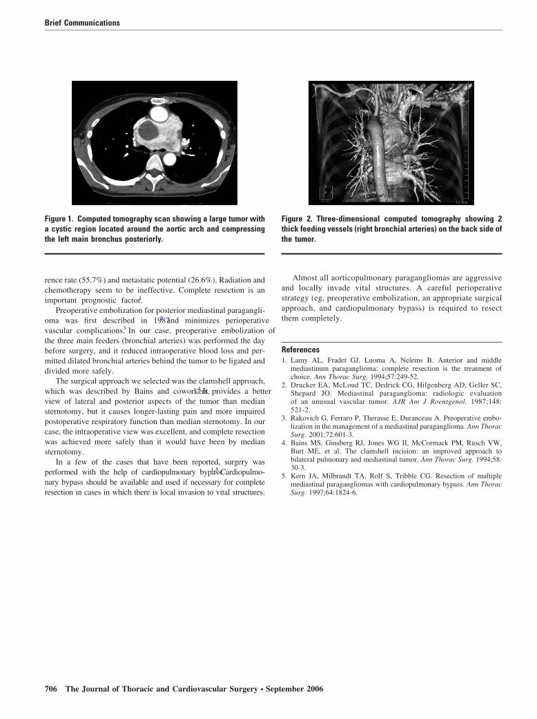
Clinical SummaryA 52-year-old woman was found to have an abnormal shadow ona chest radiograph for a medical checkup. Chest computed tomog-raphy at another hospital revealed a large well-enhanced mass witha cystic lesion located between the superior vena cava, aortic arch,right pulmonary artery, left atrium, and trachea (Figures 1 and 2).A video-assisted thoracoscopic biopsy was performed at another
hospital to make the diagnosis,but massive bleeding occurredduring the procedure, and thebiopsy was abandoned. Thepatient was then referred toour department. We stronglysuspected a paragangliomaof the middle mediastinum.The serum noradrenaline level was slightly increased, and aniodine 123–meta-iodobenzylguanidine scan showed uptake in thetumor. An angiographic study showed many feeding arteries, andthe main feeders were 3 thick bronchial arteries. Preoperativeembolization with Gelfoam (Pfizer, Ann Arbor, Mich) and severalmicrocoils was performed the day before the operation. At oper-ation, we were ready to perform cardiopulmonary bypass; how-ever, complete resection of the tumor without cardiopulmonarybypass was performed via the clamshell approach. Finally, intra-operative blood loss was reduced to 1070 mL. Histologic diagnosiswas reported as a typical paraganglioma, 7 cm in size. There wasno sign of recurrence on a computed tomography scan 1 year aftersurgery.
CommentAorticopulmonary paragangliomas are rare neoplasms; 79 anteriorand middle mediastinal paragangliomas, which represent a surgicalchallenge, were reviewed.1 Because of their location close to thegreat vessels and trachea, complete resection is very difficult.Paragangliomas are locally invasive and have a high local recur-
From the Department of Cardiothoracic Surgery, Faculty of Medicine,University of Tokyo, Tokyo, Japan.
Received for publication Jan 23, 2006; accepted for publication Feb 22,2006.
Address for reprints: Jun Matsumoto, MD, Division of Surgery, AsahiGeneral Hospital, I-1326 Asahi, Chiba, Japan (E-mail: [email protected]).
J Thorac Cardiovasc Surg 2006;132:705-6
0022-5223/$32.00
Copyright © 2006 by The American Association for Thoracic Surgery
doi:10.1016/j.jtcvs.2006.02.061
Dr Matsumoto
Brief Communications
The Journal of Thoracic and Cardiovascular Surgery ● Volume 132, Number 3 705
rence rate (55.7%) and metastatic potential (26.6%). Radiation andchemotherapy seem to be ineffective. Complete resection is animportant prognostic factor.1
Preoperative embolization for posterior mediastinal paragangli-oma was first described in 19872 and minimizes perioperativevascular complications.3 In our case, preoperative embolization ofthe three main feeders (bronchial arteries) was performed the daybefore surgery, and it reduced intraoperative blood loss and per-mitted dilated bronchial arteries behind the tumor to be ligated anddivided more safely.
The surgical approach we selected was the clamshell approach,which was described by Bains and coworkers.4 It provides a betterview of lateral and posterior aspects of the tumor than mediansternotomy, but it causes longer-lasting pain and more impairedpostoperative respiratory function than median sternotomy. In ourcase, the intraoperative view was excellent, and complete resectionwas achieved more safely than it would have been by mediansternotomy.
In a few of the cases that have been reported, surgery wasperformed with the help of cardiopulmonary bypass.1,5 Cardiopulmo-nary bypass should be available and used if necessary for completeresection in cases in which there is local invasion to vital structures.
Almost all aorticopulmonary paragangliomas are aggressiveand locally invade vital structures. A careful perioperativestrategy (eg, preoperative embolization, an appropriate surgicalapproach, and cardiopulmonary bypass) is required to resectthem completely.
References1. Lamy AL, Fradet GJ, Luoma A, Nelems B. Anterior and middle
mediastinum paraganglioma: complete resection is the treatment ofchoice. Ann Thorac Surg. 1994;57:249-52.
2. Drucker EA, McLoud TC, Dedrick CG, Hilgenberg AD, Geller SC,Shepard JO. Mediastinal paraganglioma: radiologic evaluationof an unusual vascular tumor. AJR Am J Roentgenol. 1987;148:521-2.
3. Rakovich G, Ferraro P, Therasse E, Duranceau A. Preoperative embo-lization in the management of a mediastinal paraganglioma. Ann ThoracSurg. 2001;72:601-3.
4. Bains MS, Ginsberg RJ, Jones WG II, McCormack PM, Rusch VW,Burt ME, et al. The clamshell incision: an improved approach tobilateral pulmonary and mediastinal tumor. Ann Thorac Surg. 1994;58:30-3.
5. Kern JA, Milbrandt TA, Rolf S, Tribble CG. Resection of multiplemediastinal paragangliomas with cardiopulmonary bypass. Ann ThoracSurg. 1997;64:1824-6.
Figure 1. Computed tomography scan showing a large tumor witha cystic region located around the aortic arch and compressingthe left main bronchus posteriorly.
Figure 2. Three-dimensional computed tomography showing 2thick feeding vessels (right bronchial arteries) on the back side ofthe tumor.
Brief Communications
706 The Journal of Thoracic and Cardiovascular Surgery ● September 2006